Copyright © 2011, American Society for Microbiology. All Rights Reserved.
Species-Specific Variation in RELA Underlies Differences in NF-
B
Activity: a Potential Role in African Swine Fever Pathogenesis
䌤
Christopher J. Palgrave,
1,2‡ Linzi Gilmour,
2‡ C. Stewart Lowden,
3Simon G. Lillico,
2Martha A. Mellencamp,
4§ and C. Bruce A. Whitelaw
2*
Veterinary Pathology Unit, Division of Veterinary Clinical Sciences, Royal (Dick) School of Veterinary Studies, University of Edinburgh, Easter Bush Veterinary Centre, Roslin, Midlothian EH25 9RG, United Kingdom1; Division of Developmental Biology, The Roslin
Institute and Royal (Dick) School of Veterinary Studies, University of Edinburgh, Easter Bush Campus, Roslin, Midlothian EH25 9RG, United Kingdom2; Veterinary Health Research Pty Ltd., Trevenna Rd., West Armidale,
NSW 2350, Australia3; and Genus/PIC, 100 Bluegrass Commons Blvd., Hendersonville, Tennessee 370754
Received 17 February 2011/Accepted 15 March 2011
African swine fever virus (ASFV) is a highly infectious disease of domestic pigs, with virulent isolates causing a rapidly fatal hemorrhagic fever. In contrast, the porcine species endogenous to Africa tolerate infection. The ability of the virus to persist in one host while killing another genetically related host implies that disease severity may be, in part, modulated by host genetic variation. To complement transcription profiling ap-proaches to identify the underlying genetic variation in the host response to ASFV, we have taken a candidate gene approach based on known signaling pathways that interact with the virus-encoded immunomodulatory protein A238L. We report the sequencing of these genes from different pig species and the identification and initial in vitro characterization of polymorphic variation in RELA (p65; v-rel reticuloendotheliosis viral oncogene homolog A), the major component of the NF-B transcription factor. Warthog RELA and domestic pig RELA differ at three amino acids. Transient cell transfection assays indicate that this variation is reflected in reduced NF-B activityin vitrofor warthog RELA but not for domestic pig RELA. Induction assays indicate that warthog RELA and domestic pig RELA are elevated essentially to the same extent. Finally, mutational studies indicate that the S531P site conveys the majority of the functional variation between warthog RELA and domestic pig RELA. We propose that the variation in RELA identified between the warthog and domestic pig has the potential to underlie the difference between tolerance and rapid death upon ASFV infection.
African swine fever (ASF) virus (ASFV) is a pathogen of the Suidae (domestic and wild pig species), which may be trans-mitted directly or via an arthropod vector in the form of Or-nithodorusticks (35). ASFV is highly infectious, with virulent isolates causing an acute, rapidly fatal hemorrhagic fever in domestic pigs (Sus scrofa) (10, 34). This is thought, in part, to be the result of a proinflammatory cytokine storm driven by infected macrophages (9, 15, 16, 42–44, 56). Initiation of a systemic inflammatory response results in severe hematologi-cal and vascular perturbations, ultimately leading to cardiovas-cular collapse in a manner not dissimilar to septic shock (3, 17, 18, 20, 21, 40, 52, 53). In addition to hemorrhage, severe widespread apoptosis of infected macrophages and uninfected lymphocytes is a prominent feature of the disease; this is also likely related to markedly elevated proinflammatory cytokine levels (19, 33, 37, 43). In comparison to the severe disease which occurs in domestic pigs, in its natural hosts, warthogs (Phacochoerussp.) and bushpigs (Potamochoerussp.), ASF is subclinical and persistent (2, 32, 50, 51).
ASFV is notifiable to the World Organization for Animal
Health (OIE), placing it in the highest category of infectious animal pathogens. It exhibits remarkable potential for trans-boundary spread, and outbreaks in domestic pig populations have a serious socioeconomic impact worldwide. Furthermore, ASF is considered to be the major limiting factor to pig pro-duction in Africa (34). ASFV is a large, double-stranded DNA virus and the only member of the Asfarviridae family (12), suggesting that it may carry novel genes that are not carried by other virus families. Furthermore, the ability of the virus to persist in one host while killing another genetically related host alludes to the possibility that disease severity may, in part, be modulated by host genetic variation.
Several candidate ASFV-encoded immune modulatory fac-tors have been identified, including homologues of CD2 (8-DR/CD2v) (5, 6, 41), IAP (A224L) (31, 39), Bcl-2 (A179L; 5-HL) (1, 7, 8, 30), and IB␣(A238L; 5-EL) (36, 49). Of these, A238L shares 40% sequence homology and 20% identify with domestic pig IB␣(NFKBIA) and substitutes for NFKBIA by binding to the RELA (p65; v-rel reticuloendotheliosis viral oncogene homolog A) subunit of NF-B. Thus, A238L reduces the ability of NF-B to be activated (36, 49). In addition to inhibiting host NF-B, A238L also suppresses calcineurin phosphatase activation of NFAT signaling by the following two mechanisms: direct binding to calcineurin phosphatase 3,  isoform (PPP3CB), and binding to the immunophilin carrier cyclophilin A (PPIA) in a manner similar to that of the immu-nosuppressive drug cyclosporine A (28, 29).
Various groups have initiated transcription profiling of host genes implicated in ASFV infection (15, 16, 42–44, 56). These * Corresponding author. Mailing address: Division of
Developmen-tal Biology, The Roslin Institute and Royal (Dick) School of Veteri-nary Studies, University of Edinburgh, Easter Bush Campus, Roslin, Midlothian EH25 9RG, United Kingdom. Phone: 44 (0)131 651 9175. Fax: 44 (0)131 651 9105. E-mail: [email protected].
‡ C.J.P. and L.G. contributed equally to this work.
§ Present address: Ralco Nutrition Inc., 1600 Hahn Road, Marshall, MN 56258.
䌤Published ahead of print on 30 March 2011.
6008
on November 7, 2019 by guest
http://jvi.asm.org/
studies identified numerous upregulated host genes, but to date, all are limited to analysis in domestic pig cells. In this study, we take a complementary approach to this question by the testing of variation in targeted candidate genes. Clearly A238L represents a novel and versatile immunoregulatory mechanism by which ASFV can inhibit both the NF-B and NFAT signaling pathways (11, 28, 29, 36, 49). We therefore consider the three A238L target proteins, RELA, PPP3CB, and PPIA, and the two proteins it mimics, NFKBIA and NFATC1, as candidates for the genetic variation between pig species which may contribute to species-specific responses to ASFV infection. We now report the sequencing of these genes from different pig species and identification and initialin vitro characterization of polymorphic variation in one of them.
MATERIALS AND METHODS
mRNA isolation, cDNA synthesis, and DNA sequencing.Whole blood (5 ml)
was collected into EDTA from a domestic pig (Sus scrofa, commercial pig;
United Kingdom), common warthog (Phacochoerus africanus; Rotterdam Zoo,
Holland), and babirusa (Babyrousa babyrussa; Marwell Zoo, United Kingdom)
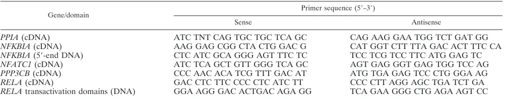
and transferred immediately into DNA/RNA stabilization reagent for blood/ bone marrow (Roche Diagnostics). This was processed using an mRNA isolation kit for white blood cells (Roche Diagnostics). Initially, cDNA libraries were synthesized using the SMART RACE (rapid amplification of cDNA ends) cDNA amplification kit (Clontech) to enable partial sequencing and design of species-specific primers (Table 1) for synthesis of individual cDNAs using
proof-reading PCR using a 1-l sample in a 25-l PCR mixture consisting of 20 pmol
of each primer in 2 mM MgCl2 and 2 mM deoxynucleoside triphosphates
(dNTPs) with 0.7 U High-Fidelity DNA polymerase (Roche Diagnostics). The PCR cycling conditions used were optimized for each gene (data not shown). PCR products were resolved on a 1% agarose gel, excised with a scalpel blade, and extracted using a QIAquick gel extraction kit (Qiagen). These were cloned into pGEM-T Easy (Promega) and sequenced in both directions. To achieve the
full coding sequence for NFKBIA, 5⬘-end fragments were amplified directly from
genomic DNA.
Genomic DNA isolation and sequencing.Samples of skeletal muscle were collected into 20% dimethyl sulfoxide (DMSO)-saturated salt (NaCl) solution
and stored at⫺70°C. DNA was extracted from 0.2 g muscle using the BACC2
extraction kit for blood and cell cultures (Nucleon Biosciences). For PCR, 50 ng
genomic DNA was used as template in a 25-l PCR mixture consisting of 20
pmol of each primer in 2 mM MgCl2and 2 mM dNTPs, with 0.7 U High-Fidelity
DNA polymerase (Roche Diagnostics).
Plasmid construction.Restriction sites were introduced into RELA products from the above-described sequencing study by nested PCR; this enabled inser-tion of domestic pig and warthog RELA genotypes into the multiple cloning site of the pFLAG-CMV-4 vector (Sigma-Aldrich). PCR products were diluted to
1:500 in sterile water, and 1l was used as a template in a 25-l PCR mixture
consisting of 20 pmol of each primer (forward HindIII [5⬘-CCA AGC TTG ACC
TCT TCC CCC TCA TCT T-3⬘] and reverse NotI [5⬘-GCG CGG CCG CTT
AGG AGC TGA TCT GA-3⬘]) in 2 mM MgCl2and 2 mM dNTPs, with 0.7 U
High-Fidelity DNA polymerase (Roche Diagnostics). Restriction sites are
un-derlined. Each⬃1.6-kbp PCR product was resolved on a 1% agarose gel, excised
with a scalpel blade, and extracted using the QIAquick gel extraction kit (Qia-gen). Following HindIII and NotI restriction digestion of the vector and RELA
PCR products, the products were ligated into the open plasmid. These constructs allowed constitutive expression of warthog RELA and domestic pig RELA driven by the cytomegalovirus (CMV) promoter. In addition, an N-terminal eight-amino-acid FLAG sequence is incorporated into the protein, which is recognized by an anti-FLAG monoclonal antibody. Site-directed mutagenesis was preformed using a QuikChange II site-directed mutagenesis kit (Stratagene). Plasmid transient transfection was performed using Lipofectamine2000
(In-vitrogen) in⬃1⫻105
cells/well of a 12-well plate in culture medium at 37°C and
5% CO2. COS-7 cells were cultured in Glasgow minimal essential medium
(GMEM; Sigma), containing 10% fetal bovine serum (Invitrogen), 1%L
-glu-tamine (Invitrogen), 1% sodium pyruvate (Invitrogen), 1% nonessential amino
acids (Invitrogen), and 0.2%-mercaptoethanol (Invitrogen). Mouse embryonic
fibroblasts (MEFs) and RELA⫺/⫺MEFs were cultured in Dulbecco’s modified
Eagle medium (DMEM; Sigma), containing 10% fetal bovine serum (Invitro-gen). Luciferase activity was determined in triplicate using the dual reporter assay system (Promega) and analyzed with Excel (Microsoft), with total protein measured with the bicinchoninic acid (BCA) protein assay kit (Pierce). The data
are presented as the means⫾standard deviations; statistical significance was
evaluated using the unpaired Studentttest, with a difference between groups
being considered statistically significant if thePvalue of the comparison was
⬍0.05.
Western blotting.For Western blot analysis, denatured protein (10g) was run in Tris-glycine-SDS running buffer (National Diagnostics) on precast Nu-Page 12% Tris-glycine gels (Invitrogen) before transfer to a nitrocellulose mem-brane (National Diagnostics). Proteins were visualized using anti-FLAG
M2-peroxidase (horseradish peroxidase [HRP]) (Sigma)-conjugated primary
antibody and mouse-actin primary antibody with goat anti-mouse IgG HRP
(Sigma)-conjugated secondary antibody and detected with Immobilon Western (Millipore) chemiluminescent HRP substrate.
RESULTS
Limited sequence variation in candidate porcine genes.
cDNAs were produced forPPIA,NFKBIA,NFATC1 (regula-tory domain),PPP3CB, andRELA. These were obtained from the following three pig species: the domestic pig (Sus scrofa), common warthog (Phacochoerus africanus), and babirusa (Babyrousa babyrussa). The domestic pig and warthog repre-sent ASFV-susceptible and ASFV-tolerant species, respec-tively. The “outspecies” is the babirusa, which is considered to be the most ancient extant species of pig; its range is restricted to the island of Sulawesi in the Indonesian archipelago. It has no common ancestor with the domestic pig more recently than approximately 10 to 19 million years ago (38). It is not known how this pig species would respond regarding ASFV infection. We generated sequence data (deposited in the EMBL data-base) (Table 2) and aligned all sequences to those of the human homologues which we used as reference sequences.
[image:2.585.42.544.81.180.2]Limited sequence variation was observed in this study (data not shown), with the following genes displaying complete ho-mology at the translated protein level between the domestic pig and warthog: PPIA, NFATC1 regulatory domain, and TABLE 1. Gene-specific PCR primers
Gene/domain Primer sequence (5⬘–3⬘)
Sense Antisense
PPIA(cDNA) ATC TNT CAG TGC TGC TCA GC CAG AAG GAA TGG TCT GAT GG
NFKBIA(cDNA) AAG GAG CGG CTA CTG GAC G CAT GGT CTT TTA GAC ACT TTC CA NFKBIA(5⬘-end DNA) CTC ATC GCA GGG AGT TTC TC TCC TCG TCC TTC ATG GAG TC NFATC1(cDNA) ATC TCA GCT GTT GGG TCA GC AGT GAG GGT GAG TGG TCC AG PPP3CB(cDNA) CCC AAC ACA TCG TTT GAC AT ATG TGA GAG TCC CTG GGA AG
RELA(cDNA) GAC CTC TTC CCC CTC ATC TT CCC CTT AGG AGC TGA TCT GA
RELAtransactivation domains (DNA) GGA AGG GAC ACTGAC AGA GG TCA GAA GGG CTG AGA AGT CC
on November 7, 2019 by guest
http://jvi.asm.org/
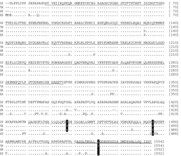
NFKBIA. In the warthog, PPP3CB contained two insertions with respect to the domestic pig. However, these were located outwith the known functional domains. Furthermore, these correlate with splice variants described in human PPP3CB (22, 27). Only in the RELA subunit of NF-B were potentially significant coding differences between the domestic pig and warthog identified (Fig. 1).
Sequence of the porcineRELAgene.TheRELAopen read-ing frame (ORF) is 1,662 nucleotides in the domestic pig and warthog and is slightly smaller at 1,556 nucleotides in the babirusa. These encode proteins which are 554 and 552 amino acids in length, respectively. HumanRELAORF and protein are the same lengths as the babirusa sequences. Due to insuf-ficient primer binding sites in the 5⬘ untranslated region
(UTR), we sequenced all but the first 6 nucleotides for all three pig species (Fig. 1).
Although the babirusa and human sequences are both 6 nucleotides shorter than the other pig sequences, the nucleo-tide deletions occur at different locations. The babirusa se-quence has a single 6-nucleotide deletion, in relation to those of the other pig species, located between the Rel homology domain and the transactivation 2 domain. HumanRELAhas two 3-nucleotide deletions, one 20 nucleotides upstream from the babirusa deletion and the other in the transactivation 2 domain.
[image:3.585.42.283.82.171.2]Of the 13 nucleotide differences between the domestic pig and warthog, only three are nonsynonymous and result in codon changes, T448A, S485P, and S531P. The threonine at 448 in the domestic pig occurs as an alanine in the warthog and babirusa and is absent in human RELA. The serine at 485 in the domestic pig sequence is a proline in the other porcine sequences and in human RELA. The serine at 531 in the domestic pig is also found in babirusa and human but is a proline in warthog RELA. All three amino acid differences occur outside the Rel homology domain, with amino acids 448 and 485 located within transactivation domain 2 and amino acid 531 within transactivation domain 1. To confirm these sequence differences, a 268-nucleotide region was amplified directly from genomic DNA and sequenced for an additional 5 TABLE 2. Sequences deposited into the EMBL database
Gene EMBL accession no.
Domestic pig Warthog Babirusa
PPIA FN401368 FN401369 FN401370
NFKBIA(cDNA) FN421467 FN421468 FN421469 NFKBIA(5⬘-end DNA) FN421464 FN421465 FN421466 NFATC1 FN421470 FN421471 FN421472 PPP3CB FN421473 FN421474 FN421475
RELA FN999988 FN999989 FN999990
FIG. 1. RELA primary protein sequence minus the first two amino acids of the porcine sequences. The Rel homology domain is underlined, the transactivation 2 domain is dashed underlined, and the transactivation 1 domain is double underlined. The nuclear localization signal (KRKR) is dotted underlined. The T448A, S485P, and S531P variations between the domestic pig and warthog are highlighted. SS,Sus scrofa; PA, Phacochoerus africanus; BB,Babyrousa babyrussa; HS,Homo sapiens(M62399).
on November 7, 2019 by guest
http://jvi.asm.org/
[image:3.585.113.477.374.688.2]domestic pigs and 11 warthogs (domestic pig sequences, EMBL accession numbers FN424224 to FN424228; warthog sequences, EMBL accession numbers FN424229 to FN424239). All domestic pig sequences were identical to each other over this region, and similarly, all warthog sequences were identical to each other over this region.
Predicted structural changes as a consequence of RELA sequence variation.Given that even single amino acid changes can have significant effects on protein structure, we performed in silico analyses on the identified changes in RELA in an attempt to determine the structural consequences of the amino acid substitutions. At the three sites, the greater hydrophobic-ity was conferred by the threonine at 448 in the domestic pig sequence and the prolines at 385 and 531 in the warthog sequence. No difference in structural disorder was evident (Phyre analysis) (25), and the only site predicted to be highly likely (0.994) to undergo phosphorylation was the domestic pig serine at 531 (485S⫽0.072; 448T⫽0.0142) (NetPhos2.0) (4). However, the Phospho.ELM database infers that the threonine at 448 could also be a target for phosphorylation (23).
Basal activity of porcine RELA variants. To determine whether the identified sequence variation between domestic pig RELA and warthog RELA affects NF-B activity, we es-tablished a cell transfection assay. Cells were transiently trans-fected in duplicate using a triple plasmid cotransfection strat-egy involving the following: (i) 1 g expression vector for FLAG-tagged domestic pig or warthog RELA or an empty vector control (pFLAG-CMV4; Sigma Aldrich); (ii) 1 g NF-B reporter plasmid comprising 4 copies of the NF-B consensus binding sequence, driving expression of firefly lucif-erase (pNFB-Luc; BD Biosciences, Clontech); and (iii) 1g transfection control vector expressing Renilla luciferase driven off the herpes simplex virus (HSV) thymidine kinase (TK) promoter (pRL-TK; Promega). Cells were harvested at 24 h posttransfection, and a dual luciferase assay (firefly luciferase activity relative to Renilla luciferase activity) was performed on quantified protein extracts. Luciferase activity values for each RELA type were averaged, and fold differences were calcu-lated against background values for empty vector transfections. This strategy resulted in the delivery of an equivalent amount
of either domestic pig or warthog RELA, as determined by Western blotting (Fig. 2A). Initially, we established the assay in COS-7 cells, which have a functional endogenousRELAgene. Repeat transfection experiments (n ⫽ 5) demonstrated that domestic pig RELA is 50% more active than warthog RELA (Fig. 2B). A similar (44%) differential activity was also evident inRELA⫺/⫺mouse embryonic fibroblasts (MEFs) that lacked endogenous RELA activity (derived fromRELA⫺/⫺murine embryos) using the same transfection regime (n⫽3) (Fig. 2C). We attempted to produce stably transfected RELA⫺/⫺ MEFs for the domestic pig and warthog RELA genes. Al-though we could generate numerous colonies for warthog RELA, we were reproducibly unsuccessful in generating those for domestic pigRELA. It is possible that the higher activity level of domestic pig RELA may not be compatible with sur-vival in these cells; a similar scenario may have occurred during a study investigating phosphorylation at the equivalent site (S529) in human RELA (54).
Effect of RELA sequence variation on induced NF-B activ-ity.To determine if the ability to induce NF-B activity was altered by the different pig RELA proteins, we stimulated transiently transfectedRELA⫺/⫺MEFs with known inducers of NF-B signaling (9, 14, 15, 16, 42–44, 56). Induction with tumor necrosis factor alpha (TNF-␣) nearly doubled (90% increase) warthog RELA activity, whereas domestic pig RELA showed a 70% increase in activity (Fig. 3). We tested a further three known NF-B-inducing agents, lipopolysaccharide, phor-bol-12-myristate-13-acetate, and hydrogen peroxide. For all three additional agents, the fold induction observed for wart-hog RELA was always greater than that of domestic pig (Fig. 3). FLAG immunostaining of COS-7 cells transfected with either domestic pig or warthog RELA and treated with phor-bol-12-myristate-13-acetate did not identify any gross differ-ences in cytoplasm-nuclear transit time (RELA of both species was in the nucleus within 1 min and back in the cytoplasm after 20 min) (data not shown).
[image:4.585.139.451.69.175.2]Identification of functional mutations in porcine RELA.In our sequence data, we identified three amino acid differences between domestic pig RELA and warthog pig RELA. To de-termine if thein vitrodifference in domestic pig and warthog FIG. 2. Effect of polymorphic RELA on basal NF-B activity. (A) Western blot of COS-7 cells at 24 h after transfection with 1g FLAG-tagged warthog (WH) or domestic pig (DP)RELAor empty FLAG vector (⫺ve) and established RELA-FLAG expressing COS-7 cells (⫹ve). (B) Fold difference between NF-B-luciferase activity in COS-7 cells after 24 h of transient transfection with 1g warthog (WH) or 1g domestic pig (DP) RELA. Activity is presented as the fold difference between RELA-induced NF-B—luciferase activity normalized to cotransfected TK-Renilla luciferase activity relative to that of the empty vector. Error bars⫽standard deviations from the means.P⬍0.03 between WH and DP. (C) Fold difference between NF-B-luciferase activities inRELA⫺/⫺MEFs after 24 h of transient transfection with warthog (WH) and domestic pig (DP)
RELAgenes. Activity is presented as the fold difference between RELA-induced NF-B-luciferase activity normalized to cotransfected TK-Renilla luciferase activity relative to that of the empty vector normalized to cotransfected TK-Renilla luciferase activity. Error bars⫽standard deviations from the means.P⬍0.05 between WH and DP.
on November 7, 2019 by guest
http://jvi.asm.org/
RELA activities was a cumulative effect of these three differ-ences, or due to one or other individual mutations, we gener-ated versions of RELA carrying the following single-base changes: T448A, S485P, and S531P. Transfection of these RELA variants into RELA⫺/⫺MEFs demonstrated that the majority of the reduced basal activity observed for warthog RELA compared to that observed for domestic pig RELA was attributed to the S531P mutation (Fig. 4).
DISCUSSION
In this study, we have taken a candidate approach, identify-ing genes which may affect the severity of the host response to ASFV infection in the highly susceptible domestic pig and ASFV-tolerant warthog. The ASFV immunomodulatory factor A238L is known to interact with components of both NF-B and NFAT host signaling pathways (11, 28, 29, 36, 49). We have sequenced five key factors in these pathways, including three A238L-targeted proteins, RELA, PPP3CB, and PPIA, and the two proteins it mimics, NFKBIA and NFATC1 (reg-ulatory domain). Modest sequence differences have been iden-tified at the cDNA (mRNA) level between the domestic pig and warthog; however, the majority of these are synonymous (silent) and do not alter the resulting amino acid sequences. We also sequenced the same genes from the ancient babirusa and observed a small number of amino acid differences be-tween this species and the other two species of pig. These differences likely reflect the long evolutionary distance that exists between these species (38).
Despite the high degree of conservation observed between warthog and domestic pig PPP3CB-, PPIA-, NFKBIA-, and NFATC1-translated protein sequences, significant variation was detected in RELA. RELA is the predominant member of the heterodimeric transcription factor NF-B (47). Moreover, the sequence variation includes a phosphorylation site in trans-activation domain 1 (position 531), which is highly conserved across mammals and has been demonstrated to modulate the activity of human RELA (equivalent to S529) (54, 55). The function of approximately one-third of all eukaryote proteins is controlled by phosphorylation; thus, the observed S531P se-quence variation represents an intriguing candidate regulator
for the reduced pathology observed in African pigs infected with ASFV. We demonstrate that the genetic variation in the RELA sequence between the domestic pig and warthog is reflected in NF-B activityin vitro, with warthog RELA dis-playing significantly reduced basal and induced NF-B activity. We discuss three (not mutually exclusive) scenarios of how the genetic variation we have identified between the domestic pig and warthog may underlie the dramatic phenotypic difference in how these two pig species respond to ASFV.
First, as suggested by ourin vitroassays, warthog RELA is inherently less active than the domestic pig RELA, exhibiting lower basal and induced levels. In a study of macrophage transcription profiles following ASFV infectionin vitro, several factors within the NF-B signaling pathway displayed elevated expression (i.e.,NFKB1,NFKBIA); however,RELAexpression was not observed to be altered (56). The impact of ASFV infection on the warthogRELAexpression level is not known, since this study used only domestic pig macrophages. There-fore, although we have not determined whether the porcine RELA variants are differentially phosphorylated, it would ap-pear that variation in domestic pig RELA activity may not be determined by altered expression levels. Alternatively, the warthog will have to function with reduced basal NF-B activ-ity, which would presumably indicate an adapted NF- B-de-pendent transcriptome between the two species.
[image:5.585.78.248.68.160.2]In the second scenario, phosphorylation of domestic pig RELA at S531 activates a set(s) of genes which are not acti-vated by warthog RELA, as it lacks this phosphorylation site. Much is known about activation of the NF-B pathway, with data coming primarily from studies of human RELA (47). NF-B activity is regulated by two different mechanisms. The classical canonical pathway involves inhibitors (e.g., NFKBIA) that sequester this transcription factor in the cytoplasm until they are proteolytically degraded by the ubiquitin pathway (24). The alternative pathway revolves around posttranscrip-tional modifications, predominantly phosphorylation of RELA (46). At least 8 inducible phosphorylation sites have been iden-tified in human RELA, which enable transcription of subsets of NF-B-dependent genes (26, 45). One such site is S529, which is equivalent to domestic pig S531, suggesting that this site may perform a similar role. Indeed, differential expression of sub-sets of immune and inflammatory proteins as a result of S531 FIG. 4. Comparison of individual allelicRELAvariation on NF-B activity. Comparison of NF-B–luciferase activity inRELA⫺/⫺MEFs
[image:5.585.335.505.69.146.2]after 24 h transient transfection with 1g warthog (WH) or 1 g domestic pig (DP) RELA or 1g of RELA variants encoding the individual amino acid substitutions T448A, S485P, and S531P. Activity is presented as the fold difference between RELA-induced NF-B– luciferase activity normalized to cotransfected TK-Renilla luciferase activity relative to that of the empty vector normalized to cotransfected TK-Renilla luciferase activity. Error bars⫽standard deviations from the means,*,P⬍0.08 versus DP.
FIG. 3. Induction ofRELAallelic variants.RELA⫺/⫺MEFs were
transiently cotransfected with 1g warthog (clear box) or 1g do-mestic pig (filled box) RELA. Cells were treated with TNF-␣ (30 ng/ml), lipopolysaccharide (LPS; 10g/ml), phorbol-12-myristate 13-acetate (PMA; 20 mM), and hydrogen peroxide (10M) immediately after and for the duration of the transfection. Cells were harvested at 24 h. RELA-induced NF-B–luciferase (1g) activity was normalized to cotransfected TK-Renilla luciferase (1g) activity and presented as the fold induction above nondrug-treated cells (relative value of 1 depicted by the line).
on November 7, 2019 by guest
http://jvi.asm.org/
phosphorylation could play a role in determining ASFV patho-genesis in the domestic pig. Furthermore, phosphorylation not only controls specific transcription profiles but also can under-lie the developmental timing in gene activation (13). It is tempting to speculate that if a similar mechanism is applied through the domestic pig S531 site, then it could also play a role in the gross physical differences that characterize the var-ious pig species.
In the third scenario, we consider whether the S531P varia-tion in RELA results in different outcomes of interacvaria-tion with A238L during ASFV infection. Phosphorylation of human S529 (equivalent to domestic pig S531) is inhibited by the interaction of RELA with NFKBIA; only upon activation and degradation of NFKBIA can S529 phosphorylation occur (55). During ASFV infection, NFKBIA is degraded and replaced by A238L, which mimics NFKBIA but is not susceptible to pro-teolytic degradation (36, 49). As a result, in domestic pig cells infected with ASFV, S531 may not be exposed for phosphor-ylation. In comparison, warthogs lack this phosphorylation site; therefore, the ability of A238L to block phosphorylation at this site is irrelevant. The role of NFKBIA is to prevent nuclear translocation of NF-B (24). We did not observe any gross temporal differences in nuclear transportation rates upon stim-ulation between cells expressing domestic pig RELA and wart-hog RELA; likewise, phosphorylation of human S529 also does not affect nuclear translocation (54). Furthermore, studies us-ing recombinant ASFV lackus-ing A238L indicate that neither nuclear import nor export of RELA is affected by this immune modulator (48). This suggests that the immunomodulatory functions of A238L are not the result of preventing nuclear translocation in a manner similar to that used by NFKBIA. Instead, A238L may inhibit NF-B-mediated transcription by other mechanisms, for example, by preventing phosphorylation of RELA in domestic pigs, as discussed above. Whether such differences in how domestic pig RELA and warthog RELA interact with A238L exist will require further investigation. Likewise, elucidating how different mechanisms governing RELA activity and NF-B-mediated transcription have evolved in these species, and determining the full extent of their functional implications on the immune system and wider transcriptome, will require additional study.
ASFV is highly infectious, with virulent isolates causing an acute, rapidly fatal hemorrhagic fever in domestic pigs (10, 34). In contrast, the porcine species endogenous to Africa tolerate the virus. Outbreaks have significant economic repercussions in addition to welfare concerns. In Africa, ASFV limits the use of the genetically improved breeding pig stock that is pervasive in the Eurasian landscape. Furthermore, ASF poses a constant threat to Europe and Asia, as documented by the list of out-breaks that have occurred over the last 30 to 40 years. As our world climate changes and the international movement of pork products continues to rise, this risk may well increase. No effective vaccine has been developed, so many look to genetic strategies to mitigate the geographical limits of pig breeding imposed by ASFV. We have demonstrated that a polymorphic RELA variant found in warthogs has the potential to underlie the difference between tolerance and rapid death upon infec-tion with ASFV.
ACKNOWLEDGMENTS
This work was supported by the BBSRC (United Kingdom) and a Genus/PIC Postgraduate Studentship. C.J.P. was also funded by a Postgraduate Centenary Fellowship (University of Edinburgh). L.G. was funded through a Faraday-BBSRC CASE studentship.
RELA⫺/⫺ MEFs were kindly donated by Ron Hay (University of
Dundee). The pcDNA-A238L expression vector was kindly donated by Linda Dixon (Institute for Animal Health, Pirbright). We are grateful to Susan Rhind, Liz Glass, Sarah Howie, and Paul Hopwood (Univer-sity of Edinburgh) for their support of this work.
REFERENCES
1.Afonso, C. L., J. G. Neilan, G. F. Kutish, and D. L. Rock.1996. An African swine fever virus Bc1-2 homolog, 5-HL, suppresses apoptotic cell death.
J. Virol.70:4858–4863.
2.Anderson, E. C., G. H. Hutchings, N. Mukarati, and P. J. Wilkinson.1998.
African swine fever infection of the bushpig (Potamochoerus porcus) and its
significance to the epidemiology of the disease. Vet. Microbiol.62:1–15.
3.Anderson, E. C., S. M. Williams, S. P. Fisher-Hoch, and P. J. Wilkinson.
1987. Arachidonic acid metabolites in the pathophysiology of
thrombocyto-penia and haemorrhage in acute African swine fever. Res. Vet. Sci.42:387–
394.
4.Blom, N., S. Gammeltoft, and S. Brunak.1999. Sequence and structure-based prediction of eukaryotic protein phosphorylation sites. J. Mol. Biol.
294:1351–1362.
5.Borca, M. V., et al.1998. Deletion of a CD2-like gene, 8-DR, from African
swine fever virus affects viral infection in domestic swine. J. Virol.72:2881–
2889.
6.Borca, M. V., et al.1994. An African swine fever virus gene with similarity to the T-lymphocyte surface antigen CD2 mediates hemadsorption. Virology
199:463–468.
7.Brun, A., C. Rivas, M. Esteban, J. M. Escribano, and C. Alonso.1996. African swine fever virus gene A179L, a viral homologue of bcl-2, protects
cells from programmed cell death. Virology225:227–230.
8.Brun, A., F. Rodriguez, J. M. Escribano, and C. Alonso.1998. Functionality and cell anchorage dependence of the African swine fever virus gene A179L,
a viral bcl-2 homolog, in insect cells. J. Virol.72:10227–10233.
9.Carrasco, L., et al.2002. African swine fever: expression of interleukin-1 alpha and tumour necrosis factor-alpha by pulmonary intravascular
macro-phages. J. Comp. Pathol.126:194–201.
10.Colgrove, G. S., E. O. Haelterman, and L. Coggins.1969. Pathogenesis of
African swine fever in young pigs. Am. J. Vet. Res.30:1343–1359.
11.Dixon, L. K., et al.2004. African swine fever virus proteins involved in
evading host defence systems. Vet. Immunol. Immunopathol.100:117–134.
12.Dixon, L. K., et al.2005. FamilyAsfarviridae, p. 135–143.InC. M. Fauquet, M. A. Mayo, J. Maniloff, U. Desselberger, and L. A. Ball (ed.), Virus taxonomy. Eighth report of the International Committee on Taxonomy of Viruses. Elsevier Academic Press, London, England.
13.Gavalda, N., H. Gutierrez, and A. M. Davies.2009. Developmental switch in
NF-kappaB signalling required for neurite growth. Development136:3405–
3412.
14.Ghosh, S., M. J. May, and E. B. Kopp.1998. NF-B and Rel proteins: evolutionary conserved mediators of immune responses. Annu. Rev.
Immu-nol.16:225–260.
15.Gil, S., et al.2003. Expression at mRNA level of cytokines and A238L gene in porcine blood-derived macrophages infected in vitro with African swine
fever virus (ASFV) isolates of different virulence. Arch. Virol.148:2077–
2097.
16.Gomez del Moral, M., et al.1999. African swine fever virus infection induces tumor necrosis factor alpha production: implications in pathogenesis. J.
Vi-rol.73:2173–2180.
17.Gomez-Villamandos, J. C., et al.1997. African swine fever virus infection of
bone marrow: lesions and pathogenesis. Vet. Pathol.34:97–107.
18.Gomez-Villamandos, J. C., et al.1998. Thrombocytopenia associated with apoptotic megakaryocytes in a viral haemorrhagic syndrome induced by a moderately virulent strain of African swine fever virus. J. Comp. Pathol.
118:1–13.
19.Gomez-Villamandos, J. C., et al.1995. Experimental African swine fever: apoptosis of lymphocytes and virus replication in other cells. J. Gen. Virol.
76(Pt. 9):2399–2405.
20.Gomez-Villamandos, J. C., et al.1995. A pathological study of the
perisi-nusoidal unit of the liver in acute African swine fever. Res. Vet. Sci.59:146–
151.
21.Gomez-Villamandos, J. C., et al.1995. Pathological changes in the renal interstitial capillaries of pigs inoculated with two different strains of African
swine fever virus. J. Comp. Pathol.112:283–298.
22.Guerini, D., and C. B. Klee.1989. Cloning of human calcineurin A: evidence for two isozymes and identification of a polyproline structural domain. Proc.
Natl. Acad. Sci. U. S. A.86:9183–9187.
on November 7, 2019 by guest
http://jvi.asm.org/
23.Hammet, A., et al.2003. FHA domains as phospho-threonine binding
mod-ules in cell signaling. IUBMB Life55:23–27.
24.Karin, M.2006. Nuclear factor-kappaB in cancer development and
progres-sion. Nature441:431–436.
25.Kelley, L. A., and M. J. Sternberg.2009. Protein structure prediction on the
Web: a case study using the Phyre server. Nat. Protoc.4:363–371.
26.Law, M., P. Corsino, N. T. Parker, and B. K. Law.2010. Identification of a small molecule inhibitor of serine 276 phosphorylation of the p65 subunit of
NF-kappaB using in silico molecular docking. Cancer Lett.291:217–224.
27.McPartlin, A. E., H. M. Barker, and P. T. Cohen.1991. Identification of a third alternatively spliced cDNA encoding the catalytic subunit of protein
phosphatase 2B beta. Biochim. Biophys. Acta1088(2):308–310.
28.Miskin, J. E., C. C. Abrams, and L. K. Dixon.2000. African swine fever virus protein A238L interacts with the cellular phosphatase calcineurin via a
binding domain similar to that of NFAT. J. Virol.74:9412–9420.
29.Miskin, J. E., C. C. Abrams, L. C. Goatley, and L. K. Dixon.1998. A viral mechanism for inhibition of the cellular phosphatase calcineurin. Science
281:562–565.
30.Neilan, J. G., et al.1993. An African swine fever virus gene with similarity to the proto-oncogene bcl-2 and the Epstein-Barr virus gene BHRF1. J. Virol.
67:4391–4394.
31.Nogal, M. L., et al.2001. African swine fever virus IAP homologue inhibits caspase activation and promotes cell survival in mammalian cells. J. Virol.
75:2535–2543.
32.Oura, C. A., P. P. Powell, E. Anderson, and R. M. Parkhouse.1998. The pathogenesis of African swine fever in the resistant bushpig. J. Gen. Virol.
79:1439–1443.
33.Oura, C. A., P. P. Powell, and R. M. Parkhouse.1998. African swine fever:
a disease characterized by apoptosis. J. Gen. Virol.79:1427–1438.
34.Penrith, M.-L., G. R. Thomson, and A. D. S. Bastos.2004. African swine
fever, p. 1088–1119.InJ. A. W. Coetzer and R. C. Tustin (ed.), Infectious
diseases of livestock, 2nd ed., vol. 2. Oxford University Press, Oxford, United Kingdom.
35.Plowright, W., J. Parker, and M. A. Pierce.1969. The epizootiology of
African swine fever in Africa. Vet. Rec.85:668–674.
36.Powell, P. P., L. K. Dixon, and R. M. Parkhouse.1996. An IkappaB homolog encoded by African swine fever virus provides a novel mechanism for down-regulation of proinflammatory cytokine responses in host macrophages.
J. Virol.70:8527–8533.
37.Ramiro-Ibanez, F., A. Ortega, A. Brun, J. M. Escribano, and C. Alonso.
1996. Apoptosis: a mechanism of cell killing and lymphoid organ impairment
during acute African swine fever virus infection. J. Gen. Virol.77(Pt. 9):
2209–2219.
38.Randi, E., V. Lucchini, and C. Hoong Diong.1996. Evolutionary genetics of the Suiformes as reconstructed using mtDNA sequencing. J. Mamm. Evol.
3:163–194.
39.Rodriguez, C. I., et al.2002. African swine fever virus IAP-like protein
induces the activation of nuclear factor kappa B. J. Virol.76:3936–3942.
40.Rodriguez, F., A. Fernandez, J. P. Martin de las Mulas, M. A. Sierra, and A. Jover.1996. African swine fever: morphopathology of a viral haemorrhagic
disease. Vet. Rec.139:249–254.
41.Rodriguez, J. M., R. J. Yanez, F. Almazan, E. Vinuela, and J. F. Rodriguez.
1993. African swine fever virus encodes a CD2 homolog responsible for the
adhesion of erythrocytes to infected cells. J. Virol.67:5312–5320.
42.Salguero, F. J., et al.2002. Changes in macrophages in spleen and lymph nodes during acute African swine fever: expression of cytokines. Vet.
Im-munol. Immunopathol.90:11–22.
43.Salguero, F. J., P. J. Sanchez-Cordon, A. Nunez, M. Fernandez de Marco, and J. C. Gomez-Villamandos.2005. Proinflammatory cytokines induce lym-phocyte apoptosis in acute African swine fever infection. J. Comp. Pathol.
132:289–302.
44.Salguero, F. J., et al.2004. Apoptosis of thymocytes in experimental African
swine fever virus infection. Histol. Histopathol.19:77–84.
45.Sasaki, C. Y., T. J. Barberi, P. Ghosh, and D. L. Longo.2005. Phosphory-lation of RelA/p65 on serine 536 defines an I{kappa}B{alpha}-independent
NF-{kappa}B pathway. J. Biol. Chem.280:34538–34547.
46.Schmitz, M. L., S. Bacher, and M. Kracht.2001. IB-independent control of
NF-B activity by modulatory phosphorylations. Trends Biochem. Sci.26(3):
186–190.
47.Schmitz, M. L., I. Mattioli, H. Buss, and M. Kracht.2004. NF-kappaB: a multifaceted transcription factor regulated at several levels. Chembiochem
5:1348–1358.
48.Silk, R. N., G. C. Bowick, C. C. Abrams, and L. K. Dixon.2007. African swine fever virus A238L inhibitor of NF-kappaB and of calcineurin phosphatase is imported actively into the nucleus and exported by a CRM1-mediated
path-way. J. Gen. Virol.88:411–419.
49.Tait, S. W. G., E. B. Reid, D. R. Greaves, T. E. Wileman, and P. P. Powell.
2000. Mechanism of inactivation of NF-B by a viral homologue of IB␣.
J. Biol. Chem.275:34656–34664.
50.Thomson, G. R.1985. The epidemiology of African swine fever: the role of
free-living hosts in Africa. Onderstepoort J. Vet. Res.52:201–209.
51.Thomson, G. R., M. D. Gainaru, and A. F. Van Dellen.1980. Experimental infection of warthogs (Phacochoerus aethiopicus) with African swine fever
virus. Onderstepoort J. Vet. Res.47:19–22.
52.Villeda, C. J., S. M. Williams, P. J. Wilkinson, and E. Vinuela.1993. Con-sumption coagulopathy associated with shock in acute African swine fever.
Arch. Virol.133:467–475.
53.Villeda, C. J., S. M. Williams, P. J. Wilkinson, and E. Vinuela.1993. Hae-mostatic abnormalities in African swine fever a comparison of two virus strains of different virulence (Dominican Republic ’78 and Malta ’78). Arch.
Virol.130:71–83.
54.Wang, D., and A. S. Baldwin, Jr.1998. Activation of nuclear factor-
B-dependent transcription by tumor necrosis factor-␣ is mediated through
phosphorylation of RelA/p65 on serine 529. J. Biol. Chem.273:29411–29416.
55.Wang, D., S. D. Westerheide, J. L. Hanson, and A. S. Baldwin, Jr.2000.
Tumor necrosis factor␣-induced phosphorylation of RelA/p65 on Ser529is
controlled by casein kinase II. J. Biol. Chem.275:32592–32597.
56.Zhang, F., et al.2006. Macrophage transcriptional responses following in vitro infection with a highly virulent African swine fever virus isolate. J.
Vi-rol.80:10514–10521.