1
EVALUATION OF CREATINE KINASE AS A DIAGNOSTIC
TOOL FOR
THYROID FUNCTION
DISSERTATION SUBMITTED FOR
M.D GENERAL MEDICINE
BRANCH – I
MAY2018
THE TAMIL NADU
2
CERTIFICATE FROM THE DEAN
This is to certify that the dissertation entitled “EVALUATION OF CREATINE KINASE AS A DIAGNOSTIC TOOL FOR THYROID FUNCTION” is the bonafide work of Dr. N.EZHILin partial fulfilment of the University regulations of the TamilNadu Dr. M.G.R Medical University, Chennai for M.D General Medicine Branch I examination to be held in May 2018.
Dr. D. MARUTHUPANDIAN M.S, FICS, FAIS
3
CERTIFICATE FROM THE HOD
This is to certify that the dissertation entitled “EVALUATION OF CREATINE KINASE AS A DIAGNOSTIC TOOL FOR THYROID FUNCTION” is the bonafide work of Dr. N.EZHILin partial fulfillment of the university regulations of the Tamil Nadu Dr. M.G.R Medical
University, Chennai for M.D General Medicine Branch I examination to be held in May 2018.
Dr. V.T. THEOPHILUS PREMKUMAR, M.D., H.O.D & Professor of Medicine
4
CERTIFICATE FROM THE GUIDE
This is to certify that the dissertation entitled “EVALUATION OF CREATINE KINASE AS A DIAGNOSTIC TOOL FOR THYROID FUNCTION” is the bonafide Work ofDr. N.EZHILin partial fulfillment of the university regulations of the Tamil Nadu Dr. M.G.R Medical
University, Chennai for M.D General Medicine Branch I examination to be held in May 2018.
Dr. G. BAGIALAKSHMI, M.D., Professor of Medicine,
5
DECLARATION
I, Dr. N.EZHILsolemnly declare that this dissertation“EVALUATION OF CREATINE KINASE AS A DIAGNOSTIC TOOL FOR THYROID FUNCTION”is a bonafide record of work done by me at the Department of General Medicine, Govt. Rajaji Hospital, Madurai under the guidance of Dr.G.BAGIALAKSHMI, MD, Professor. Department of General Medicine Madurai Medical College. Madurai.
This Dissertation is submitted to the Tamil Nadu Dr. M.G.R Medical University in partial fulfilment of the rules and regulations for the award of M.D GENERAL MEDICINE DEGREE BRANCH–I examination to be held in May 2018.
DATE:
6
ACKNOWLEDGEMENT
I would like to thank our respected DeanProf. Dr. D.MARUTHUPANDIANM.S,
FICS, FAIS, Madurai Medical College for permitting me to utilize the facilities of
Madurai Medical College and Government Rajaji Hospital for this dissertation.
I wish to express my respect and sincere gratitude to my beloved teacher
and Head of the Department, Prof. Dr. V.T. THEOPHILUS PREMKUMAR, M.D.,
and Professor of Medicine for his valuable guidance and encouragement during
the study and also throughout my course period.
I would like to express my deep sense of gratitude, respect and thanks to
my beloved Unit Chief and Professor of Medicine, Prof.Dr.G.BAGIALAKSHMI,
M.D., for her valuable suggestions, guidance and support throughout the study
and also throughout my course period.
I am greatly indebted to the Professors, Dr. R. BALAJINATHAN M.D., Dr.
M. NATARAJAN M.D, and Dr.J. SANGUMANI M.D, Dr.DHARMARAJ M.D. andDr.R.PRABHAKARAN, M.D., for their valuable suggestions throughout
thecourse of the study.
I sincerely thank the Assistant ProfessorsDr.P.SARAVANAN M.D.,
Dr.S.KRISHNASAMY PRASAD M.D. for their guidance and suggestions in this
dissertation work.
I sincerely thank all the staffs of department of medicine for the timely
7
I extend my thanks to all my friends, batch mates, my junior and senior
colleagues who have stood by me and supported me throughout my study and
course period.
Finally, I thank all the patients, who form the most vital part of my work,
for their extreme patience and co-operation without whom this project would
8
CONTENTS
Sl.
NO
CONTENTS
PAGE
NUMBER
1
INTRODUCTION
1
2
AIM OF THE STUDY
29
3
REVIEW OF LITERATURE
30
4
MATERIALS AND METHODS
52
5
RESULTS AND ANALYSIS
56
6
CONCLUSION
74
7
ANNEXURES
I. PROFORMA
II. MASTER CHART
III. ABBREVIATIONS
IV. BIBLIOGRAPHY
V. ETHICAL COMMITTEE CERTIFICATE
9
INTRODUCTION
Thyroid disorders are one of the most common endocrinological disorders among general population. Thyroid gland secretes T3(tri iodo thyronine), T4(thyroxine) hormones, which plays a role in basal metabolic rate, growth, development and tissue differentiation.
ANATOMY OF THYROID
Thyroid gland consists of
A midline isthmus lying parallel just below the cricoidcartilage
Right and left, two lateral lobes that extend superiorly, infront of neck
giving the look of a butterfly.
The gland is fully enclosed by pre tracheal fascia, under the strapmuscle,
which makes the gland move up with deglutition.
HISTOLOGY OF THYROID
Thyroid gland is divided by thin fibrous septa into pseudolobules
These pseudolobules are composed of vesicles otherwise
calledfollicles or acini, are densely surrounded by a capillary network.
Follicular walls are surrounded by cuboidal epithelium
Protenaceous colloidal material is filled within the lumen offollicles
10
The peptide sequences of T4 and T3 are stored and synthesized ascomponent of thyroglobulin.
DEVELOPMENT OF THYROID
Develops from the ectoderm of the floor of the pharynx with
somecontribution from the lateal pharyngeal pouches.
The thyroglossal duct, which extends from the foramen caecumnear the
base of the tongue to the isthmus of the thyroid arise fromdescent of the midline thyroid anlagen.
The posterior aspect of the thyroid gland becomes associated withthe
parathyroid gland and the para follicular C cells, during thedevelopment, which are derived from ultimo-bronchial body,which become incorporated into its substance.
While they experience malignant transformation, the C cells are
thesource of the calcitonin and gives to medullary thyroid carcinoma.
At about 10-12 weeks of intra uterine life, the foetal thyroid beginsto
concentrate and organify iodine.
Maternal TSH and T4 do not cross the placenta, but the maternalTRH
crosses the placenta.
The major source of thyroid hormone in the foetal life is T4 fromthe
foetal thyroid.
The functional unit is foetal pituitary – thyroid axis which isdistinct
11 PHYSIOLOGY OF THYROID GLAND
Thyroid secretes three hormones – thyroxin (T4), triiodothyronine(T3) and clacitionin. Thyroid follicles secrete the first two hormones,have similar biological activity and the term “thyroid hormones” ispertinent to these 2
hormones only. Calcitonin is chemically andbiologically dissimilar entirely and is secreted from parafollicular C cells. It regulates calcium metabolism and it is considered along with parathormone.
Thyroid hormone contains iodine. Iodine enters the thyroid in theform of inorganic or organic iodide is oxidized by a peroxidise enzyme atthe cell – colloidal interface. Subsequent reactions results in formation ofthyroxin. The only source of T4 is thyroid gland. Thyroid secretes 20% ofT3; extra glandular tissues produce the remaining amount by theperipheral conversion of T4 into T3.
CHEMISTRY AND SYNTHESIS OF THYROID HORMONE
Both T4 and T3, which is a condensation product of 2 molecules oftyrosine are iodine containing derivatives of thyronine.Thyroxine (T4) - 3, 5, 3’, 5’ – tetraiodothyronineT3 - 3, 5, 3’ – triiodothyronineThyroid hormones are
12
1. IODIDE UPTAKE OR IODIDE TRAPPING : Iodine fromperipheral circulation is taken into the follicles by active transportprocess called Na + I – symporter or NIS. Iodine content of follicleregulates the iodide trap. Meagre storage activates and largestorage inhibits this trap. This method is mediated by TSH.Percholarate, thiocyanates and nitrates inhibits this trapping.
2. OXIDATION AND IODINATION : “Iodide trapped by follicularcells is transported by one another transporter across the apicalmembrane called as “pendrin” and oxidized by thyroid peroxidaseenzyme present in
follicular membrane and forms iodinium ions(I+) or hypoiodous acid (HOI) or enzyme linked hypoiodate (E-OI)with the help of H2O2.These various forms of iodine bound avidly with thyroglobulin and forms monoiodothyrosine (MIT) and diiodotyrosine (DIT).
3. COUPLING : Pairs of iodinated tyrosine residues forms T3 and T4by coupling with one another. Coupling belongs to oxidativereaction and is catalysed by the same thyroid peroxidise. Oxidationand Coupling, both reactions are regulated by TSH.
13
5. PERIPHERAL COVERSION OF T4 TO T3: Conversion occurspredominantly in kidney and liver. One third of T4 undergoesconversion and most of T3 in plasma is derived from liver. Targetorgans take up T3 for metabolic functions except brain andpituitary which take up T4 and converts in to T3 by their owncellular mechanisms.
RELATION BETWEEN T3 AND T4
Normally thyroid secretes more amount of T4 compared to T3. Butthis
difference is reduced in iodine deficient state.
Normally T4 is the major circulating form because it is avidly
boundwith plasma proteins 15 times more.
T3 is five times more potent than T4.
T3 acts very faster than T4.
Peak effect of T3 comes earlier (1-2 days), but peak effect of T4comes
later (6-8 days).
T3 is more tightly bound to the nuclear receptors than T4 and the
T4receptor complex is not able to activate or depress the genetranscription.
About one third of T4 is converted in to T3 in peripheral tissues, inliver and kidney, by D1 type of 5’ Deiodinase (D1 type 5’ DI) andreleased in
14
called type 2 deiodinase (D2 type 5’ DI). T4 is convertedin to
metabolically active T3 or inactive reverse T3 (r T3).
T4 and T3 metabolized in liver by conjugation with glucuronate
andsulfate. Enzyme inducers such as phenobarbitone, Carbamazepine andphenytoin increase the metabolic clearance of the hormones withoutdecreasing the proportion of free hormones in the circulation.
Finally, T3 is an active form. T4 is a transport form i.e.precursor ofT3.
Normal daily secretion of T3-10-30 mcgm. T4-60-90 mcgm.
T3 and T4 bound with 3 plasma proteins – they are
Thyroxin binding globulin (TBG)
Thyroxin binding pre albumin (Transthyretin)
Albumin
Plasma t 1⁄2 of T3 is 1-2 days; of T4 is 6-7 days. The half life is
increased in hypothyroidism and shortened in hyperthyroidism due to enhanced and blunted metabolism respectively.
Thyroid is the only source of T4”.
LABORATORY EVALUATION
Measurement of Thyroid Hormones
15
and specificity of TSH assayshave greatly improved laboratory assessment of thyroid function.
Immune chemi-lluminometric assays (ICMAs) for TSH aresensitive enough to discriminate between the suppressed values that occurwith thyrotoxicosis and the lower limit of the reference range. Extremelysensitive (fourth –generation) assays can detect TSH levels 0.004 mU/L,are enough. The TRH stimulation test is now obsolete because of thewidespread availability of the TSH ICMA. Also there is often a failure ofTSH to rise after an intravenous bolus of 200-400 mcg.
The finding of an abnormal TSH level should then be followed bycirculating thyroid hormone levels to correctly diagnose hypothyroidism(elevated TSH) or hyperthyroidism (Suppressed TSH). Radioimmunoassays are widely available for serum totalT4 and totalT3. T4 andT3 are highly protein-bound. Medications, illness, genetic factors etc.
Can influence protein binding. So the free or unbound hormone levels,which correspond to the biologically available hormone pool should bemeasured next.
16
Thyroid hormones level in various clinical conditions
CONDITION FREE T3 FREE T4 TSH
Subclinical hypothyroidism
Normal Normal Increased
Subclinical hyperthyroidism
Normal Normal Low
Primary hyperthyroidism
Increased Increased Undetectable
Primary hypothyroidism
Low or normal Low High
Secondary Hyperthyroidism
(TSHoma)
Increased Increased Increased/normal
Secondary Hypothyroidism
Low/normal Low Low/normal
17
18 METABOLIC
PROCESS
INCREASED DECREASED
Binding proteins Heroin, Estrogens, Clofibrate Androgens, Glucocoricoids, Phenytoin, Carbamazepine
T4 synthesis / release Iodine Lithium, Iodide
T4 / T3 binding in serum
Frusemide, Amiodarone, mefenamic acid, beta
blockers, glucocorticoids, Salicylates T4 metabolism Rifampicin Anticonvulsants
TSH secretion Amiodarone
19 REFERENCE VALUES
T3-77-135ng/dl
T4- 5.5- 12.5mcg/dl
TSH- 0.5-5.5mIU/L
CPK- 50-150 U/L
Maternal Thyroid Function During Pregnancy
Normal pregnancy entails substantial changes in thyroid function in all animals. These phenomena have been studied most extensively in humans, but probably are similar in all mammals. Major alterations in the thyroid system during pregnancy include:
Increased blood concentrations of T4-binding globulin: TBG is one of
several proteins that transport thyroid hormones in blood, and has the highest affinity for T4 (thyroxine) of the group. Estrogens stimulate expression of TBG in liver, and the normal rise in estrogen during pregnancy induces roughly a doubling in serum TBG concentratrations.
Increased levels of TBG lead to lowered free T4 concentrations, which
20
Increased demand for iodine: This results from a significant
pregnancy-associated increase in iodide clearance by the kidney (due to increased glomerular filtration rate), and siphoning of maternal iodide by the fetus. The World Health Organization recommends increasing iodine intake from the standard 100 to 150 ug/day to at least 200 ug/day during pregnancy.
Thyroid stimulation by chorionic gonadotropin: The placentae of
humans and other primates secrete huge amounts of a hormone called chorionic gonadotropin (in the case of humans, human chorionic gonadotropin or hCG) which is very closely related to luteinizing hormone. TSH and hCG are similar enough that hCG can bind and transduce signalling from the TSH receptor on thyroid epithelial cells. Toward the end of the first trimester of pregnancy in humans, when hCG levels are highest, a significant fraction of the thyroid-stimulating activity is from hCG. During this time, blood levels of TSH often are suppressed, as depicted in the figure to the right. The thyroid-stimulating activity of hCG actually causes some women to develop transient hyperthyroidism.
Hyperestrogenemic states, including pregnancy, cause an increase in
21
in serum was explored by measuring the in vivo half-lives (t½) of TBGs with different isoelectric points. TBG in unfractionated serum and its major peaks, isolated by chromatofocusing and defined by their isoelectric points on isoelectric focusing were each injected iv into rats. The resulting TBG concentrations, measured by specific RIA in serum samples obtained at intervals after injection, were used for the calculation of the t½. TBG in serum from a pregnant woman had a significantly longer t½ of 17.2 ± 1.2 h (mean ± SD) compared to those of 13.3 ± 1.5 and 12.9 ± 0.9 h for TBG in serum from a man and a nonpregnant woman, respectively. TBG peaks II, III, IV, and V, with increasing anodal mobility, had progressively longer t½ values of 11, 13, 15, and 33 h, respectively. However, TBG peaks of the same mobility on IEF isolated from serum of pregnant or nonpregnant subjects had similar t½values. Neither the TBG concentration nor estrogen had a direct effect on the rate of TBG clearance. Indeed, the t½ of TBG from a subject with inherited TBG excess was not different from that of TBG from a nonpregnant woman or a man. Chronic treatment of rats with estradiol did not alter the rate of clearance of injected human TBG. Finally, the more heavily sialylated anodal bands of purified but unfractionated serum TBG, analyzed by Western blots, survived longer in the circulation of a rat.
These results indicate that the rate of in vivo metabolism of TBG is
22
molecules with higher sialic acid content thus contributes to the increase in the serum TBG concentration in hyperestrogenemic states”.
THYROXINE-BINDING GLOBULIN
TBG is a glycoprotein with a molecular mass of about 54 kDa, about 20% of which is carbohydrate, encoded by a 3.8-kb transcript located on the X chromosome. The protein sequence of TBG resembles that of the serpin family of serine antiproteases. Because there is one iodothyronine binding site per TBG molecule, the T4 or T3 binding capacity of TBG in normal human serum is equivalent to its concentration, which is approximately 270 nmol/L (1.5 µg/dL). The half-life of the protein in plasma is about 5 days. A congenital
deficiency of TBG is common, occurring in 1/5000 newborns, and is associated with the complete absence of the protein in males. l-Asparaginase blocks the synthesis of TBG, which accounts for the low T4concentrations in patients receiving this agent.
T3-23
binding protein, changes in TBG or its binding are paralleled by changes in total plasma T4 and T3 even though T4 and T3production is little changed.
Another post-translational modification affecting TBG occurs in septic patients or following cardiopulmonary bypass surgery. TBG is subjected to cleavage by a serine protease released from polymorphonuclear leukocytes, resulting in the release of a 5-kDa carboxy-terminal loop with a consequent decrease in affinity for T4. An analogous reaction has been described for cortisol-binding globulin, which releases cortisol at the site of inflammation. It has been postulated that the released T4might play a critical role in the response to injury perhaps by providing a supply of iodine for antibacterial purposes. The cleaved TBG of approximately 49 kDa circulates, and because it
binds T4 with lower avidity, it may explain the increased ratio of free to bound T4 in acute illness, even when TBG saturation studies or immunoassays indicate TBG concentration is normal
THYROID FUNCTION TEST IN CRITICALLY ILL
24
thyroid-stimulating hormone (TSH). This condition may affect 60 to 70% of critically ill patients. The changes in serum thyroid hormone levels in the critically ill patient seem to result from alterations in the peripheral metabolism of the thyroid hormones, in TSH regulation, in the binding of thyroid hormone to transport-protein and in receptor binding and intracellular uptake. Medications also have a very important role in these alterations. Hormonal changes can be seen within the first hours of critical illness and, interestingly, these changes correlate with final outcome. Data on the beneficial effect of thyroid hormone treatment on outcome in critically ill patients are so far controversial. Thyroid function generally returns to normal as the acute illness resolves.
DRUG INDUCED THYROID DYSFUNCTION
“Drug-induced thyroid dysfunction should be considered when thyroid
function test results are inconsistent with the clinical scenario or when a patient is taking a medication known to commonly disrupt thyroid function. Pseudo-abnormalities in thyroid function tests should be differentiated from true thyroid dysfunction. Certain drugs or agents can cause either or both of these abnormalities and understanding their potential thyroidal effects will help the clinician to appropriately manage the patient.
25
Thyroid hormone absorption (in patients already taking levothyroxine
[LT4] therapy)
Hypothalamic and pituitary regulation of thyroid hormone production
Thyroid hormone synthesis and production
Binding of T4 and T3 (triiodothyronine) to serum carrier proteins,
mainly thyroxine binding globulin (TBG)
Thyroid hormone pharmacokinetics
Thyroid hormone pharmacodynamics (e.g. interference with the
conversion of T4 to T3 in peripheral target organs)
Thyroid dysfunction can be transient or permanent, depending on the specific drug or agent, status of iodine nutrition, and presence or absence of any pre-existing autonomous thyroid nodules, subclinical thyroid dysfunction, and thyroid autoantibodies.
26
General considerations regarding drug-induced hypothyroidism
The clinical presentation of drug-induced hypothyroidism is indistinguishable from other causes of hypothyroidism. The types of drug-induced hypothyroidism are:
Impaired levothyroxine absorption arising from use of calcium, iron,
bile acid sequestrants, coffee, sulcralfate, aluminum hydroxide, and sevelamer (to minimize this, patients should be encouraged to take their levothyroxine in the morning on an empty stomach to reduce the risk of interaction)
Transient hypothyroidism, similar to the hypothyroid phase of painless
thyroiditis (silent lymphocytic thyroiditis), which normalizes after withdrawal of the drug or agent
Permanent hypothyroidism (with or without detectable thyroid
autoantibodies)
Similarly, symptoms and signs resulting from drug-induced
hyperthyroidism are indistinguishable from causes of spontaneous hyperthyroidism. The types of drug-induced hyperthyroidism are:
Transient hyperthyroidism, similar to the hyperthyroid phase of painless
thyroiditis (silent lymphocytic thyroiditis)
Hyperthyroidism due to Graves' disease (with or without positive TSH
receptor antibodies)
Hyperthyroidism arising from an iodine load in a patient with thyroid
27
Use of certain drugs may result in altered thyroid hormone metabolism and require higher doses of replacement LT4 to achieve a normal TSH.
Increased hepatic enzymes from certain antiepileptic medications
(phenobarbital, carbamazepine or phenytoin) and the antibiotic, rifampicin, may reduce the half-lives of T4 and T3
Imatinib (a tyrosine kinase inhibitor used to treat certain cancers) is
thought to increase the hepatic metabolism of thyroid hormone
Drugs that increase thyroxine binding globulin (TBG) levels (e.g.
estrogens) will reduce the availability of FT4
Amiodarone impairs the peripheral de-iodination of T4, and therefore,
the conversion of T4 to T3
Glucocorticoids and some beta blockers at high doses can also inhibit
T4 toT3 de-iodination, although these changes are not usually clinically relevant”
Creatine (CK) - also known as creatine phospho (CPK) or phospho-creatine - is an enzyme expressed by various tissues and cell types. CK catalyses the change of creatine and utilizes adenosine triphosphate (ATP) to create phosphocreatine (PCr) and adenosine diphosphate (ADP). This CK enzyme reaction is reversible and thus ATP can be generated from PCr and ADP.
28
rapid buffering and regeneration of ATP in situ, as well as for intracellular energy transport by the PCr shuttle or circuit. Thus creatine is an important enzyme in such tissues.
Clinically, creatine is assayed in blood tests as a marker of damage of CK-rich tissue such as in myocardial infarction (heart attack), rhabdomyolysis (severe muscle breakdown), muscular dystrophy, autoimmune myositides, and acute kidney injury.
Contents
TYPES
29
While mitochondrial creatine is directly involved in formation of phospho-creatine from mitochondrial ATP, cytosolic CK regenerates ATP from ADP, using PCr. This happens at intracellular sites where ATP is used in the cell, with CK acting as an in situ ATP regenerator.
Gene Protein
CKB creatine, brain, BB-CK
CKBE creatine, ectopic expression
CKM creatine, muscle, MM-CK
CKMT1A, CKMT1B creatine mitochondrial 1; ubiquitous mtCK; or mtCK
CKMT2 creatine mitochondrial 2; sarcomeric mtCK; or mtCK
30 FUNCTIONS
31 LABORATORY TESTING
CK is often determined routinely in a medical laboratory. It is used specifically in patients with chest pain but this test has been replaced by troponin. Normal values at rest are usually between 60 and 174 IU/L, where one unit is enzyme activity, more specifically the amount of enzyme that will catalyze 1 μmol of substrate per minute under specified conditions
(temperature, pH, substrate concentrations and activators.) This test is not specific for the type of CK that is elevated.
Creatine in the blood may be high in health and disease. Exercise increases the outflow of creatine to the blood stream for up to a week, and this is the most common cause of high CK in blood. Furthermore, high CK in the blood may be related to high intracellular CK such as in persons of African descent. Finally, high CK in the blood may be an indication of damage to CK-rich tissue, such as in rhabdomyolysis, myocardial infarction, myositis and myocarditis. This means creatine in blood may be elevated in a wide range of clinical conditions including the use of medication such as statins; endocrine disorders such as hypothyroidism; and skeletal muscle diseases and disorders including malignant hyperthermia, and neuroleptic malignant syndrome.
33
AIM OF THE STUDY
To evaluate the role of creatine (CK) as a diagnostic tool in thyroid
disorders
To show that increased CK in hypothyroidism is not only because of
prevalence of muscular dystrophy in hypothyroidism but also due to role of free T3 in gene expression, resulting in elevated CK in hypothyroidism and low CK in hyperthyroidism.
34
REVIEW OF LITERATURE
The decreased serum levels of triiodothyroinine (T3) and thyroxine (T4) in hypothyroid patients is well established but whether there is any correlation of creatinephospho kinase (CPK) with hypothyroidism is not well established. There is a paucity of reference on this study. Therefore a study of serum CPK and thyroid profile was carried out in thyroid diseases by the dept of biochemistry, ajmer which was published in Indian journal of biochemistry. “In hypothyroid patients T3, T4 levels in serum were found to be lowered with an increase level of thyroidstimulating hormone (TSH) associated with marked rise in serum CPK level. In hyperthyroid patients serum levels ofT3, T4 were found to be increased with decrease in TSH with significant decrease in creatine phospho level. Serum creatine phospho levels thus show an inverse relation with serum T3, T4 levels”.
Lima GH et al, evaluated retrospectively 6,230 laboratory results of TSH and CPK from 2007 to 2011. From these, 3,369 had free T4 results. They evaluated the correlation between CPK and TSH and the pathological states of the thyroid. A positive correlation was found between serum CPK and TSH, and a negative correlation between CPK and FT4. CPK was lower in the group with hyperthyroidism, and greater in that with hypothyroidism.
A study by Goldman J, Matz R, Mortimer R, Freeman R on the title “High elevations of creatine phosphokinase in hypothyroidism isoenzyme
35
160-fold) of CPK levels. Enzyme analysis showed only MM isoenzyme in four cases and MM plus trace MB isoenzyme in two patients. Hypothyroidism should, therefore, be considered when elevated CPK levels, even extreme, are found. Isoenzyme analysis in such a case will show primarily an MM pattern, although trace MB fraction can also be seen. This isoenzyme pattern suggests that the sources of the CPK elevations is skeletal muscle.
A study by KMDS Panag*, Gitanjali*, Sudeep Goyal** significant increase in CK levels was found as compared to control group (190 ± 40 IU/l in hypothyroid patients and 100 ± 70 IU/l in control group). A negative correlation was also found between FT3 and CK (r = –0.51; p < 0.005). In patients of hyperthyroidism, the levels of CK were found to be on the lower side. It was concluded that CK measurements may be useful as alternative diagnostic tool for the diagnosis of thyroid function disorders, which may be not only because of prevalence of muscular dystrophies in thyroid disorders but also due to role of FT3 in gene expression.
36
thyroid diseases. Skeletal muscle is affected by hypothyroidism more profoundly in cases of overt hypothyroidism and less so when subclinical hypothyroidism is present. Thus, it follows that assay of CK activity in serum may prove to be valuable in screening of thyroid disorders and in the present study, we tried to evaluate the role of CK as an alternative diagnostic tool in patients of thyroid disorder. The study was done at GGS Govt. Medical College, Faridkot, Punjab. The study group comprised of 100 patients randomly selected from patients coming for thyroid function tests in the biochemistry diagnostic laboratory. There were 60 hypothyroid cases and 40 hyperthyroid cases. Fifty age, sex and socioeconomic status matched persons were taken as controls. Exclusion criteria was taken to rule out other diseases which can alter the results of study like cardiovascular, neuromuscular involvements, recent cerebral stroke, gross hepatic or renal dysfunction and pulmonary infarction. All patients were screened for any drug history, especially drugs which can affect CK or thyroid hormone levels. Recent history of intramuscular injections, strenuous exercise was ruled out.
37
normalization of muscle enzymes often precedes the correction of elevations in TSH.
A study by Finsterer J1, Stöllberger C, Grossegger C, Kroiss A on the title “Hypothyroid myopathy with unusually high serum creatine kinase values,
showed that depending on the degree of hormone deficiency, skeletal muscle involvement may occur in hypothyroidism. Usually, hypothyroid myopathy is associated with creatine kinase values <5,000 U/l. they reported a 54-year-old man suffering from increasing fatigability, hoarseness, gait disturbances and a creatine kinase of 9,000 (normal: <80 U/l). He presented with bradyphrenia, macroglossia, dysarthria, myxedema, monoparesis, reduced deep tendon reflexes and stocking-type sensory disturbances. Free triiodthyronine was 0.25 pg/ml (normal: 0.6-1.9 pg/ml), free thyroxine <0.1 ng/dl (normal: 0. 6-1.8 ng/dl) and the thyroid-stimulating hormone >48.0 (normal: 0. 1-4.0 IU). Clinical neurologic examination and electromyography were compatible with myopathy and polyneuropathy. Other causes of myopathy, except hypothyroidism, were excluded. After L-thyroxine therapy (1.7 microg/kg BW/day) during 3 months, the patient's symptoms and signs vanished, except for sensory disturbances, and creatine kinase values and electromyography became normal. Severe hypothyroidism may be associated with highly elevated creatine kinase and myopathy. Adequate therapy leads to complete recovery, including myopathy.
38
as the sole manifestation of hypothyroidism is rare although muscle weakness, aches and cramps, stiffness and delayed tendon jerk relaxation are the usual features of hypothyroid associated myopathy. We describe a patient with primary hypothyroidism, presenting solely with a clinical picture of myositis with very high levels of creatine (CPK), which normalised after 12 weeks of treatment with levothyroxine
Aslam H1, Sayeed MA1, Qadeer R1, Afsar S presented a case of Hypothyroidism simulating as polymyositis. Polymyositis-like syndrome in hypothyroidism is a rare condition characterised by proximal muscle weakness and elevated muscle enzymes. Patients with this condition can initially be misdiagnosed as having polymyositis due to similar characteristics of both diseases; however a response to thyroxine is the main differentiating feature. This report highlights the case of a 30-year-old male who had severe myalgia and proximal muscle weakness. In addition to raised creatinine (CPK) levels, his biochemical profile showed hypothyroidism. Initially thought to be suffering from polymyositis, improvement in both clinical and biochemical profile with thyroxine led to the diagnosis of polymyositis-like syndrome associated with hypothyroidism
39
70 IU/l in control group). A negative correlation was also found between FT3 and CK (r = –0.51; p < 0.005). In patients of hyperthyroidism, the levels of CK were found to be on the lower side
G.Rupa, G.Assalatha, N.Geetha, in the title “AN APPROACH TO EARLY DETECTION OF THYROID DYSFUNCTION ASSOCIATED WITH MYOPATHIES” the results are as follows.Inhypothyroid patients,
T3/T4 serum levels were found lowered with increased TSH levels(100%) along with marked rise in CPK levels(84%) whereas hyperthyroid cases showed an increase(T3/T4 serum levels) with decrease in TSH( 96%) and CPK levels; and thus confirming, an inverse relation between Serum CPK levels and T3/T4 levels.
Hence concluded that, Hypothyroidism reduces ability of muscles to maintain its energetic economy leading to myopathy causing elevation of CPK levels while a decrease in thegeneration of enzyme is the cause in hyperthyroidism.
A study by shimada sl, kasai k studied A clinical evaluation of the increased serum myoglobin: creatine phosphokinase and lactic dehydrogenase in patients with thyroid disorders “Since muscle dysfunction is frequently
40
good inverse correlation with protein-bound iodine (PBI). Therefore, as part of a study of the relationship between thyroid states and muscle tissue, values of serum myoblobin (Mb) were measured by RIA. The values of Mb in untreated hyperthyroidism were significantly lower (P<0.01) and, in untreated hypothyroidism, Mb values were significantly higher (p<0.001) than in normal subjects. There was a significant inverse correlation (p<0.01) between T4 or T3 concentration and Mb levels in these subjects. The relationship found between either Mb and LDH or Mb and CPK was also studied in the present study, and it was found that Mb significantly correlated to both LDH and CPK (P<0.001). Abnormalities of these enzyme levels in serum returned to the normal range rapidly after the correction of the abnormal thyroid states in each patient””
A study by kedzia A, kryziak R, madej A, okopien B on the title “Is every case of muscle damage during hypolipemic therapy the side effect of this therapy? A case report”showed that “Hypolipemic agents, both statins and
41
hypolipemic treatment, may sometimes present with an atypical clinical presentation, making its diagnosis challenging. In this article, we present the case of a 50-year-old male physical worker presented with marked dyslipidemia, in whom myopathy was diagnosed during therapy with hypolipemic agents. Cessation of the treatment resulted in the only moderate reduction of CPKactivity. Only just the introduction of thyroid hormone supplementation led to regression of symptoms and normalization of abnormalities found in laboratory examinations including remarkable improvement in lipid profile. After several months of observation we consider that hypolipemic treatment probably revealed previously occult autoimmune thyroid disease in this patient”.
42
The type I fibers had sarcolemmal and mitochondrial accumules in 85% and 70% had areas without oxidative activity, similar to "core". In this study, we did not find any correlation between the evolution time of hypothyroidism, hormonal levels, CPK increase, and muscular weakness. The EMG was myopathic in cases with severe weakness, however, in patients with moderate weakness it could also prove abnormal. There was no correlation between the electric myopathic pattern, CPK levels and thyroid hormones.
A study by Heffron JJ, Mitchell G in the topic” Diagnostic value of
serum creatine phosphokinase activity for the porcine malignant hyperthermia syndrome” showed that “Serum creatine phosphokinase (CPK) activity was
43
CPK levels can be used as evidence of predisposition to the malignant hyperthermia syndrome but cannot be relied on as a single ultimate test.”
.
The ROC curves demonstrated a significant discriminatory ability of both increased total CPK and decreased CPK-MB% ratio for the diagnosis of E.
Determination of CPK isoenzyme fractions can significantly enhance the diagnostic value of total maternal CPK in the prediction of ectopic pregnancy.
SPECIFIC CONSIDERATIONS WHEN INTERPRETING TFT
45
46 Clinical context/thyroid status
As a general rule, thyroid function tests should only be requested when there are specific clinical features that require a primary disorder of hypothalamic–pituitary–thyroid function to be ascertained. Measuring TH and/or TSH concentrations when there is a low index of suspicion for HPT dysfunction risks the possibility of TFTs that confound, and a train of inappropriate investigations and management (e.g. in non-thyroidal illness). Accordingly, when apparently anomalous TFTs occur, the first step is to reappraise the patient's clinical status as this will help guide further management. Importantly, many clinical laboratories provide generic reference ranges for T4, T3 and TSH, despite increasing evidence that this may not be appropriate, with, for example, ethnicity, iodine intake, gender, age, and body mass index influencing the reference range of serum TSH, while pregnancy is associated with major changes in both TH and TSH concentrations.
Non-thyroidal illness
A common pitfall in the interpretation of thyroid function tests is to overlook the confounding effects of ‘non-thyroidal illness’ (NTI). NTI (or sick
47
debated, but compelling evidence for the use of T3 or T4 therapy in the majority of patients with NTI is currently lacking.
Changes in TH (especially T3) and TSH may be seen as early as 24 h after the onset of non-thyroidal illness, and have been observed in subjects with poor nutrition/starvation, sepsis, burns, malignancy, myocardial infarction, post-surgery, and with chronic liver and renal disease. NTI can be characterized by a variety of abnormal TFT patterns, which may evolve/change with progression or resolution of the underlying primary disorder. Many commercial assays for free TH typically return low (or low-normal) FT4 and FT3, with normal or low (but rarely fully suppressed) TSH. However, elevated FT4 may also be found, and it is not uncommon for the same sample to yield markedly discordant FT4 concentrations when run on different assay platforms, reflecting methodological differences/limitations. Where total TH concentrations are measured, reductions in TT4, and in particular TT3, are common even in mild NTI, and are usually more marked than the corresponding decreases in free hormone concentrations (likely reflecting reduced serum TH binding capacity in acute and chronically ill patients, secondary to a fall in TH binding protein concentrations and/or impaired T4/T3 binding). The magnitude of T4 decrease has been reported to correlate with a less favourable outcome, and mortality can be as high as 80% when TT4 drops below 26 nmol/L . When measured, reverse T3 (rT3) is usually raised.
48
within a short time frame (<2 weeks); in others, TSH may be elevated or suppressed but without accompanying abnormalities of T4 or T3.
There remains considerable debate regarding the precise mechanisms underpinning NTI, with changes noted at all levels in the pathway of TH synthesis/secretion, transport, cellular uptake and action. These include, but are not restricted to: reduced hypothalamic TRH secretion from paraventricular nuclei ; impaired pituitary TSH secretion; decreased TH binding capacity in serum ; reduced tissue/cellular uptake of T4 and T3; altered deiodinase activity with reduced DIO1, but increased DIO2 and DIO3 (although findings in DIO3 knockout and DIO1/DIO2 knockout mice suggest altered deiodinase activity may be a consequence, rather than a cause, of the changes that occur in T4 and T3) ; and altered thyroid hormone receptor expression/signalling (e.g. reduced in skeletal muscle) .
The mediators of such changes are also much debated, but pro-inflammatory cytokines (including IL-1, IL-6, TNF-α) have been implicated in NTI in a variety of infectious, inflammatory and neoplastic states. In addition, the reduction in leptin levels that accompanies malnutrition may directly impair hypothalamic TRH secretion. A role for excess endogenous glucocorticoids has also been postulated, while the use of exogenous corticosteroid therapy and dopamine (see below) in critically ill patients may further suppress pituitary TSH release.
49
for a short period of time. This rise in TSH typically precedes the increase in T4 and T3 concentrations, suggesting that it is required for the restoration of euthyroidism .It is important to be aware of this transient phenomenon in order to avoid inappropriate diagnosis and treatment.
Pregnancy
50
As hCG levels decline, FT4 decreases and this has been shown to be a genuine effect rather than the result of analytical interference. FT4 concentrations are often lower than those observed in the non-pregnant state, which may lead to concern regarding the possibility of central thyroid dysfunction if values are compared with non-pregnant reference ranges. Changes in FT3 broadly parallel those of FT4. TSH levels are restored as hCG levels fall in the second and third trimester.
In addition to the effects of fluctuating hCG levels and rising TBG, a number of other mechanisms have been proposed to contribute to the alterations in thyroid status observed in pregnancy, including an increased circulating plasma volume, enhanced DIO3 activity (placental origin: increased T4 & T3 degradation) and increased urinary iodine clearance (leading to goitre in iodine-deficient regions)
Levothyroxine therapy
Although clinical and biochemical euthyroidism is readily restored in the majority of patients treated with levothyroxine (L-T4), an important subgroup manifests apparently anomalous TFTs, which can be a source of considerable frustration and confusion both for patient and clinician alike.
51 Drug therapy
An array of commonly prescribed drugs may result in altered thyroid function/status, either by modulating the HPT axis itself, or through downstream effects on thyroid hormone transport or metabolism.
Altered TBG concentrations
52
Drugs that are recognised to increase serum TBG concentrations include oral (but not transdermal) oestrogen, raloxifene, tamoxifen, mitotane, fluorouracil, methadone and heroin. In contrast, androgens, chronic glucocorticoid therapy and nicotinic acid have all been shown to inhibit TBG synthesis.
T4 and T3 displacement from TH binding proteins
In contrast to the situation described above, where quantitative changes in binding proteins bring about changes in total but not free TH concentrations, the presence of agents in serum that are capable of displacing T4 and T3 from their binding sites can alter hormone delivery and clearance and distort diagnostic tests for FT4 and FT3. A number of commonly prescribed drugs have been shown to bring about competition for TH binding sites on TBG, albumin and transthyretin, including furosemide (especially with doses >80 mg/day and when given intravenously) aspirin, nonsteroidal anti-inflammatory agents, phenytoin and heparin.
53
by adding a similar concentration of heparin to the sample in vitro, thus confirming that this phenomenon is initiated in vivo.
Subsequent studies have shown that in heparin-treated subjects, serum non-esterified fatty acid (NEFA) concentrations may increase markedly as a consequence of heparin-induced activation of endothelial lipoprotein lipase in vivo, leading to increased NEFA generation in vitro during sample storage or incubation. In the presence of normal serum albumin concentrations, NEFA concentrations >2–3 mmol/L exceed normal serum binding capacity, resulting in direct competition for T4 and T3 binding sites on TBG either by NEFAs themselves or as a result of displacement of other ligands from the albumin sites that normally limit their free concentration.
Not surprisingly, this artefact is more pronounced when serum triglyceride concentrations are elevated, in the presence of hypoalbuminaemia, and with laboratory methods that require long incubation periods. Indeed, in the presence of sufficient triglyceride substrate, even very low dose intravenous heparin (equivalent to that used to maintain the patency of an indwelling cannula), and subcutaneous low molecular weight heparin (LMWH) prophylaxis can lead to FT4 (and FT3) elevation.
54
55
MATERIALS AND METHODS
STUDY POPULATION
This study is done in Madurai medical college, Madurai. This study group comprises of50 patients randomly coming for TFT to endocrinology OPD. There were 30 hypothyroid, 20 hyperthyroid patients. 25 age, sex, socioeconomic status matched persons were taken as controls.
All patients are interviewed and clinically examined.
Informed consent will be obtained from all subjects for clinical examination. Patient confidentiality will be maintained.
Inclusion criteria
All adult patients with newly detected hypothyroidism and hyperthyroidism based on T3, T4, TSH were taken into this study
Exclusion criteria
• Cardiovascular disease • Neuro muscular involvement • Recent stroke
• Pulmonary infarction
• Drugs affecting CK and thyroid hormone levels • Recent IM injections and exercise
56 ANTICIPATED OUTCOME
The levels of CK are likely to be elevated in hypothyroidism, reduced in hyperthyroidism as compared to normal individuals
DATA COLLECTION
A previously designed profoma will be used to collect the demographic and clinical details of the patients. A thorough clinical examination and laboratory investigations will be done after obtaining consent from the patient.
LABORATORY INVESTIGATIONS
• T3, T4, TSH
• Creatine phosphokinase • ECG
• Renal function test • Liver function test
DESIGN OF STUDY
Prospective observational study.
PERIOD OF STUDY
6 MONTHS (March 2017- August 2017)
COLLABORATING DEPARTMENTS
Department of biochemistry
Department of endocrinology
57
CONSENT: Individual written and informed consent.
ANALYSIS: STATISTICAL ANALYSIS.
CONFLICT OF INTEREST: NIL
FINANCIAL SUPPORT: NIL
PARTICIPANTS
50 patients randomly coming for TFT to endocrinology OPD. 25 age, sex, socioeconomic status matched persons coming to medicine OPD were taken as controls
PROFORMA
Name:
Age / Sex:
Occupation:
Presenting complaints:
H/o fatigability, H/o cold intolerance, H/o constipation ,
H/o palpitations, weight loss, tremors
H/o swelling of legs/abdominal distension, H/o burning micturition, H/o jaundice
Past History:
H/o DM, HT, CKD, DRUG INTAKE, Thyroid disorders, Alcohol intake
58 General Examination
Consciousness, Pallor, jaundice, Clubbing, Lymphadenopathy, hydration status
Vitals:
PR
BP
RR
SpO2
Systemic examination:
CVS:
RS:
ABDOMEN:
CNS:
Laboratory investigations
• T3, T4, TSH
• Creatine phosphokinase • ECG
59
RESULTS AND ANALYSIS
This is a prospective observation study, data was collected for controls from medicine OPD in govt rajaji hospital, Madurai. Data from cases were selected from endocrinology OPD
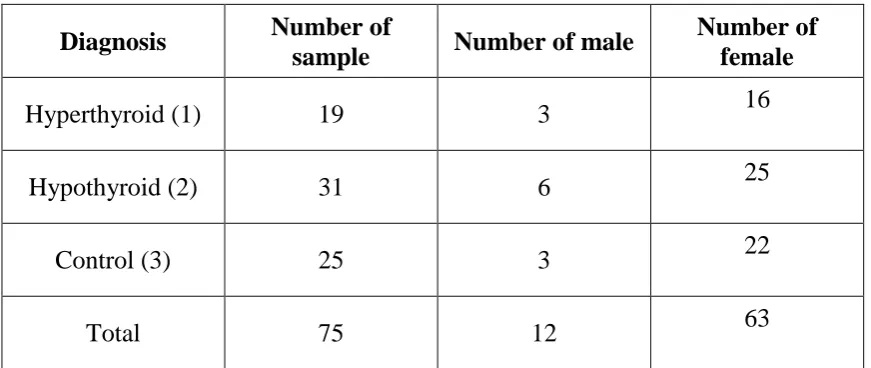
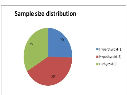
This study group comprises 30 hypothyroid, 20 hyperthyroid patients and 25 age and sex matched controls. Sample distribution in the study group is shown in table 1 and figure 1
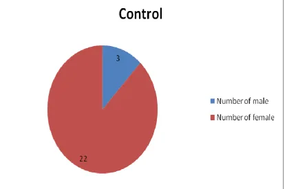
Their sex distribution is shown in figure 2,3,4
[image:59.595.99.535.439.623.2]Sample size distribution in the study group{table 1}
Table 1
Diagnosis Number of
sample Number of male
Number of female
Hyperthyroid (1) 19 3 16
Hypothyroid (2) 31 6 25
Control (3) 25 3 22
61
63
64
The data was collected and entered into excel sheet. The descriptive and inferential statistics was done using SPSS software. The data was segregated based on the diagnosis into 3 categories: hypothyroidism, hyperthyroidism and control. The summary of description for each diagnostic category was given below.
Descriptive statistics of the data {Table 2}
Diagnosis N Mean age
Mean of T3
Mean of T4
Mean of TSH
Mean of CPK
Hyperthyroid 20 35.53 159.16 38.21 0.07 78.26
Hypothyroid 30 32.84 43.68 1.99 48.48 195.81
Control 25 33.32 98.08 7.59 2.39 90.44
REFERENCE VALUES
T3-77-135ng/dl
T4- 5.5- 12.5mcg/dl
TSH- 0.5-5.5mIU/L
65
On inferential analysis, the data (T3, T4, TSH, CPK) under each diagnostic category (hyperthyroid, hypothyroid and control) was checked for normality, and depending on whether the data is normally distributed or not, selection of analysis was done. When the bivariate data was normally distributed, Pearson correlation was done and when the bivariate data was not distributed normally, spearman rank correlation was done between T3, T4, TSH and CPK and tabulated individually.
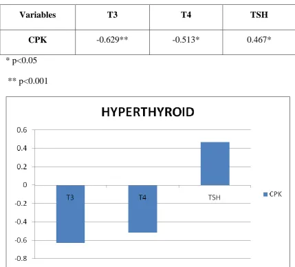
HYPERTHYROIDISM ANALYSIS
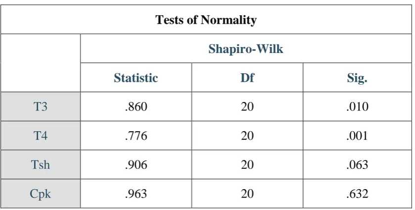
The data was analyzed through SPSS and Shapiro-Wilk test of normality was done for T3, T4, TSH and CPK. From Shapiro-Wilk test, TSH and CPK were normally distributed as significance value is more than0.05 but T3 and T4 were not as the significance value is less than 0.05.
Test of normality for T3, T4, TSH and CPK for sample with hyperthyroidism.
[image:65.595.107.527.516.728.2]Table - 3
Tests of Normality
Shapiro-Wilk
Statistic Df Sig.
T3 .860 20 .010
T4 .776 20 .001
Tsh .906 20 .063
66
Pearson correlation was done between TSH and CPK, whereas Spearman correlation was done between T3, T4 and CPK.
Table - 4
Correlation co-efficient table for T3, T4, TSH and CPK for sample with
hyperthyroidism.
Variables T3 T4 TSH
CPK -0.629** -0.513* 0.467* * p<0.05
[image:66.595.100.530.233.621.2]** p<0.001
Figure 5
From the table… showing the correlation coefficient of different
67
respectively. Whereas, there is a strong positive correlation between TSH and CPK values (r=.467) at p < 0.05 level of significance in this given sample.
HYPOTHYROIDISM
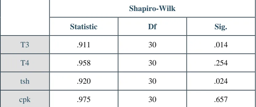
[image:67.595.107.525.388.563.2]The data was analyzed through SPSS and Shapiro-Wilk test of normality was done for T3, T4, TSH and CPK. From Shapiro-Wilk test, T3 and TSH were not normally distributed whereas, T4 and CPK were normally distributed.
Table - 5
Test of normality for T3, T4, TSH and CPK for sample with
hypothyroidism.
Shapiro-Wilk
Statistic Df Sig.
T3 .911 30 .014
T4 .958 30 .254
tsh .920 30 .024
cpk .975 30 .657
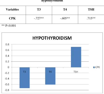
68 Table - 6
Correlation co-efficient table for T3, T4, TSH and CPK for sample with
hypothyroidism
Variables T3 T4 TSH
CPK -.727** -.605** .715** ** P<0.001
Figure 6
69 CONTROL….
[image:69.595.109.526.275.458.2]The data was analyzed through SPSS and Shapiro-Wilk test of normality was done for T3, T4, TSH and CPK. From Shapiro-Wilk test, T3, T4, TSH and CPK were normally distributed as significance value is more than0.05
Table - 7
Test of normality for T3, T4, TSH and CPK for sample with control
Shapiro-Wilk
Statistic Df Sig.
T3 .936 25 .121
T4 .946 25 .199
tsh .946 25 .205
cpk .974 25 .752
Pearson correlation was done to compare between T3, T4, TSH and CPK as all the data were normally distributed
Table - 8
Correlation co-efficient table for T3, T4, TSH and CPK for sample with
control
Variables T3 T4 TSH
[image:69.595.99.532.659.730.2]70
From the above table… showing the correlation coefficient of different variables indicates that there is no correlation between T3, T4, TSH and CPK (r= 0.002; r= 0.197; r= 0.144 respectively)
Distribution of CPK normality, increase, decrease in hypothyroidism and hyperthyroidism were studied and tabulated so as to analyse their usefulness as a diagnostic tool.
CPK in hypothyroidism
[image:70.595.112.520.421.683.2]30 cases of newly detected hypothyroid patients were included in the study and Evaluated. About 73.33% of hypothyroid cases had elevated CPK levels. 26.67% of hypothyroid cases had normal CPK values.{figure 7}
71 CPK in hyperthyroidism
[image:71.595.112.522.231.515.2]20 newly diagnosed hyperthyroid patients were taken into our study and evaluated. 85% of cases had normal CPK values and 15% of cases had low CPK values.
72 CPK in controls
Figure 9
73
DISCUSSION
Based on the results of the study mean age of study population is 34 yrs. Controls were age and sex matched.pearson correlation was done between TSH and CPK and showed a strong positive correlation (i-e as TSH increase in hypothyroidism CPK proportionally increases)
Spearman correlation was done between T3 T4 and CPK, correlation coefficient was calculated which established a negative correlation between T3 T4 and CPK. The statistical significance of the results were analyzed with chi-square method.
In this study neuromuscular disorders were excluded, but still CPK values were high, this may be due to subclinical myopathy or the role of T3 in gene expression of CPK which explains its elevation even in the absence of muscle disease.
Similar study was done by KMDS panag, geetanjali, sudeep goyal from department of biochemistry. The study showed similar results but ths study was performed in a larger population and they correlated CPK with FT3.
Hekimsoy et al in a study conducted in 2005, found that skeletal muscle is affected by
74
measurements may be useful as alternative diagnostic tool for the diagnosis of thyroid function disorders, which may be not only because of prevalence of muscular dystrophies in thyroid disorders but also due to role of FT3 in gene expression.
75
levels of CK which resolved after treatment for hypothyroidismand in a patient of Grave’s disease, patient developed myalgia with high level of CK after total
76 LIMITATIONS OF THE STUDY
• The sample size is less
• The ratio of control: case is less. If the control population was more
the power of the study would have been improved • Isoenzymes of CPK could not be done
77
CONCLUSION
Its concluded from the above study that CPK may be used as a supportive marker to diagnose hypothyroidism, especially in situations where measured thyroid hormone levels are likely to vary like pregnancy, oral contraceptives, drugs like heparin, protein wasting diseases etc.
78 FUTURE RECOMMENDATIONS
Molecular studies are required to prove the role of T3 in gene
expression
Large study population may show that CPK decreases in
hyperthyroidism at par with other studies
Quantitative range of CPK elevation in hypothyroidism may be studied
79
PROFORMA
Name: Age / Sex: Occupation:
Presenting complaints:
H/o fatigability, H/o cold intolerance, H/o constipation , H/o palpitations, weight loss, tremors
H/o swelling of legs/abdominal distension, H/o burning micturition, H/o jaundice
Past History:
H/o DM, HT, CKD, drug intake, Thyroid disorders, Alcohol intake Clinical Examination:
General Examination:
Consciousness, Pallor, jaundice, Clubbing, Lymphadenopathy, hydration status Vitals:
PR BP RR SpO2
Systemic examination: CVS:
RS:
80
CNS:
Laboratory investigations: • T3, T4, TSH
• Creatine phosphokinase • ECG
81
ANNEXURE
MASTER CHART
CASE AGE SEX T3 T4 TSH CPK DIAGNOSIS
82
lakshmi .p 41 F 22 1.5 78 254 hypo Sangeetha 36 F 51 4.2 48 178 hypo Lathamary 19 F 44 1.4 70 204 hypo Lathiffa 23 F 56 2.8 30 140 hypo mariyabeevi 37 F 67 2.65 15 67 hypo Nancy 27 F 39 3.2 56 186 hypo Rani 33 F 60 1.25 28 240 hypo Abinaya 26 F 15 0.06 96 284 hypo Ganesan 29 M 9 0.01 100 310 hypo Chandra 39 F 41 1.5 58 168 hypo Jesika 25 F 52 1.9 45 175 hypo Jeslin 40 M 54 3.4 18 98 hypo Nivetha 20 F 65 2.5 22 80 hypo Saroja 41 F 32 0.96 65 254 hypo Christi 37 F 33 1.45 42 178 hypo Mupudathi 30 M 35 1.56 74 240 hypo Hariharan 38 M 65 3.4 14 52 hypo Salma 22 F 45 2.4 42 196 hypo Catherine 38 F 10 0.12 100 408 hypo Preethi 18 F 32 0.56 50 225 hypo meenakshi .m 29 F 64 1.5 14 194 hypo marudhayee 40 F 12 1.8 86 250 hypo Karthika 35 F 56 2.1 32 190 hypo Nirmala 52 F 65 3.3 10 128 hypo Tessy 25 F 45 4.2 28 156 hypo CONTROLS
83
84
ABBREVIATIONS
TSH - Thyroid stimulating hormone T3 - Triiiodothyronine
T4 - Tetraiodothyronine TH - Thyroid hormone
r T3 - Reverse triiodothyronine CPK - Creatine phosphokinase FT3 - Free T3
FT4 - Free T4
CK - Creatine kinase TFT - Thyroid function test TBG - Thyroid binding globulin NEFA - Non esterified fatty acid L-T4 - Levothyroxine
HPT - Hypothalamo pituitary thyroid DIO - Deiodinase
NTI - Non thyroidal illness EMG - Electromyography
85
BIBLIOGRAPHY
1. RashmiRanka and RatiMathur.“Serum creatine phosphokinase in Thy.Disorders”.Indian Journal of clinical Biochemistry; 2003, 18(1)
107-110.
2. Lochmuller H; Reimers CD, Fischer P et al. Exercise inducedmyalgia in hypothyroidism. Clin. Invest; 1993;71:999-1001.
3. Kung AW, Ma JT, Wang CC. “Myopathy in acute hypothyroidism”. Postgraduate Medical journal. 1987; 63: 661-63
4. Fessel W J. Hoffman's syndrome psedohyportrophic myopathy as initial manifestation of hypothyroidism. Case report.“Myopathy of hypothyroidism”. Ann. Rheum. Disease.1968; 27: 590-96.
5. Saha M, Sarkar P, Chattopadhyay R, Mukhopadhyaya M, Bhowmick K. Role of creatine kinase and its coenzymesas surrogate markers of thyroid function. IJMB 2009;13(2):10-4.
6. Spencer CA. Strategy for use of serum thyrotrophin vs. free thyroxine measurement in thyroid testing. AACC Endo 1991;10:917.
7. Whitley RJ. Thyroid functions. In:Teitz Fundamentals of Clinical Chemistry. 5th edition, Burtis CA, Ashwood ER (Eds.) WB Saunders 2001:p.842.
86
9. Rosalki SB. Enzyme assays in diseases of the heart and skeletal muscle. J ClinPatholSuppl (AssocClinPathol) 1970;4:60-70.
10.Andersen S., Pedersen K.M., Bruun N.H. Narrow individual variations in serum T(4) and T(3) in normal subjects: a clue to the understanding of subclinical thyroid disease. Journal of Clinical Endocrinology and Metabolism. 2002;87:1068–1072.
11.Visser W.E., Friesema E.C., Visser T.J. Minireview: thyroid hormone transporters: the knowns and the unknowns. Molecular Endocrinology. 2011;25:1–14.
12.Trajkovic-Arsic M., Müller J., Darras V.M. Impact of monocarboxylate transporter-8 deficiency on the hypothalamus-pituitary-thyroid axis in mice. Endocrinology. 2010;151:5053–5062.
13.Bianco A.C., Kim B.W. Deiodinases: implications of the local control of thyroid hormone action. Journal of Clinical Investigation. 2006;116:2571–
14.2579.. Davis P.J., Davis F.B. Nongenomic actions of thyroid hormone on the heart. Thyroid. 2002;12:459–466.
15.Gurnell M., Visser T., Beck-Peccoz P. Resistance to thyroid hormone. In: Jameson J.L., De Groot L.J., editors. Endocrinology. 6th ed. Saunders Elsevier; Philadelphia, PA: 2010. pp. 1745–1759.
87
17.Vadiveloo T., Donnan P.T., Murphy M.J. Age- and gender-specific TSH reference intervals in people with no obvious thyroid disease in Tayside, Scotland: the thyroid epidemiology, audit, and research study (TEARS) Journal of Clinical Endocrinology and Metabolism. 2013;98:1147–1153. 18.Biondi B. The normal TSH reference range: what has changed in the last
decade? Journal of Clinical Endocrinology and Metabolism. 2013;98:3584–3587.
19.Lazarus J.H., Soldin O.P., Evans C. Assessing thyroid function in pregnancy. In: Brent G.A., editor. Thyroid function testing. Springer; New York: 2010. pp. 209–233.
20.Farwell A.P. Thyroid hormone therapy is not indicated in the majority of patients with the sick euthyroid syndrome. Endocrine Practice. 2008;14:1180–1187.
21.Kaptein E.M. In: Thyroid hormone metabolism. Hennemann G., editor. Marcel Dekker; New York: 1986. pp. 297–334.
22.Docter R., Krenning E.P., de Jong M. The sick euthyroid syndrome: changes in thyroid hormone serum parameters and hormone metabolism. Clinical Endocrinology. 1993;39:499–518.
23.Warner M.H., Beckett G.J. Mechanisms behind the non-thyroidal illness syndrome: an update. Journal of Endocrinology. 2010;205:1–13.
88
(SimulTRAC) for free thyroxine compared. Annals of Clinical Biochemistry. 1991;28:335–344.
25.Beckett G.J. Thyroid function and thyroid function tests in non-thyroidal illness. CPD Bulletin: Clinical Biochemistry. 2006;7:107–116.
26.Wartofsky L., Burman K.D. Alterations in thyroid function in patients with systemic illness: the “euthyroid sick syndrome” Endocrine Reviews. 1982;3:164–217.
27.De Groot L.J. Dangerous dogmas in medicine: the nonthyroidal illness syndrome. Journal of Clinical Endocrinology and Metabolism. 1999;84:151–164.
28.Arem R., Cusi K. Thyroid function testing in psychiatric illness: usefulness and limitations. Trends in Endocrinology and Metabolism. 1997;8:282–287.
29.Fliers E., Guldenaar S.E., Wiersinga W.M. Decreased hypothalamic thyrotropin-releasing hormone gene expression in patients with nonthyroidal illness. Journal of Clinical Endocrinology and Metabolism. 1997;82:4032–4036.
30.Jirasakuldech B., Schussler G.C., Yap M.G. A characteristic serpin cleavage product of thyroxine-binding globulin appears in sepsis sera. Journal of Clinical Endocrinology and Metabolism. 2000;85:3996–3999. 31.Finsterer, J. Stellberger, C., Grossege, C., Koroiss, A. (1999):
89
32.Wan Nazaimoon, W.M., Siano, F.S., Sheriff, I.H., Faridah, I., Khalid, IA (2001): Serum creatine kinase an adjunct unit biochemical index of subclinical thyrotoxicosis. Ann. Clin. Biochem. 38(Pt-l), 57–8.
33.Graig, F.A. and Ross, G. (1963): Serum creatine phosphokinase in thyroid disease. Metabolism 12(1), 57–59.
34.Graig, F.A. and Smith, J.C. (1965): Serum creatine phosphokinase activity in altered thyroid states. J. Clin. Endocri. 25, 723.
35.Go