0022-538X/10/$12.00 doi:10.1128/JVI.01571-09
Copyright © 2010, American Society for Microbiology. All Rights Reserved.
Localization of Mammalian Orthoreovirus Proteins to Cytoplasmic
Factory-Like Structures via Nonoverlapping Regions of
NS
䌤
Cathy L. Miller,
1,2* Michelle M. Arnold,
1,3‡ Teresa J. Broering,
1†
Craig E. Hastings,
2and Max L. Nibert
1,3*
Department of Microbiology and Molecular Genetics, Harvard Medical School, 200 Longwood Avenue,1and Program in Virology,
Division of Medical Sciences, Harvard Medical School, 240 Longwood Avenue,3Boston, Massachusetts 02115, and
Department of Veterinary Microbiology and Preventive Medicine, College of Veterinary Medicine, Iowa State University, Ames, Iowa 500112
Received 29 July 2009/Accepted 29 October 2009
Virally induced structures called viral factories form throughout the cytoplasm of cells infected with mammalian orthoreoviruses (MRV). When expressed alone in cells, MRV nonstructural proteinNS forms factory-like structures very similar in appearance to viral factories, suggesting that it is involved in forming the structural matrix of these structures.NS also associates with MRV core particles; the core proteins2,1,
2,3, and2; and the RNA-binding nonstructural proteinNS. These multiple associations result in the recruitment or retention of these viral proteins or particles at factory-like structures. In this study, we identified the regions ofNS necessary and sufficient for these associations and additionally examined the localization of viral RNA synthesis in infected cells. We found that short regions within the amino-terminal 220 residues ofNS are necessary for associations with core particles and necessary and sufficient for associations with the proteins2,1,2,2, andNS. We also found that only the3 protein associates with the carboxyl-terminal one-third ofNS and that viral RNA is synthesized within viral factories. These results suggest thatNS may act as a cytoplasmic scaffolding protein involved in localizing and coordinating viral replication or assembly interme-diates for the efficient production of progeny core particles during MRV infection.
Mammalian orthoreoviruses (MRV) are members of the family Reoviridae, which includes important human, animal, and plant pathogens (e.g., rotavirus, bluetongue virus, and rice dwarf virus). All members of the family Reoviridae share a number of characteristics including a similar genome com-prised of 9 to 12 segments of double-stranded RNA (dsRNA). These segments are enclosed within a multilayered protein capsid, including one or more inner layers that contact the genome and play roles in viral RNA synthesis and one or more outer layers that mediate virus attachment and entry into the host cell (17, 37, 41). During the entry process, the outer capsid(s) is largely removed from these viruses, and the inner capsid(s) is released into the cytoplasm. Upon release, the genome-enclosing inner capsid(s) serves as the viral transcrip-tase particle, synthesizing and capping viral plus-strand RNAs, which are then released into the cytoplasm for translation into viral proteins (17, 37, 41).
At early times after entry, distinctive structures, which grow
in size over time, appear throughout the cytoplasm of infected cells. These cytoplasmic structures are variously termed viral factories (VF) (MRV), viroplasms (rotavirus and phytoreovi-rus), or viral inclusion bodies (VIB) (orbivirus). In each case, they contain many viral proteins, particles, and dsRNAs and are thought to be the locations of viral RNA replication and packaging into progeny particles (13, 15, 31, 42, 43, 46).
In previous studies, either one or two nonstructural proteins of each virus were shown to be required for forming the cyto-plasmic structures. In MRV and avian orthoreoviruses, the nonstructural proteinNS expressed alone in cells forms fac-tory-like structures (FLS) that appear to be similar by light microscopy to VF formed in infected cells (4, 8, 49). Likewise, orbiviruses such as bluetongue virus and epizootic hemor-rhagic disease virus encode a single nonstructural protein, NS2, that forms VIB-like structures when expressed alone in cells (25, 47, 48). In phytoreoviruses such as rice dwarf virus, the nonstructural protein Pns12 expressed alone in cells forms viroplasm-like structures (VLS) (51). Rotaviruses, on the other hand, encode two nonstructural proteins, NSP2 and NSP5, which must be coexpressed under most circumstances to form VLS (16, 18, 34).
In MRV, theNS sequences required for forming FLS have been carefully examined. The carboxyl-terminal (C-terminal) 250 amino acids (aa) ofNS are sufficient to form FLS, with four distinct regions within this portion of the protein shown to be necessary (5). These regions include two previously pre-dicted coiled-coil domains (30), a linker region containing a putative zinc hook between the coiled coils, and a short C-terminal tail region.
Importantly, the capacity of MRV to form VF is necessary
* Corresponding author. Mailing address for Cathy L. Miller: Dept. of Veterinary Microbiology and Preventive Medicine, College of Vet-erinary Medicine, Iowa State University, Ames, IA 50011. Phone: (515) 294-4797. Fax: (515) 294-1401. E-mail: clm@iastate.edu. Mailing address for Max L. Nibert: Dept. of Microbiology and Molecular Genetics, Harvard Medical School, 200 Longwood Ave., Boston, MA 02115. Phone: (617) 645-3680. Fax: (617) 738-7664. E-mail: mnibert @hms.harvard.edu.
† Present address: Massachusetts Biologic Laboratories, Jamaica Plain, MA 02130.
‡ Present address: Laboratory of Infectious Diseases, National Institute of Allergy and Infectious Diseases, National Institutes of Health, Be-thesda, MD 20892.
䌤Published ahead of print on 4 November 2009.
867
on November 8, 2019 by guest
http://jvi.asm.org/
for viral replication. WhenNS expression is knocked down by RNA interference, viral growth is severely inhibited (1, 27). Moreover, when wild-typeNS is plasmid expressed intrans, viral growth is rescued (1, 27). Plasmid-expressed NS with mutations in the putative zinc hook or the C-terminal tail, in contrast, does not rescue viral growth (1, 28). These results strongly suggest that the formation of VF is an important and necessary function for successful MRV multiplication.
Previous studies have shown that NS associates with six other MRV proteins: five structural proteins that make up the core particle (1,2,3,2, and2) and the single-stranded RNA (ssRNA) binding nonstructural proteinNS (4, 6, 8, 32, 33). Limited mapping of NS associations with other viral proteins has shown thatNS aa 14 to 41 are necessary and that aa 1 to 41 are sufficient for the association with the minor core protein2 (8). In addition,NS aa 1 to 13 are necessary for the association with the nonstructural proteinNS (33), and aa 1 to 41 are dispensable for the association with the core surface proteins1,2, and2 (6).NS additionally associates with MRV core particles in vitro and in cells (6, 7).
In light of these data, we have hypothesized that in addition to its role in forming the structural matrix of VF, a second role forNS in MRV infection is to act as a type of cytoplasmic scaffolding protein for the coordinated recruitment and assem-bly of MRV replication complexes within VF. Based on our previously reported data, we have developed a model of MRV VF assembly. In this model, following entry and release into the cytoplasm, the MRV core particle begins transcribing the viral plus-strand RNAs. MRV proteins, including NS, are synthesized by the cellular translational machinery, either co-transcriptionally or adjacent to the site of core RNA transcrip-tion. Following translation,NS may associate with the adja-cent core particle to seed a new VF, orNS may self-associate first to form a small VF, which then further associates with the adjacent MRV core. Proteins required for the assembly of progeny core particles (1,2,3,2, and2), as well asNS, also associate withNS either by direct association withNS in the VF matrix or in the cytoplasm with subsequent recruit-ment to VF byNS. The VF-localized core particle continues to transcribe the MRV plus-strand RNAs, some of which are now bound by adjacent viral proteins to form replication and assembly complexes for the production of progeny core parti-cles and virions within the VF. The VF is additionally tethered to the cellular cytoskeleton viaNS associations with a micro-tubule-associated viral protein,2. This association allows VF to move toward the perinuclear region of the cell, merging with other VF during the journey, ultimately forming the large perinuclear VF that are characteristic of MRV-infected cells. We previously developed an assay that exploits the charac-teristic ability ofNS, as well as the rotavirus protein NSP5 fused at its amino terminus (N terminus) to enhanced green fluorescent protein (EGFP) (34), to form distinctive structures in the cytoplasm in order to identify and map protein-protein interactions (32). In the current study, we modified and made extensive use of this novel assay to explore further the associ-ations betweenNS and other MRV proteins. In new exper-iments, we first defined the region ofNS that is necessary for associations with individual MRV proteins, as well as the MRV core particle, by determining the ability of these proteins to be recruited to FLS formed by a series ofNS deletion mutants.
We then constructed a number of plasmids expressing NS fragments fused to EGFP/NSP5 as a method to map regions of
NS sufficient for associations with each MRV protein. In sum, we found that small, largely nonoverlapping regions of
NS are necessary and sufficient for the association with indi-vidual MRV proteins and necessary for the association with core particles. Because the MRV core particle transcribes viral plus-strand RNAs (22), we additionally examined the localiza-tion of viral RNA synthesis and found that it too occurs in VF. These results advance our understanding of how a viral non-structural protein, MRVNS, has evolved to build discrete regions of cytoplasmic scaffolding within which critical viral activities occur.
MATERIALS AND METHODS
Cells, viruses, antibodies, and other reagents.CV-1 cells were maintained in Dulbecco’s modified Eagle’s medium (DMEM) (Invitrogen) containing 10% fetal bovine serum (HyClone) and 10g/ml gentamicin (Invitrogen). MRV strain type 1 Lang was our laboratory stock, originally obtained from B. N. Fields. MRV strain type 3 Dearing was obtained from the laboratory of L. W. Cash-dollar (Medical College of Wisconsin). Virus was propagated on L929 cells and plaque purified as previously described (19). Top-component infectious sub-virion particles (ISVPs) were made by chymotrypsin digestion (200 g/ml; Sigma) of purified top-component virions (1⫻1013particles/ml) for 10 min at
32°C. The reaction was quenched by the addition of 2 mM phenylmethylsulfonyl fluoride (Sigma). Rabbit polyclonal antiserum and purified polyclonal antibodies specific forNS,2, and MRV core particles were described previously (7, 8, 11, 26). Monoclonal antibodies againstNS (3E10) and2 (7F4) were also de-scribed previously (3, 50). Monoclonal antibody HA.11, specific for the influenza virus hemagglutinin (HA) epitope, was obtained from Covance Research Prod-ucts. In the indicated experiments, protein A-purified rabbit anti-NS immuno-globulin G (IgG) was conjugated to Texas Red by using a kit obtained from Molecular Probes. Monoclonal antibody against bromodeoxyuridine was pur-chased from Sigma. The following secondary antibodies were used as appropri-ate for different experiments: Alexa 488- or Alexa 594-conjugappropri-ated goat anti-mouse or anti-rabbit IgG (Molecular Probes) and horseradish peroxidase (HRP)-conjugated donkey anti-mouse or anti-rabbit IgG (Pierce). For micros-copy, antibodies were titrated to optimize signal-to-noise ratios. Bromouridine (BrU) was purchased from Sigma. Actinomycin D was purchased from Sigma. All restriction enzymes were obtained from New England Biolabs.
Plasmid constructions.pCI-NS, pCI-NS, pCI-2, pCI-1, pCI-2, and
pCI-2, expressing NS,NS, 2,1,2, and 2, respectively, were described previously but originally named by their genes, M3, S3, M1, pCI-L3, pCI-L2, and pCI-S2, respectively (6, 8, 33, 38). pCI-NS(14-721),
pCI-NS(41-721), pCI-NS(173-721), pCI-NS(221-721), and pCI-NS(471-721) were described previously but were originally named M3(14-721), pCI-M3(41-721), pCI-M3(173-721), pCI-M3(221-721), and pCI-M3(471-721), respec-tively (5, 8, 33). pCI-3/HA expressing a C-terminally HA-tagged version of3 was described previously (32). pEGFP/NS(471-721) was described previously but was originally named pEGFP-C1-M3(471-721) (5). pEGFP/NSP5 was obtained from Oscar Burrone and was described previously (16). To makeNS deletion plasmids pCI-NS(20-721), pCI-NS(26-721), pCI-NS(55-721), pCI-NS(65-721),
pCI-NS(75-721), pCI-NS(85-721), and pCI-NS(95-721), PCR was performed by using pCI-NS as a template. The PCR product was purified, digested with NheI and BlpI or EcoRI and NotI, and ligated into appropriately digested pCI-NS. To make the NS fragment fusion plasmids p-NS(41-221)/EGFP/NSP5, p-NS (55-221)/EGFP/NSP5, p-NS(173-221)/EGFP/NSP5, p-NS(1-20)/EGFP/NSP5, p-NS(41-173)/EGFP/NSP5, p-NS(55-173)/EGFP/NSP5, and p-NS(41-110)/ EGFP/NSP5, PCR was performed by using pCI-NS as a template. The PCR product was purified, digested with AgeI, and ligated into AgeI-digested pEGFP/ NSP5. Orientation was determined by restriction digestion. p-NS(1-227)/EGFP/ NSP5 was made in two steps. First, pGEM-M3 was digested with BsaHI to liberate a fragment containing the coding region forNS aa 1 to 227. This fragment was gel purified, the overhanging ends were filled in with Klenow fragment, and a HindIII digestion was performed on the resulting fragment, which was then ligated into HindIII/SmaI-digested pEGFP-N1 to make p-NS(1-227)/EGFP. Second, p-NS(1-227)/EGFP was digested with NdeI and BsrGI and ligated into NdeI/BsrGI-digested EGFP/NSP5 to make p-NS(1-227)/EGFP/NSP5. To make p-NS(95-221)/EGFP/NSP5, p-
on November 8, 2019 by guest
http://jvi.asm.org/
41)/EGFP/NSP5, p-NS(20-41)/EGFP/NSP5, and p-NS(95-173)/EGFP/NSP5, PCR was performed by using pCI-NS as a template. The PCR product was purified, digested with NheI and AgeI, and ligated into NheI/AgeI-digested p-NS(1-227)/EGFP/NSP5. p-NS(1-12)/EGFP/NSP5 was made by digesting p-NS(1-12)/EGFP/NS(471-721) and EGFP/NSP5 with NdeI and BsrGI fol-lowed by ligating the appropriate purified products. p-NS(1-41)/EGFP/NSP5 was made by digesting p-NS(1-41)/EGFP/NS(471-721) and EGFP/NSP5 with NdeI and BsrGI, followed by ligating the appropriate purified products. p-NS/ EGFP was made by PCR using pCI-NS as a template. The PCR product was purified, digested with EcoRI and SacII, and ligated into EcoRI/SacII-digested pEGFP-N1. p-NS/EGFP/NSP5 was made by digesting p-NS/EGFP and pEGFP/NSP5 with NdeI and BsrGI, followed by ligating the appropriate purified products. p-3/EGFP/NSP5 was made by PCR using pCI-3/HA as a template. The PCR product was purified, digested with AgeI, and ligated into AgeI-digested pEGFP/NSP5. Orientation was confirmed by restriction digestion. All plasmid sequences were confirmed by sequencing. Primer sequences used in PCRs are available upon request.
Transfections and infections.For transfections, a total of 4g plasmid DNA was mixed with 10l Lipofectamine (Invitrogen) in Optimem (Invitrogen). After a 20-min incubation, the mixture was added to cells containing DMEM with 10% fetal calf serum (FCS) but lacking antibiotics and incubated for 18 h at 37°C as suggested by the manufacturer. After incubation, cells were processed for im-munostaining and microscopy. For other transfections, 8g plasmid DNA was mixed with 20l Lipofectamine in Optimem. All other steps of the above-described protocol were then followed except that cells were processed for immunoprecipitation and immunoblotting.
For immunostaining studies, 2⫻105
CV-1 cells were seeded the day before transfection in six-well plates (9.6 cm2per well) containing 18-mm round glass
coverslips. For immunoprecipitation studies, 4⫻105cells were seeded onto
60-mm dishes the day before transfection. Infections were performed by remov-ing media from cells and overlayremov-ing cells with virus diluted in phosphate-buffered saline (PBS) (137 mM NaCl, 3 mM KCl, 8 mM Na2HPO4[pH 7.5])
supple-mented with 2 mM MgCl2for 1 h at room temperature to allow virus to adsorb
to cells. Cells were then refed with medium and allowed to incubate further at 37°C. Top-component ISVP infection of cells transfected withNS expression plasmids was described previously (6).
Immunostaining and microscopy.Infected or transfected cells were fixed for 20 min with 2% paraformaldehyde in PBS, except for BrU experiments, in which cells were fixed by incubation for 3 min in 100% methanol. Fixed cells were washed three times with PBS and permeabilized for 5 min with 0.2% Triton X-100 in PBS. Cells were again washed three times with PBS and blocked for 5 min with 2% bovine serum albumin in PBS. Primary and secondary antibodies were diluted in 2% bovine serum albumin in PBS. After blocking, cells were incubated for 1 h with primary antibodies, washed three times with PBS, and incubated for 1 h with secondary antibodies. Immunostained cells were washed a final three times with PBS and mounted onto slides with Prolong Plus reagent (Molecular Probes). Immunostained samples were examined by using an Optiphot-2 epifluorescence upright microscope (Nikon) or an Axiovert 200 inverted fluorescence microscope (Zeiss). Images were collected digitally by using either a Photometrics CoolSnapcfcamera (Roper Scientific) and MetaVue imaging
software (Molecular Devices) or an AxioCam MR color camera and Axiovision AC imaging software (Zeiss). Images were prepared for presentation by using Photoshop and Illustrator (Adobe Systems).
Core immunoprecipitation assay.Transfected cells were lysed by incubation for 30 min on ice in nondenaturing lysis (Raf) buffer (20 mM Tris [pH 8.0], 137 mM NaCl, 10% [vol/vol] glycerol, 1% [vol/vol] NP-40) containing protease in-hibitors (Roche Biochemicals). 7F4 (2-specific) monoclonal antibodies that had been incubated for 2 h with protein A-conjugated magnetic beads (Dynal Bio-tech) and washed six times with Raf buffer were separated into two aliquots. MRV core particles, prepared as previously described (11), were added to one of the two 7F4-coated bead aliquots at a concentration of 1⫻1012cores/ml and
incubated with rotation for an additional 2 h at 4°C. 7F4/core-coated beads were washed an additional six times with Raf buffer. 7F4-coated or 7F4/core-coated beads were added in equal volumes to theNS protein-containing cell lysates, which were then rotated overnight at 4°C. Immunoprecipitated proteins were separated from the cell lysate and washed four times with Raf buffer. Sample buffer (125 mM Tris [pH 6.8], 10% [wt/vol] sucrose, 1% [wt/vol] sodium dodecyl sulfate, 0.02% [vol/vol] -mercaptoethanol, 0.01% bromophenol blue) was added to both the immunoprecipitated proteins and the postbinding supernatant. Samples were then boiled for 3 min and separated on denaturing 10% (wt/vol) polyacrylamide gels containing 0.01% (wt/vol) sodium dodecyl sulfate. Proteins were transferred from the gels onto pieces of nitrocellulose by electroblotting in transfer buffer (25 mM Tris, 192 mM glycine, 20% [vol/vol] methanol [pH 8.3]).
Nitrocellulose was then blocked by incubation with 5% milk in Tris-buffered saline (20 mM Tris [pH 7.6], 137 mM NaCl) containing 0.5% Tween 20 (Sigma) (TBS-T) for 30 min at room temperature. Primary antibodies were allowed to bind during incubation overnight at room temperature in TBS-T containing 1% milk. HRP-conjugated secondary antibodies were allowed to bind during a 2-h incubation at room temperature in TBS-T containing 1% milk. Before and after the incubation with secondary antibodies, the nitrocellulose was washed three times with TBS-T. The binding of HRP conjugates was detected by incubation with chemiluminescence substrate (Bio-Rad) according to the manufacturer’s recommendations, followed by exposure to film (Fuji).
BrU assay.CV-1 cells were plated onto 8-well chamber slides at a density of 1⫻104cells per well and then infected with type 1 Lang or type 3 Dearing
ISVPs, prepared as previously described (19), at a multiplicity of infection of 100 PFU/cell. Infection was allowed to proceed for 2, 3, 4, or 6 h, at which time cells were treated for 30 min with 10g/ml actinomycin D to inhibit cellular RNA polymerase II (52). Cells were then transfected with 10 mM BrU in 3.7l Lipofectamine in 30l Optimem in the presence or absence of 10g/ml acti-nomycin D and incubated for an additional 60 min before fixation and immu-nostaining.
RESULTS
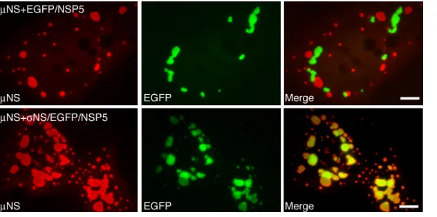
NS and EGFP-fused NSP5 form nonoverlapping cytoplas-mic structures.To establish the feasibility of our new ap-proach for defining regions ofNS sufficient for associating with other MRV proteins, we examined its capacity to detect such a known association. A plasmid expressingNS was co-transfected into cells with a plasmid expressing either EGFP/ NSP5 or EGFP/NSP5 fused to the NS-associating protein
NS. At 18 h posttransfection (p.t.), cells were fixed and stained with antibodies against NS. The inherent fluores-cence of EGFP was used to detect EGFP/NSP5 orNS/EGFP/ NSP5. Interestingly, when the FLS-formingNS was coexpressed with the VLS-forming EGFP/NSP5, the proteins formed distinc-tive structures in cells as previously described (8, 32, 34); how-ever, the respective structures did not colocalize, suggesting that the proteins do not associate (Fig. 1, top). In contrast, whenNS was coexpressed withNS/EGFP/NSP5, the respec-tive structures completely colocalized in cells (Fig. 1, bottom), reflecting the known association ofNS withNS and validat-ing the use of EGFP/NSP5-formed VLS as a way to identify regions ofNS that are sufficient for associations with other MRV proteins.
NS aa 20 to 25 are necessary and aa 14 to 41 are sufficient for associations with2.The MRV2 protein was previously shown to be a strain-specific microtubule-associated protein (38). When expressed alone in transfected cells, 2 derived from most MRV strains localizes to cellular microtubules and the nucleus (38). When coexpressed withNS (Fig. 2A, top) and in infected cells,2 andNS from these strains are asso-ciated with microtubules (8, 38). For occasional MRV strains in which2 does not associate with microtubules, it colocalizes withNS in FLS and VF, suggesting that the association be-tween NS and 2 is independent of the 2 microtubule association but necessary forNS microtubule localization (8). To identify the regions ofNS that are necessary and sufficient for the association with2 more precisely, we created a series of NS deletion mutants and N-terminal fusions to EGFP/ NSP5 and examined the ability of2 to associate with these proteins following plasmid cotransfections into cells. We de-fined the region ofNS necessary for the association with2 by creating deletions from the N terminus ofNS. We cotrans-fected cells with plasmids expressing2 and theNS deletion
on November 8, 2019 by guest
http://jvi.asm.org/
mutants and examined the localization of2 andNS within the cells at 18 h p.t. by immunofluorescence microscopy. In cells coexpressing2 andNS(20-721),NS colocalized with
2 on cellular microtubules, suggesting that this deletion did not affect their association (Fig. 2A, middle). In cells coex-pressing2 andNS(26-721), while2 localized to both the nucleus and cellular microtubules (Fig. 2, bottom right),
NS(26-721) was found in FLS that did not associate with microtubules (Fig. 2A, bottom left), suggesting thatNS aa 20 to 25 are necessary for the2 association.
To identify regions ofNS sufficient for the association with
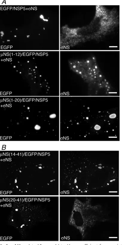
2, we created a series of plasmids expressing fusion proteins connecting fragments of the N-terminal 41 aa of NS to EGFP/NSP5. We then tested the ability of2 to associate with each of these fusion proteins in transfected cells. In each case, protein localization was examined at 18 h p.t. by immunofluo-rescence microscopy.2 did not associate with either NS(1-12)/EGFP/NSP5 (Fig. 2B, second row) orNS(1-20)/EGFP/ NSP5 (Fig. 2B, third row) but did associate withNS(1-41)/ EGFP/NSP5 (Fig. 2B, bottom row), mirroring our previous findings that these N-terminal amino acids of NS are not necessary for the2 association and that NS(1-41) is suffi-cient for the 2 association (8, 33). Additional N-terminal deletions were made from theNS(1-41)/EGFP/NSP5 fusion protein to determine the smallest region ofNS sufficient for the association with 2. When 2 was coexpressed with
NS(14-41)/EGFP/NSP5, the two proteins completely colocal-ized on cellular microtubules (Fig. 2C, top); however,2 did not efficiently associate with NS(20-41)/EGFP/NSP5 (Fig. 2C, bottom). Taken together, these data suggest thatNS aa 14 to 41 are sufficient for the association with2.
NS aa 1 to 12 and aa 14 to 41 are sufficient for association withNS.Our previously described deletion of 13 aa from the
NS N terminus (33) disrupted its association withNS, and because this was already a small region, we did not create additional deletions to dissect it further. These previous find-ings suggested thatNS might associate with the N-terminal 12 aa ofNS. Indeed, whenNS was coexpressed with NS(1-12)/EGFP/NSP5 in cells, we found that the two proteins colo-calized in VLS, although colocalization was incomplete (Fig.
3A, second row). When an additional 8 aa fromNS were added to the EGFP/NSP5 fusion protein,NS colocalized with VLS in every cell (Fig. 3A, third row), suggesting thatNS aa 13 to 19 contribute to the NS association. When the N-terminal 13 aa were deleted from theNS fusion protein,NS also completely colocalized with VLS in every cell (Fig. 3B, top).NS aa 14 to 19 again appear to be important for this putative binding region forNS, because when deleted in the fusion protein, there was no association between the two pro-teins (Fig. 3B, bottom). These findings suggest thatNS may bind independently to two different regions within the N-ter-minal 41 aa ofNS: aa 1 to 12 and aa 14 to 41.
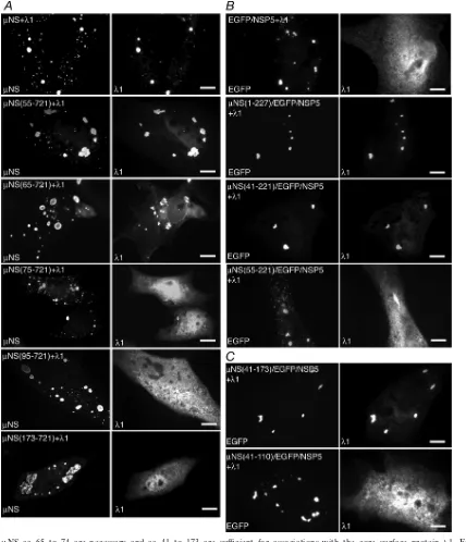
NS aa 65 to 74 are necessary and aa 41 to 173 are sufficient for associations with 1. A previous study showed that the deletion of the N-terminal 41 aa ofNS does not disrupt its associations with the viral core surface protein 1 (6). To identify the region ofNS that associates with1 more pre-cisely, we utilized a series of N-terminal deletion mutants of
NS. We cotransfected plasmids expressing each deletion mu-tant with plasmids expressing1 in cells and then visualized the localization of1 relative to FLS at 18 h p.t. by immunofluo-rescence microscopy. We found that the deletion of 55 aa from the N terminus ofNS did not disrupt the association with1 (Fig. 4A, second row). The deletion of an additional 10 aa from
NS did not completely abrogate the1 association; however,
1 formed aggregates in these cells, which appeared to localize around FLS, and a portion of1 was diffusely distributed in cells (Fig. 4A, third row). The deletion of 74 aa from theNS N terminus resulted in the loss of an association with1 (Fig. 4A, fourth row), suggesting thatNS aa 65 to 74 are necessary for the1 association. FurtherNS deletions of 94 and 172 aa also resulted in the loss of an association with1 (Fig. 4A, fifth and bottom rows).
[image:4.585.135.451.68.225.2]We were next interested in identifying the NS regions sufficient for the association with1. To identify these regions, we constructed plasmids to express a series of EGFP/NSP5 fusion proteins containing contiguous short regions of NS. Work with our deletion mutants suggested that the N-terminal 221 aa ofNS are necessary for the association with each of the tested proteins except the viral RNA-dependent RNA
FIG. 1. MRVNS and EGFP-fused rotavirus NSP5 form nonoverlapping structures that can be made to colocalize through a known protein-protein association. CV-1 cells cotransfected with a plasmid expressingNS and either EGFP/NSP5 (top) orNS/EGFP/NSP5 (bottom) were fixed at 18 h p.t. FLS were visualized by staining withNS-specific polyclonal antibodies followed by Alexa 594-conjugated goat anti-rabbit IgG (left). VLS were visualized by the inherent fluorescence of EGFP (middle). Merged images are also shown (right). Bar, 10m.
on November 8, 2019 by guest
http://jvi.asm.org/
polymerase (RdRp)3 (see below), and thus, we restricted our
NS/EGFP/NSP5 fusions to the N-terminal 227 aa of NS. We cotransfected a plasmid expressing each of the fusion pro-teins with a plasmid expressing1 and visualized the protein
[image:5.585.79.506.66.559.2]localizations at 18 h p.t. by immunofluorescence microscopy. We found that1 associated with the EGFP/NSP5 fusion con-taining eitherNS(1-227) (Fig. 4B, second row) or NS(41-221) (Fig. 4B, third row) but not with the fusion containing
FIG. 2.NS aa 20 to 25 are necessary and aa 14 to 41 are sufficient for associations with minor core protein2. For each experiment, cells were processed for fluorescence microscopy at 18 h p.t. (A) CV-1 cells were cotransfected with plasmids expressing2 and eitherNS (top),
NS(20-721) (middle), orNS(26-721) (bottom). After fixation, cells were stained with rabbit polyclonal antibodies against2 followed by Texas Red-conjugatedNS-specific rabbit IgG to visualizeNS (left) and Alexa 488-conjugated goat anti-rabbit IgG to visualize2 (right). (B) CV-1 cells were cotransfected with plasmids expressing2 and either EGFP/NSP5 (top row),NS(1-12)/EGFP/NSP5 (second row),NS(1-20)/EGFP/ NSP5 (third row), orNS(1-41)/EGFP/NSP5 (bottom row). After fixation, cells were stained with rabbit polyclonal antibodies against2 followed by Alexa 594-conjugated goat anti-rabbit IgG to visualize2 (right). The inherent fluorescence of EGFP was used to visualize each of the fusion proteins (left). (C) CV-1 cells were cotransfected with plasmids expressing2 andNS(14-41)/EGFP/NSP5 (top) orNS(20-41)/EGFP/NSP5 (bottom). After fixation, cells were stained with rabbit polyclonal antibodies against2 followed by Alexa 594-conjugated goat anti-rabbit IgG to visualize2 (right). The inherent fluorescence of EGFP was used to visualize each of the fusion proteins (left). Bar, 10m.
on November 8, 2019 by guest
http://jvi.asm.org/
NS(55-221) (Fig. 4B, bottom row). In contrast to what was found for theNS N-terminal deletion mutants, these findings suggest that in the absence of theNS C terminus, aa 42 to 54 are necessary for the1 association. We additionally examined the localization of1 when coexpressed with EGFP/NSP5 fu-sions from which an additional 49 aa and 63 aa were deleted from the C terminus of theNS fragment. In these experi-ments,1 associated with the fusion containing NS(41-173)
(Fig. 4C, top) but not with that containingNS(41-110) (Fig. 4C, bottom), suggesting that amino acids in the region of aa 111 to 173 ofNS are important for the1 association. These findings suggest thatNS aa 41 to 173 are sufficient for the1 association.
NS aa 75 to 84 are necessary and aa 41 to 173 are sufficient for associations with2.Similar to the case for1, we previ-ously found that the N-terminal 41 aa ofNS are not necessary for the association with2 (6). We next investigated the region ofNS that was necessary for the association with2 by indi-vidually cotransfecting a plasmid expressing2 with a panel of
NS N-terminal deletion mutants and examining the localiza-tion of2 relative to FLS by immunofluorescence microscopy. We found that2 associated with FLS formed byNS(55-721) (Fig. 5A, second row) andNS(65-721) (Fig. 5A, third row) was less completely associated to FLS formed byNS(75-721) (Fig. 5A, fourth row) and did not colocalize with FLS formed by NS(85-721) or NS(95-721) (Fig. 5A, fifth and bottom rows). These findings suggest thatNS aa 75 to 84 are neces-sary for the2 association withNS.
To identify the region ofNS sufficient for the association with 2, we examined the localization of this protein when coexpressed with our panel ofNS/EGFP/NSP5 fusion pro-teins. We found that2 associated with the EGFP/NSP5 fusion containing NS(1-227) (Fig. 5B, second row), NS(41-221) (Fig. 5B, third row), orNS(55-221) (Fig. 5B, fourth row) but not with that containingNS(95-221) (Fig. 5B, bottom row). We then tested its association with the fusions from which an additional 48 aa were deleted from the C terminus of theNS fragment and found that2 associated with that containing
NS(55-173) (Fig. 5C, top) but not with that containing
NS(95-173) (Fig. 5C, middle). The deletion of 63 aa more from the C terminus of theNS fragment abrogated colocal-ization with2 (Fig. 5C, bottom), suggesting that amino acids in the region of aa 110 to 173 ofNS are important for the2 association. These findings suggest thatNS aa 55 to 173 are sufficient for the2 association.
NS aa 173 to 220 are necessary and sufficient for associ-ations with2.As found for the core surface proteins1 and
2, our previous studies have shown that the N-terminal 41 aa ofNS are not required for its association with the other core surface protein,2 (6). To map the region ofNS necessary for the association with2 more precisely, we cotransfected a plasmid expressing 2 with plasmids expressing NS N-terminal deletion mutants and then examined the localiza-tion of 2 relative to FLS formed by each of the NS deletions at 18 h p.t. by immunofluorescence microscopy. We found that2 colocalized with FLS formed by NS(173-721) (Fig. 6A, middle) but was diffusely distributed through-out cells expressing NS(221-721), even though the latter deletion continued to form distinctive FLS (Fig. 6A, bot-tom). These data suggest thatNS aa 173 to 220 are nec-essary for the association with2.
[image:6.585.61.265.71.478.2]To identify the region ofNS sufficient for the association with 2, we determined the localization of 2 when coex-pressed with plasmids expressingNS/EGFP/NSP5 fusion pro-teins. Cells were transfected, and the localization of2 relative to VLS formed by the fusion proteins was examined at 18 h p.t. by immunofluorescence microscopy. Consistent with results described above for nonfusedNS deletions, we found that2
FIG. 3.NS aa 1 to 12 or aa 14 to 41 are sufficient for associations with the nonstructural proteinNS. For each experiment, cells were processed for fluorescence microscopy at 18 h p.t. (A) CV-1 cells were cotransfected with plasmids expressingNS and either EGFP/NSP5 (top), NS(1-12)/EGFP/NSP5 (middle), or NS(1-20)/EGFP/NSP5 (bottom). After fixation, cells were stained with mouse monoclonal antibody 3E10 againstNS followed by Alexa 594-conjugated goat anti-mouse IgG to visualizeNS (right). The inherent fluorescence of EGFP was used to visualize each of the fusion proteins (left). (B) CV-1 cells were cotransfected with plasmids expressing NS and either
NS(14-41)/EGFP/NSP5 (top) orNS(20-41)/EGFP/NSP5 (bottom). After fixation, cells were stained with mouse monoclonal antibody 3E10 againstNS followed by Alexa 594-conjugated goat anti-mouse IgG to visualizeNS (right). The inherent fluorescence of EGFP was used to visualize each of the fusion proteins (left). Bar, 10m.
on November 8, 2019 by guest
http://jvi.asm.org/
FIG. 4.NS aa 65 to 74 are necessary and aa 41 to 173 are sufficient for associations with the core surface protein 1. For each experiment, cells were processed for fluorescence microscopy at 18 h p.t. (A) CV-1 cells were cotransfected with plasmids expressing1 and either NS (top row), NS(55-721) (second row), NS(65-721) (third row), NS(75-721) (fourth row), NS(95-721) (fifth row), or
NS(173-721) (bottom row). After fixation, cells were stained with rabbit polyclonal antibodies against MRV cores followed by Texas Red-conjugated NS-specific rabbit IgG to visualize NS (left) and Alexa 488-conjugated goat anti-rabbit IgG to visualize1 (right). (B) CV-1 cells were cotransfected with plasmids expressing1 and either EGFP/NSP5 (top row),NS(1-227)/EGFP/NSP5 (second row),
NS(41-221)/EGFP/NSP5 (third row), orNS(55-221)/EGFP/NSP5 (bottom row). After fixation, cells were stained with rabbit polyclonal antibodies against MRV cores followed by Alexa 594-conjugated goat anti-rabbit IgG to visualize1 (right). The inherent fluorescence of EGFP was used to visualize each of the fusion proteins (left). (C) CV-1 cells were cotransfected with plasmids expressing1 and either
NS(41-173)/EGFP/NSP5 (top) orNS(41-110)/EGFP/NSP5 (bottom). After fixation, cells were stained with rabbit polyclonal antibodies against MRV cores followed by Alexa 594-conjugated goat anti-rabbit IgG to visualize1 (right). The inherent fluorescence of EGFP was used to visualize each of the fusion proteins (left). Bar, 10m.
on November 8, 2019 by guest
http://jvi.asm.org/
on November 8, 2019 by guest
http://jvi.asm.org/
localized to VLS formed byNS(173-221)/EGFP/NSP5 (Fig. 6B, bottom), suggesting thatNS aa 173 to 220 are sufficient for the association with2.
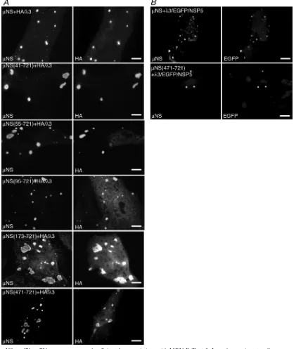
The C-terminal 250 aa ofNS are sufficient for associations with3.Although our previous studies have shown that full-lengthNS associates with the MRV RdRp3 (32), this pro-tein was not examined for its ability to associate withNSC, a second form of NS expressed in cells that is missing the N-terminal 41 aa (29). We utilized our new panel of NS deletion mutants in this study to identify the region ofNS necessary for the association with3. A plasmid expressing an HA-tagged form of 3 was cotransfected with plasmids ex-pressing each of theNS deletions. The localization of HA-tagged3 was then examined relative to the FLS formed by each of theNS deletions at 18 h p.t. by immunofluorescence microscopy. We found that3/HA associated completely with
NS(41-721) (Fig. 7A, second row) andNS(55-721) (Fig. 7A, third row) and continued to colocalize partially with NS(95-721) (Fig. 7A, fourth row),NS(173-721) (Fig. 7A, fifth row), orNS(471-721) (Fig. 7A, bottom row), suggesting thatNS aa 471 to 721 are sufficient for the association with3/HA. Since further deletions from the N terminus ofNS result in the loss of FLS formation (8), such mutants could not be examined using this assay.
Because the localization of 3/HA to FLS appeared to be partially diminished with some of theNS deletion mutants, we utilized an additional assay to confirm thatNS aa 471 to 721 are sufficient for the association with3-HA. Our previous experiments showed that when coexpressed in cells, MRVNS and rotavirus EGFP/NSP5 do not colocalize and instead form nonoverlapping FLS and VLS, respectively (Fig. 1, top). We therefore cloned the L1 gene, encoding the 3 protein, up-stream of the sequences encoding EGFP/NSP5 to form a plas-mid expressing 3/EGFP/NSP5. This plasmid was then co-transfected with a plasmid expressing either full-lengthNS or
NS(471-721), and the localization of3/EGFP/NSP5 relative toNS(1-721) orNS(471-721) FLS was examined at 18 h p.t. by immunofluorescence microscopy to determine if the addi-tion of3 to EGFP/NSP5 would cause a coalescence of FLS and VLS. As expected,3/EGFP/NSP5 associated with NS(1-721) (Fig. 7B, top). Importantly, the 3/EGFP/NSP5 fusion also completely colocalized withNS(471-721) (Fig. 7B, bot-tom), confirming that the C-terminal 250 aa ofNS are suffi-cient for the association with3.
We also attempted to examine the association of3/HA with fusion proteins consisting of fragments ofNS fused to EGFP/
NSP5 as we had done to define regions ofNS sufficient for associations with other MRV proteins. In this case, however, we found that3/HA colocalized with EGFP/NSP5 VLS even in the absence ofNS fusions to the latter protein. This is perhaps not completely unexpected, as the MRV RdRp 3 shares notable homology with the rotavirus RdRp VP1, a known NSP5-associating protein (2).
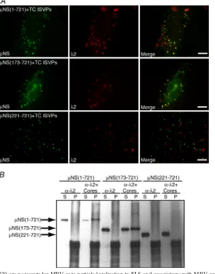
Core particles associate withNS aa 173 to 220.We have previously shown that in addition to recruiting each of the viral core proteins andNS to VF, parental input core particles are recruited to FLS when cells transfected with a plasmid express-ing full-lengthNS are infected with top-component (genome-minus) ISVPs of MRV (6). To identify the region of NS necessary for the recruitment of core particles to FLS, we transfected cells with a plasmid expressing either full-length
NS or the deletion mutantNS(41-721),NS(173-721), or
NS(221-721). At 6 h p.t., we infected the transfected cells with top-component ISVPs at 1,000 PFU/cell in the presence of cycloheximide to prevent protein synthesis. At 90 min postinfection (p.i.), cells were fixed and stained with antibodies againstNS and MRV core particles, and the localization of cores relative to FLS was examined by immunofluorescence microscopy. We found that cores localized to FLS in cells expressing both full-lengthNS (Fig. 8A, top) (as previously shown [6]) as well as the deletion mutantNS(173-721) (Fig. 8A, middle). In cells expressingNS(221-721), however, core particles did not associate with FLS (Fig. 8A, bottom), sug-gesting thatNS aa 173 to 220 are necessary for core recruit-ment to FLS.
A second experimental approach was used to confirm the region ofNS necessary for the association with parental core particles. Cells transfected with a plasmid expressing full-lengthNS or the deletion mutantNS(173-721) or NS(221-721) were incubated for 18 h p.t. and then collected and lysed. Magnetic beads conjugated with protein A were incubated with monoclonal antibody 7F4 against the core surface protein
2 and then split into two aliquots. One aliquot (bead/2) was set aside, and the other aliquot was incubated with purified MRV core particles (bead/2/cores). The 7F4-bound beads, bound to cores or not, were then incubated for 4 h with wild-type or mutantNS-containing lysates, and beads and associ-ated proteins were then separassoci-ated from the supernatant and washed. Proteins contained in the bead (pellet) fraction as well as in the postbinding supernatant fraction were separated by SDS-PAGE, and anyNS protein associated with the samples was visualized by immunoblotting usingNS antiserum. When
FIG. 5.NS aa 75 to 84 are necessary and aa 55 to 173 are sufficient for associations with the core surface protein2. For each experiment, cells were processed for fluorescence microscopy at 18 h p.t. (A) CV-1 cells were cotransfected with plasmids expressing2 and eitherNS (top row),NS(55-721) (second row),NS(65-721) (third row),NS(75-721) (fourth row),NS(85-721) (fifth row), orNS(95-721) (bottom row). After fixation, cells were stained with rabbit polyclonal antibodies againstNS and mouse monoclonal antibody 7F4 against2 followed by Alexa 594-conjugated goat anti-rabbit IgG to visualizeNS (left) and Alexa 488-conjugated goat anti-mouse IgG to visualize2 (right). (B) CV-1 cells were cotransfected with plasmids expressing2 and either EGFP/NSP5 (top row),NS(1-227)/EGFP/NSP5 (second row),NS(41-221)/EGFP/ NSP5 (third row),NS(55-221)/EGFP/NSP5 (fourth row), orNS(95-221)/EGFP/NSP5 (bottom row). After fixation, cells were stained with mouse monoclonal antibody 7F4 against 2 followed by Alexa 594-conjugated goat anti-mouse IgG to visualize 2 (right). The inherent fluorescence of EGFP was used to visualize each of the fusion proteins (left). (C) CV-1 cells were cotransfected with plasmids expressing2 and eitherNS(55-173)/EGFP/NSP5 (top),NS(95-173)/EGFP/NSP5 (middle), orNS(41-110)/EGFP/NSP5 (bottom). After fixation, cells were stained with mouse monoclonal antibody 7F4 against2 followed by Alexa 594-conjugated goat anti-mouse IgG to visualize2 (right). The inherent fluorescence of EGFP was used to visualize each of the fusion proteins (left). Bar, 10m.
on November 8, 2019 by guest
http://jvi.asm.org/
bead/7F4 complexes were used to immunoprecipitate associ-ated proteins, full-lengthNS was found entirely in the super-natant (Fig. 8B, lanes 1 and 2), reflecting the inability of the
2-specific antibody to immunoprecipitate NS from the ly-sate. However, when bead/7F4/core complexes were incubated with lysate containing full-lengthNS, mostNS was found in the pellet (Fig. 8B, lanes 3 and 4), reflecting the specific
inter-action ofNS with core particles. Similar results were seen when lysate containing NS(173-721) was used, with NS found entirely in the supernatant following incubation with bead/7F4 complexes (Fig. 8B, lanes 5 and 6) and in both the supernatant and pellet fraction following incubation with bead/ 7F4/core complexes (Fig. 8B, lanes 7 and 8). This suggests that the N-terminal 172 aa ofNS are not required for the associ-ation with core particles. In the lysate expressing NS(221-721), in contrast, a different result was seen. When incubated with either bead/7F4 complexes or bead/7F4/core complexes,
NS(221-721) was found entirely in the supernatant fraction of the immunoprecipitation (Fig. 8B, lanes 9 to 12). This finding suggests thatNS(221-721) is not able to bind core particles and concurs with our immunofluorescence findings showing thatNS aa 173 to 220 are necessary for the association with MRV core particles.
Newly synthesized MRV RNA is localized to VF in infected cells.Our previous results showing that parental core particles localize to FLS formed byNS (6), coupled with the above-described new results defining a region ofNS necessary for this localization, suggest that MRV core particles are embed-ded in VF in infected cells. Because cores synthesize the plus-strand RNAs of MRV (22), we hypothesized that the localiza-tion of cores to VF may result in the produclocaliza-tion of MRV plus-strand RNAs within VF during infection. To test this hypothesis, we infected cells with ISVPs and at early times p.i. transfected the infected cells with BrU, a uridine analog that is efficiently incorporated into RNA as it is synthesized (24), in the presence or absence of actinomycin D, an inhibitor of cellular RNA polymerase II (52), to prevent cellular transcrip-tion. At 60 min p.t., cells were fixed and stained with antibodies againstNS to visualize VF as well as with antibodies against bromodeoxyuridine, which cross-react with BrU (23), to vi-sualize newly synthesized RNA. The localization of newly synthesized RNA relative to VF was visualized by immuno-fluorescence microscopy. We found that in infected cells pulse-labeled with BrU (Fig. 9, top), the localization of newly synthesized BrU-containing RNA was concentrated in VF, supporting our hypothesis that MRV plus-strand RNAs are synthesized by core particles localized within these struc-tures. The BrU staining in VF was not a result of nonspecific antibody binding, as no staining was seen in the absence of BrU transfection (Fig. 9, middle). The presence of newly tran-scribed RNA was not seen in actinomycin D-treated, unin-fected cells (Fig. 9, bottom left), and an alternative pattern of nuclear BrU staining was seen in uninfected cells without ac-tinomycin D treatment (Fig. 9, bottom right).
DISCUSSION
[image:10.585.60.266.71.478.2]Early studies found thatNS is a major protein component of replication complexes associated with MRV RNAs (35, 36). Subsequent studies have shown thatNS associates with each of the viral core proteins, NS, and core particles and that these associations result in their recruitment to or retention in FLS (4, 6–8, 32, 33). The consequent hypothesis is thatNS serves as a cytoplasmic scaffolding protein that organizes core proteins,NS, and core particles such that viral replication and assembly intermediates can efficiently form in VF, which are sites of MRV genome replication and progeny core assembly
FIG. 6.NS aa 173 to 220 are necessary and aa 173 to 221 are sufficient for associations with the core surface protein2. For each experiment, cells were processed for fluorescence microscopy at 18 h p.t. (A) CV-1 cells were cotransfected with plasmids expressing2 and eitherNS (top),NS(173-721) (middle), orNS(221-721) (bottom). After fixation, cells were stained with rabbit polyclonal antibodies against MRV cores followed by Texas Red-conjugatedNS-specific rabbit IgG to visualize NS (left) and Alexa 488-conjugated goat anti-rabbit IgG to visualize2 (right). (B) CV-1 cells were cotrans-fected with plasmids expressing2 and either EGFP/NSP5 (top) or
NS(173-221)/EGFP/NSP5 (bottom). After fixation, cells were stained with rabbit polyclonal antibodies against MRV cores followed by Alexa 594-conjugated goat anti-rabbit IgG to visualize2 (right). The inher-ent fluorescence of EGFP was used to visualize each of the fusion proteins (left). Bar, 10m.
on November 8, 2019 by guest
http://jvi.asm.org/
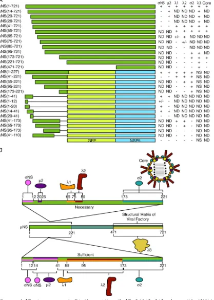
(14, 40, 44). Our new results, which show that each associating protein or particle utilizes distinct primary sequence regions withinNS, support this hypothesis by providing evidence that a single molecule ofNS may be capable of associating with multiple MRV proteins at the same time (see Fig. 10 for a
[image:11.585.78.516.71.568.2]summary of results). However, additional work is needed to determine if a single molecule ofNS can bind all, a subset, or just one of its identified binding partners or, alternatively, if the binding of one protein interferes with the binding of others. The sequential association of a particularNS molecule with
FIG. 7.NS aa 471 to 721 are necessary and sufficient for associations with MRV RdRp3. In each experiment, cells were processed for fluorescence microscopy at 18 h p.t. (A) CV-1 cells were cotransfected with plasmids expressing3/HA and eitherNS (top row),NS(41-721) (second row),NS(55-721) (third row),NS(95-721) (fourth row),NS(173-721) (fifth row), orNS(471-721) (bottom row). After fixation, cells were stained with rabbit polyclonal antibodies againstNS and mouse monoclonal antibody against HA followed by Alexa 594-conjugated goat anti-rabbit IgG to visualizeNS (left) and Alexa 488-conjugated goat anti-mouse IgG to visualize HA-tagged3 (right). (B) CV-1 cells were cotransfected with plasmids expressing3/EGFP/NSP5 and eitherNS (top) orNS(471-721) (bottom). After fixation, cells were stained with rabbit polyclonal antibodies againstNS followed by Alexa 594-conjugated goat anti-rabbit IgG to visualizeNS (left). The inherent fluorescence of EGFP was used to visualize3/EGFP/NSP5 (right). Bar, 10m.
on November 8, 2019 by guest
http://jvi.asm.org/
different proteins over the course of the process of replication and assembly is another interesting possibility.
We previously reported that upon entering cells, MRV core particles are rapidly localized to FLS that have been preformed by plasmid-expressed NS and thus might be similarly re-cruited into a newly forming VF during the course of normal infection (6). It was not known, however, if cores are recruited to VF in infected cells. The core serves as the transcriptase particle for the production of MRV plus-strand RNAs (22); thus, if cores localize to VF during infection, it would be expected that newly synthesized MRV RNAs are located
[image:12.585.135.450.72.472.2]within VF. In this study, we found that MRV RNAs are local-ized to VF from very early times after infection (starting at 3 h p.i.), suggesting that entering cores are recruited to VF as they form at early times in infected cells or, alternatively, that en-tering cores may seed VF at early times in infection. Together with the localization of all five core structural proteins, as well as the ssRNA-binding nonstructural protein NS, which is thought to play a role in genome replication and/or packaging (3, 12, 20, 21, 39), this localization of cores to VF may be a particularly efficient way for the virus to ensure that newly transcribed MRV plus-strand RNAs are retained close to the
FIG. 8.NS aa 173 to 220 are necessary for MRV core particle localization to FLS and associations with MRV cores. (A) CV-1 cells were transfected with a plasmid expressing eitherNS (top row),NS(173-721) (middle row), orNS(221-721) (bottom row). At 6 h p.t., 100g/ml of cycloheximide (CHX) was added to the cells for 30 min, at which point the cells were incubated (still in the presence of cycloheximide) with type 1 Lang top-component (TC) ISVPs (1,000 PFU/cell) at 4°C for 30 min and then shifted to 37°C for 90 min and fixed. Cells were stained with mouse monoclonal antibody 7F4 against 2 followed by Texas Red-conjugated NS-specific rabbit IgG to visualize NS (left) and Alexa 488-conjugated goat anti-mouse IgG to visualize2 (middle). Merged images are also shown (right). Bars, 10m. (B) CV-1 cells transfected with a plasmid expressing eitherNS (lanes 1 to 4),NS(173-221) (lanes 5 to 8), orNS(221-721) (lanes 9 to 12) were lysed at 18 h p.t., and the proteins from the resulting lysates were immunoprecipitated with magnetic beads preincubated with mouse monoclonal antibody 7F4 against2 either without (lanes 1, 2, 5, 6, 9, and 10) or with (lanes 3, 4, 7, 8, 11, and 12) an additional incubation with MRV core particles. Postbinding supernatants (S) and immunoprecipitated proteins (P) were then separated by SDS-PAGE, and associatedNS proteins were visualized by immunoblotting using rabbit polyclonal antibodies againstNS followed by HRP-conjugated goat anti-rabbit IgG antibody to visualizeNS.NS,NS(173-221), andNS(221-721) are indicated by arrows.
on November 8, 2019 by guest
http://jvi.asm.org/
proteins necessary for replication and core assembly. The lo-calization of newly synthesized RNAs at viroplasms was also shown previously for rotavirus- and phytoreovirus-infected cells (45, 51), suggesting that diverse members of the family
Reoviridaeare similar in this regard.
The region of NS mapped as necessary for associations with core particles is the same one mapped as both necessary and sufficient for the association with the core surface protein
2. This suggests that cores may be localized to FLS by the association ofNS with2 on the surface of the core particle; however, we have not yet identified the region of2 involved in the association withNS and therefore do not yet know if this region is surface exposed in the context of the assembled core. We have also recently found thatNS associates with a small region of the core surface protein 2 that is indeed surface exposed in the assembled core (C. L. Miller and M. L. Nibert, unpublished results), therefore raising the possibility that core particles may also associate withNS via2. Fur-thermore, we have not yet addressed the region ofNS that is sufficient for the association of cores, leaving open the possi-bility that more than one region of NS can associate with cores such that they localize to FLS.
LikeNS, orbivirus NS2, phytoreovirus Pns12, and rotavirus NSP2/NSP5 have been shown to be the only viral proteins whose expression is required for forming VIB-like structures or VLS in transfected cells (18, 47, 48, 51). Thus, other, and perhaps all, members of the familyReoviridaeencode specific proteins that function in part to form the organizational matrix of these cytoplasmic structures. In addition, there is a good
deal of evidence suggesting that these other proteins, likeNS, are involved in recruiting or retaining other viral proteins to their respective structures. For example, most of the proteins that make up the bluetongue virus (orbivirus) core and outer capsid are localized in or near VIBs in infected cells (9). Sim-ilarly, several recent studies have shown that in addition to its function in forming the matrix of VIBs, the NS2 protein is involved in recruiting or retaining the bluetongue virus core proteins VP1 (RdRp), VP3 (core shell protein), VP4 (mRNA capping enzyme), and VP6 (dsRNA helicase) to VIBs (25). The remaining core protein (VP7) appears to be recruited to VIBs through its association with VP3 (25). The regions within the NS2 protein necessary and sufficient for each of these functions have not yet been identified, but it would be inter-esting if, likeNS, NS2 associates with each of the proteins utilizing short, largely nonoverlapping sequences, supporting our hypothesis that these proteins act as cytoplasmic scaffold-ing for buildscaffold-ing replication or assembly complexes in cells.
Unlike MRV, orbiviruses, and phytoreoviruses, rotaviruses encode two proteins that must normally associate to form viroplasms in infected cells (18). Whether these two proteins “split” the functional duties of NS, NS2, or Pns12 is not known. NSP5 and NSP2 have both been reported to associate with the rotavirus RdRp VP1 (2), suggesting that NSP5 and NSP2 may work together to recruit other rotavirus proteins to VLS. A determination of other rotavirus protein associations with VLS, along with the mapping of these associations, will shed light on other potential similarities and differences be-tweenNS and these proteins.
FIG. 9. Newly synthesized MRV RNA is localized to VF in infected cells. CV-1 cells were infected with type 1 Lang (T1L) ISVPs at 100 PFU/cell (top and middle) or not infected (bottom), and at 6 h p.i., cells were treated (⫹ActD) or not (no ActD), as indicated, with actinomycin D to inhibit transcription by cellular RNA polymerase II. Cells were then transfected (⫹BrU) or not transfected (no BrU), as indicated, with BrU. At 60 min p.t., cells were fixed and stained with rabbit polyclonal antibodies againstNS and a mouse monoclonal antibody against bromode-oxyuridine, which also binds to BrU, followed by Alexa 594-conjugated goat anti-rabbit IgG to visualizeNS (top and middle, left) and Alexa 488-conjugated goat anti-mouse IgG to visualize BrU (top, middle, and bottom rows, middle column, and bottom row, left column). Merged images of the top and middle panels are also shown (right). Bar, 10M.
on November 8, 2019 by guest
http://jvi.asm.org/
FIG. 10. Summary ofNS regions necessary and sufficient for associations withNS,2,1,2,2,3, and core particles. (A) List of allNS deletion mutants and EGFP/NSP5 fusion proteins tested in this study and the results of their association with the indicated MRV proteins and MRV cores. ND, not determined; NS, nonspecific association. (B) Schematic representation ofNS showing regions of the protein necessary and sufficient for associations with each MRV protein and core particles.
on November 8, 2019 by guest
http://jvi.asm.org/
When coexpressed in cells,NS and EGFP/NSP5 form non-overlapping structures. Little is known about the cellular pro-teins that may be involved in forming either FLS or VLS; however, this result suggests that either the two proteins do not require a shared set of cellular proteins to form the structures or any shared cellular proteins that they do require are abun-dant enough for both structures to form independently in cells. The identity and importance of cellular proteins that associate withNS are currently under investigation.
One cellular system that does appear to play some role in both VF/FLS and viroplasm/VLS formation is the microtubule network. When cells are treated with the microtubule-depoly-merizing drug nocodazole, VF (in infected cells) and FLS (in cells expressingNS) remain small and diffusely distributed in the cytoplasm (compared to the large, perinuclear structures that are normally seen) (8, 38). This finding suggests that VF movement on microtubules plays a role in VF coalescence. Similarly, a recent study showed that both NSP2 and NSP5 associate with cellular microtubules and that nocodazole treat-ment of rotavirus-infected cells results in small, diffuse viro-plasms that do not develop into larger, more perinuclear struc-tures (10). Nonetheless, while some FLS and VLS localize near each other in the cytoplasm (perhaps suggesting the movement of both of them on the same microtubule track), this associa-tion with microtubules does not result in coalescence events betweenNS and EGFP/NSP5. UnlikeNS and NSP2/NSP5, it was recently reported that the formation of VIB-like struc-tures by bluetongue virus NS2 is not affected by a disruption of the microtubule network (25), suggesting that these cytoplas-mic scaffolding proteins from the different viruses may exploit different cellular mechanisms for cytoplasmic trafficking.
ACKNOWLEDGMENTS
We thank Oscar Burrone for the EGFP/NSP5 plasmid, Elaine Frei-mont for technical assistance, and other members of our laboratories for helpful discussions.
This work was supported by grants F32 AI56939 to C.L.M. and R01 AI47904 and R56 AI067445 to M.L.N. from the U.S. National Insti-tutes of Health. M.M.A. is a Ph.D. graduate of the Harvard Virology Program, which is partially supported by training grant T32 AI07245 from the U.S. National Institutes of Health. In addition, T.J.B. was partially supported by a postdoctoral fellowship from that training grant. Other assistance to C.L.M. was provided by the Office of the Dean, Iowa State University College of Veterinary Medicine.
REFERENCES
1.Arnold, M. M., K. E. Murray, and M. L. Nibert.2008. Formation of the factory matrix is an important, though not a sufficient function of nonstruc-tural proteinNS during reovirus infection. Virology375:412–423. 2.Arnoldi, F., M. Campagna, C. Eichwald, U. Desselberger, and O. R.
Bur-rone.2007. Interaction of rotavirus polymerase VP1 with nonstructural pro-tein NSP5 is stronger than that with NSP2. J. Virol.81:2128–2137. 3.Becker, M. M., M. I. Goral, P. R. Hazelton, G. S. Baer, S. E. Rodgers, E. G.
Brown, K. M. Coombs, and T. S. Dermody.2001. ReovirusNS protein is required for nucleation of viral assembly complexes and formation of viral inclusions. J. Virol.75:1459–1475.
4.Becker, M. M., T. R. Peters, and T. S. Dermody.2003. ReovirusNS and
NS proteins form cytoplasmic inclusion structures in the absence of viral infection. J. Virol.77:5948–5963.
5.Broering, T. J., M. M. Arnold, C. L. Miller, J. A. Hurt, P. L. Joyce, and M. L. Nibert.2005. Carboxyl-proximal regions of reovirus nonstructural protein
NS necessary and sufficient for forming factory-like inclusions. J. Virol.
79:6194–6206.
6.Broering, T. J., J. Kim, C. L. Miller, C. D. Piggott, J. B. Dinoso, M. L. Nibert, and J. S. L. Parker.2004. Reovirus nonstructural proteinNS recruits viral core surface proteins and entering core particles to factory-like inclusions. J. Virol.78:1882–1892.
7.Broering, T. J., A. M. McCutcheon, V. E. Centonze, and M. L. Nibert.2000. Reovirus nonstructural proteinNS binds to core particles but does not inhibit their transcription and capping activities. J. Virol.74:5516–5524. 8.Broering, T. J., J. S. L. Parker, P. L. Joyce, J. Kim, and M. L. Nibert.2002.
Mammalian reovirus nonstructural proteinNS forms large inclusions and colocalizes with reovirus microtubule-associated protein2 in transfected cells. J. Virol.76:8285–8297.
9.Brookes, S. M., A. D. Hyatt, and B. T. Eaton.1993. Characterization of virus inclusion bodies in bluetongue virus-infected cells. J. Gen. Virol.74:525–530. 10.Cabral-Romero, C., and L. Padilla-Noriega.2006. Association of rotavirus viroplasms with microtubules through NSP2 and NSP5. Mem. Inst. Oswaldo Cruz101:603–611.
11.Chandran, K., S. B. Walker, Y. Chen, C. M. Contreras, L. A. Schiff, T. S. Baker, and M. L. Nibert.1999. In vitro recoating of reovirus cores with baculovirus-expressed outer-capsid proteins1 and3. J. Virol.73:3941– 3950.
12.Cross, R. K., and B. N. Fields.1972. Temperature-sensitive mutants of reovirus type 3: studies on the synthesis of viral RNA. Virology50:799–809. 13.Dales, S.1965. Replication of animal viruses as studied by electron
micros-copy. Am. J. Med.38:699–715.
14.Dales, S., P. Gomatos, and K. C. Hsu.1965. The uptake and development of reovirus in strain L cells followed with labelled viral ribonucleic acid and ferritin-antibody conjugates. Virology25:193–211.
15.Eaton, B. T., A. D. Hyatt, and J. R. White.1987. Association of bluetongue virus with the cytoskeleton. Virology157:107–116.
16.Eichwald, C., J. F. Rodriguez, and O. R. Burrone.2004. Characterization of rotavirus NSP2/NSP5 interactions and the dynamics of viroplasm formation. J. Gen. Virol.85:625–634.
17.Estes, M. K.2001. Rotaviruses and their replication, p. 1747–1786.InD. M. Knipe, P. M. Howley, D. E. Griffin, R. A. Lamb, M. A. Martin, B. Roizman, and S. E. Straus (ed.), Fields virology, 4th ed. Lippincott Williams & Wilkins, Philadelphia, PA.
18.Fabbretti, E., I. Afrikanova, F. Vascotto, and O. R. Burrone.1999. Two non-structural rotavirus proteins, NSP2 and NSP5, form viroplasm-like structures in vivo. J. Gen. Virol.80:333–339.
19.Furlong, D. B., M. L. Nibert, and B. N. Fields.1988. Sigma 1 protein of mammalian reoviruses extends from the surfaces of viral particles. J. Virol.
62:246–256.
20.Gillian, A. L., and M. L. Nibert.1998. Amino terminus of reovirus nonstruc-tural proteinNS is important for ssRNA binding and nucleoprotein com-plex formation. Virology240:1–11.
21.Gillian, A. L., S. C. Schmechel, J. Livny, L. A. Schiff, and M. L. Nibert.2000. Reovirus proteinNS binds in multiple copies to single-stranded RNA and shares properties with single-stranded DNA binding proteins. J. Virol.74:
5939–5948.
22.Gillies, S., S. Bullivant, and A. R. Bellamy.1971. Viral RNA polymerases: electron microscopy of reovirus reaction cores. Science174:694–696. 23.Jackson, D. A., A. B. Hassan, R. J. Errington, and P. R. Cook.1993.
Visu-alization of focal sites of transcription within human nuclei. EMBO J.12:
1059–1065.
24.Javed, A., S. K. Zaidi, S. E. Gutierrez, C. J. Lengner, K. S. Harrington, H. Hovhannisyan, B. C. Cho, J. Pratap, S. M. Pockwinse, M. Montecino, A. J. van Wijnen, J. B. Lian, J. L. Stein, and G. S. Stein.2004. In situ immuno-fluorescence analysis: analyzing RNA synthesis by 5-bromouridine-5⬘ -triphosphate labeling. Methods Mol. Biol.285:29–31.
25.Kar, A. K., B. Bhattacharya, and P. Roy.2007. Bluetongue virus RNA binding protein NS2 is a modulator of viral replication and assembly. BMC Mol. Biol.8:4.
26.Kim, J., X. Zhang, V. E. Centonze, V. D. Bowman, S. Noble, T. S. Baker, and M. L. Nibert.2002. The hydrophilic amino-terminal arm of reovirus core shell protein 1 is dispensable for particle assembly. J. Virol.76:12211– 12222.
27.Kobayashi, T., J. D. Chappell, P. Danthi, and T. S. Dermody.2006. Gene-specific inhibition of reovirus replication by RNA interference. J. Virol.
80:9053–9063.
28.Kobayashi, T., L. S. Ooms, J. D. Chappell, and T. S. Dermody.2009. Iden-tification of functional domains in reovirus replication proteinsNS and2. J. Virol.83:2892–2906.
29.Lee, P. W. K., E. C. Hayes, and W. K. Joklik.1981. Characterization of anti-reovirus immunoglobulins secreted by cloned hybridoma cell lines. Vi-rology108:134–146.
30.McCutcheon, A. M., T. J. Broering, and M. L. Nibert.1999. Mammalian reovirus M3 gene sequences and conservation of coiled-coil motifs near the carboxyl terminus of theNS protein. Virology264:16–24.
31.McNulty, M. S., W. L. Curran, and J. B. McFerran.1976. The morphogen-esis of a cytopathic bovine rotavirus in Madin-Darby bovine kidney cells. J. Gen. Virol.33:503–508.
32.Miller, C. L., M. M. Arnold, T. J. Broering, C. Eichwald, J. Kim, J. B. Dinoso, and M. L. Nibert.2007. Virus-derived platforms for visualizing protein associations inside cells. Mol. Cell. Proteomics6:1027–1038. 33.Miller, C. L., T. J. Broering, J. S. L. Parker, M. M. Arnold, and M. L. Nibert.
on November 8, 2019 by guest
http://jvi.asm.org/
2003. ReovirusNS protein localizes to inclusions through an association requiring theNS amino terminus. J. Virol.77:4566–4576.
34.Mohan, K. V., J. Muller, I. Som, and C. D. Atreya.2003. The N- and C-terminal regions of rotavirus NSP5 are the critical determinants for the formation of viroplasm-like structures independent of NSP2. J. Virol.77:
12184–12192.
35.Morgan, E. M., and H. J. Zweerink.1975. Characterization of transcriptase and replicase particles isolated from reovirus-infected cells. Virology68:455– 466.
36.Morgan, E. M., and H. J. Zweerink.1977. Characterization of the double-stranded RNA in replicase particles in reovirus-infected cells. Virology77:
421–423.
37.Nibert, M. L., and L. A. Schiff.2001. Reoviruses and their replication, p. 793–842.InD. M. Knipe, P. M. Howley, D. E. Griffin, R. A. Lamb, M. A. Martin, B. Roizman, and S. E. Straus (ed.), Fields virology, 4th ed. Lippin-cott Williams & Wilkins, Philadelphia, PA.
38.Parker, J. S. L., T. J. Broering, J. Kim, D. E. Higgins, and M. L. Nibert.2002. Reovirus core protein2 determines the filamentous morphology of viral inclusion bodies by interacting with and stabilizing microtubules. J. Virol.
76:4483–4496.
39.Ramig, R. F., R. Ahmed, and B. N. Fields.1983. A genetic map of reovirus: assignment of the newly defined mutant groups H, I, and J. to genome segments. Virology.125:299–313.
40.Rhim, J. S., L. E. Jordan, and H. D. Mayor.1962. Cytochemical, fluorescent-antibody and electron microscopic studies on the growth of reovirus (ECHO 10) in tissue culture. Virology17:342–355.
41.Roy, P. 2001. Orbiviruses and their replication, p. 1835–1869.InD. M. Knipe, P. M. Howley, D. E. Griffin, R. A. Lamb, M. A. Martin, B. Roizman, and S. E. Straus (ed.), Fields virology, 4th ed. Lippincott Williams & Wilkins, Philadelphia, PA.
42.Sharpe, A. H., L. B. Chen, and B. N. Fields. 1982. The interaction of mammalian reoviruses with the cytoskeleton of monkey kidney CV-1 cells. Virology120:399–411.
43.Shimizu, T., M. Yoshii, T. Wei, H. Hirochika, and T. Omura.2009. Silencing by RNAi of the gene for Pns12, a viroplasm matrix protein of rice dwarf virus, results in strong resistance of transgenic rice plants to the virus. Plant Biotechnol. J.7:24–32.
44.Silverstein, S. C., and P. H. Schur.1970. Immunofluorescent localization of double-stranded RNA in reovirus-infected cells. Virology41:564–566. 45.Silvestri, L. S., Z. F. Taraporewala, and J. T. Patton.2004. Rotavirus
rep-lication: plus-sense templates for double-stranded RNA synthesis are made in viroplasms. J. Virol.78:7763–7774.
46.Spendlove, R. S., E. H. Lennette, C. O. Knight, and J. H. Chin.1963. Development of viral antigen and infectious virus on HeLa cells infected with reovirus. J. Immunol.90:548–553.
47.Theron, J., H. Huismans, and L. H. Nel.1996. Site-specific mutations in the NS2 protein of epizootic haemorrhagic disease virus markedly affect the formation of cytoplasmic inclusion bodies. Arch. Virol.141:1143–1151. 48.Thomas, C. P., T. F. Booth, and P. Roy. 1990. Synthesis of bluetongue
virus-encoded phosphoprotein and formation of inclusion bodies by recom-binant baculovirus in insect cells: it binds the single-stranded RNA species. J. Gen. Virol.71:2073–2083.
49.Touris-Otero, F., M. Cortez-San Martin, J. Martinez-Costas, and J. Benavente.2004. Avian reovirus morphogenesis occurs within viral factories and begins with the selective recruitment ofNS andA toNS inclusions. J. Mol. Biol.341:361–374.
50.Virgin, H. W., IV, M. A. Mann, B. N. Fields, and K. L. Tyler.1991. Mono-clonal antibodies to reovirus reveal structure/function relationships between capsid proteins and genetics of susceptibility to antibody action. J. Virol.
65:6772–6781.
51.Wei, T., T. Shimizu, K. Hagiwara, A. Kikuchi, Y. Moriyasu, N. Suzuki, H. Chen, and T. Omura.2006. Pns12 protein of rice dwarf virus is essential for formation of viroplasms and nucleation of viral-assembly complexes. J. Gen. Virol.87:429–438.
52.Yu, F. L.1980. Selective inhibition of rat liver nuclear RNA polymerase II by actinomycin D in vivo. Carcinogenesis1:577–581.