0022-538X/09/$12.00 doi:10.1128/JVI.00691-09
Copyright © 2009, American Society for Microbiology. All Rights Reserved.
Characterization of Determinants Important for Hepatitis C Virus p7
Function in Morphogenesis by Using
trans
-Complementation
䌤
Christiane Brohm,
1,5† Eike Steinmann,
1,5† Martina Friesland,
5Ivo C. Lorenz,
2‡ Arvind Patel,
3Francois Penin,
4Ralf Bartenschlager,
1and Thomas Pietschmann
1,5*
Department of Molecular Virology, University Heidelberg, Im Neuenheimer Feld 345, 69120 Heidelberg, Germany1; Center for the Study of
Hepatitis C, Laboratory of Virology and Infectious Disease, The Rockefeller University, 1230 York Avenue, New York, New York 100652;
MRC Virology Unit, Institute of Virology, University of Glasgow, Church Street, Glasgow G11 5JR, United Kingdom3; Institut et
Chimie des Proteines, UMR 5086 CNRS, Universite Lyon, IFR 128 BioSciences Lyon-Gerland, Lyon, France4; and Division of
Experimental Virology, Twincore, Centre for Experimental and Clinical Infection Research, Medical School Hannover (MHH) and
the Helmholtz Centre for Infection Research (HZI), Feodor-Lynen-Str. 7, 30625 Hannover, Germany5
Received 3 April 2009/Accepted 26 August 2009
Hepatitis C virus (HCV) p7 is an integral membrane protein that forms ion channels in vitro and that is crucial for the efficient assembly and release of infectious virions. Due to these properties, p7 was included in the family of viroporins that comprises proteins like influenza A virus M2 and human immunodeficiency virus type 1 (HIV-1) vpu, which alter membrane permeability and facilitate the release of infectious viruses. p7 from different HCV isolates sustains virus production with variable efficiency. Moreover, p7 determinants modulate processing at the E2/p7 and the p7/NS2 signal peptidase cleavage sites, and E2/p7 cleavage is incomplete. Consequently, it was unclear if a differential ability to sustain virus production was due to variable ion channel activity or due to alternate processing at these sites. Therefore, we developed atrans-complementation assay permitting the analysis of p7 outside of the HCV polyprotein and thus independently of processing. The rescue of p7-defective HCV genomes was accomplished by providing E2, p7, and NS2, or, in some cases, by p7 alone both in a transient complementation assay as well as in stable cell lines. In contrast, neither influenza A virus M2 nor HIV-1 vpu compensated for defective p7 in HCV morphogenesis. Thus, p7 is absolutely essential for the production of infectious HCV particles. Moreover, our data indicate that p7 can operate independently of an upstream signal sequence, and that a tyrosine residue close to the conserved dibasic motif of p7 is important for optimal virus production in the context of genotype 2a viruses. The experimental system described here should be helpful to investigate further key determinants of p7 that are essential for its structure and function in the absence of secondary effects caused by altered polyprotein processing.
Hepatitis C virus (HCV) is a highly variable enveloped virus. It is the sole member of the genusHepaciviruswithin the family
Flaviviridae(36). Based on sequence homology, patient isolates
are classified into seven genotypes and more than 100 subtypes (17, 52).
The genome of HCV is a single-stranded RNA molecule of positive polarity with a size of⬃9.6 kb. It encodes a polypro-tein of ca. 3,000 amino acids and contains nontranslated re-gions (NTRs) at both the 5⬘ and 3⬘ termini that are required for translation and RNA replication (33). Cellular and two viral proteases, NS2-3 and NS3-4A, liberate the individual viral proteins. The N-terminal portion of the polyprotein contains the structural proteins core and envelope glycoproteins 1 and 2 (E1, E2), which constitute the virus particle. These proteins are cleaved from the polyprotein by the host cell signal pepti-dase (18, 24). In the case of the core protein, an additional cleavage step mediated by the signal peptide peptidase
liber-ates its mature C terminus (41). Further downstream of the structural proteins the polyprotein harbors p7, a short mem-brane-associated polypeptide required for virus assembly and release (27, 55), and the nonstructural (NS) proteins NS2, NS3, NS4A, NS4B, NS5A, and NS5B. Proteins NS3 to NS5B are the minimal components of the membrane-bound replication com-plexes that catalyze RNA replication (16, 38).
Using the novel JFH1-based HCV infection model (35, 61, 65), it has been demonstrated recently that besides the canon-ical structural proteins core, E1, and E2, NS5A, p7, NS3, and NS2 also are crucial for the production of infectious HCV particles (1, 26, 27, 39, 40, 55, 57). These data highlight that HCV assembly and release is a coordinated process involving both structural and nonstructural proteins. However, how the aforementioned proteins contribute to the production of in-fectious virus particles remains poorly understood.
HCV p7 comprises two helical domains connected by a polar loop. Studies with epitope-tagged p7 variants indicate that both termini of the protein are resident in the lumen of the endoplasmic reticulum (ER) (4) or that, in addition, a second alternative topology with the C terminus exposed to the cyto-plasm can be adopted (25). Using such constructs for fluores-cent microscopy, a complex localization of p7 was revealed. While most prominent staining generally was observed at the ER (4, 19, 23), pools of p7 also were detected at mitochondria (19) and at the plasma membrane (4). These data suggest that * Corresponding author. Mailing address: Twincore Center,
Depart-ment of ExperiDepart-mental Virology, Feodor-Lynen-Str. 7, Hannover 30625, Germany. Phone: 49 511 220027 130. Fax: 49 511 220027 186. E-mail: [email protected].
† These authors contributed equally to this work.
‡ Present address: International AIDS Vaccine Initiative, AIDS Vaccine Design & Development Laboratory, Brooklyn, New York 11220.
䌤Published ahead of print on 2 September 2009.
11682
on November 8, 2019 by guest
http://jvi.asm.org/
p7 influences virus replication at various sites within infected cells, and that the function and/or localization of p7 is regu-lated by different trafficking signals that could be exposed in a topology-dependent manner. However, caution is warranted since, due to the lack of antibodies, epitope-tagged p7 variants had to be employed for most analyses, and since localization studies of virus-producing cells with functional p7 still are lacking.
One hallmark of p7 is its ability to form cation-selective channels in artificial membranes (20, 46, 49), a property that likely depends on the oligomerization of the protein (7, 21). There are intriguing correlations that link p7’s function as an ion channel protein in vitro to its role in the assembly and release of infectious HCV particles in tissue culture. First, the mutation of the conserved dibasic motif in the polar loop of p7 abrogates ion channel activity and interferes with virus produc-tion in tissue culture (20, 27, 55). Second, iminosugars coupled to long alkyl chains likeN-nonyl deoxygalactonojirimycin (NN-DGJ) not only interfere with ion channel activity but also repress the release of infectious particles from transfected Huh-7 cells (46, 56). Taken together, these data suggest that the ion channel activity of p7 is crucial for its role in the late steps of the HCV replication cycle, and that this function is amenable to the development of selective inhibitors for anti-viral therapy. However, presently it is unknown how mecha-nistically p7, as an ion channel protein, facilitates HCV assem-bly and release or if p7 also is a component of virus particles and participates in entry.
Besides its function as an ion channel, p7 harbors a signal-like sequence in its C-terminal domain that directs the inser-tion of the N terminus of NS2 into the lumen of the ER (4). Strikingly, due to structural determinants within the C termi-nus of E2, p7, and the N termitermi-nus of NS2, signalase cleavages at the E2/p7 and the p7/NS2 sites are incomplete, thus yielding E2-p7-NS2 and E2-p7 precursor proteins (3, 18, 34, 42). Al-though these precursors are not absolutely essential for the production of infectious HCV particles (26, 27), a defined ratio between mature and precursor proteins might play a role to orchestrate optimal virus assembly. Given these circumstances, genetic studies of p7 function are complicated, since mutations may, on the one hand, affect ion channel activity, and on the other hand influence processing at the E2-p7 and p7-NS2 junc-tions.
To circumvent this problem, in this study we developed a complementation system that permits the rescue of genomes with defects in p7 by the ectopic expression of p7 intrans. This enabled us to directly assess the function of p7 in the absence of secondary effects caused by aberrant polyprotein cleavage. Using this approach, we analyzed the role of the native signal sequence of p7 and p7-containing precursor proteins. In addi-tion, we investigated key determinants that are essential for the optimal function of p7 in the course of HCV infectious particle production.
MATERIALS AND METHODS
Cell culture and cell lines.All cell lines were grown in Dulbecco’s modified minimal essential medium (DMEM; Invitrogen, Karlsruhe, Germany)
supple-mented with 2 mML-glutamine, nonessential amino acids, 100 U/ml of penicillin,
100g/ml of streptomycin, and 10% fetal calf serum (DMEM complete).
Huh7-Lunet and Huh-7.5 cells (subclones of the Huh-7 hepatoma cell line [2, 13]),
which are highly permissive for HCV RNA replication, and in the case of the
latter also for HCV infection, served as cell lines for transienttrans
-complemen-tation assays and for the construction of cells expressing HCV p7 and precursor proteins, respectively. Stable cell lines were cultivated in DMEM complete
sup-plemented with blasticidin at a concentration of 5g/ml.
Plasmid construction.The plasmids pFK-Jc1 (48), pFK-Jc1-⌬p7half,
pFK-Jc1-KR33,35QQ, and pFK-Jc1-E1-TMD(E1-K370Q) have been described recently
(55). Plasmid pFK-Jc1⌬p7full, which carries a complete deletion of the p7 coding
region, was created by using PCR mutagenesis. The plasmid pFK PI-EI-NS3-5B/JFH1 is a bicistronic helper replicon designed for the insertion of various transgenes (e.g., Jc1-derived genome segments for the expression of p7 and p7-containing precursor proteins) into an autonomously replicating JFH1-based replicon. The basic helper replicon vector was engineered with a poliovirus-derived internal ribosome entry site (IRES) (designated PI) downstream of the
JFH1-derived 5⬘-nontranslated region (5⬘NTR) (nucleotides 1 to 341 of JFH1)
and separated from the HCV 5⬘NTR by a spacer region of 72 nucleotides.
Unique restriction sites BglII, SwaI, and NotI were inserted downstream of the PI and are followed by the encephalomyocarditis virus IRES (EI) that expresses JFH1-derived NS3 to NS5B proteins. The design of this construct is analogous to that of Con1-derived replicons described previously (14) and was necessary to permit the efficient expression of proteins without HCV core protein fusion in the first cistron (note that a fully functional HCV IRES extends into the coding region of the core protein). The coding region of the gaussia luciferase (G-luc) or Jc1-derived p7 and p7-containing precursor proteins were amplified by PCR from plasmid pCMV-G-Luc1 or pFK-Jc1, respectively, and inserted into the parental pFK PI-EI-NS3-5B/JFH1 vector. Resulting replicon vectors were des-ignated pFK PI-Spp7/J6 EI NS3-5B/JFH1, pFK PI-Spp7-NS2/Jc1 EI NS3-5B/ JFH1, pFK PI-SpE2p7/J6 EI NS3-5B/JFH1, pFK PI-SpE2/J6 EI NS3-5B/JFH1, pFK PI-SpE2p7NS2/Jc1 EI NS3-5B/JFH1, and pFK PI-G-Luc EI NS3-5B/JFH1. Influenza A virus M2 (A/FPV/Rostock/34/H7N1) and human immunodeficiency
virus type 1 (HIV-1) vpu (HIV-1NL4-3) were amplified using appropriate primers
and were inserted into pFK PI-5B/JFH1 to create pFK PI-M2 EI-NS3-5B/JFH1 and pFK PI-vpu EI-NS3-EI-NS3-5B/JFH1, respectively. For the construction of HCV stable cell lines, we used a derivative of the lentiviral self-inactivating
vector pWPI (47) that encodes the blasticidin S deaminase (BSD) from
Aspergil-lus terreus, which confers resistance to blasticidin in place of green fluorescent protein and which we designated pWPI-BSD (54). Using this vector, we inserted the same coding regions as those used in the replicon described above. The plasmids are designated Spp7/J6-BSD, Spp7NS2/Jc1-BSD, pWPI-SpE2p7/J6-BSD, pWPISpE2/J6-BSD, and pWPI-SpE2p7NS2/Jc1-BSD. In these constructs, we used the native signal sequences (C-terminal domains of E1 and E2) corresponding to codons 370 to 383 and 734 to 750, respectively, of the J6CF isolate (63). To analyze the possible modulation of p7 function by upstream signal sequences, we created additional constructs either lacking a signal se-quence (pFK PI-p7/J6 EI NS3-5B/JFH1) or encoding an extended form of the native signal sequence (60 C-terminal residues of E2; codons 691 to 750; pFK PI-Sp60p7/J6 EI NS3-5B/JFH1) or a construct with p7 fused to the signal se-quence of the murine immunoglobulin (Ig) kappa chain signal peptide (pFK
PI-SIgp7/J6 EI NS3-5B/JFH1). Inserts generated by a PCR-based strategy were
verified by automated nucleotide sequencing. Further details regarding the clon-ing strategies and exact nucleotide sequences are available upon request.
In vitro transcription, electroporation, and transient HCV replication assays using luciferase reporter genomes.In vitro transcripts of the individual
con-structs were generated by linearizing 5 to 10g of the respective plasmid by
digestion for 1 h with MluI. Plasmid DNA was extracted with phenol and chloroform and after precipitation with ethanol dissolved in RNase-free water. In vitro transcription reaction mixtures contained 80 mM HEPES (pH 7.5), 12
mM MgCl2, 2 mM spermidine, 40 mM dithiothreitol (DTT), a 3.125 mM
con-centration of each ribonucleoside triphosphate, 1 U of RNasin (Promega,
Mann-heim, Germany) perl, 0.1g plasmid DNA/l, and 0.6 U of T7 RNA
poly-merase (Promega) perl. After incubation for 2 h at 37°C, an additional 0.3 U
of T7 RNA polymerase/l of reaction mixture was added, followed by another
2 h of incubation at 37°C. Transcription was terminated by the addition of 1.2 U
of RNase-free DNase (Promega) perg of plasmid DNA and 30 min of
incu-bation at 37°C. The RNA was extracted with acidic phenol and chloroform, precipitated with isopropanol, and dissolved in RNase-free water. The concen-tration was determined by the measurement of the optical density at 260 nm. Denaturing agarose gel electrophoresis was used to check RNA integrity.
For the electroporation of HCV RNA into Huh7-Lunet cells, single-cell sus-pensions were prepared by the trypsinization of monolayers and subsequent resuspension with DMEM complete. Huh7-Lunet cells were washed with
phos-phate-buffered saline (PBS), counted, and resuspended at 1⫻107cells per ml in
Cytomix (59) containing 2 mM ATP and 5 mM glutathione, whereas Huh-7.5
on November 8, 2019 by guest
http://jvi.asm.org/
cells were resuspended at 1.5⫻107
cells per ml. Unless otherwise stated, 10g
of in vitro-transcribed RNA was mixed with 400l cell suspension by pipetting
and then electroporated with a Gene Pulser system (Bio-Rad, Munich,
Ger-many) in a cuvette with a gap width of 0.4 cm (Bio-Rad) at 975F and 270 V.
Cells were immediately transferred to 16 ml DMEM complete, and 2 ml of the cell suspension was seeded per well of a six-well plate. In the case of p7 rescue
assays, we transfected a total amount of 10g RNA and used a ratio of helper
replicon to p7-defective genome of 1:1.
The quantification of luciferase reporter activity was used to determine tran-sient HCV RNA replication as described previously (32). For assaying the lucif-erase activity, cells were washed once with PBS, lysed directly on the plate with 1 ml of ice-cold lysis buffer (1% Triton X-100, 25 mM glycylglycine, 15 mM
MgSO4, 4 mM EGTA, and 1 mM DTT, pH 7.8), and frozen. After being thawed,
lysates were resuspended by pipetting. For each well, 100l lysate was mixed
with 360l assay buffer (25 mM glycylglycine, 15 mM MgSO4, 4 mM EGTA, 1
mM DTT, 2 mM ATP, and 15 mM K2PO4, pH 7.8) and, after the addition of 200
l of a luciferin solution (200M luciferin, 25 mM glycylglycine, pH 8.0),
measured for 20 s in a luminometer (Lumat LB9507; Berthold, Freiburg, Ger-many). The kinetics of replication was determined by normalizing the relative light units of the different time points to the respective 4-h value. All luciferase assays were done at least in duplicate measurements.
Western blot analysis. Forty-eight hours after electroporation, cells were washed once with PBS, detached from the wells of a six-well plate by trypsiniza-tion, and subsequently resuspended in DMEM complete. Cells were centrifuged,
and the cell pellet was harvested in 120l 2⫻sample buffer (400 mM Tris, pH
8.8, 10 mM EDTA, 0.2% bromophenol blue, 20% sucrose, 3% sodium dodecyl
sulfate [SDS], 2% ß-mercaptoethanol, 1⫻10⫺4
U/ml aprotinin, 4g/ml
leu-peptin, 1 mM phenylmethylsulfonyl fluoride). After incubation at 37°C for 30 min with 50 to 75 U benzonase (Merck, Darmstadt, Germany), samples were heated for 5 min at 95°C and loaded onto a 15% polyacrylamide–SDS gel. After electrophoresis, proteins were transferred to a polyvinylidene difluoride mem-brane using a semidry blotter (Bio-Rad, Munich, Germany) according to the manufacturer’s instructions. The membrane was blocked in PBS supplemented with 0.5% Tween (PBS-T) and 5% milk for 1 h. Upon being blocked, the membrane was incubated with primary antibodies diluted in PBS-T supple-mented with 5% milk, and after being extensively washed with PBS-T, it was incubated with horseradish peroxidase-conjugated secondary antibodies diluted in PBS-T supplemented with 5% milk. As the primary antibody, the NS5A-specific 9E10 mouse monoclonal antibody was used at a dilution of 1:1,000. Bound antibodies were detected after washes with the ECL Plus Western blot-ting detection system (GE Healthcare Europe, Freiburg, Germany).
Lentiviral gene transfer.HIV-based pseudotypes bearing vesicular stomatitis virus glycoprotein were generated by the Lipofectamine 2000-based
cotransfec-tion of 293T cells (10). Briefly, 1.8⫻106293T cells were seeded in
6-cm-diameter plates 1 day before transfection with 1g of envelope protein
expres-sion construct (pczVSV-G [29]), 3g of HIV-Gag-Pol expression construct
(pCMV⌬R8.74 [11]), and 3g of the lentiviral vector pWPI (47). The medium
was replaced 6 h after transfection. Supernatants containing the pseudoparticles were harvested 48 h later. Target cells were transduced with filtered supernatants
and selected by the addition of 5g/ml blasticidin 72 h postinoculation.
Luciferase infection assay.Huh-7.5 cells were seeded at a density of 8⫻104
per well of a 12-well plate 24 h prior to inoculation with 350l of virus
prepa-ration. Cells were inoculated for 4 h, washed, and lysed in 350l of lysis buffer
72 h later. Luciferase activity was determined as described above.
Immunohistochemical staining and virus titration.Virus titers were deter-mined as described elsewhere, with slight modifications (35). In brief, Huh-7.5
cells were seeded in 96-well plates at a density of 1⫻104
cells per well 24 h prior to inoculation with dilutions of filtered cell culture supernatant (at least six wells were used per dilution). Two to 3 days later, cells were washed with PBS, fixed
for 20 min with ice-cold methanol at⫺20°C, washed three times with PBS, and
then permeabilized and blocked for 1 h with PBS containing 0.5% saponin, 1% bovine serum albumin, 0.2% dried skim milk, and 0.02% sodium acid. Endoge-nous peroxidases were blocked by incubating cells for 5 min with PBS containing 0.3% hydrogen peroxide. After three washes with PBS and one wash with PBS containing 0.5% saponin (PBS-saponin), NS5A was detected with a 1:1,000 dilution of hybridoma supernatant 9E10 (35) in PBS-saponin for 1 h at room temperature or overnight at 4°C. Cells were washed as described above, and bound 9E10 was detected by incubation with peroxidase-conjugated antibodies specific to murine IgG (Sigma-Aldrich, Steinheim, Germany) diluted 1:200 in PBS-saponin. After 1 h of incubation at room temperature, cells were washed as specified above. Finally, peroxidase activity was detected by using the Vector NovaRED substrate kit (Linaris Biologische Produkte GmbH, Wertheim,
Ger-many). Virus titers (50% tissue culture infective dose [TCID50/ml]) were
calcu-lated based on the method of Spearman and Ka¨rber (31, 53).
Northern blot analysis.Total RNA was prepared by a single-step isolation method as described previously (5). Three micrograms of total RNA was mixed with glyoxal, dimethyl sulfoxide, and sodium phosphate buffer (pH 7.0) at final concentrations of 5.9%, 50%, and 10 mM, respectively, and denatured for 1 h at 50°C. Samples were separated by denaturing agarose gel electrophoresis, and
RNA was transferred to positively charged nylon membranes (Hybond-N⫹;
Amersham Biosciences, Freiburg, Germany) with 50 mM NaOH and cross-linked by UV irradiation. Positive-strand HCV RNA was detected by
hybridiza-tion with a32P-labeled negative-sense riboprobe complementary either to the
poliovirus IRES element or to the HCV region encoding the core and E1
proteins. Hybridization with a-actin-specific riboprobe was used to monitor
equal sample loading in each lane of the gel. Serial dilutions of a poliovirus IRES-carrying helper replicon and an HCV full-length genome were loaded in parallel to ensure the specificity of the respective probes.
Metabolic radiolabeling of proteins and immunoprecipitation.Twenty-four hours after electroporation, Huh-7.5 cells were washed with PBS, starved in cysteine-free medium for 1 h, and incubated for 6 h in
methionine-cysteine-free DMEM supplemented with 100Ci/ml of Express Protein labeling
mix (Perkin Elmer, Rodgau-Ju¨gesheim, Germany). Cell lysates were prepared by
using 1 ml of ice-cold NPB per well of a six-well plate (50 mM Tris-Cl [pH 7.5], 150 mM NaCl, 1% Nonidet P-40, 1% sodium deoxycholate, and 0.1% SDS) supplemented with a complete protease inhibitor cocktail, as recommended by
the manufacturer (Roche). Lysates were cleared by centrifugation at 13,800⫻g
for 15 min at 4°C. The cleared lysates were used for immunoprecipitation using the E2-specific antibody AP33 (8), the R1233 polyclonal rabbit serum specific to E1 (62), or the NS2-specific mouse monoclonal antibody 6H6 (Dentzer, Lorenz, Evans, and Rice, in preparation). Immune complexes were resolved by denatur-ing SDS-polyacrylamide gel electrophoresis (SDS-PAGE) and detected by au-toradiography.
RESULTS
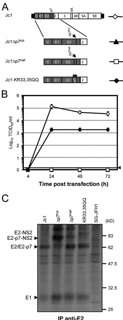
Virus production and polyprotein processing of HCV ge-nomes with defective p7.To establish a complementation sys-tem for the rescue of p7-defective HCV genomes, we initially characterized Jc1-derived constructs with various defects in p7 for the production of infectious HCV particles and polyprotein processing in the E2-p7-NS2 region of the polyprotein (Fig. 1). Constructs Jc1⌬p7half and Jc1-KR33,35QQ have been
de-scribed recently and lack residues 1 to 32 of p7 or carry a double mutation of the highly conserved basic residues KR33/35 to QQ, respectively (55). In addition, a genome with a deletion of the entire p7 coding region (Jc1⌬p7full) was constructed (Fig. 1A).
These mutants were transfected into the highly permissive human hepatoma cell line Huh7-Lunet (13), and cell-free cul-ture fluids were harvested at different time points after trans-fection to quantify the number of infectious particles released. Transfection efficiency was monitored by using immunofluo-rescence analysis (data not shown). In line with our previous findings (55), Jc1-KR33,35QQ was heavily impaired, yielding ca. 100-fold less infectious virus than the parental Jc1 genome (Fig. 1B). The mutants with the partial or complete deletion of p7 did not release detectable infectious particles. We also analyzed E2-p7-NS2 processing by the labeling of Huh7-Lunet cells transfected with different p7 mutants and immunoprecipita-tions using an E2-specific monoclonal antibody (Fig. 1C). The complete deletion of p7 (Jc1⌬p7full) displayed the strongest
pro-cessing defect, with a significant proportion of unprocessed E2-NS2. In the case of Jc1⌬p7half, signalase cleavage also was
im-paired compared to that of the parental Jc1 genome and p7 mutant Jc1-KR33, 35QQ.
Congruently with previous findings, these data show that p7 is crucial for the production of infectious particles (27, 55).
on November 8, 2019 by guest
http://jvi.asm.org/
However, at the same time substantial effects on E2-p7-NS2 processing, particularly for constructs with p7 deletions, also were evident and may influence virus particle formation.
Transient trans-complementation of p7-defective genomes.
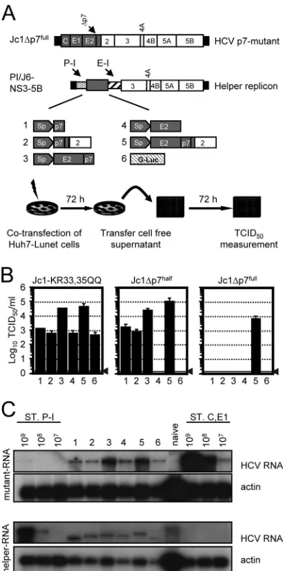
To explore if the production of infectious particles of the described p7 mutants can be rescued, a transienttrans -comple-mentation system was established. In this assay, HCV genomes with defective p7 are cotransfected with bicistronic JFH1-de-rived helper replicons expressing either p7 alone or p7-con-taining precursor proteins in the first cistron and JFH1 pro-teins NS3 to NS5B in the second (Fig. 2A). To permit the flexible insertion of genes into the first cistron of the helper, we used an internal poliovirus IRES following the HCV 5⬘NTR and a spacer of 72 nucleotides. Owing to this design, which has been used previously in Con1 replicons (14), genes in the first cistron are expressed independently of the HCV IRES and therefore without fusion to the N-terminal portion of the core protein, which constitutes part of the HCV IRES.
Of note, we transferred Jc1-derived polyprotein segments (48) into the helper replicon to match the genomes with de-fective p7 protein, which also are derived from Jc1. In addition, we utilized the native signal sequences for the insertion of E2 (C terminus of E1) and p7 (C terminus of E2) into the ER lumen (Fig. 2A; also see Materials and Methods). As a control, we employed a helper replicon expressing G-luc, which should be unable to complement defective p7. After the cotransfec-tion of Huh7-Lunet cells with the p7-defective genome and the respective helper replicon, cell-free culture fluids were har-vested 72 h later, and the accumulation of infectivity was mea-sured by the inoculation of naïve Huh-7.5 cells (Fig. 2B). As expected from our previous results (Fig. 1B), Jc1-KR33,35QQ yielded an infectivity titer of only about 103TCID
50/ml when
cotransfected with the G-luc helper replicon (Fig. 2B). How-ever, infectious virus production was increased by 30- to 50-fold by the cotransfection of helper RNAs expressing E2-p7 and E2-p7-NS2, while the coexpression of p7 alone did not significantly elevate the production of infectious particles (Fig. 2B). The infectivity of Jc1⌬p7half was rescued by all helper
RNAs expressing p7, albeit with quite different levels of effi-ciency. More specifically, while the coexpression of p7 alone restored virus production to a level of 103 TCID
50/ml, the
cotransfection of E2-p7- or E2-p7-NS2-expressing constructs yielded even higher virus titers, reaching 3⫻104and 1⫻105
TCID50/ml, respectively. Importantly, neither the G-luc helper
RNA nor the coexpression of E2 protein alone was able to rescue virus production, ruling out that the elevated expression of E2 alone is sufficient to restore virus production. In the case of the mutant Jc1⌬p7full, which showed the strongest defect in
[image:4.585.58.264.68.604.2]precursor processing, complementation was achieved only by the coexpression of E2-p7-NS2.
FIG. 1. Analysis of virus production and polyprotein processing of Jc1 genomes with different p7 mutations. (A) Schematic representa-tion of the chimeric HCV genome Jc1 consisting of J6CF (gray) and JFH1-derived (unshaded) genome segments that are fused to each other in the coding region of NS2 (48). HCV 5⬘- and 3⬘NTRs are denoted as black bars. The Jc1 chimera was used to create p7-defective HCV genomes. Shown are Jc1⌬p7full, lacking the complete p7
se-quence, Jc1⌬p7half, with a deletion of residues 1 to 32, and
Jc1-KR33,35QQ, in which two conserved basic residues were replaced by glutamine. (B) Huh7-Lunet cells were electroporated with the given HCV genomes, and the infectivity released from transfected cells was determined by a limiting dilution assay. For symbols see panel A. The gray horizontal bar and the gray arrow denote the detection limit of the
assay. (C) Huh7-Lunet cells were transfected with the indicated con-structs. Twenty-four hours posttransfection cells were labeled with [35S]methionine/cysteine-containing medium overnight and lysed
im-mediately. HCV proteins were immunoprecipitated (IP) using an an-tibody specific for E2. Arrowheads to the left point to the respective precursors and mature HCV proteins detected. SG-JFH1 indicates control cells transfected with a subgenomic JFH1 luciferase replicon expressing NS3 to NS5B proteins only.
on November 8, 2019 by guest
http://jvi.asm.org/
It is well established that when two HCV RNAs coreplicate in one cell, they compete for host cell factors, thus limiting the RNA replication efficiency (12, 37, 54). To rule out that either one of the transfected RNAs was displaced due to replication competition, thus precluding the rescue of virus production, we analyzed the RNA replication of both transfected RNAs by Northern blot hybridization. To unambiguously detect the dif-ferent replicating RNA molecules, we utilized probes specific for the HCV core-E1 region (detecting the p7 defective ge-nomes) and the poliovirus IRES (PI; it recognizes the PI-containing helper replicons). As is shown in Fig. 2C for the cotransfection experiment of Jc1⌬p7full with the individual
helper replicons, in each case both the mutant genome and the helper RNA replicated efficiently, yielding comparable quan-tities 72 h posttransfection. These data rule out that the dis-placement of either the helper or the Jc1⌬p7full RNA has
precluded the rescue of p7 trans-complementation in cases where p7, p7-NS2, E2-p7, or E2 was coexpressed. We there-fore conclude that the extent of the aberrant processing of Jc1⌬p7full(Fig. 1C) prevents rescue by helper RNAs that do
not express the complete E2-p7-NS2 region.
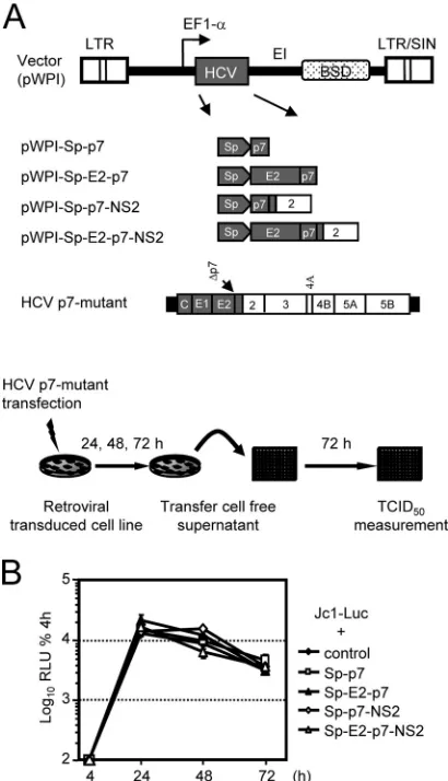
trans-complementation of p7-defective genomes in stable cell lines.In the transient transfection assay, it is difficult to pre-cisely control the equal replication efficiency of the different helper RNA molecules and the p7 mutants, which in principle could partly be responsible for different levels of rescue effi-ciency. Therefore, we wished to confirm our observations by a second assay that is independent of the different replication efficiencies of the helper RNAs and that is not influenced by replication competition between two HCV RNA molecules. To this end, we used lentiviral gene transfer and constructed a set of Huh7.5 cell lines constitutively expressing p7 alone or the p7-containing precursor proteins described above (Fig. 3A). A cell line transduced with the empty lentiviral vector served as the control. In the absence of sensitive tools to detect p7 protein expression, we verified similar transgene levels using quantitative real-time reverse transcription-PCR (RT-PCR) (data not shown). In addition, we confirmed that HCV RNA replication efficiency in these cell lines was similar by compar-ing Luc-Jc1 (32) reporter virus replication between the cell lines (Fig. 3B). The rescue of HCV particle production was assessed after the transfection of the respective cell lines with the individual p7-defective HCV genomes and the analysis of infectivity released into the culture fluid of transfected cells (Fig. 4A).
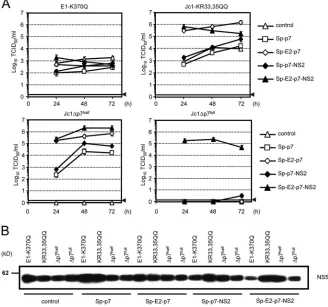
An HCV genome with a point mutation in the E1 trans-membrane domain (E1-K370Q) known to disturb E1-E2 het-erodimerization (9, 55) was used as a control to ensure that rescue is specific for defects in p7. As expected, titers of the E1-TMD mutant were similar irrespective of which cell line was transfected with the construct (Fig. 4A, upper left). In contrast, Jc1-KR33,35QQ produced much more infectious vi-rus when transfected into Huh7.5-E2-p7 or Huh7.5-E2-p7-NS2 cells, with peak titers reaching 106 and 5 ⫻ 105 TCID
50/ml,
respectively, compared to only 104TCID
50/ml for the control
cell line (Fig. 4A). Similarly to the transient assays, the virus production of Jc1⌬p7halfwas rescued in all cell lines expressing
p7 (Fig. 4A), whereas in the case of Jc1⌬p7full, infectious
particles were produced only when the construct was trans-fected into cells providing E2, p7, and NS2 (Fig. 4A). In agree-FIG. 2. Helper RNA-dependent p7trans-complementation. (A)
Sche-matic representation of the HCV genome Jc1⌬p7fulllacking the
com-plete p7 sequence. A set of JFH1-based helper replicons encoding p7 alone, various combinations of E2, p7, and NS2, or G-luc in the first cistron (gray box) were created. To permit the expression of these proteins independently of the N-terminal residues of the HCV core protein, an IRES of the poliovirus (P-I; hatched bar) was inserted downstream of the HCV 5⬘NTR. The IRES of encephalomyocarditis virus (E-I) is given as a hatched bar and directs the translation of the JFH1 NS3 to NS5B proteins. (B)trans-complementation of p7-defec-tive Jc1 genomes was analyzed by the cotransfection of each RNA helper construct and the respective p7 mutant. The release of infec-tious particles was quantified 72 h posttransfection by a limiting-dilu-tion assay using Huh-7.5 target cells. The background level of the assay is indicated by a horizontal black bar and a gray arrowhead. (C) Anal-ysis of RNA replication of Jc1⌬p7fulland helper replicons by Northern
blot hybridization. Huh7-Lunet cells were cotransfected with helper construct and p7 mutant. Total cellular RNA was prepared 48 h post-transfection and analyzed for HCV RNA using a helper replicon-specific probe (directed against the poliovirus IRES) or a core-E1-replicon-specific probe selectively reacting with the Jc1⌬p7fullgenome. Probe specificity
was controlled by loading total RNA of naïve Huh7-Lunet cells and serial dilutions of the respective in vitro transcripts in parallel. Equal sample loading was monitored by the detection of actin mRNA. ST.P-I, standard for the poliovirus IRES probe; ST. C, E1, standard for the core-E1-specific probe.
on November 8, 2019 by guest
http://jvi.asm.org/
[image:5.585.60.265.70.483.2]ment with the previous data, the rescue of Jc1⌬p7halfwas most
efficient when E2-p7 or E2-p7-NS2 was provided intrans. Since RNA replication efficiency in the different cell lines is compa-rable (Fig. 3B), and since during the rescue experiment similar quantities of HCV protein expression were detected irrespec-tive of the transfected p7-defecirrespec-tive genome and cell line (Fig. 4B), it is unlikely that these differences are attributable to the divergent replication of the HCV genome or alternative trans-gene expression levels. Rather, we conclude that the efficiency
of rescue depends on the specific p7 mutation and its effect on p7 function and polyprotein processing, which determines whether the coexpression of p7 alone or complementation by the complete set of E2-p7-NS2 proteins is necessary for rescue.
Influenza A virus M2 or HIV-1 vpu proteins are unable to substitute for p7 function in HCV assembly and release.HCV p7 has been included in the family of viroporins that includes, among others, influenza A virus M2 and HIV-1 vpu (15). All three proteins assemble ion channels and promote the release of infectious particles of the respective virus. While the proton channel activity of M2 is important to prevent the acidification of thetrans-Golgi network, thus avoiding the premature con-formational changes of influenza hemagglutinin in virus-pro-ducing cells (6), vpu interferes with host proteins (tetherin and calcium-modulating cyclophilin ligand) that otherwise restrict the release of HIV-1 particles (45, 60). In fact, it has been postulated that p7 acts in an M2-like fashion, since p7 was able to substitute for M2 in a cell-based assay (21). In addition, by counteracting tetherin, vpu facilitates not only HIV-1 release but also the release of other retro- and filoviruses (28, 44, 45). Therefore, we assessed if M2 or vpu is able to rescue the virus production of an HCV genome lacking functional p7. How-ever, when cotransfecting Huh7-Lunet cells with Jc1⌬p7half
and helper replicons expressing p7, M2, or vpu, only p7 was able to restore the production of infectious HCV particles (Fig. 5). These data indicate that p7 function is distinct from those of both M2 and vpu.
p7trans-complementation is isolate dependent but indepen-dent of upstream signal sequences.When p7 is expressed using its E2-derived signal sequence, the analysis of p7 function is, strictly speaking, not completely independent of processing, since p7 mutations may modulate the cleavage of the signal peptide and, in turn, affect p7 function. Therefore, we wanted to find out if and to what extent p7 function is modulated by upstream signal sequences. To this end, we constructed various helper replicons expressing J6CF-derived p7 either with an extended signal sequence containing 60 C-terminal E2-derived amino acids (Sp60 p7-J6), the minimal E2-derived signal se-quence (Sp p7-J6), a heterologous mouse Ig kappa chain-derived signal peptide (Igp7-J6), or no signal sequence at all (p7-J6). Interestingly, we observed only minor variations in the efficiency of trans-complementation between these con-structs, indicating that the signal sequence does not grossly affect the ability of wild-type p7 to rescue the virus production of Jc1⌬p7half(Fig. 5A). These data are in line with the
obser-vation by Jones and colleagues, who noted that the insertion of an IRES between E2 and p7 without a signal sequence up-stream of p7 permitted virus production (27). Taken together, these results confirm that p7 is able to function independently of an upstream signal peptide.
[image:6.585.60.265.69.426.2]Recently, we have reported that p7 variants from alternative HCV isolates differ with regard to their ability to promote virus production (55). Specifically, the transfer of JFH1-p7 into the Jc1 background delayed the accumulation of infectivity and reduced the peak titer by about 10-fold compared to that of Jc1. In contrast, the insertion of J6CF-derived p7 into JFH1 increased the peak titers of JFH1 and kinetics of virus release, indicating that the latter p7 variant supports virus production with greater efficiency than the cognate JFH1-p7. Further-more, the insertion of Con1-derived p7 (genotype 1b [GT1b]) FIG. 3. Construction of stable cell lines expressing HCV p7 and
various combinations of E2, p7, and NS2. (A) Construct design and assay setup. The Jc1 chimera depicted in Fig. 1 was utilized to create pWPI-Sp-p7, pWPI-SP-E2-p7, pWPI-Sp-p7-NS2, and pWPI-Sp-E2-p7-NS2 vectors encoding parts of or the complete E2-pWPI-Sp-E2-p7-NS2 coding region. To ensure the proper insertion of E2 and p7 into the ER, the respective minimal signal sequences were employed. For further de-tails of pWPI vector features, see Materials and Methods and refer-ence 54. EF1-a, elongation factor 1-␣promoter; LTR, long terminal repeat; SIN, self inactivating. The mutant Jc1⌬p7fullis shown as an
example for p7 mutants used fortrans-complementation. The experi-mental setup to rescue the infectious particle production of p7-defec-tive Jc1 genomes in stable cell lines is given at the bottom. (B) Tran-sient replication of Luc-Jc1 after the transfection of stable cell lines expressing various HCV proteins. Luc-Jc1 RNA was transfected into the indicated cell lines; cells were harvested at the time points given to determine reporter gene activity. Mean values of duplicate measure-ments are expressed relative to the quantity of luciferase activity mea-sured 4 h posttransfection, which was set to 100%.
on November 8, 2019 by guest
http://jvi.asm.org/
into the Jc1 chimera almost completely abrogated the produc-tion of infectious particles. Although in our previous study we ruled out that these effects are linked to grossly aberrant pro-cessing at the E2-p7 and p7-NS2 junction, the assay used had
limited sensitivity so that we could not rule out that subtle changes of processing may, at least in part, be responsible for these observations. In fact, the replacement of Jc1-p7 by JFH1-p7 or Con1-p7 caused a visible change in processing in the E2-p7-NS2 region (55). Given these circumstances, we wanted to reassess the genotype dependence of p7 function using an approach that is independent of polyprotein process-ing to fully exclude that processprocess-ing effects rather than intrinsic properties of the respective p7 variants are responsible for the observed differences in virus production. To this end, we com-pared the rescue of Jc1⌬p7halfby a helper replicon expressing
either J6CF-, JFH1-, or Con1-dervied p7 together with their respective natural E2 protein-derived signal sequences, the heterologous signal sequence, or no signal peptide at all (Fig. 6B). In line with our previous finding in the context of full-length Jc1 (55), in thetrans-complementation assay and irre-spective of signal sequence, Con1-p7 did not rescue the virus production of Jc1⌬p7half, and JFH1-derived p7 supported less
efficient virus production than J6CF-p7, reaching ca. threefold lower peak virus titers. Due to the limited efficiency of the p7
trans-complementation of JFH1 genomes or Con1 chimeric
[image:7.585.125.453.72.379.2]viruses, we were unable to analyze the reverse situation of rescuing p7-defective JFH1 or Con1 constructs. Nevertheless, these results argue that the intrinsic properties of J6CF-p7, FIG. 4.trans-complementation of Jc1 p7 mutants in stable Huh-7.5 cell lines expressing different portions of the E2-p7-NS2 region or the empty lentiviral vector (control). (A) Cell lines as indicated on the right were transfected with the constructs shown above the respective panel. Infectivity released after transfection was determined by using a limiting dilution assay with Huh-7.5 target cells. The gray horizontal bar with the arrowhead indicates the background level of the assay. Open triangles denote infectivity titers obtained from control cells expressing the lentiviral vector without the HCV transgene. (B) Analysis of the transient replication of Jc1 p7 mutants in transfected cells. Cell lysates of transfected cell lines indicated below the lanes were separated by denaturing SDS-PAGE, blotted, and analyzed with an HCV NS5A-specific antibody.
FIG. 5. Influenza A virus M2 and HIV-1-derived vpu are unable to complement p7 function in HCV assembly and release. Huh7-Lunet cells were transfected with Jc1⌬p7halfand helper replicons expressing p7, M2, or
vpu. The production of infectious particles was quantified using the limiting dilution assay. The gray horizontal bar, including the arrowhead, depicts the background of the assay.
on November 8, 2019 by guest
http://jvi.asm.org/
[image:7.585.60.267.530.669.2]independently of different types of processing at the E2-p7 and p7-NS2 junctions, permit more robust virus production than JFH1-p7, and that the likely genetic incompatibility of Con1-p7 precludes the rescue of GT2a virus production.
Analysis of J6CF-p7 determinants important for highly ef-ficient virus production.Searching for features that differ be-tween JFH1-p7 and J6CF-p7 and that may be crucial in medi-ating a high efficiency of virus production, we focused our analysis on the cyclic reside at position 31 of the primary sequence of p7. This amino acid is in close proximity to the highly conserved dibasic motif of p7 that is crucial for infec-tivity in vivo, as well as ion channel acinfec-tivity and the function of the protein in virus release (21, 50, 55). To this end, we
changed the histidine residue of JFH1 at position 31 of p7 (position 781 of the JFH1 polyprotein) to tyrosine, which is encoded by J6CF, and introduced the reverse mutation into the J6CF-p7 protein. At first we assessed the effect of these mutations on polyprotein processing and virus production in the context of JFH1 and Jc1 full-length virus genomes (JFH1/ H781Y and Jc1/Y781H). As can be seen in the radioimmuno-precipitation experiment depicted in Fig. 7A, an E1-specific polyclonal serum precipitated comparable quantities of E1 from JFH1- and Jc1-transfected cells as well as from cells harboring the mutant viruses JFH1/H781Y and Jc1/Y781H. Similarly, immunoprecipitation with E2-specific antibodies or a monoclonal antibody against NS2 did not reveal a marked change of processing between these viruses at the E2-p7 and p7-NS2 junction. Although there was some variability in the quantities of E2, NS2, and E1, and for Jc1/Y781H a slight shift in the mobility of E1, we did not observe strong alterations with regard to the degree of the coprecipitation of NS2 and E1 with E2 and of E2 and E1 with NS2. Therefore, these data argue that the introduced changes did not grossly alter polyprotein processing and the formation of E1-E2-NS2-con-taining complexes.
We next assessed the impact of these mutations on RNA replication, the release of virus particles, and infectivity (Fig. 7B). As expected, RNA replication in the cells transfected was indistinguishable between the viruses, as is evidenced by equal quantities of intracellular core protein between 4 and 48 h after transfection. Interestingly, the JFH1/H781Y mutant yielded ca. three- to fivefold more core protein in the culture fluid than did JFH1, indicating that this mutation moderately increased the assembly and release of JFH1 virus particles. In line with this finding, infectivity also was elevated for the JFH1/H781Y mutant, with titers 5- to 10-fold higher than those of JFH1. In contrast, the exchange of the J6CF-encoded tyrosine at posi-tion 31 of p7 for the JFH1-specific histidine (Jc1/Y781H) did not detectably affect core release at 24 and 48 h posttransfec-tion (Fig. 7B). However, infectivity at early time points (8 and 24 h posttransfection), when core protein release is below the detection limit of the assay, was slightly lower compared to that of Jc1, arguing that the JFH1-specific residue in J6CF-p7 slightly reduced kinetics of virus production. Taken together, these data imply that the cyclic amino acid residue at the end of the first transmembrane helix of p7 modulates the efficiency of p7-dependent virus production.
To distinguish if this residue directly alters p7 function, possibly by modifying ion channel activity and thus changing virus production, or if the quantity of released particles is affected by altered processing at the E2-p7 or p7-NS2 junction, we analyzed the function of these p7 variants in the trans -complementation assay. Also in this context, tyrosine at posi-tion 31 of p7 permitted the more efficient release of infectious virus particles (Fig. 7C). In fact, the efficiency of JFH1-p7 to rescue Jc1⌬p7half was fully restored to the level of J6CF-p7
upon the exchange of histidine 31 to tyrosine, and J6CF-p7 encoding a histidine at position 31 was only as efficient as wild-type JFH1-p7. Taken together, these results argue that tyrosine 31 is critical for optimal p7 function in the context of infectious JFH1 and Jc1 genomes by directly modulating p7 properties rather than by polyprotein cleavage.
FIG. 6. p7 function is independent of signal sequences but is isolate specific. (A) Jc1⌬p7half was cotransfected with helper replicons
ex-pressing J6CF-derived p7 expressed with either an extended signal sequence encompassing the 60 C-terminal residues of E2 (Sp60 p7-J6), a minimal E2-derived signal sequence (Sp p7-J6), a heterologous sig-nal sequence derived from the murine IgG kappa chain (IgGp7-J6), or with no signal sequence at all (⫺p7-J6). Cells cotransfected with a helper replicon expressing G-Luc and Jc1⌬p7halfserved as the control.
Released infectivity was determined by using the limiting dilution assay with Huh-7.5 target cells. Mean values including the standard deviations from three independent experiments are shown. (B)trans -complementa-tion of Jc1⌬p7halfby helper replicons with J6CF-p7, Con1-p7, or JFH1-p7
proteins. The experimental setup was the same as that described for panel A. Individual p7 proteins were expressed without signal sequence, a min-imal signal sequence of the respective JFH1-, Con1-, or J6CF-derived E2 proteins, or the IgGsignal sequence. The gray horizontal bar and the arrow indicate the detection limit of the assay.
on November 8, 2019 by guest
http://jvi.asm.org/
DISCUSSION
In this study, we developed a new assay that permits the evaluation of p7 function in virus-producing cells indepen-dently of secondary effects on polyprotein processing. In a first set of experiments, we observed that Jc1 genomes with either a point mutation of the conserved dibasic motif of p7 or the partial or complete deletion of p7 differ in their ability to produce infectious virus particles. While the mutation of the dibasic motif reduced viral titers almost 100-fold (see reference 55), half or complete deletions of the p7 coding region entirely
abolished the production of infectious viruses. The analyses of the polyprotein processing of these constructs revealed strong cleavage defects in the case of the p7 deletion mutants that likely directly impact virus production, for example, by de-creasing the amounts of free E2 or NS2 protein or an altered relative ratio between these two proteins.
In line with this interpretation, the rescue of the virus release of Jc1⌬p7full occurred only when the complete
E2-p7-NS2 coding region was provided in trans. In contrast, Jc1⌬p7half was rescued irrespective of whether p7 was
ex-FIG. 7. Isolate-specific function of the cyclic residue at position 31 of p7. (A) Cells were transfected with the given constructs and labeled with [35S]methionine/cysteine-containing medium overnight and lysed immediately. Viral proteins were precipitated using an E1-specific polyclonal
serum or E2- or NS2-specific monoclonal antibody. Immune complexes were resolved by denaturing SDS-PAGE and detected by autoradiography. Viral proteins are indicated at the left with black arrowheads. (B) Time course of intracellular core protein expression (left), extracellular core quantities (middle), and infectivity (right) produced after the transfection of the given full-length virus constructs. (C) Rescue of Jc1⌬p7halfby the
cotransfection of helper replicons expressing given p7 variants. The release of infectivity was determined using the limiting dilution assay with Huh7.5 target cells. Mean values with the standard errors of the means from two independent experiments are given.
on November 8, 2019 by guest
http://jvi.asm.org/
pressed alone or together with E2, NS2, or both E2 and NS2. Notably, however, the efficiency oftrans-complementation var-ied depending on which proteins were expressed. The highest efficiency was observed when E2-p7-NS2 was provided, while E2-p7 yielded ca. 3-fold and p7 alone ca. 100-fold lower levels
of trans-complementation. Similarly, the rescue of the
Jc1-KR33,35QQ mutant was most effective with E2-p7 or E2-p7-NS2, while the coexpression of p7 alone or p7-NS2 did not improve virus production beyond the level attained by Jc1-KR33,35QQ. Importantly, we have ruled out that these qual-itative and quantqual-itative differences between the requirements for thetrans-complementation of the Jc1 genomes with differ-ent p7 mutations are due to differences in RNA replication between the constructs or due to variable competition between the helper replicon and the full-length HCV genomes. First, using Northern blot analysis, we excluded that the lack of the
trans-complementation of Jc1⌬p7full by helper replicons
ex-pressing E2-p7, p7-NS2, or p7 was due to competition between the replicating RNAs that, in principle, may have eliminated either one of the RNAs, thus precludingtrans -complementa-tion. Second, we confirmed that the differences between the complementation of the individual p7 mutant full-length ge-nomes in stable cell lines expressing the respective transgenes mirror the results observed in the cotransfection experiments. Taken together, these data indicate that the type of p7 muta-tion and the degree of indirect effects on polyprotein process-ing determine if the restoration of virus production requires the expression of p7, E2, and NS2 or if p7 alone is sufficient. Moreover, it is interesting that in the context of the Jc1 mutant lacking the dibasic motif of p7 (Jc1-KR33,35QQ), the ectopic expression of E2, p7, or p7-NS2 did not increase virus production beyond the level supported by the mutant full-length genome itself. These data imply that in this setting, the abundance of these proteins does not limit virus assembly and release. Interestingly, however, the ectopic expression of E2-p7 or E2-p7-NS2 clearly enhanced virus titers of Jc1-KR33,35QQ. Although Jones et al. observed that the insertion of an IRES between these proteins does not abrogate virus production (27), thus clearly showing that an E2-p7 fusion protein is not essential, thetrans-complementation data nevertheless suggest that a specific amount of E2-p7 protein is important for a high level of virus production. For instance, it is possible that a certain degree of unprocessed E2-p7 facilitates the concentra-tion of active p7 ion channels directly at the site of glycoprotein folding and virus budding, thus increasing its overall efficiency. The observation that the Jc1⌬p7halfmutant, which is unable
to produce infectious HCV particles, is rescued by the coex-pression of p7 alone either with or without signal sequence is important for two reasons. First, this result provides strong evidence that the function of p7 is absolutely required for HCV production. Second, this property of p7 allowed us to study p7 independently of additional indirect effects on HCV polypro-tein cleavage. The former feature distinguishes HCV p7 from other members of the family of viroporins (16) like, for in-stance, M2 of influenza A virus or HIV-1-derived vpu, both of which facilitate the release of infectious particles; however, they are not absolutely required. These proteins increase virus release by two different mechanisms, either preventing prema-ture conformational changes of influenza hemagglutinin in
vi-rus-producing cells (6) or counteracting cellular restriction fac-tors (45, 60).
Like influenza A virus, HCV relies on a low-pH-triggered fusion mechanism to infect host cells (32, 58). Therefore, pH equilibration in the secretory compartment of HCV-producing cells by p7 may protect the HCV glycoproteins from low-pH exposure, thus contributing to virus release. Alternatively or in addition, HCV may encounter host restriction factors limiting virus assembly and release similarly to retroviruses, filoviruses, and Lassa virus (30, 45, 51). However, neither M2 nor vpu rescued an HCV p7 defect, suggesting that HCV does not rely on the equilibration of low pH in the secretory compartment for virus production, and that HCV release is not restricted by factors that are counteracted by vpu. In any case, these results indicate that p7 function is distinct from that of vpu and M2. In line with this notion, vpu is broadly active and rescues not only HIV-1 but also other retroviruses and filoviruses from teth-erin-dependent restriction. In contrast, p7 functions in an iso-late-specific manner even within the genusHepacivirus. First, the insertion of J6CF-p7 (GT2a) into JFH1 (GT2a) increased virus production, whereas the incorporation of JFH1-p7 into a virus with J6CF structural proteins and N terminus had the opposite effect, arguing for isolate-specific differences of p7 function (55). Second, the incorporation of the J6CF p7, which is highly efficient in GT2a viruses, into a chimeric virus with GT1b structural proteins and the N-terminal domain of NS2 (Con1/C3) (53) almost completely abolishes the production of HCV particles (55). Similarly, Haqshenas et al. noted a de-layed virus production when JFH1 p7 was replaced by p7 of an Australian GT1b isolate (22). Taken together, these results highlight that p7 function is context dependent and therefore argue that p7 acts in concert with other viral structural proteins (core, E1, and E2) and/or NS2. Genetic evidence based on a core protein mutation that was rescued by a compensatory mutation in p7 (43) and on a H77-JFH1 (GT1a-2a) chimeric virus with a crossover between these isolates within NS2, which acquired a compensatory mutation in p7, suggest that the core and/or NS2 protein is an interaction partner for p7 (64). In summary, these findings point to an integral role of p7 in the course of assembly and release that may depend on interac-tions with additional viral factors.
In this study, we utilized the novel trans-complementation system for the analysis of p7 function in two different ways. First, we investigated the importance of signal sequences for p7 function. In line with previous findings by Jones et al. (27), we observed that p7 functions together with a heterologous signal sequence and even in the absence of an upstream signal se-quence. Second, we employed p7 trans-complementation to evaluate isolate-dependent p7 properties that are crucial for efficient HCV particle production. Notably, when using helper replicons expressing either J6CF-p7 or JFH1-p7 together with their respective minimal signal sequences, the rescue of Jc1⌬p7half was more efficient with J6CF-p7. This result
con-firms our findings in the full-length polyprotein context (55) and argues for intrinsic properties of J6CF-p7 that are inde-pendent of polyprotein processing and that are important for high yields of infectious HCV particles. In search for such J6CF-specific determinants, we investigated the role of the cyclic amino acid residue at position 31 of p7, a tyrosine in the case of J6CF and a histidine in the case of JFH1-p7. Although
on November 8, 2019 by guest
http://jvi.asm.org/
the mutation of this residue, which is in close proximity to the dibasic motif of p7, did not detectably affect polyprotein pro-cessing, the introduction of the J6CF-specific tyrosine into JFH1-p7 enhanced, while the replacement of this residue for the JFH1-typic histidine in the context of Jc1 slightly delayed, the kinetics of infectious particle release. Taken together, these results indicate that this residue is at least in part respon-sible for the different efficiency of virus production sustained by J6CF compared to that of JFH1-p7 in the context of GT2a full-length viruses.
In summary, we have analyzed determinants of p7 that are essential for its structure and function by using a novel p7-basedtrans-complementation approach. This assay permits the analysis of p7 function outside of the HCV polyprotein and independently of secondary effects on polyprotein processing. Our data indicate that unlike other viroporins, p7 is absolutely required for HCV particle production and further highlight the role of a cyclic residue close to the dibasic motif of p7 for protein function. The p7trans-complementation system should be a useful instrument to further dissect p7 properties crucial for virus morphogenesis and may help to develop new reagents to monitor p7 localization and interactions, which in turn should help to better define the mechanisms of p7 function.
ACKNOWLEDGMENTS
We are grateful to Takaji Wakita for the gift of the JFH1 isolate, to Jens Bukh for the J6CF strain, to Didier Trono for the provision of the pWPI and pCMV⌬R8.74 constructs, to Chris Brown for Venus-GFP, to Charles Rice for Huh-7.5 cells and the 9E10 monoclonal antibody, to Oliver Keppler for HIV-1NL4-3-derived vpu, and to Gert Zimmer for
A/FPV/Rostock/34/H7N1-derived M2. Moreover, we thank Ulrike Herian for excellent technical assistance. We also acknowledge Volker Lohmann for help with the construction of helper replicons and Jan Mu¨nch for the kind gift of the G-luc expression construct pCMV-G-Luc1.
This work was supported by an Emmy Noether fellowship from the Deutsche Forschungsgemeinschaft to T.P. (PI 734/1-1) and by a grant from the Helmholtz Association (SO-024).
REFERENCES
1.Appel, N., M. Zayas, S. Miller, J. Krijnse-Locker, T. Schaller, P. Friebe, S. Kallis, U. Engel, and R. Bartenschlager.2008. Essential role of domain III of nonstructural protein 5A for hepatitis C virus infectious particle assembly.
PLoS Pathog.4:e1000035.
2.Blight, K. J., J. A. McKeating, and C. M. Rice.2002. Highly permissive cell lines for subgenomic and genomic hepatitis C virus RNA replication. J.
Vi-rol.76:13001–13014.
3.Carre`re-Kremer, S., C. Montpellier, L. Lorenzo, B. Brulin, L. Cocquerel, S. Belouzard, F. Penin, and J. Dubuisson.2004. Regulation of hepatitis C virus polyprotein processing by signal peptidase involves structural determinants
at the p7 sequence junctions. J. Biol. Chem.279:41384–41392.
4.Carre`re-Kremer, S., C. Montpellier-Pala, L. Cocquerel, C. Wychowski, F. Penin, and J. Dubuisson.2002. Subcellular localization and topology of the
p7 polypeptide of hepatitis C virus. J. Virol.76:3720–3730.
5.Chomczynski, P., and N. Sacchi.1987. Single-step method of RNA isolation by acid guanidinium thiocyanate-phenol-chloroform extraction. Anal.
Bio-chem.162:156–159.
6.Ciampor, F., P. M. Bayley, M. V. Nermut, E. M. Hirst, R. J. Sugrue, and A. J.
Hay.1992. Evidence that the amantadine-induced, M2-mediated conversion
of influenza A virus hemagglutinin to the low pH conformation occurs in an
acidic trans Golgi compartment. Virology188:14–24.
7.Clarke, D., S. Griffin, L. Beales, C. S. Gelais, S. Burgess, M. Harris, and D. Rowlands.2006. Evidence for the formation of a heptameric ion channel
complex by the hepatitis C virus p7 protein in vitro. J. Biol. Chem.281:
37057–37068.
8.Clayton, R. F., A. Owsianka, J. Aitken, S. Graham, D. Bhella, and A. H. Patel.2002. Analysis of antigenicity and topology of E2 glycoprotein present
on recombinant hepatitis C virus-like particles. J. Virol.76:7672–7682.
9.Cocquerel, L., C. Wychowski, F. Minner, F. Penin, and J. Dubuisson.2000. Charged residues in the transmembrane domains of hepatitis C virus
glyco-proteins play a major role in the processing, subcellular localization, and
assembly of these envelope proteins. J. Virol.74:3623–3633.
10.DuBridge, R. B., P. Tang, H. C. Hsia, P. M. Leong, J. H. Miller, and M. P. Calos.1987. Analysis of mutation in human cells by using an Epstein-Barr
virus shuttle system. Mol. Cell. Biol.7:379–387.
11.Dull, T., R. Zufferey, M. Kelly, R. J. Mandel, M. Nguyen, D. Trono, and L. Naldini.1998. A third-generation lentivirus vector with a conditional
pack-aging system. J. Virol.72:8463–8471.
12.Evans, M. J., C. M. Rice, and S. P. Goff.2004. Genetic interactions between
hepatitis C virus replicons. J. Virol.78:12085–12089.
13.Friebe, P., J. Boudet, J. P. Simorre, and R. Bartenschlager.2005.
Kissing-loop interaction in the 3⬘end of the hepatitis C virus genome essential for
RNA replication. J. Virol.79:380–392.
14.Friebe, P., V. Lohmann, N. Krieger, and R. Bartenschlager.2001. Sequences
in the 5⬘nontranslated region of hepatitis C virus required for RNA
repli-cation. J. Virol.75:12047–12057.
15.Gonzalez, M. E., and L. Carrasco.2003. Viroporins. FEBS Lett.552:28–34. 16.Gosert, R., D. Egger, V. Lohmann, R. Bartenschlager, H. E. Blum, K. Bienz, and D. Moradpour.2003. Identification of the hepatitis C virus RNA repli-cation complex in huh-7 cells harboring subgenomic replicons. J. Virol.
77:5487–5492.
17.Gottwein, J. M., T. K. Scheel, T. B. Jensen, J. B. Lademann, J. C. Prentoe, M. L. Knudsen, A. M. Hoegh, and J. Bukh.2009. Development and char-acterization of hepatitis C virus genotype 1-7 cell culture systems: role of CD81 and scavenger receptor class B type I and effect of antiviral drugs.
Hepatology49:364–377.
18.Grakoui, A., C. Wychowski, C. Lin, S. M. Feinstone, and C. M. Rice.1993. Expression and identification of hepatitis C virus polyprotein cleavage
prod-ucts. J. Virol.67:1385–1395.
19.Griffin, S., D. Clarke, C. McCormick, D. Rowlands, and M. Harris.2005. Signal peptide cleavage and internal targeting signals direct the hepatitis C
virus p7 protein to distinct intracellular membranes. J. Virol.79:15525–
15536.
20.Griffin, S. D., L. P. Beales, D. S. Clarke, O. Worsfold, S. D. Evans, J. Jaeger, M. P. Harris, and D. J. Rowlands.2003. The p7 protein of hepatitis C virus forms an ion channel that is blocked by the antiviral drug, amantadine. FEBS
Lett.535:34–38.
21.Griffin, S. D., R. Harvey, D. S. Clarke, W. S. Barclay, M. Harris, and D. J. Rowlands.2004. A conserved basic loop in hepatitis C virus p7 protein is required for amantadine-sensitive ion channel activity in mammalian cells
but is dispensable for localization to mitochondria. J. Gen. Virol.85:451–
461.
22.Haqshenas, G., X. Dong, G. Ewart, S. Bowden, and E. J. Gowans.2007. A 2a/1b full-length p7 inter-genotypic chimeric genome of hepatitis C virus is
infectious in vitro. Virology360:17–26.
23.Haqshenas, G., J. M. Mackenzie, X. Dong, and E. J. Gowans.2007. Hepatitis C virus p7 protein is localized in the endoplasmic reticulum when it is
encoded by a replication-competent genome. J. Gen. Virol.88:134–142.
24.Hijikata, M., N. Kato, Y. Ootsuyama, M. Nakagawa, and K. Shimotohno.
1991. Gene mapping of the putative structural region of the hepatitis C virus
genome by in vitro processing analysis. Proc. Natl. Acad. Sci. USA88:5547–
5551.
25.Isherwood, B. J., and A. H. Patel.2005. Analysis of the processing and transmembrane topology of the E2p7 protein of hepatitis C virus. J. Gen.
Virol.86:667–676.
26.Jirasko, V., R. Montserret, N. Appel, A. Janvier, L. Eustachi, C. Brohm, E. Steinmann, T. Pietschmann, F. Penin, and R. Bartenschlager.2008. Struc-tural and functional characterization of non-strucStruc-tural protein 2 for its role
in hepatitis C virus assembly. J. Biol. Chem.283:28546–28562.
27.Jones, C. T., C. L. Murray, D. K. Eastman, J. Tassello, and C. M. Rice.2007. Hepatitis C virus p7 and NS2 proteins are essential for production of
infec-tious virus. J. Virol.81:8374–8383.
28.Jouvenet, N., S. J. Neil, M. Zhadina, T. Zang, Z. Kratovac, Y. Lee, M. McNatt, T. Hatziioannou, and P. D. Bieniasz.2009. Broad-spectrum
inhibi-tion of retroviral and filoviral particle release by tetherin. J. Virol.83:1837–
1844.
29.Kalajzic, I., M. L. Stover, P. Liu, Z. Kalajzic, D. W. Rowe, and A. C. Lichtler.
2001. Use of VSV-G pseudotyped retroviral vectors to target murine
osteo-progenitor cells. Virology284:37–45.
30.Kaletsky, R. L., J. R. Francica, C. Agrawal-Gamse, and P. Bates.2009. Tetherin-mediated restriction of filovirus budding is antagonized by the
Ebola glycoprotein. Proc. Natl. Acad. Sci. USA106:2886–2891.
31.Ka¨rber, G.1931. Beitrag zur kollektiven Behandlung pharmakologischer
Reihenversuche. Arch. Exp. Pathol. Pharmacol.162:480–487.
32.Koutsoudakis, G., A. Kaul, E. Steinmann, S. Kallis, V. Lohmann, T. Pi-etschmann, and R. Bartenschlager.2006. Characterization of the early steps of hepatitis C virus infection by using luciferase reporter viruses. J. Virol.
80:5308–5320.
33.Lemon, S. M., C. M. Walker, M. J. Alter, and M. Yi.2007. Hepatitis C virus,
p. 1253–1304.InD. M. Knipe and P. M. Howley (ed.), Fields virology.
Lipincott Williams & Wilkins, Philadelphia, PA.
34.Lin, C., B. D. Lindenbach, B. M. Pragai, D. W. McCourt, and C. M. Rice.