R E S E A R C H A R T I C L E
Open Access
Stage-specific expression of protease genes in the
apicomplexan parasite,
Eimeria tenella
Marilyn Katrib
1, Rowan J Ikin
1, Fabien Brossier
3, Michelle Robinson
1, Iveta Slapetova
1, Philippa A Sharman
2,
Robert A Walker
2, Sabina I Belli
1, Fiona M Tomley
4and Nicholas C Smith
2*Abstract
Background:Proteases regulate pathogenesis in apicomplexan parasites but investigations of proteases have been largely confined to the asexual stages ofPlasmodium falciparumandToxoplasma gondii. Thus, little is known about proteases in other Apicomplexa, particularly in the sexual stages. We screened theEimeria tenellagenome database for proteases, classified these into families and determined their stage specific expression.
Results:Over forty protease genes were identified in theE. tenellagenome. These were distributed across aspartic (three genes), cysteine (sixteen), metallo (fourteen) and serine (twelve) proteases. Expression of at least fifteen protease genes was upregulated in merozoites including homologs of genes known to be important in host cell invasion, remodelling and egress inP. falciparumand/orT. gondii. Thirteen protease genes were specifically
expressed or upregulated in gametocytes; five of these were in two families of serine proteases (S1 and S8) that are over-represented in the coccidian parasites,E. tenellaandT. gondii,distinctive within the Apicomplexa because of their hard-walled oocysts. Serine protease inhibitors prevented processing of EtGAM56, a protein fromE. tenella
gametocytes that gives rise to tyrosine-rich peptides that are incorporated into the oocyst wall.
Conclusion:Eimeria tenellapossesses a large number of protease genes. Expression of many of these genes is upregulated in asexual stages. However, expression of almost one-third of protease genes is upregulated in, or confined to gametocytes; some of these appear to be unique to the Coccidia and may play key roles in the formation of the oocyst wall, a defining feature of this group of parasites.
Keywords:Eimeria, Apicomplexa, Protease, Protease inhibitors, Gametocyte, Oocyst wall
Background
Proteases are essential regulators of pathogenesis in the Apicomplexa, a phylum that includes obligate, intracel-lular protozoan parasites of great human health (e.g., Plasmodium species, causing malaria,Toxoplasma
gondii, causing toxoplasmosis, and Cryptosporidium,
causing cryptosporidiosis) and agricultural and economic significance (e.g.,Neospora caninum, the cause of foetal abortion in cattle, and Eimeria species, the causative agents of coccidiosis in poultry, cattle, sheep and rabbits). Extensive study of Plasmodium species and T. gondii has established that proteases help to coordinate and regulate the lifecycles of these parasites, playing key
roles in host cell invasion, general catabolism, host cell remodelling and egress from host cells [1]. These processes are all associated with the asexual stages of apicomplexan parasites. By contrast, relatively little is known about what roles proteases may play in the sexual phase of the apicomplexan lifecycle though it is known that a subtilisin 2 is detected specifically in the gametocyte proteome [2] and expression offalcipain 1is upregulated in gametocytes [3] ofP. falciparum.Moreover, it has been demonstrated that the cysteine protease inhibitor, E64d, or the targeted genetic disruption offalcipain 1can inhibit oocyst production in P. falciparum [3,4]. Likewise, the proteosome inhibitors, epoxomicin and thiostrepin, ex-hibit gametocytocidal activity [5,6].
In comparison to P. falciparum and T. gondii, pro-teases from Eimeria species have been studied far less intensively, despite the economic importance of this genus of parasites. Thus, homologs or orthologs of * Correspondence:nicholas.smith@jcu.edu.au
2Queensland Tropical Health Alliance Research Laboratory, Faculty of Medicine, Health and Molecular Sciences, James Cook University, Cairns Campus, McGregor Road, Smithfield, QLD 4878, Australia
Full list of author information is available at the end of the article
several classes of proteases found in P. falciparum and/ orT. gondii have also been identified inEimeria species including an aspartyl protease [7-10], an aminopeptidase [11], a rhomboid protease [12,13], a subtilisin 2-like pro-tease [10,13,14], three cathepsin Cs [15], a cathepsin L [15] and an orthologue of toxopain, a cathepsin B cyst-eine protease [14,15]. As forP. falciparumandT. gondii, these proteases have been found in the asexual stages of Eimeria and are mostly predicted to play roles in host cell invasion, though expression of some of these enzymes is associated with the sporulation of the devel-oping oocyst [11,13,15]. However, it is hypothesized that proteolytic processing of two proteins from the wall forming bodies of the macrogametocytes of Eimeria – GAM56 and GAM82 – is essential for the subsequent incorporation of tyrosine-rich peptides into the oocyst wall [16].
In this study, we screened theE.tenellagenomic data-base for genes encoding proteases, classified these into clans and families and designed PCR probes for them. Using cDNA produced from E. tenella stage specific mRNA, we carried out semi-quantitative PCR to deter-mine the stage specificity of expression of the protease genes, especially to identify protease mRNAs that were upregulated in gametocytes. In order to further resolve which of these may be involved in oocyst wall formation, we carried out a processing assay using gametocyte extracts ofE.tenella, whereby a variety of specific prote-ase inhibitors were tested for their ability to inhibit the processing of GAM56 into smaller, putative oocyst wall proteins.
Results
Identification of potential protease genes inEimeria tenella
The genome of E. tenella (Houghton strain) was
sequenced by the Parasite Genomics Group at the
Well-come Trust Sanger Institute and provided
pre-publication for the current analysis. The Parasite Gen-omics Group plan to publish the annotated sequence in a peer-reviewed journal in the coming future. The E. tenellagenome database (http://www.genedb.org/Home-page/Etenella) was explored to identify genes that were automatically predicted to code for aspartic, cysteine, metallo and serine proteases. Database mining revealed over 60 gene sequences whose predicted open reading frames were associated with potential peptidase activity. Manual annotation of the genes was performed by BLAST search of apicomplexan genome databases to identify phylogenetically closely related nucleotide sequences and by BLAST search of various protein data-bases to identify the most closely related, experimentally characterized homologs available (Table 1). Additionally, the predicted proteins were analysed for conserved motifs and domains to further validate protein function
(Table 1). Each predicted protein was then assigned a five-tiered level of confidence for function using an Evidence Rating (ER) system (Table 1). The evidence rat-ing system, described previously [17], allocates genes an overall score (ER1-5), indicating how compelling the bioinformatic and experimental evidence is for protein function. An ER1 rating signifies extremely reliable experimental data to support protein function in the particular species being investigated, in this case Eimeria,whereas ER5 indicates no experimental or bio-informatic evidence for gene function. Genes with an ER5 were eliminated from further investigation. After this validation process was performed, 45 putative prote-ase genes remained and these could be classified into clans and families of aspartic, cysteine, metallo and serine proteases (Table 1), including: three aspartic teases, all within family A1 in clan AA; 16 cysteine pro-teases, the vast majority (15) of which were in clan CA, five being cathepsins (family C1), one calpain (family C2), eight ubiquitinyl hydrolases (family C19) and one OTU protease (family C88), as well as a single clan CF pyroglutamyl peptidase (family C15); 14 metallo pro-teases, distributed over five clans (MA (6), ME (5), MF (1), MK (1) and MM (1)) and seven families (M1 (2), M41 (3), M48 (1), M16 (5), M17 (1), M22 (1) and M50 (1)); and 12 serine proteases in clan PA (three trypsin-like proteases in family S1), clan SB (six subtilisin-trypsin-like proteases in family S8), clan SC (one prolyl endopeptid-ase in family S33), clan SK (one Clp proteendopeptid-ase in family S14) and clan ST (a rhomboid protease – rhomboid protease 1 – in family S54). Three additional rhomboid proteases were identified in theE. tenella genome data-base by using BLASTP to search the datadata-base using, as queries, homologs described in T. gondii: rhomboid protease 3 (ETH_00032220, Supercontig_69: 140161– 141340; 4.0e52); rhomboid protease 4 (ETH_00009820, Supercontig_44: 17996–24858; 9.8e-164); and rhomboid
protease 5 (ETH_00040480, contig NODE_916_
length_3953_cov_17.775614: 53–3466; 7.2e-65). How-ever, we were unable to confirm coding sequences or stage-specific expression for any of these three genes.
Stage-specific protease gene expression
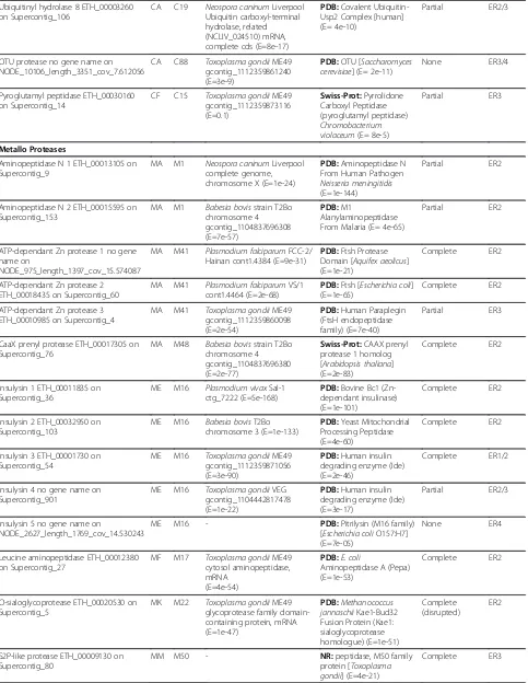
Table 1 Protease genes identified in theEimeria tenellagenome database
Protease/Gene Identifier/Contig Clan Family BLAST Apicomplexa (Database: nucleotide)
BLAST NCBI (Database: PDB, Swissprot, NR)
Family Domains (Pfam, MEROPS, InterProScan) Evidence Rating Aspartic Proteases
Eimepsin 1 ETH_00001725 on Supercontig_54
AA A1 Theileria annulatastrain Ankara genomic DNA chromosome 3 (E=2e-17)
PDB:Porcine Pepsin (E=4e-11)
partial ER1
Eimepsin 2 ETH_00007420 on Supercontig_38
AA A1 Toxoplasma gondiiME49 gcontig_1112359860822 (E=7e-36)
NR:aspartic protease 7 [Toxoplasma gondii] (E=3e-37)
none ER3/4
Eimepsin 3 ETH_00008525 on Supercontig_8
AA A1 Plasmodium bergheiwhole genome shotgun assembly, contig PB_RP2841 (E=1e-93)
PDB:Human pepsin (E=7e-56)
complete ER2
Cysteine Proteases
Cathepsin B ETH_00003570 on Supercontig_23
CA C1 Toxoplasma gondii GAB2-2007-GAL-DOM2 contig00350 (E= 1e-12)
PDB:Human Recombinant Procathepsin B (E=2e-58)
Complete ER2
Cathepsin L ETH_00033530 on
NODE_2923_length_1315_cov_12.253232
CA C1 Toxoplasma gondiiME49 gcontig_1112359872114 (E= 9e-43)
PDB:Toxoplasma gondii Cathepsin L (Tgcpl) (E=1e-64)
Complete ER2
Cathepsin C1 ETH_00019750 on Supercontig_2
CA C1 Toxoplasma gondiiME49 gcontig_1112359873648 (E= 5e-46)
PDB:Porcine Cathepsin H (E=3e-11)
Partial ER2/3
Cathepsin C2 ETH_0005000 on
NODE_22022_length_2554_cov_8.124119
CA C1 Toxoplasma gondiiGT1 gcontig_1107000835548 (E=2e-11)
PDB:Human Dipeptidyl Peptidase I (Cathepsin C) (E=0.016)
Partial ER3/4
Cathepsin C3 ETH_00001590, ETH_00001595 and ETH_00001600 on Supercontig_115
CA C1 Cryptosporidium hominisstrain TU502 chromosome 4 CHRO014106 (E=1e-34)
PDB:Cathepsin CRattus norvegicus(E= 3e-13)
partial ER2
Calpain ETH_00004075 on Supercontig_49
CA C2 Toxoplasma gondiiME49 gcontig_1112359873650 (E=5e-96)
PDB:Human Calpain 8 (E= 1e-16)
Complete ER2
Ubiquitinyl hydrolase 1 ETH_00012075 on Supercontig_122
CA C19 Cryptosporidium murisRN66 gcontig_1106632353963 (E=2e-87)
Swiss-Prot:ubiquitin specific peptidase 39 [Mus musculus] (E=1e-116)
Complete ER2
Ubiquitinyl hydrolase 2 ETH_00034675 on Supercontig_3
CA C19 Cryptosporidium murisRN66 gcontig_1106632353937 (E=3e-35)
Swiss-Prot:ubiquitin specific peptidase 5 (isopeptidase T)[Mus musculus] (E=7e-81)
Partial ER2
Ubiquitinyl hydrolase 3 ETH_00001555 on Supercontig_115
CA C19 Neospora caninumLiverpool ubiquitin carboxyl-terminal hydrolase, related
(NCLIV_041690) mRNA, partial cds (E=63e-94)
PDB:Ubp-Family Deubiquitinating Enzyme [human] (E= 5e-40)
Complete ER2
Ubiquitinyl hydrolase 4 ETH_00007310 on Supercontig_39
CA C19 Cryptosporidium murisRN66 gcontig_1106632353835 (E=1e-60)
PDB:Usp14, A Proteasome-Associated Deubiquitinating Enzyme (E= 5e-40) Complete (disrupted) ER2
Ubiquitinyl hydrolase 5 ETH_00003260 on Supercontig_106
CA C19 Plasmodium falciparumVS/1 cont1.2577 (E=2e-49)
PDB:Human Ubiquitin Carboxyl-Terminal Hydrolase 8 (E=2e-37)
Partial ER2
Ubiquitinyl hydrolase 6 ETH_00020635 on Supercontig_5
CA C19 Toxoplasma gondiiGT1 gcontig_1107000919460 (E=1e-12)
Swiss-Prot:Ubiquitin carboxyl-terminal hydrolase 26Arabidopsis thaliana
(E= 2e-12)
Partial ER2/3
Ubiquitinyl hydrolase 7 ETH_00008925 on Supercontig_8
CA C19 Plasmodium vivaxSaI-1 ctg_6569 (E=1e-30)
PDB:Ubiquitin-Usp2 Complex [human] (E= 2e-15)
Table 1 Protease genes identified in theEimeria tenellagenome database(Continued) Ubiquitinyl hydrolase 8 ETH_00003260
on Supercontig_106
CA C19 Neospora caninumLiverpool Ubiquitin carboxyl-terminal hydrolase, related (NCLIV_024510) mRNA, complete cds (E=8e-17)
PDB:Covalent Ubiquitin-Usp2 Complex [human] (E= 4e-10)
Partial ER2/3
OTU protease no gene name on NODE_10106_length_3351_cov_7.612056
CA C88 Toxoplasma gondiiME49 gcontig_1112359861240 (E=3e-9)
PDB:OTU [Saccharomyces cerevisiae] (E= 2e-11)
None ER3/4
Pyroglutamyl peptidase ETH_00030160 on Supercontig_14
CF C15 Toxoplasma gondiiME49 gcontig_1112359873116 (E=0.1) Swiss-Prot:Pyrrolidone Carboxyl Peptidase (pyroglutamyl peptidase) Chromobacterium violaceum(E= 8e-5)
Partial ER3
Metallo Proteases
Aminopeptidase N 1 ETH_00013105 on Supercontig_9
MA M1 Neospora caninumLiverpool complete genome, chromosome X (E=1e-24)
PDB:Aminopeptidase N From Human Pathogen Neisseria meningitidis (E=1e-144)
Partial ER2
Aminopeptidase N 2 ETH_00015595 on Supercontig_153
MA M1 Babesia bovisstrain T2Bo chromosome 4 gcontig_1104837696308 (E=7e-57)
PDB:M1
Alanylaminopeptidase From Malaria (E= 4e-65)
Partial ER2
ATP-dependant Zn protease 1 no gene name on
NODE_975_length_1397_cov_15.574087
MA M41 Plasmodium falciparumFCC-2/ Hainan cont1.4384 (E=9e-31)
PDB:Ftsh Protease Domain [Aquifex aeolicus] (E=1e-21)
Complete ER2
ATP-dependant Zn protease 2 ETH_00018435 on Supercontig_60
MA M41 Plasmodium falciparumVS/1 cont1.4464 (E=2e-68)
PDB:Ftsh [Escherichia coli] (E=1e-65)
Complete ER2
ATP-dependant Zn protease 3 ETH_00010985 on Supercontig_4
MA M41 Toxoplasma gondiiME49 gcontig_1112359860098 (E=2e-54)
PDB:Human Paraplegin (FtsH endopeptidase family) (E=7e-40)
Partial ER3
CaaX prenyl protease ETH_00017305 on Supercontig_76
MA M48 Babesia bovisstrain T2Bo chromosome 4 gcontig_1104837696380 (E=2e-77)
Swiss-Prot:CAAX prenyl protease 1 homolog [Arabidopsis thaliana] (E=2e-83)
Complete ER2
Insulysin 1 ETH_00011835 on Supercontig_36
ME M16 Plasmodium vivaxSaI-1 ctg_7222 (E=5e-168)
PDB:Bovine Bc1 (Zn-dependant insulinase) (E=1e-101)
Complete ER2
Insulysin 2 ETH_00032950 on Supercontig_103
ME M16 Babesia bovisT2Bo chromosome 3 (E=1e-133)
PDB:Yeast Mitochondrial Processing Peptidase (E=4e-60)
Complete ER2
Insulysin 3 ETH_00001730 on Supercontig_54
ME M16 Toxoplasma gondiiME49 gcontig_1112359871056 (E=3e-90)
PDB:Human insulin degrading enzyme (Ide) (E=2e-46)
Complete ER1/2
Insulysin 4 no gene name on Supercontig_901
ME M16 Toxoplasma gondiiVEG gcontig_1104442817478 (E=1e-22)
PDB:Human insulin degrading enzyme (Ide) (E=3e-17)
Partial ER2/3
Insulysin 5 no gene name on
NODE_2627_length_1769_cov_14.530243
ME M16 - PDB:Pitrilysin (M16 family)
[Escherichia coliO157:H7] (E=7e-05)
None ER4
Leucine aminopeptidase ETH_00012380 on Supercontig_27
MF M17 Toxoplasma gondiiME49 cytosol aminopeptidase, mRNA
(E=4e-54)
PDB:E. coli
Aminopeptidase A (Pepa) (E=1e-53)
Complete ER2
O-sialoglycoprotease ETH_00020530 on Supercontig_5
MK M22 Toxoplasma gondiiME49 glycoprotease family domain-containing protein, mRNA (E=1e-47)
PDB:Methanococcus jannaschiiKae1-Bud32 Fusion Protein (Kae1: sialoglycoprotease homologue) (E=1e-51)
Complete (disrupted)
ER2
S2P-like protease ETH_00009130 on Supercontig_80
MM M50 - NR:peptidase, M50 family
protein [Toxoplasma gondii] (E=4e-21)
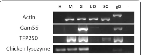
optimize relative amounts of parasite starting material as described previously [18]. TheE. tenellaβ-actin gene was amplified from each of the parasite lifecycle cDNA sam-ples and quantification of bands visualized by agarose gel electrophoresis allowed the specificE. tenellacDNA to be standardized to each other accordingly. The E. tenella gam56gene product, which is predominantly expressed in gametocytes but largely down-regulated in unsporulated oocysts, confirmed the quality of gametocyte cDNA and
[image:5.595.58.540.102.528.2]served as a gametocyte-specific positive control, establish-ing the lack of gametocytes in merozoite and oocyst sam-ples. The amplification of the tfp250 gene, specifically expressed in the asexual stages [19], indicated contaminat-ing merozoite cDNA in the gametocyte cDNA sample, as anticipated, at the 134 h time point. Furthermore, amplifi-cation of a chicken host-specific lysozyme gene indicated host cDNA was present in both merozoite and gametocyte preparations, also as anticipated.
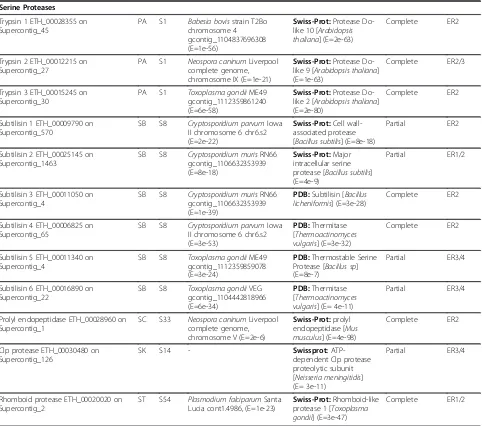
Table 1 Protease genes identified in theEimeria tenellagenome database(Continued)
Serine Proteases
Trypsin 1 ETH_00028355 on Supercontig_45
PA S1 Babesia bovisstrain T2Bo chromosome 4 gcontig_1104837696308 (E=1e-56)
Swiss-Prot:Protease Do-like 10 [Arabidopsis thaliana] (E=2e-63)
Complete ER2
Trypsin 2 ETH_00012215 on Supercontig_27
PA S1 Neospora caninumLiverpool complete genome, chromosome IX (E=1e-21)
Swiss-Prot:Protease Do-like 9 [Arabidopsis thaliana] (E=1e-63)
Complete ER2/3
Trypsin 3 ETH_00015245 on Supercontig_30
PA S1 Toxoplasma gondiiME49 gcontig_1112359861240 (E=6e-58)
Swiss-Prot:Protease Do-like 2 [Arabidopsis thaliana] (E=2e-80)
Complete ER2
Subtilisin 1 ETH_00009790 on Supercontig_570
SB S8 Cryptosporidium parvumIowa II chromosome 6 chr6.s2 (E=2e-22)
Swiss-Prot:Cell wall-associated protease [Bacillus subtilis] (E=8e-18)
Partial ER2
Subtilisin 2 ETH_00025145 on Supercontig_1463
SB S8 Cryptosporidium murisRN66 gcontig_1106632353939 (E=8e-18)
Swiss-Prot:Major intracellular serine protease [Bacillus subtilis] (E=4e-9)
Partial ER1/2
Subtilisin 3 ETH_00011050 on Supercontig_4
SB S8 Cryptosporidium murisRN66 gcontig_1106632353939 (E=1e-39)
PDB:Subtilisin [Bacillus licheniformis] (E=3e-28)
Complete ER2
Subtilisin 4 ETH_00006825 on Supercontig_65
SB S8 Cryptosporidium parvumIowa II chromosome 6 chr6.s2 (E=3e-53)
PDB:Thermitase [Thermoactinomyces vulgaris] (E=3e-32)
Complete ER2
Subtilisin 5 ETH_00011340 on Supercontig_4
SB S8 Toxoplasma gondiiME49 gcontig_1112359859078 (E=3e-24)
PDB:Thermostable Serine Protease [Bacillus sp] (E=8e-7)
Partial ER3/4
Subtilisin 6 ETH_00016890 on Supercontig_22
SB S8 Toxoplasma gondiiVEG gcontig_1104442818966 (E=6e-34)
PDB:Thermitase [Thermoactinomyces vulgaris] (E= 4e-11)
Partial ER3/4
Prolyl endopeptidase ETH_00028960 on Supercontig_1
SC S33 Neospora caninumLiverpool complete genome, chromosome V (E=2e-6)
Swiss-Prot:prolyl endopeptidase [Mus musculus] (E=4e-98)
Complete ER2
Clp protease ETH_00030480 on Supercontig_126
SK S14 - Swissprot:
ATP-dependent Clp protease proteolytic subunit [Neisseria meningitidis] (E= 3e-11)
Partial ER3/4
Rhomboid protease ETH_00020020 on Supercontig_2
ST S54 Plasmodium falciparumSanta Lucia cont1.4986, (E=1e-23)
Swiss-Prot:Rhomboid-like protease 1 [Toxoplasma gondii] (E=3e-47)
Complete ER1/2
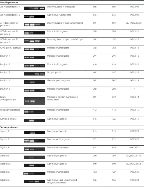
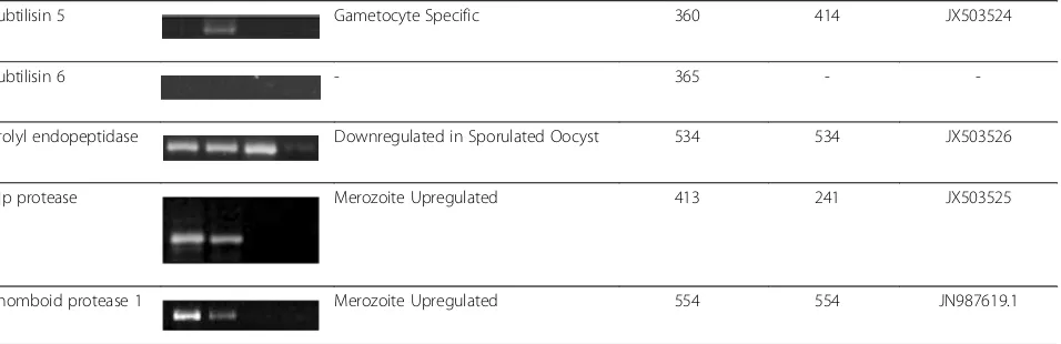
After optimisation of parasite lifecycle stage cDNA samples, primer pairs were designed to generate PCR products from exons of less than 1 kb in size, where possible. PCRs were performed at optimal annealing temperatures specific for the individual primer pairs and annealing times optimal for predicted cDNA sized pro-ducts. PCRs were performed at least twice (and normally three times) for each gene product, by different researchers each time. In the case of failed PCRs, primer pairs were redesigned and retested. Results of PCR on the different lifecycle stages of E. tenella indicated that 40 of the 45 protease genes could be amplified from parasite cDNA (Table 2). The five PCRs that failed to amplify a product from cDNA were for three of the eight ubiquitinyl hydrolases, the single OTU protease and one of the six subtilisins. However, it was possible to amplify PCR products from gDNA for all five of these proteases that, when sequenced, confirmed primer speci-ficity (data not shown). The failure to amplify a product from cDNA for these genes may be due to genome annotation problems; possibly the sequence targeted by our primers is not transcribed or falls in unpredicted in-tronic regions. Alternatively, a low abundance of these transcripts may have contributed to the failure to detect cDNA amplification products. Further work will be required to characterize these genes. All other PCR pro-ducts from cDNA from the four E. tenella lifecycle stages were directly sequenced to confirm the correct coding sequence. Expected and actual cDNA amplicon sizes and their corresponding sequence accession num-bers are shown in Table 2.
The majority of the protease genes were expressed in more than one of the four parasite stages investigated (Table 2). However, stage-specific up- or downregulation of protease gene expression was evident. Thus, taking into account that merozoite cDNA contaminates the
gametocyte samples, it is safe to conclude that there were a large number (at least 15, probably 17) of prote-ase genes whose expression was upregulated in mero-zoites including eimepsin 3, cathepsin C1, calpain, several of the ubiquitinyl hydrolases, an ATP-dependent Zn protease, the CAAX prenyl protease, three of the five insulysins, the leucyl aminopeptidase, the O-sialoglyco-protease, one of the trypsins, a subtilisin, the Clp prote-ase and rhomboid proteprote-ase 1. Aminopeptidprote-ase N1 appeared to be downregulated specifically in merozoites. Gametocyte-specific or gametocyte-upregulated pro-teases were also common, with thirteen in all, also dis-tributed across the four groups of proteases, including eimepsin 2, cathepsin C2, ubiquitinyl hydrolase 2 and 5, the pyroglutamyl peptidase, aminopeptidase N2, insuly-sin 4, the S2P-like metalloprotease, two trypinsuly-sin-like proteases and three of the subtilisins. Additionally, two other proteases were upregulated or specific for the sexual phase of the lifecycle (i.e., gametocytes and unsporulated oocysts), namely, cathepsin C3 and subtili-sin 4. Cathepsubtili-sin L appeared to be downregulated specifically in gametocytes. Only two protease genes, a pepsin-like protease with high homology to eimepsin (eimepsin 1) and an insulysin, were switched on exclu-sively in oocyst lifecycle stages. In contrast, numerous protease genes appeared to be downregulated in sporu-lated oocysts (Table 2).
Protease processing of GAM56
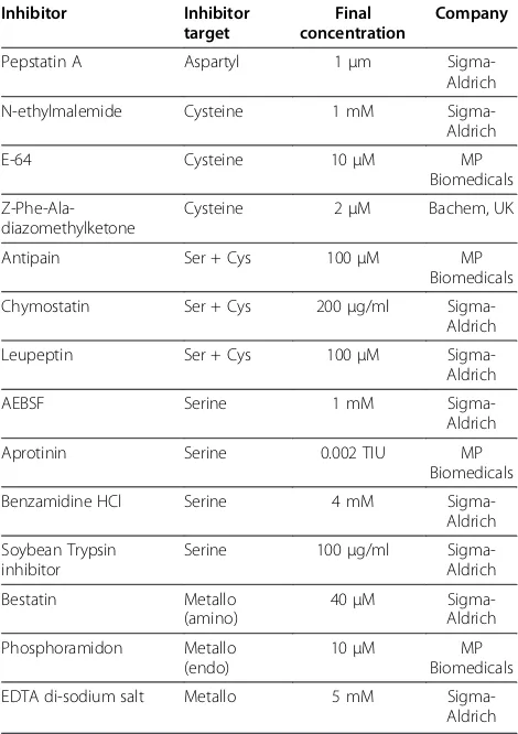
[image:6.595.57.291.89.181.2]Gametocytes from E. tenella-infected caeca were lysed and immediately incubated with or without protease inhibitors for various lengths of time, and the native GAM56 protein analysed by SDS-PAGE and western blotting with anti-GAM56 antibodies, as described previously [20,21], to track the disappearance of the pro-tein to determine whether any inhibitors could prevent the degradation observed in the presence of native gam-etocyte proteases. The precise epitopes recognised by the anti-GAM56 polyclonal antibodies are not known for E. tenellathough there is some evidence, from work with E. maxima [21], that they are located in the con-served amino-terminus of the protein. The anti-GAM56 antibodies are, thus, very useful for sensitive and specific tracking of the degradation of GAM56. No disappear-ance of GAM56 was apparent after 2, 4, 6, 8, 10, 12 or 16 h (data not shown) but was obvious at 24h (Figure 2). The 24 h assay was therefore repeated three times with a comprehensive range of protease inhibitors (Table 3) targeting the four protease families identified in the gen-ome. The aspartyl protease inhibitor, pepstatin A, had no effect on GAM56 disappearance (Figure 2). None of three cysteine protease inhibitors investigated, Z-Phe-Ala-diazomethylketone (data not shown), N-ethylmalemide (data not shown) or E64 (Figure 2) Figure 1RT-PCR assessment of purity ofEimeria tenella
merozoites, gametocytes and oocysts.Total RNA was extracted fromE. tenellamerozoite (M), gametocyte (G), unsporulated oocyst (UO) and sporulated oocyst (SO) preparations. cDNA was synthesized and stage-specific parasite genes (GAM56 for
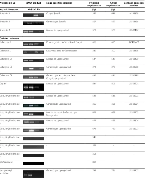
Table 2 Expression of protease genes in merozoites, gametocytes, unsporulated oocysts and sporulated oocysts of Eimeria tenella
Protease group cDNA product Stage specific expression Predicted amplicon size
Actual amplicon size
Genbank accession number
Aspartic Proteases M G UO SO (bp) (bp)
Eimepsin 1 Oocyst Specific 357 659 AJ293829
Eimepsin 2 Gametocyte Specific 467 467 JX503496
Eimepsin 3 Merozoite Upregulated 578 578 JX503497
Cysteine proteases
Cathepsin B Downregulated in Sporulated Oocyst 656 656 JN641867.1
Cathepsin L Downregulated in Gametocytes 330 300 JX503498
Cathepsin C1 Merozoite Upregulated 547 547 JX503499
Cathepsin C2 Gametocyte Upregulated 273 273 JX503500
Cathepsin C3 Gametocyte and Unsporulated
Oocyst Upregulated
436 436 JX546580
Calpain Merozoite Upregulated 831 806 JX503501
Ubiquitinyl hydrolase 1
Merozoite Upregulated 548 548 JX503503
Ubiquitinyl hydrolase 2
Gametocyte Upregulated 395 395 JX503504
Ubiquitinyl hydrolase 3
Merozoite, possibly Gametocyte Upregulated
698 698 JX503505
Ubiquitinyl hydrolase 4
Merozoite Upregulated 499 499 JX503506
Ubiquitinyl hydrolase 5
Gametocyte Upregulated 674 719 JX503507
Ubiquitinyl hydrolase 6
- 546 -
-Ubiquitinyl hydrolase 7
- 529 -
-Ubiquitinyl hydrolase 8
- 303 -
-OTU protease - 802 -
-Pyroglutamyl peptidase
Table 2 Expression of protease genes in merozoites, gametocytes, unsporulated oocysts and sporulated oocysts of
Eimeria tenella(Continued)
Metalloproteases
Aminopeptidase N 1 Downregulated in Merozoite 602 602 JX503508
Aminopeptidase N 2 Gametocyte Upregulated 636 636 JX503509
ATP-dependant Zn protease 1
Downregulated in Sporulated Oocyst 604 604 XM_001238661.1
ATP-dependant Zn protease 2
Merozoite Upregulated 389 389 JX503516
ATP-dependant Zn protease 3
Downregulated in Sporulated Oocyst 567 1045 JX503517
CAAX prenyl protease Merozoite Upregulated 368 368 JX503518
Insulysin 1 Merozoite Upregulated 294 294 JX503510
Insulysin 2 Merozoite Upregulated 416 416 JX503511
Insulysin 3 Oocyst Specific 402 697 JX503512
Insulysin 4 Gametocyte Upregulated 367 367 JX503513
Insulysin 5 Merozoite Upregulated 202 202 *
Leucyl aminopeptidase
Merozoite, possibly Gametocyte Upregulated
804 859 JX503514
O-sialoglycoprotease Merozoite Upregulated 412 412 JX503515
S2P-like protease Gametocyte Specific 616 1027 JX503519
Serine proteases
Trypsin 1 Gametocyte Specific 613 613 JX503520
Trypsin 2 Gametocyte Upregulated 721 721 JX503521
Trypsin 3 Merozoite Upregulated 654 849 JN987371.1
Subtilisin 1 Gametocyte Specific 260 260 XM_001238722.1
Subtilisin 2 Gametocyte Specific 690 690 XM_001238654.1
Subtilisin 3 Merozoite Upregulated 1171 1084 JX503522
Subtilisin 4 Gametocyte and Unsporulated
Oocyst Upregulated
inhibited GAM56 disappearance. The serine/cysteine protease inhibitor, chymostatin (data not shown) and leupeptin (Figure 2), inhibited GAM56 disappearance but another inhibitor with the same specificity, antipain, did not (data not shown). The serine protease specific inhibitors, benzamidine HCL (data not shown), soybean trypsin inhibitor (data not shown) and aprotinin (Figure 2) all inhibited the disappearance of GAM56 but AEBSF did not (Figure 2). The metal chelating agent, EDTA, also inhibited the disappearance of GAM56 but more specific metalloprotease inhibitors, bestatin and phosphoramidon, did not (Figure 2).
Discussion
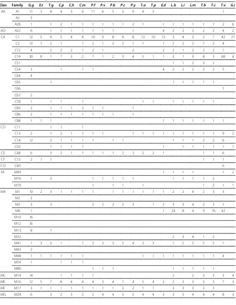
Mining of the E. tenella genome database has revealed over 40 protease transcripts distributed over 13 clans and 18 families of aspartic, cysteine, metallo and serine proteases. Such diversity of proteases is not unusual, in-deed it may be an underestimate of the true number of protease genes in this parasite since other apicomplexan parasites are known to possess substantially more prote-ase genes (Table 4); thus, for example, there are at least 70 in Cryposporidium parvum, more than 80 in P. falciparum and over 90 in T. gondii, though other api-complexan parasites possess similar numbers of protease genes asE. tenella. Eimeria tenellaalso has lower num-bers of protease genes than protozoan parasites like
Leishmania, TrypanosomaandTrichomonas(though the
[image:9.595.62.540.114.269.2]latter is known to have an unusually expanded genome in general [22] and, apparently, in C1 and C19 cysteine proteases and M8 metalloproteases, in particular; Table 4). But, again,E. tenellahas a broadly similar total number of protease genes to Entamoeba dispar and Giardia intestinalis, which are also intestinal parasites. However, the fact that our dataset for E. tenella lacks protease genes for several families, across all four types of proteases, that are represented in all other Apicom-plexa and most other protozoan parasites, including A28, A22, C12, C85, C86, C13, C14, C50, C48, M24, M18, M67, S9, S26 and S16, provides reason to believe that someE. tenellaprotease genes remain unannotated. The apparent stage-specific regulation of protease genes inE. tenellais striking and intriguing. Most inves-tigations of parasitic protozoan proteases have focused on the asexual stages of the apicomplexan parasites,T. gondii and P. falciparum, establishing crucial roles for proteases in host cell invasion, remodelling and egress by the asexual stages of these parasites [1]. Our finding that expression of up to 17 of 40 protease genes Table 2 Expression of protease genes in merozoites, gametocytes, unsporulated oocysts and sporulated oocysts of
Eimeria tenella(Continued)
Subtilisin 5 Gametocyte Specific 360 414 JX503524
Subtilisin 6 - 365 -
-Prolyl endopeptidase Downregulated in Sporulated Oocyst 534 534 JX503526
Clp protease Merozoite Upregulated 413 241 JX503525
Rhomboid protease 1 Merozoite Upregulated 554 554 JN987619.1
MMerozoites,GGametocytes,UOUnsporulated Oocysts,SOSporulated Oocysts. *= short sequence not accepted for submission.
Figure 2The effect of protease inhibitors on degradation of GAM56 inEimeria tenellagametocytes.A sample of purifiedE. tenellagametocytes was lysed and equal volumes of lysate added to a range of protease inhibitors (see Table 3 for details on
[image:9.595.58.292.490.587.2]examined in E. tenellais upregulated in merozoites fur-ther underscores the importance of proteases in the biol-ogy of the asexual stages of apicomplexan parasites. Not surprisingly, therefore, an eimepsin, several cathepsins, a calpain, a trypsin-like protease, subtilisins, Clp and a rhomboid protease are upregulated in the asexual stages ofE. tenella(Table 2). Likewise, eimepsin1 and insulysin 3 are expressed specifically in oocysts and may play an important role in the first steps of the parasite lifecycle, such as host cell invasion; they are, therefore, worthy of further research. The downregulation of several pro-teases (including cathepsin B, ATP-depenedent ZN proteases 1 and 3, and a prolyl endopeptidase) in sporu-lated oocysts may be, in part, attributed to the dormancy of this lifecycle stage, yet still warrants further investigation.
Perhaps the most significant finding of our stage-specific expression study was the relatively large number of protease genes whose expression is upregulated spe-cifically in the gametocytes stage –a total of at least 13 genes, including six that are only expressed in gameto-cyte (Table 2). This observation becomes even more intriguing when examined in the context of the
distribution of different families of proteases across para-sitic protozoa (Table 4). Four classes of proteases stand out amongst the protozoa because they are only found, or are“over-represented”in the two Coccidian parasites, E. tenella andT. gondii–families C15, M50, S1 and S8. Eimeria tenellacontains a total of eleven protease genes distributed unevenly across these families, with only one in C15 and M50 and three and six in the serine protease families, S1 and S8, respectively. But, even more signifi-cantly, all but three of these unique protease genes are upregulated or confined in expression to the gametocyte stage of the parasite. Thus, expression of a pyroglutamyl peptidase, a trypsin-like protease and subtilisin 4 is upre-gulated in gametocytes whilst expression of an SP2-like protease, a trypsin 1-like protease and three subtilisins is entirely gametocyte specific.
[image:10.595.56.291.112.445.2]One of the defining features of the Coccidia is the possession of a hard-walled oocyst that originates from specialized organelles (wall forming bodies) in macroga-metocytes. It is hypothesized [16] that degradation of two proteins found in the wall forming bodies of macro-gametocytes ofEimeria,namely GAM56 and GAM82, is integral to oocyst wall formation; tyrosine-rich peptides formed by the degradation of these two proteins are believed to be subsequently cross-linked via dityrosine bonds [23], giving the oocyst wall its renowned strength and resistance to environmental and chemical insults [24]. To test this hypothesis, we designed an assay to fol-low the degradation of GAM56 in freshly harvested gametocytes (Figure 2). This assay has certain inherent limitations: first, it relies on sensitive antibodies for de-tection of specific degradation of GAM56 and, unfortu-nately, the lack of suitable antibodies for detection of GAM82 inE. tenella[21] meant that we were unable to run confirmatory experiments with this protein; and, second, the only controls possible are a zero time point and a cocktail of protease inhibitors designed to prevent all proteolytic activity. These limitations require us to be cautious in our interpretations; none-the-less, the inhib-ition of degradation of native GAM56 by a very specific group of protease inhibitors reveals that this function may be carried out by subtilisin-like proteases. Thus, degradation of GAM56 was inhibited by the serine/cyst-eine protease inhibitors, chymostatin and leupeptin, and the serine protease specific inhibitors, benzamidine HCL, soybean trypsin inhibitor and aprotinin but not by AEBSF (Figure 2). Intriguingly, the metal chelating agent, EDTA, also inhibited degradation of GAM56. This profile indicates that serine proteases are critical for degradation of GAM56 but it seems to rule out participation of rhomboid pro-teases, which are unaffected by EDTA, aprotonin, leupeptin and chymostatin [25]. Trypsin-like proteases can, perhaps, not be completely ruled out of this process but the inhibi-tory profile, particularly the lack of inhibition by AEBSF Table 3 Protease inhibitors used in theEimeria tenella
GAM56 processing assay Inhibitor Inhibitor
target
Final concentration
Company
Pepstatin A Aspartyl 1μm
Sigma-Aldrich
N-ethylmalemide Cysteine 1 mM
Sigma-Aldrich
E-64 Cysteine 10μM MP
Biomedicals
Z-Phe-Ala-diazomethylketone
Cysteine 2μM Bachem, UK
Antipain Ser + Cys 100μM MP
Biomedicals
Chymostatin Ser + Cys 200μg/ml
Sigma-Aldrich
Leupeptin Ser + Cys 100μM
Sigma-Aldrich
AEBSF Serine 1 mM
Sigma-Aldrich
Aprotinin Serine 0.002 TIU MP
Biomedicals
Benzamidine HCl Serine 4 mM
Sigma-Aldrich
Soybean Trypsin inhibitor
Serine 100μg/ml
Sigma-Aldrich
Bestatin Metallo
(amino)
40μM
Sigma-Aldrich
Phosphoramidon Metallo
(endo)
10μM MP
Biomedicals
EDTA di-sodium salt Metallo 5 mM
Table 4 Distribution of protease clans and families inEimeria tenella, its host and other protozoan parasites
Clan Family G.g E.t T.g C.p C.h C.m P.f P.v P.b P.c P.y T.a T.p E.d L.b L.i L.m T.b T.c T.v G.i
AA A1 11 3 8 4 3 6 11 6 5 3 9 4 3 1
A2 3
A28 1 1 2 1 1 1 1 1 1 2 1 1 1 1 1 1 1 2 6
AD A22 6 1 1 1 1 1 1 1 1 4 2 2 2 2 2 4 2
CA C1 12 5 4 5 4 4 10 9 8 4 8 12 10 15 3 4 3 2 7 42 21
C2 12 1 2 1 1 2 1 2 2 1 1 1 2 2 2 1 2 4
C12 4 2 2 2 1 2 1 2 2 2 1 2 2 2 1
C19 30 8 1 7 3 2 7 1 2 3 4 5 1 1 3 1 9 8 3 68 4
C51 1 1 2 3 2
C54 3 1 1 1 4 2 2 2 2 2 5
C64 4
C65 1 1 1 1 1 1
C66 1
C67 2
C78 1 1 1 1 1 1 1 1 1 1 1
C85 2 1 1 1 2 1 1 1
C86 2 1 1 1 1 1 1 1 1
C88 1 1 1 1 1 1 1 1 1 1
CD C11 1 1 1
C13 2 1 2 1 1 1 1 1 1 1 1 1 1 1 1 1 9 2
C14 12 2 1 1 1 1 1 1 1 1 1 2 2 6
C50 1 1 1 1 1 1 1 1 1 1 1
CE C48 6 3 2 1 1 1 1 1 2 2 2 2 1 1 1 6
CF C15 2 1 1 1 1 1
CO C40 6
M- M49 1 1 1 1 1 2
M76 1 3 1 1 1 1 1 1 1 1 1 2
M79 1 1 1 1 1 2 1 1
MA M1 10 2 3 1 1 1 1 1 1 1 1 1 1 2 2 4 2 3 3
M2 3
M3 3 3 2 2 2 2 3 1 2 3 3 4 2 3 1
M8 1 1 24 8 4 9 76 42
M10 16
M12 35
M13 8 1
M32 2 3 4 1 2
M41 1 3 3 1 1 3 3 3 3 4 3 3 1 5 5 5 5 1
M43 2
M48 1 1 1 1 1 1 1 1 1 1 1 1 1 1 4
M54 1 1 1 1
M80 1 1 1 1 1 1 1 1
MC M14 14 1 1 1 1 2 2 3 3 3 4
ME M16 12 5 7 8 4 4 4 3 4 1 4 5 4 2 2 3 3 3 3 7 3
MF M17 3 1 1 1 1 1 1 1 1 3 2 1 1 3 3 3 3 3
coupled with the inhibitory effect of EDTA, points to a sub-tilisin or subsub-tilisins asP. falciparumsubtilisin 1 is inhibited in exactly the same fashion [26].
Subtilisins are further implicated in the formation of the oocyst wall of Eimeria through analogy with their known role in the formation of the cuticle of nematodes. Thus, the assembly of collagens to form the cuticle involves a number of molecular events that strikingly resemble our model of oocyst wall formation pathways: first, collagens are the re-sult of degradation of proproteins by a subtilisin-like prote-ase [27-30]; and, second, these collagens are subsequently bonded together by di- and tri-tyrosine crosslinks [31]. A failure in either of these steps, results in a malformed cu-ticle and parasite death [31]. Subtilisins are currently being further investigated as potential candidates in the catalytic cleavage of the oocyst wall precursor proteins.
Conclusion
Eimeria tenellapossesses a large number of genes coding for proteolytic enzymes, which display a remarkable pattern
of stage specific expression. As in other apicomplexan para-sites such as P. falciparum and T. gondii, expression of many of these genes is upregulated in the asexual, invasive stages, possibly indicating important roles in host cell inva-sion, remodelling and egress. However, expression of al-most one-third of the protease genes identified in the E. tenella genome is upregulated or confined to the sexual gametocyte stage of this parasite’s lifecycle; some of these appear to be unique to Coccidia and may play key roles in the formation of the resilient oocyst wall, a defining feature of this group of important parasites.
Methods
Data-base mining
[image:12.595.57.539.100.419.2]Eimeria tenellagenome sequences and gene models were downloaded from GeneDB (http://www.genedb.org/Home page/Etenella). The genome ofE. tenella(Houghton strain) was produced by the Parasite Genomics Group at the Well-come Trust Sanger Institute (http://www.sanger.ac.uk/re-search/projects/parasitegenomics/) and has been provided Table 4 Distribution of protease clans and families inEimeria tenella, its host and other protozoan parasites(Continued)
MH M18 1 1 1 1 1 1 1 1 1 1 1 1 1 1 1 1 1 1 5
M20 4 1 1 6 3 4 7 4 8 10 3
M28 10 1 1
MJ M19 1
M38 4
MK M22 2 1 1 1 1 1 2 1 1 1 2 2 1 1 1 1 1 1 1 1 2
MM M50 1 1 1
MP M67 7 2 2 2 1 1 1 1 1 1 2 2 2 2 2 3 1 3 4 2
PA S1 66 3 4 1 2 1
SB S8 10 6 11 1 1 3 3 2 2 2 1 2 2 11
SC S9 15 4 10 3 2 3 1 1 2 2 1 2 5 4 7 8 8 8 5
S10 2 1 1 1 3 1
S15 1 1 1 1 1
S28 1 1 2 1 8 2
S33 3 1 2 3 1 6 1 1 4 1 1 2 1 2 1 2 13 7
SE S12 1
SF S26 4 1 1 1 1 1 1 1 2 2 1 2 2 2 2 1 3
SJ S16 2 1 1 1 1 1 1 1 1
SK S14 1 1 1 1 1 1 2 1
S41 1
SP S59 1 1 1 1
SR S60 1
ST S54 6 1 6 3 2 3 7 2 4 2 5 1 1 1 2
Clan Family G.g E.t T.g C.p C.h C.m P.f P.v P.b P.c P.y T.a T.p E.d L.b L.i L.m T.b T.c T.v G.i
Table 5 Primers forEimeria tenellaprotease genes for the amplified coding sequences
Protease gene Primer Primer sequence 50to 30
Aspartic Proteases
Eimepsin 1 F ATC ACC ACA CCA CCA TGG G
R CAA GAT TCG AGC AGT TCT CAG CA
Eimepsin 2 F AGC AAA CAG CTG CAG ATG TTC C
R CGA AAA TAG GTC TAG GGG CCC
Eimepsin 3 F AGA AGT CCT TTC CTC CGT CAC G
R GGC GAA GTG TAT TGA AAG CTC G
Cysteine Proteases
Cathepsin B F TGA CGT GGG AAG CAG AAG TGT C
R ACT GTA TGC ACT CGT GGC GAA A
Cathepsin L F CAA CCA ACA AGG TCA CTC TTA C
R CCC TGG AGG GCC CCC GTG CTC G
Cathepsin C1 F AAC GGA AGT GGA GAA GAC AGC A
R CGG GGT CAA TGA AGG AAG TTT G
Cathepsin C2 F GGC TTT GAA GTT TGG CAG CG
R TGC ATC TGA GCA GCA GAA GAG
Cathepsin C3 F CGC TCA GGA GTA CAA CTA CGT GGG TGG
R GCT GCT GCA CAA GCA GCG CTG CTC TGC C
Calpain F CGA CTG CTC CTT CCT CTC
R GCA TCC TTT AGC TGC TGG
Ubiquitinyl hydrolase 1 F CTG GCT CTT GAA TGT GCT GCA T
R CTT CCA TTT CGT GCC ATT CG
Ubiquitinyl hydrolase 2 F TTG GAC TTC AGC CCG AGG AG
R CCG CTC GTC AAA TCC CAA AG
Ubiquitinyl hydrolase 3 F AGG TGG TGC CCC TCG ACA TA
R TTC TGC TGC CGT GCT CTT TG
Ubiquitinyl hydrolase 4 F GTG AAT GTG GGC AGC ACC AG
R GCT TTC CTG CAA GGC GAA GA
Ubiquitinyl hydrolase 5 F TCT CTT CCA GGG GCA GTA CAG G
R GCT GCT CGT AGG CCA TAT TTG AAC
Ubiquitinyl hydrolase 6 F GGT GGC CAG CAT AGA CGA GAG T
R CCG TAG GTT TGC TGC GAT CTC T
Ubiquitinyl hydrolase 7 F CGA CAG TCC CGT TGT TGC TG
R AGC AAG CCG GGG AAA GAG AC
Ubiquitinyl hydrolase 8 F TCC TGG AGG CCT TGA GCA GT
R TGC TGC TCA CTG GGA TGT GG
OTU protease F ATG GAC GTA GCA ATC CGT TAC
R GTT GCT GCA CAT AGT CCC AAG
Pyroglutamyl peptidase F ATG GGA CAA CCT ACA GGC GAG
R GAG GAA GTT ACA AAC GAA GCA GC
Metallo Proteases
Aminopeptidase N 1 F CAA GCA GTA CAC TCC AGC AAC TC
Table 5 Primers forEimeria tenellaprotease genes for the amplified coding sequences(Continued)
Aminopeptidase N 2 F GAT ACA TCG CAA GGA CTA CAG C
R CAT TGT GCG GAA GGA GTC TG
ATP-dependant Zn protease 1 F CTT CGA CCA GCT GAA GAT CCT G
R TGT CTC CGC GCT AAT GCT G
ATP-dependant Zn protease 2 F CTC AAT GGT GGA TTT CAG CAA G
R CAT CAT TCC CTG GCT TGC G
ATP-dependant Zn protease 3 F GTC AAG AAA GCA GAT TGT CAG G
R CTC GAC AGC CAT CTC AAA GTC
CAAX prenyl protease F GAA TGT CCC AGG AGA GCT ATG C
R GTT CAC AGC GCA CAG AAT TGG
Insulysin 1 F CAC TTC CTG GAG CAC ATG G
R GTG CAG CCT GTC GAA GAT G
Insulysin 2 F CCG CGG TAG CCT CTCA GC
R CTC CGC TGT TGC TCA CAT CTT C
Insulysin 3 F GAT TGA CGC CGA GCA CCA G
R GCT AGG CGA GCT GCT TCA TCC
Insulysin 4 F GTC GTG CAG AGA GGC TGG
R GAG GTA GCT GAG TCG GAG GG
Insulysin 5 F ATG ATA TTT CTG AAG TCT GC
R GAG GAT TCA AAA GAT GGT C
Leucine aminopeptidase F CAA GAT CTA GTT GTT TGA GGA GCC C
R GCT GTT CCA CTT CAC TCG TCT TC
O-sialoglycoprotease F GCA AAT GTG ACG CAG CTG ATT CG
R CGA ACT TGA GGG CCA TTT GCT C
S2P-like protease F GCC CTC GTA TAA CAG CAA C
R GAA CGA CTA ACT TCC TCT TGC
Serine Proteases
Trypsin 1 F GTA GGC AAC GGA ACT CCA GC
R CGA GGA CTA CAA CTG TCT CC
Trypsin 2 F ATG GAA GCG TCT GGT TGG GAC
R GCT TTT GCT GCA TGC ACT C
Trypsin 3 F GAC AAA CAA GTT CAA CGA GCA CTG
R GCT TCC CTC TCC GCA GAA TTG
Subtilisin 1 F TAA TTA CCT CCA TCC CGA ACT G
R CCA GAA TCT TCA GCG CCA TCA C
Subtilisin 2 F GCA GCA GCA AAT GTT GAA GAC CC
R ATA AGT GCT GCT GCC AAC CAC C
Subtilisin 3 F AGA GCT TTT GTC CGT GGT GGA G
R AAA GAC CCC GAA AAC CAA TGC T
Subtilisin 4 F CCT TTG TGG CGT GTT CGT GAG
R CCA GCA GAA GCA GTA CCG TGG CC
Subtilisin 5 F TTG AAG CCG ACA GGA CGT GG
R CCG CGT AGT CAA GAG CAG GGA T
Subtilisin 6 F AGC GGC TGC GAC TTG AAC C
prepublication. TheE. tenellagenome database was searched for genes predicted to code for proteins with peptidase ac-tivity. All auto-annotated peptidase genes identified were manually curated by performing BLAST analysis against apicomplexan genome sequence databases and against vari-ous protein databases [32] such as the protein data bank (PDB), Swiss-Prot and non-redundant (NR) protein se-quence databases. In addition, signature protein motifs for the protein sequence of each gene were identified through Pfam (http://pfam.sanger.ac.uk/search; [33]), InterproScan (http://www.ebi.ac.uk/Tools/pfa/iprscan/) and the MER-OPS databases (http://merops.sanger.ac.uk/; [34]). Further gene sequence manipulations, such as translation into amino acid sequences and ClustalW alignments, were per-formed using the DNASTAR Lasergene™ 9 Core Suite. After the bioinformatic information was collated, genes were assigned a five-tiered level of confidence for gene function using an Evidence Rating (ER) system giving an overall score of ER1-5, where ER1 indicates extremely reli-able experimental data to support function and ER5 indi-cates no evidence for gene function [17].
Animals and parasites
One day old chicks (Australorp; Barter and Sons Hatchery, Luddenham, Australia) were housed at the Ernst Facility Animal House (University of Technology, Sydney), under heat lamps for the first 2 weeks of their life and, thereafter, at 21°C with a 12 hour light/dark cycle with free access to food and water. Chickens were infected orally at 4.5 weeks of age with 2.5 × 10³sporulated oocysts ofEimeria tenella (Houghton strain originally provided by Janene Bumstead, Institute for Animal Health, Compton, UK). FreshE. tenella oocysts were harvested 7 days post infection from the caeca following protocols published previously [35]. Sporulation of oocysts was carried out at 28°C for 72–120 hours using a low-pressure aquarium pump to aerate the suspension. Sporulated oocysts were then treated with 2.8 M NaCl and 2% sodium hypochlorite (Milton solution) and stored in 2% potassium dichromate at 4°C until required. Unsporulated oocysts were also treated with Milton solution and stored at
−80°C. Merozoites (112 h p.i.) and gametocytes (134 h p.i.) were isolated from infected chicken caecae following tech-niques published previously [36,37]. Aliquots of parasites were either frozen at −80°C as pellets or were stored in
TRIzolW reagent (Invitrogen) at −80°C for further use in RNA purification.
RNA purification, cDNA synthesis and cDNA standardisation
To isolate total RNA, purified merozoites (1 × 10⁷) and gametocytes (1 × 10⁶) were resuspended in 1 ml
TRIzolW Reagent and homogenized by pipetting.
Unsporulated oocysts (2 × 10⁵) and sporulated oocysts (5 × 10⁵) were resuspended in 1 ml TRIzolW Reagent and one volume of glass beads were added to the sam-ple, which were then vortexed for 1 min intervals until disruption of oocyst was confirmed by bright field mi-croscopy. All TRIzolW treated samples were left at room temperature for 10 min and total RNA isolated by chloroform extraction and isopropanol precipitation.
RNA was quantified using a NanoDrop™ ND-1000
Spectrophotometer and cDNA was synthesized using SuperScript™ III Reverse Transcriptase (Invitrogen) according to manufacturer’s instructions.
Parasite cDNA samples were standardized by relative quantification of an E. tenella β-actin PCR product.
β-actin forward primer E0043 (50 ggaattcgttggccgcccaa gaatcc 30) and reverse primer E0044 (50 gctctaga ttagctcggcccagactcatc 30) were used to generate the 1020 bpβ-actin cDNA PCR product. Each PCR reaction contained 50 ng of parasite stage specific cDNA, 0.2μM forward primer, 0.2μM reverse primer, 1 × AccuPrime™ reaction mix, and AccuPrime™ Pfx DNA polymerase (Invitrogen). The PCR reaction was carried out as follows: initial denaturation 95°C for 3 min; 95°C for 30 s; 61°C for 1 min; 68°C for 1.5 min, for 25 cycles with a final extension at 68°C for 10 min. All products were electrophoresed on a 1% agarose gel and visualized using Gel Red™ (Biotium). The net intensity of each band was determined using the Kodak EDAS 290 Electrophoresis Documentation and Analysis System and serial dilutions performed until rela-tive intensity of PCR products were equal.
In addition, three control genes were amplified to de-termine the purity of parasite lifecycle stages. The GAM56 gene was used as a gametocyte specific gene.
GAM56 forward primer E0030 (50 catatggtggagaa
[image:15.595.56.538.103.189.2]cacggtgcac 30) and reverse primer E0031 (50 ctcgagttagt accagctggaggagta 30) were designed to amplify a 906 bp Table 5 Primers forEimeria tenellaprotease genes for the amplified coding sequences(Continued)
Prolyl endopeptidase F ACA GCC AGG CAC ATC AAT GGT
R GCC AAA CCC AAG CCC AGA TAG
Clp protease F GCT GCA CTT CCA GAA GCG G
R CCT CCG CAG AGA AGA CTT TGC
Rhomboid protease 1 F GGT TGT CCG CAC GTT GGC AG
gametocyte cDNA product at an annealing temperature
of 61°C. The EtTFP250 gene, a homolog of an E.
maxima gene encoding a microneme protein, was used
as an asexual stage control. The EtTFP250 forward pri-mer Et250F (50gcaaggacgttgacgagtgtg 30) and Et250RV1 (50gttctctccgcaatcgtcagc 30) were designed to amplify an 805 bp cDNA product, at an annealing temperature of 60°C. The chicken lysozyme gene was used to determine relative quantities of contaminating host cDNA. The for-ward primer RW3F (50 acaaagggaaaacgttcacgattggc 30) and reverse primer RW4R (50 tgcgttgttcacaccctgcatatgcc 30) were designed to amplify a 280 bp host cDNA prod-uct at an annealing temperature of 60°C.
Semi-quantitative PCR
The predicted coding regions of each protease gene were examined for potential primer sites within 1 kb of each other where possible. Primers were designed as detailed in Table 5.
PCRs were conducted on cDNA samples fromE. tenella merozoites, gametocytes, unsporulated and sporulated oocysts. PCR were optimized to produce cDNA sized pro-ducts. Negative controls of no DNA template and host cDNA were run alongside a positive genomic DNA control. When genomic DNA products were not amplified, a repeat PCR was performed at longer annealing times to produce the often much larger genomic DNA product. A typical PCR was as follows: 1μL of standardized cDNA sample, 0.2 μM forward primer, 0.2μM reverse primer, 1 × Accu-Prime™ reaction mix, and AccuPrime™ Pfx DNA poly-merase (Invitrogen). Cycling conditions typically involved an initial denaturation at 95°C for 3 min, followed by 25 cycles of denaturation 95°C for 30 s; annealing at Tm-5 for 1 min; extension at 68°C for 1.5 min. When products were to be sequenced, a final extension at 68°C for 10 min was performed at the end of the PCR reaction. PCRs were per-formed at least twice and, generally, three times for each gene product by a different researcher each time.
All amplified products were gel purified using a QIAquickWGel Extraction Kit (QIAGEN) according to the manufacturer’s instructions and sequenced (Australian Genome Research Facility, Queensland). When cDNA pro-ducts were amplified from different parasite stages, these were pooled and used in sequencing reactions. When cDNA products were not obtained, additional primers were designed and used. If a cDNA product was still unable to be amplified with the second primer pair, genomic DNA products were sequenced to confirm primer specificity. Sequences were analysed using DNASTAR Lasergene™9 Core suite.
GAM56 processing assay
A frozen sample of purified E. tenella gametocytes (1 × 10⁶ cells in 100μL) was resuspended in PBS (145 mM
NaCl, 7.5 mM Na2HPO4, 2.5 mM NaH2PO4.2H2O, pH 7.4) to a final volume of 500 μL. Glass beads (250μL of 710-1180μm, Sigma) were added to the suspension and vortexed at full speed for three 1 min pulses with a 1 min pause on ice between each pulse. After three vortex cycles, the sample was centrifuged and the lysate trans-ferred to a clean tube. Equal aliquots of the gametocyte extract (18μL) were immediately added to either 2μL of 10× protease inhibitor (Table 3) or PBS. A zero time sample was taken from the PBS control and immediately added to Laemmli sample buffer [38] and frozen. The assay tubes were incubated at 37°C for 2, 4, 6, 8, 10, 12, 16 or 24 h, after which Laemmli sample buffer was
added and samples stored at −20°C for further
assessment.
SDS-PAGE and immunoblotting were carried out as described previously [20,21]. Briefly, gametocyte assay samples, resuspended in Laemmli sample buffer (5 μL), were boiled for 5 min at 100°C prior to SDS-PAGE on a NuPAGEW Novex 4-12% Bis-Tris Gel (Invitrogen). SeeBlueW Plus2 Pre-Stained Standards (Invitrogen) were used as a marker. Proteins separated by SDS-PAGE were transferred electrophoretically (100 V, 1 hour, 4°C) to Immobilon-P membrane (Millipore) in transfer buffer (25 mM Tris–HCl, 192 mM glycine, 10% methanol). Membranes were rinsed in methanol and water then soaked for 10 min in transfer buffer prior to transfer. Gels were pre-soaked for 15 min in transfer buffer. After transfer, membranes were incubated overnight in block-ing solution (5% w/v skim milk powder in PBS) at 4°C and then incubated with primary antibody (1:1000 rabbit anti-EmGAM56) for 2 h at room temperature. Each membrane was washed twice for 5 min and twice for 10 min in 0.05% Tween 20 in PBS then incubated with sec-ondary antibody (1:1000 anti-rabbit IgG conjugated to alkaline phosphatase; Sigma) for 2 h at RT. Membranes were washed as above and bands visualized with SIGMA
FAST™BCIP/NBT buffered substrate (Sigma).
Competing interests
The authors declare they have no competing interests.
Authors’contributions
NCS, MK and SIB conceived the study. MK coordinated the study and performed data analysis, along with FB, FMT and NCS. MK, RJI, MR, IS, PAS and RAW carried out the laboratory work. NCS, MK, FMT and FB drafted the manuscript. All authors read and approved the final manuscript.
Acknowledgements
Institute for providing us pre-publication access to theE. tenellagenome database.
Author details
1Institute for the Biotechnology of Infectious Diseases, University of Technology, Sydney, Broadway, N.S.W. 2007, Australia.2Queensland Tropical Health Alliance Research Laboratory, Faculty of Medicine, Health and Molecular Sciences, James Cook University, Cairns Campus, McGregor Road, Smithfield, QLD 4878, Australia.3INRA UR1282, Equipe Pathogenèse des Coccidioses, Infectiologie Animale et Santé Publique, 37380, Nouzilly, France. 4Royal Veterinary College, Hawkshead Lane, North Mymms, Hatfield, Hertfordshire AL9 7TA, UK.
Received: 31 August 2012 Accepted: 4 December 2012 Published: 7 December 2012
References
1. Li H, Child MA, Bogyo M:Proteases as regulators of pathogenesis: examples from the Apicomplexa.Biochim Biophys Acta2012,1824:177–185. 2. Florens L, Washburn MP, Raine JD, Anthony RM, Grainger M, Haynes JD,
Moch JK, Muster N, Sacci JB, Tabb DL, Witney AA, Wolters D, Wu Y, Gardner MJ, Holder AA, Sinden RE, Yates JR, Carucci DJ:A proteomic view of the Plasmodium falciparumlife cycle.Nature2002,419:520–526.
3. Eksi S, Czesny B, Greenbaum DC, Bogyo M, Williamson KC:Targeted disruption ofPlasmodium falciparumcysteine protease, falcipain 1, reduces oocyst production, not erythrocytic stage growth.Mol Microbiol 2004,53:243–250.
4. Eksi S, Czesny B, van Gemeet G-J, Sauerwien RW, Eling W, Williamson KC:
Inhibition ofPlasmodium falciparumoocyst production by membrane-permeant cysteine protease inhibitor E64d.Antimicrob Agents Chemother 2007,51:1064–1070.
5. Czesny B, Gosh S, Cook JL, Williamson KC:The proteasome inhibitor epoxomicin has potentPlasmodium falciparumgametocytocidal activity. Antimicrob Agents Chemother2009,53:4080–4085.
6. Aminake MN, Schoof S, Sologub L, Leubner M, Kirschner M, Arndt H-D, Pradel G:Thiostrepton and derivative exhibit antimalarial and gametocytocidal activity by dually targeting parasite proteosome and apicoplast.Antimicrob Agents Chemother2011,55:1338–1348.
7. Laurent F, Bourdieu C, Kaga M, Chilmonczyk S, Zgrzebski G, Yvore P, Pery P:
Cloning and characterization of anEimeria acervulinasporozoite gene homologous to aspartyl proteinases.Mol Biochem Parasitol1993,
62:303–312.
8. Jean L, Grosclaude J, Labbe M, Tomley F, Pery P:Differential localisation of anEimeria tenellaaspartyl proteinase during the infection process.Int J Parasitol2000,30:1099–1107.
9. Jean L, Pery P, Dunn P, Bumstead J, Billington K, Ryan R, Tomley F:Genomic organisation and developmentally regulated expression of an apicomplexan aspartyl proteinase.Gene2001,262:129–136.
10. de Venevelles P, Chich JF, Faigle W, Lombard B, Loew D, Pery P, Labbe M:
Study of proteins associated with theEimeria tenellarefractile body by a proteomic approach.Int J Parasitol2006,36:1399–1407.
11. Fetterer RH, Miska KB, Barfield RC:Partial purification and characterization of an aminopeptidase fromEimeria tenella.J Parasitol2005,91:1280–1286. 12. Li J, Zhang X, Liu Q, Yin J, Yang J:Eimeria tenella:Cloning of a novel
Eimeria tenellacDNA encoding a protein related to rhomboid family from F2 hybrid strain.Exp Parasitol2006,113:215–220.
13. Fetterer RH, Miska KB, Lillehoj H, Barfield RC:Serine protease activity in developmental stages ofEimeria tenella.J Parasitol2007,93:333–340. 14. Lal K, Bromley E, Oakes R, Prieto JH, Sanderson SJ, Kurian D, Hunt L, Yates JR
III, Wastling JM, Sinden RE, Tomley FM:Proteomic comparison of four Eimeria tenellalife-cycle stages: unsporulated oocyst, sporulated oocyst, sporozoite and second-generation merozoite.Proteomics2009,
9:4566–4576.
15. Rieux A, Gras S, Lecaille F, Niepceron A, Katrib M, Smith NC, Lalmanach G, Brossier F:Eimeripain, a cysteine cathepsin B-like protease, expressed throughout sporulation of the apicomplexan parasiteEimeria tenella. PLoS One2012,7:e31914.
16. Belli SI, Smith NC, Ferguson DJP:The Coccidian oocyst: a tough nut to crack!Trends Parasitol2006,22:416–423.
17. Allen MA, Lauro FM, Williams TJ, Burg D, Siddiqui KS, De Francisci D, Chong KW, Pilak O, Chew HH, De Maere MZ, Ting L, Katrib M, Ng C, Sowers KR,
Galperin MY, Anderson IJ, Ivanova N, Dalin E, Martinez M, Lapidus A, Hauser L, Land M, Thomas T, Cavicchioli R:The genome sequence of the psychrophilic achaeon,Methanococcoides burtonii:the role of genome evolution in cold adaptation.ISME J2009,3:1012–1035.
18. Walker RA, Slapetova I, Slapeta J, Miller CM, Smith NC:The glycosylation pathway ofEimeria tenellais upregulated during gametocyte
development and may play a role in oocyst wall formation.Euk Cell2010,
9:127–135.
19. Witcombe DM, Belli SI, Wallach MG, Smith NC:EmTFP250: a novel member of the TRAP protein family implicated in maternal immunity toEimeria maxima.Int J Parasitol2003,33:691–702.
20. Belli SI, Lee M, Thebo P, Wallach MG, Schwartsburd B, Smith NC:
Biochemical characterisation of the 56 and 82 kilodalton immunodominant gametocyte antigens fromEimeria maxima. Int J Parasitol2002,32:805–816.
21. Belli SI, Ferguson DJP, Katrib M, Slapetova I, Mai K, Slapeta J, Flowers SA, Miska KB, Tomley FM, Shirley MW, Wallach MG, Smith NC:Conservation of proteins involved in oocyst wall formation inEimeria maxima,Eimeria tenella and Eimeria acervulina.Int J Parasitol2009,39:1063–1070. 22. Carlton JM, Hirt RP, Silva JC, Delcher AL, Schatz M, Zhao Q, Wortman JR,
Bidwell SL, Alsmark UC, Besteiro S, Sicheritz-Ponten T, Noel CJ, Dacks JB, Foster PG, Simillion C, Van de Peer Y, Miranda-Saavedra D, Barton GJ, Westrop GD, Müller S, Dessi D, Fiori PL, Ren Q, Paulsen I, Zhang H, Bastida-Corcuera FD, Simoes-Barbosa A, Brown MT, Hayes RD, Mukherjee M, Okumura CY, Schneider R, Smith AJ, Vanacova S, Villalvazo M, Haas BJ, Pertea M, Feldblyum TV, Utterback TR, Shu CL, Osoegawa K, de Jong PJ, Hrdy I, Horvathova L, Zubacova Z, Dolezal P, Malik SB, Logsdon JM Jr, Henze K, Gupta A, Wang CC, Dunne RL, Upcroft JA, Upcroft P, White O, Salzberg SL, Tang P, Chiu CH, Lee YS, Embley TM, Coombs GH, Mottram JC, Tachezy J, Fraser-Liggett CM, Johnson PJ:Draft genome sequence of the sexually transmitted pathogenTrichomonas vaginalis.Science2007,315:207–212. 23. Mai K, Smith NC, Feng ZP, Katrib M, Slapeta J, Slapetova I, Wallach MG,
Luxford C, Davies MJ, Zhang X, Norton RS, Belli SI:Peroxidase catalysed cross-linking of an intrinsically unstructured protein via dityrosine bonds in the oocyst wall of the apicomplexan parasite, Eimeria maxima.Int J Parasitol2011,41:1157–1164.
24. Mai K, Sharman PA, Walker RA, Katrib M, DeSouza D, McConville MJ, Wallach MG, Belli SI, Ferguson DJP, Smith NC:Oocyst wall formation and composition in Coccidian parasites.Memorias do Instituto Oswaldo Cruz2009,10:281–289. 25. Urban S, Wolfe MS:Reconstitution of intramembrane proteolysisin vitro
reveals that pure rhomboid is sufficient for catalysis and specificity. Proc Natl Acad Sci USA2005,102:1883–1888.
26. Withers-Martinez C, Saldanha JW, Ely B, Hackett F, O’Connor T, Blackman MJ:
Expression of recombinantPlasmodium falciparumsubtilisin-like protease-1 in insect cells. Characterization, comparison with the parasite protease, and homology modeling.J Biol Chem2002,277:29698–29709. 27. Thacker C, Peters K, Srayko M, Rose AM:The bli-4 locus ofCaenorhabditis
elegansencodes structurally distinct kex2/subtilisin-like endoproteases essential for early development and adult morphology.Genes Dev1995,
9:956–971.
28. Thacker C, Rose AM:A look at theCaenorhabditis elegans Kex2/Subtilisin-like proprotein convertase family.Bioessays2000,22:545–553.
29. Thacker C, Srayko M, Rose AM:Mutational analysis of bli-4/kpc-4 reveals critical residues required for proprotein convertase function inC. elegans.Gene2000,252:15–25.
30. Thacker C, Sheps JA, Rose AM:Caenorhabditis elegansdpy-5 is a cuticle procollagen processed by a proprotein convertase.Cell Mol Life Sci2006,
63:1193–1204.
31. Page AP, Winter AD:Enzymes involved in the biogenesis of the nematode cuticle.Adv Parasitol2003,53:85–148.
32. Altschul SF, Gish W, Miller W, Myers EW, Lipman DJ:Basic local alignment search tool.Mol Biol1990,215:403–410.
33. Punta M, Coggill PC, Eberhardt RY, Mistry J, Tate J, Boursnell C, Pang N, Forslund K, Ceric G, Clements J, Heger A, Holm L, Sonnhammer EL, Eddy SR, Bateman A, Finn RD:The Pfam protein families database.Nucleic Acids Res 2012,40:D290–D301.
34. Rawlings ND, Barrett AJ, Bateman A:MEROPS:the peptidase database. Nucleic Acids Res2010,38:D227–D233.
36. Smith NC, Wallach M, Miller CMD, Morgenstern R, Braun R, Eckert J:
Maternal transmission of immunity toEimeria maxima: ELISA analysis of protective antibodies induced by infection.Infect Immun1994,
62:1348–1357.
37. Wallach MG, Mencher D, Yarus S, Pillemer G, Halabi A, Pugatsch T:
Eimeria maxima:Identification of gametocyte protein antigens. Exp Parasitol1989,68:49–56.
38. Laemmli UK:Cleavage of structural proteins during the assembly of the head of bacteriophage T4.Nature1970,227:680–685.
doi:10.1186/1471-2164-13-685
Cite this article as:Katribet al.:Stage-specific expression of protease genes in the apicomplexan parasite,Eimeria tenella.BMC Genomics2012 13:685.
Submit your next manuscript to BioMed Central and take full advantage of:
• Convenient online submission
• Thorough peer review
• No space constraints or color figure charges
• Immediate publication on acceptance
• Inclusion in PubMed, CAS, Scopus and Google Scholar
• Research which is freely available for redistribution