A Dissertation on
‘’A STUDY OF PATHOPHYSIOLOGY, MANAGEMENT AND FACTORS INFLUENCING DIABETIC FOOT ULCERS AMONG
DIABETIC PATIENTS’’
Dissertation Submitted to
THE TAMIL NADU Dr.M.G.R. MEDICAL UNIVERSITY
CHENNAI- 600032
with partial fulfillment of the regulations for the award of the degree of
M.S. GENERAL SURGERY
(BRANCH 1)
COIMBATORE MEDICAL COLLEGE,
COIMBATORE
CERTIFICATE
Certified that this is the Bonafide Dissertation in ‘’A STUDY OF
PATHOPHYSIOLOGY, MANAGEMENT AND FACTORS
INFLUENCING DIABETIC FOOT ULCERS AMONG DIABETIC
PATIENTS’’ was a work done by Dr.ARUNKUMAR.N.R. and
submitted in partial fulfillment of the requirements for the Degree of
M.S.General Surgery, Branch I of The Tamil nadu Dr.M.G.R Medical
University, Chennai.
Date: Professor and Unit Chief
Department of General Surgery Coimbatore Medical College.
Date: Professor and HOD
Department of General Surgery Coimbatore Medical College.
Date: The DEAN
DECLARATION
I Solemnly declare that the Dissertation titled ‘’A STUDY OF
PATHOPHYSIOLOGY, MANAGEMENT AND FACTORS
INFLUENCING DIABETIC FOOT ULCERS AMONG DIABETIC
PATIENTS’’ was done by me at Coimbatore Medical College during the
academic year July 2016 – June 2017 under the guidance of
Prof. Dr.V.Elango, M.S. this Dissertation is submitted to the Tamilnadu
Dr.M.G.R Medical University towards the fulfillment of the requirement for the award of M.S. Degree in General Surgery (Branch ).
PLACE: Dr.ARUNKUMAR.N.R
DATE:
ACKNOWLEDGEMENT
I express my gratitude to Dr.B.Asokan, M.S, MCh, the Dean,
Coimbatore Medical College Hospital for providing facilities to carry out my project work successfully.
I sincerely thank Prof. Dr. V.Elango, M.S, Chief and HOD, Department of General Surgery for his constant guidance and encouragement throughout the period of my study.
I would like to express my gratitude to my guides
Prof. Dr. Ranganathan M.S, Prof. Dr. Natarajan M.S,
Prof. Dr. Shanthi M.S and Prof. Dr. Nirmala M.S, for their valuable
guidance and encouragement throughout the period of my study.
I would like to give special thanks to Dr. Narayanamoorthy M.S, Dr.
Sumitra M.S, DGO and Dr. Jayalakshmi M.S, Assistant Professors,
Department of General Surgery for their voluntary and useful guidance and support.
And also my special thanks to Dr Veena for completing my study.
CERTIFICATE – II
This is to certify that this dissertation work titled ‘’A STUDY OF
PATHOPHYSIOLOGY, MANAGEMENT AND FACTORS
INFLUENCING DIABETIC FOOT ULCERS AMONG DIABETIC
PATIENTS’’ of the candidate DR.ARUNKUMAR.N.R with
registration Number 221511303 for the award of M.S in the branch of
General Surgery,I personally verified the urkund.com website for the
purpose of plagiarism Check. I found that the uploaded thesis file contains 96 pages from introduction to conclusion and the result shows
0% (Zero) percentage of plagiarism in the dissertation.
ABBREVIATIONS
CMCH - Coimbatore Medical College Hospital IMV - Infectious Mononucleosis
HLA - Human Leukocyte Antigen HDL - High Density Lipoprotein LDL - Low Density Lipoprotein DNA - Deoxyribonucleic Acid DM - Diabetes Mellitus
IV - Intravenous
OGT - Oral Glucose Tolerance Test GTT - Glucose Tolerance Test CBP - Complete Blood Picture SPP - Systemic Pulse Pressure SVR - Systemic Venous Resistance DKA - Diabetic Ketoacidosis
ABI - Ankle Brachial Index VAC - Vacuum Assisted Closure LA - Local Anaesthetic
AKA - Above Knee Amputation BKA - Below Knee Amputation SSG - Split Skin Graft
PUFA - Polyunsaturated Fatty Acid I&D - Incision & Drainage
LIST OF FIGURES
S. No. Figure
Page
No.
1. Bones of Foot 12
2. Gangrene Foot 19
3. Debridement 49
4. Neuropathic Ulcer involving heel 50
5. Granulation Tissue 51
6. Split Skin Grafting 52
[image:11.595.144.490.166.446.2]INDEX
SR.NO CONTENT PAGE NO.
I INTRODUCTION 1
II AIMS AND OBJECTIVES 4
III REVIEW OF LITERATURE 6
IV MATERIALS AND METHODS 62
V OBSERVATIONS AND
RESULTS
66
VI DISCUSSION 82
VII SUMMARY 94
VIII CONCLUSION 95
IX BIBLIOGRAPHY 97
X
ANNEXURES PROFORMA CONSENT FORM MASTER CHART
INTRODUCTION
Diabetes is an endocrine disorder that has reached epidemic proportions worldwide. Overall 15 % of individuals with diabetes mellitus will have foot ulcer in their life time and the annual incidence of 2-5%. Diabetic foot is becoming a major concern of diabetic patients and those who treat them from quality of life, social and economic stand point.
The word “Diabetic foot” means that the pathophysiological process of diabetes mellitus do something to foot, that puts at increased risk of tissue damage resulting to ulcer formation. (Payne & Florkowski, 1998).
Foot damages such as ulceration, infection and gangrene are one of the important causes of hospital admission in patients with diabetic mellitus.(1)
Natural history of diabetic foot.
Evidence that the pathological process of Diabetes have put the
Epidemiology: (2)
3-5% of those with diabetes have a foot ulcer.
15% of all those with diabetes will, during their lifetime develops an ulcer.
4-5% of foot ulcers are increased by external trauma.
Up to 20% undergo same side amputation within 12 months.
Up to 50% undergo opposite side amputation within 1-3 yrs; 75% within 5 years.
3years increased death rate after complication is 20-50%.
Most important risk factors are:
Loss of sensations.
Longer duration of diabetes.
High foot pressure.
In 2000, The International Diabetic Federation endorsed the International Working on the Diabetic foot as a Consultative Section on the Diabetic foot. Organizations altogether established goals for the future of diabetic care worldwide (3).
Goals
To increase alertness of the diabetic foot among those at threat and those in place to act.
To influence healthcare decision makers that action is both achievable and reasonably priced.
To caution health care decision makers of the problems of not taking action.
To inform people with diabetes of the actions they can avoid foot complications.
Multidisciplinary Management
The plan in supervising diabetic foot is always to keep the patient at as low a phase possible. At each of the diabetic foot, it is essential to interfere early and to manage the foot to prevent further progression. No one person can manage the diabetic foot. Members of the team will include physician, general surgeon, orthopedic surgeon, radiologist, expert nurse and podiatrist. (4)
AIMS AND OBJECTIVES
AIM
1) The aim of this study is to study the current trends concerning the pathology, complications and treatment of diabetic foot ulcers. 2) To study the co-relation between atherosclerotic changes in the
blood vessels of the lower limb & diabetic ulcers.
3) To study the bacterial flora & evolution of the ulcer with the relation to rigorousness of diabetes.
OBJECTIVES
1. PRIMARY
To assess the prevalence of diabetic foot ulcer and relative distribution according to age, sex, occupation and other factors among diabetic patients in patients attending CMCH.
2. SECONDARY
a. To study the mode of presentation and appearance of diabetic foot ulcers
c. To understand the pathology of diabetic foot ulcer and early recognition of complications of peripheral neuropathy and ischemia and using a multidisciplinary approach.
d. To avert the various complications due to diabetic foot ulcers and early management of its complications.
e. To study the different treatment modalities in management of diabetic foot ulcers.
REVIEW OF LITERATURE
There is considerable geographic variation in the incidence of both type 1 and type 2 Diabetes mellitus. The variability is likely due to genetic, behavioural, and environmental factor. Diabetes mellitus prevalence also varies among different ethnic populations within a given country (5).
Diabetes mellitus
Diabetes mellitus is the most common endocrine disorder 6 characterized by metabolic abnormalities and by long term complications involving eye, kidney, nerves and blood vessels.
W.H.O CLASSIFICATION OF DIABETES MELLITUS ( 6)
Primary
Type 1 – Insulin dependent diabetes mellitus (IDDM).
Type 2 – Non insulin dependent diabetes mellitus (NIDDM).
Secondary
Diabetes caused by pancreatic disorder.
Diabetes caused by hormonal abnormalities.
Chemical induced diabetes mellitus.
Diabetes caused by insulin receptor abnormalities.
Diabetes associated with genetic syndromes.
ENVIRONMENTAL EVENT
The fact that monozygotic twin of IDDM patient may remain asymptomatic proves that there are other factors other than genome in the pathology of IDDM. Environmental factors also play a role. In many cases it is a viral infection of beta cells. Mumps, Hepatitis, IMN, Congenital Rubella. Coxsackie virus may trigger the disease. It is also postulated that exposure to cow s milk products early in life predispose to autoimmune diabetes. The proposed mechanism is bovine albumin acting through mechanism of molecular mimicry.
NIDDM is 7-8 times more common than IDDM (7).
PATHOGENESIS OF IDDM (8)
IDDM probably involves more than one gene. Candidate loci are proposed in chromosomes 2, 6, 11 and 15. It is HLA associated.
PATHOGENESIS OF NIDDM
Aetiopathogenesis include both insulin resistance and beta cell defects.
Major environmental factor is obesity.
Genetics: Maturity onset diabetes of the young shows autosomal dominant penitence. Other varieties of NIDDM are polygenic.
PATHOPHYSIOLOGY
Patient with Type 2 NIDDM have physiological defects. Abnormal insulin secretion and insulin resistance.
CLINICAL FEATURES OF DIABETES MELLITUS
IDDM: It usually begins below 40 years. Peak incidence is within 14
years. Onset of symptoms is often abrupt with thirst, excessive urination or increased appetite.
NIDDM: Usually begins in middle life or later. Patient is often obese.
General characteristics OF NIDDM AND IDDM are given below (9):
Characteristics IDDM NIDDM
General factors Chromosome 6 Unknown
Age of onset Below 40 More than 40
Body habitués Normal or wasted Obese
Plasma insulin Low or absent Normal or high Acute complication Ketoacidosis Hyperosmolar coma Insulin therapy Responsive Responsive to resistant Response to Sulfonyl
urea therapy
Unresponsive Responsive
SURGICAL ANATOMY OF FOOT (10)
SKIN AND NAILS
NERVES
Cutaneous nerves are arranged in the following way. The medial plantar nerve supplies the three and half digits on the big side of the foot. The lateral plantar nerve supplies one and half digits. The medial calcaneal branches of the posterior tibial nerve supply the skin under the heel.
The motor and sensory components of the sciatic nerve supply the foot. The innervations to the sole is from medial calcaneal branch of tibial nerve. The dorsum of the toes is supplied by the digital branches of these nerves except the saphenous on the terminal phalanges the supply is from the plantar nerves.
VASCULATURE OF FOOT
towards the base of the fifth metatarsal bone. The plantar arch curves convexly forward across the base of fourth, third and second metatarsals and is joined in the proximal part of the first inter-metatarsal space by the dorsalis pedis artery from the convexity of the plantar arch, plantar metatarsal arteries run forward and bifurcate to supply the four web spaces and digits. The veins accompaning perforating arteries take most of the blood from the sole and from the interosseous muscles to the dorsal venous arch.
MUSCLES OF THE FOOT
The muscles in the extensor group are located anteriorly in the leg, they include tibialis anterior, extensor hallucis longus, laterally are the peroneal muscles. The flexors are in the posterior compartment of the leg. The deep fascia encloses the muscles in the leg. The gastrocnemus arises on the distal posterior femur. In the sole of the foot the plantar aponeurosis is the most superficial layer, the fibers of plantar fascia is divided into five processes beneath fascia the fascia. The muscles in the sole of the foot are categorized into four layers only the muscles of first layer cover the whole extent of the foot muscles in the first layer include flexor digitorum brevis, abductor hallucis and abductor digiti minimi.
layer are flexor hallucis brevis, abductor hallucis and flexor digiti minimum brevis. In the fourth layer is peroneous longus tendon, of the tibialis posterior, four dorsal interossei and three plantar interossei.
PERIOSTEUM
Periosteum is a fibrous membrane investing the bones except at their articular surfaces. It is adherent to the bone and varies in different places. In adult bone it is firmly adherent and especially so at the insertion of tendons and ligaments where more periosteal, fibers penetrate into bone as the perforating fibers of Sharpey.
Periosteum consists of two layers. The outer layer is composed of coarse fibrous connective tissue containing few cells but as numerous blood vessels and nerves. The inner layer is less vascular but more cellular and contain many elastic fibers.
The bones of the foot are the tarsal bones, metatarsals and the phalanges. The tarsal bones are the calcaneum, the talus, the navicular, the cuboid and the three cuneiform bones. Calcaneum is the largest bone of the foot and forms the prominence of the heel it articulates with talus above and cuboid in front. Talus carries the whole body weight. It lies on weight bearing calcaneum below the tibia and communicates thrust from one to the other. Navicular bone can be seen and felt on the medial border of the foot. Cuboid bone is rather wedge shaped narrowest at the lateral margin and broadest medially, where it articulates with lateral cuneiform.
Cuneiform bones, all three are wedge shaped. Medial is largest and the edge lies upwards. Intermediate is smallest. All the three articulates posteriorly with navicular and anteriorly with metatarsal bones. This completes medial longitudinal arch.
METATARSAL BONES AND PHALANGES
PATHOPHYSIOLOGY OF DIABETIC FOOT
It has been recognized that persons with diabetes are prone to foot problems.
Recent advances in molecular biology have added substantial insight into the pathophysiology of the disease and opened new avenues for treatment (11).
The predisposing factors to pathologic changes in the foot of a diabetic are:
1. Metabolic factors – hyperglycemia 2. Vascular changes
3. Neuropathy 4. Infection
Polyol pathway.
Glucose → sorbitol → accumulation in nerves, retina, kidney.
Glucose + protein amino group ↓
Early glycosylation products (poorly irreversible) ↓
Advanced glycosylation products (completely irreversible) ↓ ↓ ↓
Endothelium Macrophages Extracellular matrix protein
↑Procoagulant ↑ chemotaxis ↑ cross linking of collagen
Activity GF synthesis Trapping of serum proteins
↑Permeability Monokinins Susceptibility to enzymatic
Secretion degradation ↑Activation of NF-KB
Risk factors: (12)
Hypertriglyceridemia (VLDL)
Low levels of HDL
Pathogenesis : Enhanced non – enzymatic glycosylation of lipoprotein has been shown to impair the binding of glycosylated LDL to the LDL receptor (13). Glycosylated LDL enhances the formation of cholesteryl ester and accumulation human macrophages – formation of foam cells characteristic of early atheromatous lesion (14).
It is also noted that, vascular smooth muscle cells exhibit increased growth on exposure to high glucose in vitro.
Endothelium Proliferation (15)
Polyol pathway DNA changes
Advanced glycation products Matrix protein synthesis
Diacyl glycerol protein kinase Permeability
Pathway Coagulation
Microangiopathy:
Hyperglycemia causes thickening of basement membrane of small vessels and capillaries due to incorporation of carbohydrates into basement membrane by induction of enzymes such as glycosyl, gactosyl transferase. The chemical changes in the basement membrane are:
Increased hydroxylysine and glucose disaccharide content
Increase in collagen type 4
Decrease in lysine
Decrease in laminin
Thickening interferes with transfer of oxygen and nutrients to the tissues migration of leucocytes to area of sepsis, there by delaying wound healing (16).
Hematological Changes:
The haemotological abnormalities are increased plasma and blood viscosity such as alteration in the plasma protein profile and disturbance in erythrocyte behavior. Erythrocytes are prone to increased aggregation and also showed reduced deformability (17).
Haemostatic imbalances originate from acquired coagulation defects. The abnormalities of haemostatic system in DM are:
Endothelium - ↓ Prostacyclin
Platelets - ↓ Tissue factor production
↓ Hypersensitivity to agonists
↓ Aggregation
↓ Platelet volume
Coagulation abnormalities are :
Coagulation factors :
↑ Fibrinogen
↑ Factor 7 and 8
↑ Von willebrand factor
Coagulation inhibitors:
↓ Antithrombin III activity
↓ Heparin cofactor II activity
↓ Thrombin – anti thrombin complex levels
↓Protein C levels
Fibrinolysis abnormalities:
↑ Plasminogen activator inhibitor
Megakaryocyte platelet system is activated in diabetes mellitus.
Gangrene
Type I- Patchy gangrene
Gangrene Foot
Neuropathy in the diabetic foot
Peripheral neuropathies are found in 55% of diabetes. The incidence of neuropathies increases with duration of disease and episodes of neuropathies increases with duration of disease and episodes of hyperglycemia. Peripheral neuropathy clearly renders the patient to unrecognized injury, which potentiates the risk of bacterial invasion and infection (18).
DEFINITION OF DIABETIC NEUROPATHY: Is controversial,
Signs & Symptoms- Paraesthesia, Hyperaesthesia, Hypoaesthesia, Radicular pain, Loss of deep tendon reflexes, Loss of vibratory and position sensation, Anhydrosis, Heavy callus formation over pressure points, Infection complication of trophic ulcers, Foot drop, changes in bones and joints (20).
Radiographic changes- Demineralization, Osteolysis, Charcot joint
Staging system (21)
Stage 0 : No neuropathy ( no symptoms and fewer than 2 abnormalities on testing)
Stage 1 : No symptoms, but 2 or more abnormalities of functional testing
Stage 2 : Symptoms of lesser degree than stage III along with 2 or more functional abnormalities
Stage 3 : (Disabling neuropathy) Disabling symptoms and 2 or more functional abnormalities.
The functional tests done are- Nerve conduction, Neurologic examination, Quantitative nerve testing of muscle strength, Threshold of vibratory ,cooling or warming sensation and Autonomic function.
1) Symmetrical polyneuropathies
Sensory or sensimotor polyneuropathy
Symmetrical proximal lower limb motor neuropathy
Acute or sub acute distal motor neuropathy
Autonomic neuropathy 2) Focal or multifocal neuropathies
Cranial neuropathy
Traumatic and limb mononeuropathy
Asymmetrical lower limb motor neuropathy
Mixed forms
Risk factors- Duration of diabetes and neuropathy, Male and tall
individuals, Elderly diabetics, Excessive consumption of alcohol in diabetes, Smoking tobaccoand Diabetes with lower limb ischemia caused by peripheral vascular disease.
Treatment of diabetic neuropathy:
a) Aldose reductase inhibitors
Glucose aldose sorbitol
Reductase
b) Gangliosides: results in some degree of recovery following nerve injury by stimulating sprouting mechanisms, activation of Na+ - K+ ATPase and promotion of production of nerve growth factors by Schwann cells.(22)
c) The attainment and maintenance of normal blood glucose & lipids, in conjugation with ideal body weight remain the corner
stone of improvement of DM.
Symptomatic treatment for painful diabetic neuropathy. Simple reassurance is the best treatment. Simple analgesics for mild cases
Drugs used are-
a. Tricyclic antidepressents - Amitryptaline, Despiramine, Fluphenazine b. Anticonvulsants – Carbamazepine and Phenytoin
c. Baclofen d. Clonidine
e. Lidocaine, Mexiletine
f. Transcutaneous electrical nerve stimulator
Infections: The patients with DM are prone to infection than normal
1. Skin temperature is much lower than optimum for many human pathogens
2. Metabolic products of skin have antimicrobial chemical effect
3. Acid surface of dorsum of foot and lower leg, making cervical dependent on the ability of various microbes to resist drying.
4. Thick stratum corneum.
Of all the infections seen in diabetic patient, bacterial and fungal infections of the skin are most common.
Predisposing factors- Vascular deficiency, Neuropathy, Resistance to
infection could be due to leucocyte mobilization, Defective chemotaxis, Neutrophil bactericidal defects, Defect in formation of reactive oxygen metabolites (23)
Arterial insufficiency locally tissue pressure& metabolism ↓ ↓
Increased tissue demand for oxygen ↓
Increased extra vascular tension ↓
Local production of tissue destructive enzymes ↓
Local thrombosis and small vessel occlusion ↓
Common organisms are: aerobes/anaerobes
Aerobes:
1. Gram negative bacilli - P.mirabilis, E.coli, P. Aeroginosa, E.aerogenes
2. Gram positive bacilli - Enterococcus, S.Aureus, Group B streptococcus
Anaerobes:
1. Gram negative bacilli - B.fragilis, B.ovatus, B.ureolyticus 2. Gram positive bacilli - P.magnus, P.anaerobes, C.bifirmentans
The infections are polymicrobial in DM.
Laboratory Diagnosis:
Gram stains, aerobic & anaerobic bacterial culture should be obtained from all suspected foot infections. Materials collected by curettage of the case of the ulcer are best specimens.
Anaerobic samples should be taken using a syringe, injecting the sample through the diaphragm in the tube cap and transported in oxygen free medium.
Initial broad spectrum I.V antibiotics coverage for all of these bacteria is indicated on admission. Appropriate changes are made according to bacterial sensitivity studies.
Commonly used antimicrobial agents are- Ciprofloxacin, Ampicillin clavulanate, Ampicillin sulbactum, Ceftazidime, Ceftriaxone, Cefoperazone, Piperacillin tazobactum, Imipenam cilastatin, Clindamycin and Metronidazole.
Aggressive nutrition support, Complete rest of injured part, Extensive debridement, adequate dependent drainage, appropriate arterial reconstruction and well chosen, conservative amputation are mandatory.
Development and complication of diabetic foot ulcers:
Neuropathy and ischemia are the 2 important predisposing factors for the formation of diabetic ulcer. Physical or mechanical stress is required for an ulcer to develop.
Predisposing factors :
increase in pressure under metatarsal heads & heal, ultimately leading to ulceration (1).
2. Sensory neuropathy: The major factor in neuropathy is loss of protective pain sensation and repetitive injuries to an insensitive extremity.
3. Autonomic neuropathy: cause central complications such as postural hypotension.
Effect on neuropathy on circulation:
Loss of sympathetic tone, Peripheral flow, loss of postural vasoconstriction, Pressure in capillaries, Basement membrane thickening, AV shunting.
Sensory component is assessed by “Bio Thesiometer”. This measures vibration perception threshold.
The readings are taken at medial malleous & pulp of the great toe to produce mean score. Inability to feel vibration at 35 V or more is indicative of peripheral neuropathy.
EXAMINATION OF FOOT IN DIABETIC PATIENTS
Clinical examination Objective testing Shape & deformities Toe deformities, prominent
metatarsal heads, hallux valgus, callus, Charcot’s deformity
Radiograph of foot Foot pressure studies
Sensory function Vibration, thermal proprioception, Semmes Weinstein filament
Biothesiometry thermal threshold testing
Motor function Wasting weakness Ankle reflexes
Electrophysiological tests
Autonomic function Reduced sweating, callus, warm foot, distended dorsal foot veins
Quantitative sweat test Thermograph of skin temperature Vascular status Foot pulses, pallor cold feet,
oedema
Non- invasive Doppler studies TcPO2
Ischemia : These ulcers are relatively uncommon. Common over 1st and 5th metatarsal heads. In the lower limb, vessels most commonly affected are the distal superficial femoral, tibial and peroneal arteries.
Initiation of ulceration:
Wagner diabetic foot lesion grading system.
Wagner (1983) grades lesions of diabetic foot from 0-5 by depth and extent.
Grade Description
O No ulcer but high risk foot
1 Superficial ulcer (commonest site is head of 1st Metatarsal)
2 Deep ulcer with no bony involvement
3 Abscess with bony involvement
4 Localised gangrene
5 Gangrene of whole foot
Liverpool classification system
Classification Description
Primary Neuropathic
Ischemic Combined
Secondary Uncomplicated & Complicated
DIABETIC FOOT COMPLICATIONS
Osteomyelitis is a common problem in the diabetic foot. This compromises blood supply and decreased sensation in the diabetic foot makes diagnosis and treatment of osteomyelitis difficult.
Stage 1 - It is a simple infection with no permanent anatomic damage. This is medullary osteomyelitis in a bone, acute septic arthritis of a joint or cellulitis of soft tissue.
Stage 2 - Is superficial periosteal or cortical osteomyelitis, chondrolysis, sub acute septic remains arthritis, or ulcerated soft tissue.
Stage 3 - Infection is deeper but remains localized. It involves both the cortex and medullary canal for osteomyelitis, bone about the the joint for septic arthritis, or an abscess in soft tissue.
The UTMB Staging system for Musculoskeletal infections
Anatomic type Bone OM Septic arthritis Soft tissue Cellulitis
I Simple Medullary OM Simple SA Cellulitis II Superficial Superficial OM SA with
chondrolysis
Ulcer
III Localized Local OM SA with localized OM
Abscess
IV Diffused Diffuse OM Unstable joint Permeative Physiologic class
A Host Good immune system and delivery
B Host Compromised locally (B1) or Systematically (Bs) C Host Requires suppressive or no treatment, minimal Disability, treatment worse than disease; not a Surgical candidate
Clinical Stage
Type + class = Clinical stage
Example : Stage IV (Bs) Osteomyelitis = a diffuse lesion in a
Systematically compromised host.
Abbrevations : OM – Osteomyelitis, SA – Septic arthritis
Causes for Charcot’s joint
Charcot s joint is a type neurogenic arthropathy.
1. Diabetic neuropathy
2. Autonomic nervous system dysfunction 3. Sepsis of the part
4. Ischemia of the part
INVESTIGATIONS
The following investigations are to be done for the diagnosis and treatment of diabetic foot.
To demonstrate the extent and severity of the disease process.
To screen diabetic patients for peripheral vascular insufficiency.
To confirm and control the intercurrent diseases interfering with the healing process.
URINE EXAMINATION
ALBUMIN SUGAR- Microalbuminuria > 300mg/dl indicates future risk for Diabetic Nephropathy.
glucose oxidase test which oxidase glucose to gluconic acid and liberates hydrogen peroxide, which is measured. This test applies either to blood or urine.
KETONURIA : If glucose is present in urine ketone bodies should also be determined can be detected by Rothera’s acetone test. It is the first sign to be recognized in ketosis.
BLOOD EXAMINATION
FASTING BLOOD SUGAR : Hyperglycemia is most decisive indication of diabetes. It is estimated by folin Wu or Somgy’s nelson method. Fasting blood sugar more than 120 mg% is indicative of diabetes.
POST PRANDIAL BLOOD SUGAR: After overnight fast, the patient is given breakfast of 100gm of carbohydrates or 100gm of glucose load then after venous blood is checked for glucose level every half hour for two hours, if it exceeds 180mg% is indicative of diabetes meliitus.
In normal subject fasting blood sugar is 80- 120 mg% and peak of the curve is not above 180gm%. The blood sugar value returns to normal fasting level or slightly lower at the end of two hours and there is no sugar in any sample.
INTRAVENOUS GLUCOSE TOLERANCE TEST: Intravenous GTT is indicated in certain conditions where there is adequate absorption of glucose from intestine as in steatorrhea, pancreatic islet cell tumors, Addison’s disease, hypopituitary states or post gastrectomy syndrome.
A sterile glucose solution of 20 – 30 gm(20%w/v) over one to three minutes period in an amount of 0.5gm/kg bodyweight is administered intravenously.
Blood samples were taken thereafter and estimated for sugar. In normal individuals, the fasting level is regained in about 1 hour where as in diabetes; the blood glucose level remains high as in OGT.
concentration of 140mg% or higher with 2 hour specimen. Follow up studies are necessary for such individuals.
In diabetes the blood glucose may rise to a high peak value of 300mg% or even more subsequently very slow fall sets in, so that may hours may relapses before the fasting blood sugar levels is recognized is regained and urine samples contain sugar. Cholesterol and triglycerides usually raised, can be assessed chemically or by electrophoresis.
BLOOD UREA:
Indicates the renal function but may vary with the hydration of the patient.
SERUM CREATININE
This is more sensitive indicator of the renal function.
COMPLETE BLOOD PICTURE:
It is essential to have it as a baseline investigation. It may provide information about concomitant anemia requiring correction by blood transfusion. Repeated assessment of CBP is important when uncontrolled infection is suspected, as leucocytosis is invariably present in such cases.
LIPID PROFILE:
BLEEDING AND CLOTTING TIME:
Not only as a routine, these investigations have specific relevance with regard to platelet function and response to the vessels and blood corpuscles. They may require correction when contemplating may surgery on the patient.
CULTURE AND SENSITIVITY TESTS:
Pus from infected area is cultured for microorganisms and their sensitivity to various antibiotics is tested so that appropriate antibiotic can be administered to control the infection.
X-RAY
X – ray of the foot should be taken if there is any suspicious infection deep to the foot, e.g: abscess or osteomyelitis. The sign, which suggests the presence of osteomyelitis, is destruction of bone commonly seen at metatarsophalangeal joint or in the interphalangeal joint of the great toe.
NON-INVASIVE EVALUATION
1) Toe pressure
2) Duplex scanning with ultrasound analysis (Doppler study) The recorded Doppler signal is used in two ways:
To measure segmental systolic pressure
To provide flow velocity wave form pattern for analysis.
This combines B- mode anatomic capabilities of revealing the location and amount of vessel lumen and stenosis can be recorded with Doppler derived velocity recordings.
3) Others
PHOTOPLETHYSMOGRAPHY SEGMENTAL PRESSURE WAVEFORM EVALUATION
INVASIVE TECHNIQUES
1. Angiography
Percutaneous femoral angiography
Complications- Local bleeding and hematoma, Thrombosis, Peripheral embolization, High doses of contrast will cause reduced renal function, Idiosyncratic or allergic reaction to contrast mainly Nausea, vomiting and itching, Hypertension, laryngeal edema and Bronchospasm
2. Digital subtraction angiography
The term digital subtraction angiography refers to visualization of vessels using digital fluoroscopic techniques for image enhancement.
Advantages:
Digital subtraction angiography has the following advantages:
a) Digital subtraction angiography accomplishes significantly better contrast resolution; it does with a sacrifice of spatial resolution. b) Firstly, the highly sensitive screening technique for diagnosing for
carotids and lower limb vessels.
c) When compared to conventional angiography cost is less.
d) Digital subtraction angiography can be performed routinely on out- patient basis.
e) IV digital subtraction angiography is probably less dangerous than conventional angiographic procedures.
Disadvantages
a) By comparison to conventional angiography digital subtraction angiography has poor spatial resolution.
b) With digital subtraction angiography biplane capability is harder to achieve.
c) Overlapping vessels have proven to be a significant problem.
d) Patient motion is also problem with digital subtraction angiography.
3. Radionucleotide bone Scintigraphy:
Bone scanning using Technitium 99m phosphonates is useful in identifying early osteomyelitis.
Gallium accumulates in areas of active inflammation
Sequential gallium scan are useful in monitoring the response to treatment for chronic osteomyelitis.
4. Computed tomography
Well suited for imaging complex articulations and numerous soft tissue structure.
Can identify and characterize the extent of soft tissue infection.
5. Magnetic resonance imaging
Displays the contrast between soft tissue, medullary tissue and cortex with clarity
Chance of Ischemic rest pain
Ankle pressure Unlikely Probable Likely
Non diabetic More than 55 35-55 Less than 35 Diabetic More than 80 55- 80 Less than 55
Prediction of healing of ulcer
Ankle pressure Likely Probable Unlikely Non-diabetic More than 65 55-65 Less than 55 Diabetic More than 90 80-90 Less than 80
Chance below knee amputation healing
Diabetics Likely Probable Unlikely
Calf pressure More than 65 More than 65 Less than 65 Ankle pressure More than SO More than SO Less than SO
CHART FOR MANAGEMENT OF DIABETIC FOOT
Diabetic foot ↓
Measure SPP, SVR ↓
Healing likely ↙ ↘
Yes No
Arterial surgery not required Angiography ↓ ↓
Rest, antibiotics, Arterial recontrucion Drainage of abscess ↙ ↘
Yes No
MANAGEMENT OF DIABETIC FOOT
MEDICAL MANAGEMENT
CONTROL OF DIABETES
Patient education
Dietary modification
Pharmacological therapy
Treatment of DKA
Patient education :
Patient education goes a long way in the accurate control of diabetic status.
Dietary modification:
Composition and timing of meals are particularly important for these patients. Caloric goal should be supply roughly about 35 Kcal/kg/day.
Regarding food composition, it is prudent to maintain a balance of carbohydrates, proteins and fat, i.e
Pharmacologic therapy:
INSULIN REQUIREMENTS AND ADMINISTRATION:
May rise to 1 or 2 units/kg daily acute illness (basal=0.5 or 0.6 units/kg/day). Adjust the insulin dosage in the dextrose insulin infusion based on blood glucose monitoring. An additional single bolus of regular insulin may be given intravenously or subcutaneously for rapid lowering of the blood glucose level to below 300mg/dl.
For total parenteral nutrition, add regular insulin 15-40 units to each litre of solution infused over 8 hour period.
Blood glucose monitoring, four to six time per 24hours, is needed to provide guidance for insulin dosage in all systems of insulin delivery during total parenteral nutrition or insulin resistant stages.
Separate infusions of glucose and insulin are used in a piggy back system (5 to 7.5g of glucose/hr and 1-3 units /hr of insulin by pump) for severe problems or for emergency surgery. Increments of insulin are made when urine is positive: regular blood and urine sugar measurements must be done even after the patient is stabilized.
Split normal dose regimen:
2) Administer insulin, half the usual morning dose is given subcutaneously as NPH
3) Start an infusion of 10 dl 5% dextrose normal saline solution at 1 dl/hr.
4) During the operation, a separate intravenous line for IV fluids may be needed.
5) Monitor blood glucose levels intra operatively during long operations, in recovery room, and four times daily thereafter. 6) If the blood glucose level greater than 250mg/dl give 10% to 15% of the total daily insulin requirements as regular insulin
subcutaneously.
7) Give a second dose of NPH half the usual dose, 8-12 hours later. 8) Continue administrating IV dextrose 5% solution with 20 meq of
potassium chloride. Total daily dextrose intake is 150 g or more. Potassium chloride additions are based on potassium monitoring. 9) A split dose of NPH should be continued daily after surgery. Add regular insulin if the blood glucose level is elevated.
10) For unstable patients, change to the dextrose insulin infusions method or to separate iv insulin infusion.
GLUCOSE INSULIN INFUSION METHOD FOR INSULIN
DEPENDENT DIABETICS:
Schedule surgery in the morning, if possible obtain plasma and electrolyte determinations. Start an infusion of 10 dl 5% dextrose in 0.9% sodium chloride (DNS) at 1 dl /hr with regular insulin added as follows.
Daily insulin requirement units/24hr
Insulin (units/1 D5 NS)
Infusion rate units/hr
40 or less 10 1.0
40 – 80 15 1.5
Over 80 20 2.0
The infusion rate may be increased during the operation if the fluid requirements are elevated.
Check the blood glucose level in the recovery room and four times daily thereafter. Adjust the insulin dose based on the blood glucose levels follows:
Blood glucose levels Adjust insulin dose ˂ 90 mg/dl Reduce dose by 5 units
90- 199 Continue same dose
200- 280 Add 5 units to infusion
Add potassium chloride , 20 meq to infusion post operatively .If serum potassium level ˂3.5, increase potassium chloride to 40 – 80 meq/L; if serum potassium level ˃5, no potassium chloride is added.
Post operative management: Administer dextrose insulin potassium infusion at rate to maintain hydration usually 3L daily in half normal saline solution; Add multivitamin preparation. Adjust insulin and potassium chloride based on daily monitoring data.
Glucose – insulin infusion for non- insulin dependent diabetics:
Monitor the plasma glucose level in recovery room and four times daily thereafter.
Blood glucose level
Adjust insulin dose
˂ 90 mg/dl No insulin
90 – 199 Continue same dose
200- 280 Add 5 units to infusion
˃280 10 units infusion and give 5 units subcutaneous
Add potassium chloride (20 meq) to the dextrose – insulin infusion post operatively.
Adjust regular insulin and potassium chloride additions based on monitoring data.
At discharge from hospital, when we contemplate on reinstituting dietary therapy and or oral hypoglycemic drugs for diabetic control.
TREATMENT OF DIABETIC KETOACIDOSIS:
This should include-
Restoration if volume with appropriate fluids and electrolytes as fluid deficit of 4-5 litres.
Reversal of acidosis and ketosis.
Control of plasma glucose
Stabilization of patients in shock and management of coma should proceed without delay.
1) INSULIN REPLACEMENT
Initially started with a loading dose of 0.3units /kg as a bolus to prime tissue insulin receptors followed by 0.1 unit/kg/hr; either continuously infused or given intramuscularly doesn’t improve.
2) CONTROL OF INFECTION:
role in the choice of antimicrobial therapy. The antibiotic can be changed based on culture reports.
Mild infections: (minimal cellulitis, mild purulence in a pre-existent ulcer, or an infected blister)
If there are no clinical manifestations of sepsis, monoantibiotic therapy may be instituted while awaiting culture and sensitivity reports. In the absence of necrotic tissue, foul smelling discharge and frank gangrene, it is more common to isolate single microorganisms and anaerobes are relatively uncommon (24). In this gram + aerobic cocci are usually dominant organisms.
These include - Staphylococcus aureus, Coagulase negative staphylococci, Nongroup D streptococci, Enterococci..
GENERAL SUPPORTIVE CARE:
a) To increase local tissue perfusion by administrating tab Pentoxyphylline 400mg/PO/TID for 8-10weeks.
b) General improvement in nutrition by replacing vitamins orally or parenterally along with a balanced caloric diet.
SURGICAL MANAGEMENT:
1. TREATMENT OF CORNS AND CALLUSES
Corns calluses are due to friction and pressure, most often from improperly fitting shoes and stockings. Wear proper fitting shoes. Do not tear it off. Do not cut corns or calluses unless there is underlying infection.
Excision and drooling can be done and granulation scraped if there is infection.
2. TREATMENT OF FISSURES:
Fissures are more frequently caused by dry skin especially in the area of the heel around calluses or in the presence of chronic fungal infections. They must be treated with regular applications of moisturizing and/or antifungal creams like Eucerin, hydrated lanoline, antifungal like Tolnaftate.
3. TREATMENT OF BLISTERS:
Blisters must be opened or unroofed to relieve the pressure on the underlying tissue and to permit culture of the exudates.
4 . LOCAL WOUND CARE:
coverage that also serves to clean and debride the ulcer. The primary goal in the treatment of diabetic foot is to obtain wound closure.
MINOR ULCERS
For a minor ulcer topical medication covered with a dry gauge pad changed once or twice daily is adequate. Topical medications frequently used are 5 % Povidine iodine solution, 1 % silver Sulphadiazine cream, Gentamicin cream, Bacitracin ointment, Hydrogen peroxide, EUSOL.
MAJOR ULCERS
Infected lesions need hospitalization and adequate debridement.
METHODS:
Superficial debridement
Incision and drainage of abscess
Debridement
These Principles to be followed during a debridement. i. All areas of dead tissue should be removed.
ii. The margins of the ulcer should be trimmed up to the point, where it bleeds freely.
iii. An open wound particularly near a joint should be probed to see for communication.
iv. Radical debridement including the dorsal skin should be performed if skin of the dorsal surface following cellulitis get necrosed.
v. Palmar surface drainage with amputation of the necrotic toes in case of central palmar space abscess.
vi. Tissues left after debridement should not be damaged by forceps or any other crushing instruments.
vii. If pus or fluid is present it should be sent for culture and sensitivity.
Neuropathic ulcer involving Heel
The main objective in the treatment of neuropathic ulcers is to reduce the mechanical stress of the insensitive feet is by giving rest, maintaining ambulation, elevation of foot and relief of pressure.
The cast is applied in such a way that it spread the excess pressure throughout foot and lower leg and allowing the ulcer to heal quickly. It also protects the foot from further damage, reduce edema. It should be reapplied weekly.
Granulation Tissue
SOFT TISSUE TECHNIQUES TO SALVAGE DIABETIC FOOT
1. Skin grafts are 2 types
b) Full thickness graft - epidermis and all of dermis
Split Skin Grafting
2. Local flaps- 2 types
a) Rotation flap- rotated around a pivot joint
Transposition flap
Limberg flap
Z plasty
Interpolation flap
Island flap
b) Advancement flap – advanced forward from their base
V.Y f
3. Muscle/musculocutaneous flap
Abductor hallucis brevis flap
FOB flap
4. Faciocutaneous flap
Lateral Calcaneal artery FC flap
Plantar flap
RECONSTRUCTIVE ARTERIAL SURGERY:
It should be considered only after measuring the ankle systolic pressure by Doppler flow meter. If an ankle pressure is ˂ 2/3 of the arm pressure, there will be large vessel disease where reconstructive arterial surgery for limb salvage is worth trying.
The surgery carried out will be depending on the extent of arterial occlusion which can be find out by arteriogram.
INDICATIONS OF PERIPHERAL VASCULAR SURGERY IN THE DIABETIC FOOT- Nocturnal pain, Rest pain, Foot ulcers not responding to treatment, Infections not responding to treatment, Incipient gangrene, Severe disabling intermittent claudication.
NON-OPERATIVE CORRECTION OF SELECTED ARTERIAL
BLOCKAGES: ANGIOGRAPHY
Transluminal balloon dilatation is done by means of a small balloon catheter which is carefully introduced into site of stenosis and distended under fluoroscopy, thereby relieving the obstruction.
MINOR AMPUTATIONS:
The principles regarding local amputations are: 1. Infected areas should be widely opened
2. Tourniquet should be avoided.
3. No place for small incisions, as if the infections usually will be deep which requires wide incisions and excision of devitalized tissues.
4. The other foot must be taken care of because this may be the only one the patient will have.
VARIOUS TYPES OF LOCAL AMPUTATION
1. Transphalangeal amputation of a toe
2. Amputation of a single toe and Ray amputation 3. Transmetatarsal amputation
4. Syme’s amputation
This is the most commonly performed amputation. If there is evidence of arterial insufficiency, adequate collateral circulation as shown by a venous filling time of 20 sec or less is necessary. The lesion must be in the distal 1/3 of the toe, leaving reasonably healthy skin at the time of incision.There should be no dependent redness of the proximal part of the toe and all cellulitis and lymphangitis should have cleared prior to operation.
RAY AMPUTATION
This operation removes one toe and distal half metatarsal shaft including the head through a racquet shaped incision, this leaves a residual space which cannot be surgically closed and healed by secondary intention with scar formation which eventually pulls the adjacent metatarsal together. The adjacent joint capsules are avascular structures which further delay healing, the operation is therefore reserved for those patients with quite good collateral circulation. This procedure is more useful in the neuropathic rather than the ischemic foot and hence mainly done for ulcers under the first or fifth metatarsal heads or beneath one of the other metatarsal head
Transmetatarsal amputation may have to be done if more than one toe is involved or there is persistent recurrent plantar ulcer. In this, one removes all of the toes and metatarsal heads, using a plantar flap resembling a Turkish slipper for closure.
Infection must be controlled and the skin of the dorsum and the sole of the foot must be healthy. It may take upto 3 weeks of care in the hospital to prepare the patient for the operation. Pulses need not to be present in the foot but the collateral with a venous filling time of less than 25 seconds and absence of dependent redness at the level of incision.
SYME ˈS AMPUTATION
This is indicated where more than one toe is involved, sepsis is confined to distal half of the foot and the large vessel disease is absent. The role of this amputation is diabetic foot however still remains controversial. The main reason for its advocacy is that the patient will be able to walk on his own leg (feeling of earth) and without shortening of his limb.
BELOW KNEE AMPUTATION:
at or below the ankle level. The amputation is usually accomplished slightly below the mid lower leg level using short and equal anterior or posterior flaps, dividing the fibula one inch higher that the tibia, and the tibia at a suitable level to permit easy closure
ABOVE KNEE AMPUTATION
The operations most commonly performed are:
1. Through knee disarticulation 2. Supracondylar articulation 3. Mid – thigh amputation
The first two operations are likely to heal well due to presence of collateral vessels in the area of the knee joint and the subcutaneous tissue. Both operations involve division of only small amount of muscle tissues which has the practical advantage of reduced anaerobic sepsis. Both have a disadvantage of difficulty in fitting a proper prosthesis. Of the three the supracondylar amputation offers the ideal stump.
RECENT TRENDS
Clean, non- healing deep cavity wounds may respond to repeated treatments by application of negative pressure under an occlusive wound dressing (VAC).
Vaccum assisted closure
Hydrotherapy
Intractable, infected, cavity wounds so metimes improve with hydrotherapy using saline pulse lavage under pressure.
Skin grafts
Tissue cultured skin substitutes
Dermagraft: is a cryopreserved human fibroblast- derived
dermal substitute produced by seeding neonatal foreskin fibroblasts onto a bioabsorbable polyglactin mesh scaffold. It is used for managing full thickness chronic diabetic foot ulcers, which is not appropriate for infected ulcers, those involving bone or tendon or those having sinus tracts.
Apligraf (Organogenesis): is a living, bilayered human skin
substitute. It is not used for infected ulcers, with involvement of tendon or bone or those having sinus tracts.
Surgical wound closure
Delayed primary closure of a chronic wound requires well- vascularized clean tissues and tension free apposition. It usually requires undermining and mobilization of adjacent tissue planes by creation of skin flaps or myocutaneous flaps.
Hyperbaric oxygen treatment:
This is rarely used, not a substitute for revascularization. In the presence of intractable wound and associated non correctible ischemic arterial disease, the hyperbaric oxygen therapy may be beneficial
The following are done in our study:
a) Protective shoes – protect from further trauma and helps in effective weight bearing.
b) Crutches – after amputation, gait training is given with this
c) Prosthesis – Absolute minimum prosthetic requirement for the AKA is a wheel chair or a set of walking crutches.
FOLLOW-UP
Health education regarding the foot care and diabetic control were given at the time of discharge. All the patients were asked to review after 1 week during the follow up, diabetic status was checked and detailed general and local examination was done.
PREVENTION (DIABETIC FOOT CARE)
Care of foot takes place at three level:
1. The patient must take routine measures to take care of his foot
2. Early lesions require expert care either from a podiatrist or from an experienced doctor in the care of the patient.
3. Advanced lesions require specialized care.
Nurse, Endocrinologist, Podiatrist, Neurologist, Vascular surgeon, Orthopedist, Physiotherapist, Social workers, Home care nurse.
Patient education is important in case of diabetic foot.
The advice given to patient include (25)
Do not smoke
Inspect the feet daily for blisters, cuts and scratches. With the use of a mirror can see the bottom of feet, also check in between the toes.
Wash feet daily, dry carefully, especially between the toes.
Avoid extreme temperature. Test water with hand, elbow or thermometer before bathing
If feet feel cold at night wear socks. Do not apply hot bottle water or heating pads.
Do not walk on hot surfaces.
Do not walk barefoot or soak feet.
Do not use chemical agents for removal of corns, calluses
Inspect the feet daily for foreign objects, nail points, rough areas
Do not wear sandals with thongs between the toes
MATERIALS AND METHODS
TITLE OF THE STUDY: A STUDY OF PATHOPHYSIOLOGY,
MANAGEMENT AND FACTORS INFLUENCING DIABETIC FOOT ULCERS AMONG DIABETIC PATIENTS.
STUDY DESIGN : Observational study
METHODOLOGY
Sample size –
n = (Zα)2
pq / d2 Zα2
=1.96
Where p- prevalence and q = 100 – p;
d = maximum allowable limit of error over prevalence; i.e. (20% of p)
p = 29 % ; q =71% and d = 20% of p = 5.8
Hence sample size n = 250
STUDY POPULATION
INCLUSION CRITERIA
250 patients with age > 13 years presenting to surgical op/ emergency department (1 B) with diabetic foot ulcer
EXCLUSION CRITERIA
Patients below 13 years
Pregnant females
Psychiatric patients.
Diabetic patients who have ulcer other than diabetic ulcer for example traumatic ulcer, venous ulcer.
METHODS OF COLLECTION OF DATA
After registration and admission , detailed clinical history of patient taken, followed by a detailed clinical examination.
The name, age, sex, address, profession of the patient is noted. The symptoms and clinical features are recorded in chronological order.
swelling, pain, wound, discoloration andPersonal habits such as smoking and alcoholism.
General examination of the patient includes all vitals and other system examination.
Clinical features of neuropathic foot are- Warm with intact pulses, Diminished sensations, callus, Ulceration, Sepsis, Local necrosis, Edema, Charcot’s joints.
Clinical features of ischaemic or neuro-ishaemic foot are Cold with absent pulse, diminished sensations, Ulceration and Necrosis or gangrene
In the examination of the feet, the following points are to be noted- Types of lesion and extent, evidence of any predisposing factors, any changes suggestive of neuropathy or vascular involvement
The neurological status of the lower limb assessed to rule out diabetic neuropathy. All the sensations, power, reflexes, and neurological deficit were noted.
OUTCOME
Enhancing the importance of patient’s knowledge in self-care practice and regular diabetic foot evaluation.
Reduce the morbidity of patients with diabetic foot ulcer and enhance early healing of ulcer.
OBSERVATIONS AND RESULTS
An analysis of 250 cases of diabetic foot was done. These cases were treated in different surgical units in the Department of Surgery, Coimbatore Medical College Hospital from July 2016 to June 2017.
In our study 250 patients of diabetic foot lesions were studied. In 35 patients only incision and drainage and fasciotomy was done, healing of wound occurred without complications. In 125 patients debridement was done as the definite treatment, as a groundwork to amputation. Skin grafting was well thought-out in 40 patients once the wound was clean and granulating.
1.Age distribution
Table :-1 Showing the Age distribution
Age (years) No. of patients Percentage (%)
0-10 - -
11-20 2 1
21-30 5 2
31-40 25 10
41-50 55 22
51-60 80 32
61-70 66 26
71-80 15 6
81-90 2 1
Total 250 100
Of 250 cases studied, youngest patient was 19 years and oldest was 84 years of age. Highest number of cases was found in the age group 51-60 years (31%) followed by 61- 70 years (27%). Maximum number of diabetic foot i.e 80% are between the age group of 41-70 years.
Age distribution
0-10
11-20
21-30
31-40
41-50
51-60
2. Sex
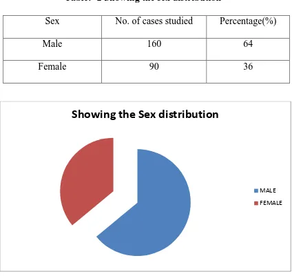
Table:- 2 Showing the sex distribution
Sex No. of cases studied Percentage(%)
Male 160 64
Female 90 36
Of the 250 cases studied in this series, 160 (64%) cases were male and 90(36%)
Showing the Sex distribution
MALE
3.Occupation
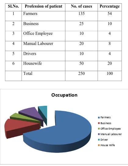
Table 3:- Occupation
Sl.No. Profession of patient No. of cases Percentage
1 Farmers 135 54
2 Business 25 10
3 Office Employee 10 4
4 Manual Labourer 20 8
5 Drivers 10 4
6 Housewife 50 20
Total 250 100
In this study maximum patients were farmers (54%) and minimum patients were drivers and office employee (4%).
Occupation
Farmers
Business
Office Employee
Manual Labourer
Driver
4.Type of diabetes
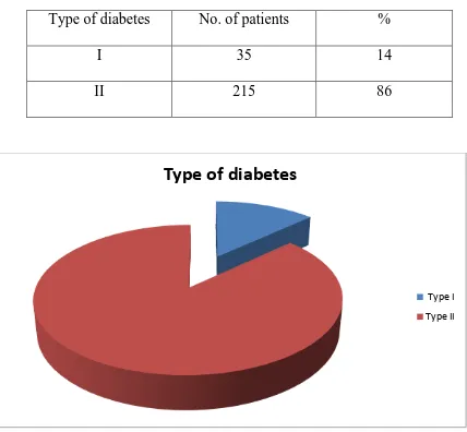
Table:- 4- Type of diabetes
Type of diabetes No. of patients %
I 35 14
II 215 86
In this study 35 patients had type I diabetes, remaining 215 patients had type II diabetes.
Type of diabetes
Type I
5.Incidence of Trauma
Chart 5:- Incidence of Trauma
In this study 64 cases exposed a history of some kind of trauma before the onset of lesion.
[image:84.595.107.543.113.325.2]6. Mode of presentation
Table 6 :- Showing incidence of various types of lesions.
Mode of presentation No. of cases %
Ulcer 120 48
Gangrene 50 20
Abscess 15 6
Cellulitis 65 26
Showing the incidence of trauma
YES
The different types of lesions seen including ulcer, cellulitis, abscess and gangrene. Most of the patients present with more than one lesions. Only major lesion is considered here. Ulcer was the major lesion seen and is present in 120 patients. 15 patients presents as a abscess was the least common lesion.
In above patients x-ray of 30 cases showed changes of Osteomyelitis. 15 patients present with Charcot’s joint.
Doppler studied in 10 patient showed atherosclerotic changes with low volume flow in anterior and posterior tibial arteries.
7. Duration of diabetes
Duration is not accurately known, as few patients were unaware of being diabetics and were newly diagnosed as suffering from diabetes on admission with complaints of non- healing ulcer or other complications of Diabetes.
Mode of presentation
ULCER
GANGRENE
ABSCESS
Table 7 :- Showing duration of Diabetes
Duration of diabetes in years No. of patients %
0-1 45 18
2-5 30 12
6-10 80 32
11-15 40 16
16-20 35 14
˃20 20 8
Total 250 100
Showing duration of diabetes:-
In our study 18% presented with duration less than or equal to 1 year. Most of these patients were diagnosed post admission. Only 20 patients had diabetes of more than 20 years. Maximum 80 patients in our study were diabetes of 6- 10 years.
In the present series 10 patients were detected as a diabetic at the time of admission.
8.Culture and Sensitivity
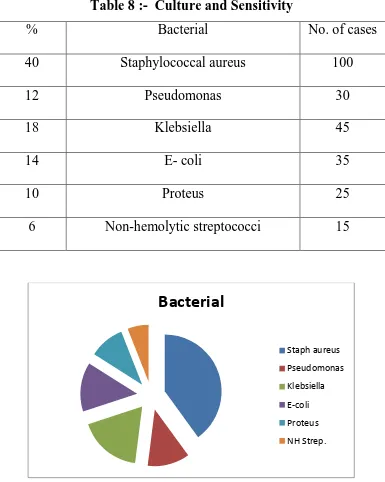
Table 8 :- Culture and Sensitivity
% Bacterial No. of cases
40 Staphylococcal aureus 100
12 Pseudomonas 30
18 Klebsiella 45
14 E- coli 35
10 Proteus 25
6 Non-hemolytic streptococci 15
In our study majority of septic lesions yielded Staphylococcus aureus in about 40% on culture of pus. Other organisms were isolated are Pseudomonas 12%, Klebsiella 18%, E-coli 14%, Proteus 10%.
Bacterial
Staph aureus
Pseudomonas
Klebsiella
E-coli
Proteus
9. Sensitivity report
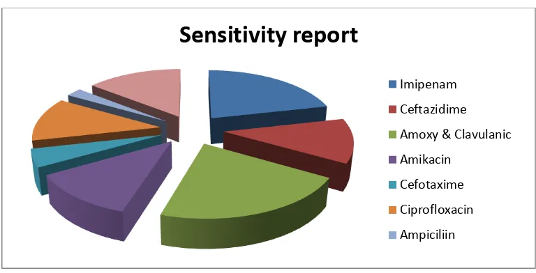
Table 9:- Showing Antibiotic Sensitivity according to Culture.
Antibiotics % of patients
Imipenam 45
Ceftazidime 25
Amoxycillin and Clavulanic acid 45
Amikacin 25
Cefotaxime 10
Ciprofloxacin 25
Ampicillin 5
Vancomycin 30
Imipenem and Amoxicillin & Clavulanic acid were sensitive against most of the organisms as they cover a wide range of organisms.
Sensitivity report
Imipenam
Ceftazidime
Amoxy & Clavulanic
Amikacin
Cefotaxime
Ciprofloxacin
10. Neuropathic lesions
Table 10:- Neuropathic lesions
Neuropathic lesions No. of cases %
Yes 130 52
No 120 48
In the present study 130 cases were found to have neuropathy. Patients with neuropathy varied from 35-80 years. Majority had history of diabetes more than 5 years. This shows that peripheral neuropathy is common in long standing diabetic patients. 50 patients had gangrene.
Neuropathic lesion
YES