Copyright © 2010, American Society for Microbiology. All Rights Reserved.
Lambda Interferon Renders Epithelial Cells of the Respiratory and
Gastrointestinal Tracts Resistant to Viral Infections
䌤
†
Markus Mordstein,
1,2Eva Neugebauer,
3Vanessa Ditt,
3Birthe Jessen,
4Toni Rieger,
5Valeria Falcone,
1Frederic Sorgeloos,
6Stephan Ehl,
4Daniel Mayer,
1Georg Kochs,
1Martin Schwemmle,
1Stephan Gu
¨nther,
5Christian Drosten,
3Thomas Michiels,
6and Peter Staeheli
1*
Department of Virology1and Spemann Graduate School of Biology and Medicine (SGBM),2University of Freiburg, D-79104 Freiburg,
Germany; Institute of Virology, University Medical Centre, D-53127 Bonn, Germany3; Centre of Chronic Immunodeficiency,
University of Freiburg, D-79106 Freiburg, Germany4; Bernhard-Nocht-Institute for Tropical Medicine, D-20359 Hamburg,
Germany5; and de Duve Institute, Universite´ catholique de Louvain, B-1200 Brussels, Belgium6
Received 5 February 2010/Accepted 18 March 2010
Virus-infected cells secrete a broad range of interferons (IFN) which confer resistance to yet uninfected cells by triggering the synthesis of antiviral factors. The relative contributions of the various IFN subtypes to innate immunity against virus infections remain elusive. IFN-␣, IFN-, and other type I IFN molecules signal through a common, universally expressed cell surface receptor, whereas type III IFN (IFN-) uses a distinct cell-type-specific receptor complex for signaling. Using mice lacking functional receptors for type I IFN, type III IFN, or both, we found that IFN-plays an important role in the defense against several human pathogens that infect the respiratory tract, such as influenza A virus, influenza B virus, respiratory syncytial virus, human metapneumovirus, and severe acute respiratory syndrome (SARS) coronavirus. These viruses were more pathogenic and replicated to higher titers in the lungs of mice lacking both IFN receptors than in mice with single IFN receptor defects. In contrast, Lassa fever virus, which infects via the respiratory tract but primarily replicates in the liver, was not influenced by the IFN- receptor defect. Careful analysis revealed that expression of functional IFN-receptor complexes in the lung and intestinal tract is restricted to epithelial cells and a few other, undefined cell types. Interestingly, we found that SARS coronavirus was present in feces from infected mice lacking receptors for both type I and type III IFN but not in those from mice lacking single receptors, supporting the view that IFN-contributes to the control of viral infections in epithelial cells of both respiratory and gastrointestinal tracts.
The interferon (IFN) system represents a major element of the innate immune response against viral infections (10, 13, 14). Virus-induced IFN is a complex mixture of biologically active molecules, which includes type I and type III IFN. Type
I IFN consists of 14 different IFN-␣subtypes in the mouse as
well as IFN-, IFN-, IFN-ε, and limitin, which all signal
through the same universally expressed cell surface receptor
complex (IFNAR) (30). Type III IFN includes IFN-1,
IFN-2, and IFN-3 (21, 28), of which only the latter two are
encoded by genes that are expressed in the mouse (22). Type III IFN uses a distinct receptor complex (IL28R) for signaling (21, 28), which appears to be expressed on only a few cell types, including epithelial cells (29). Binding of type I IFN and type III IFN to their cognate receptor complexes triggers signaling cascades that result in the activation of a large number of genes, many of which encode antiviral proteins (10, 32). Type I IFN and type III IFN trigger highly similar gene expression profiles in responsive cells, suggesting that both IFN types might serve similar functions. However, it has to date been
largely unclear to which extent IFN-might contribute to
in-nate immunity.
Using knockout mouse strains that lack receptors for
type I IFN (IFNAR10/0), type III IFN (IL28R␣0/0), or both
(IFNAR10/0IL28R␣0/0), we have recently shown that IFN-
contributes to resistance against influenza A virus (FLUAV) (26). Here, we used the same mouse strains to investigate
the relative contribution of IFN-in resistance against
ad-ditional viral pathogens that infect the respiratory and
gas-trointestinal tract and to visualize IFN--responsive cells.
We found that the double-knockout mice showed enhanced susceptibility to various viruses that primarily replicate in lung epithelial cells. Our analysis further revealed that ep-ithelial cells of both lung and gastrointestinal tracts can
strongly respond to IFN- and that IFN- inhibited the
replication of severe acute respiratory syndrome coronavi-rus (SARS-CoV) in both lung and gastrointestinal tracts.
MATERIALS AND METHODS
Ethics statement.All animal experiments were performed in compliance with the German animal protection law (TierSchG). The mice were housed and handled in accordance with good animal practice as defined by FELASA (www .felasa.eu/guidelines.php) and the national animal welfare body GV-SOLAS (www.gv-solas.de/index.html). The animal welfare committees of the universities of Freiburg, Bonn, and Hamburg as well as the local authorities (Regierung-spra¨sidium Freiburg; Landesamt fu¨r Natur, Umwelt und Verbraucherschutz Nordrhein-Westfalen; and Beho¨rde fu¨r Soziales, Familie, Gesundheit und Ver-braucherschutz, Hamburg) approved all animal experiments.
* Corresponding author. Mailing address: Department of Virology, University of Freiburg, Hermann-Herder-Strasse 11, D-79104 Freiburg, Germany. Phone: 49-761-203-6579. Fax: 49-761-203-5350. E-mail: peter .staeheli@uniklinik-freiburg.de.
† Supplemental material for this article may be found at http://jvi .asm.org/.
䌤Published ahead of print on 24 March 2010.
5670
on November 8, 2019 by guest
http://jvi.asm.org/
Viruses.Influenza A viruses included the pandemic swine origin H1N1 isolate A/HH/05/2009 (a kind gift from Markus Eickmann, Marburg, Germany) (7) and mutant laboratory strain SC35M-⌬NS1 (H7N7), which lacks the IFN-antagonis-tic factor NS1 (19). We further used influenza B virus (FLUBV) strain B/Lee/40 (a kind gift from Thorsten Wolff), human respiratory syncytial virus (RSV) strain A2 (originally obtained from Peter Openshaw, Imperial College, London, United Kingdom), human metapneumovirus (HMPV) strain DO3-574 (17), SARS-CoV strain Frankfurt-1 (9), and Lassa fever virus strain AV (12). For systemic induction of IFN, we made use of the attenuated “clone 13” strain of Rift Valley fever virus (RVFV), which lacks functional IFN-antagonistic factor NSs (4). Experiments with SARS-CoV and Lassa fever virus were performed under BSL3 and BSL4 conditions, respectively. All other viruses used in this study are classified as BSL2 pathogens in Germany. Stocks of the various influ-enza A virus strains were prepared in MDCK cells, stocks of RSV in Hep-2 cells, stocks of HMPV in LLC-MK2 cells, stocks of SARS-CoV and RVFV in Vero cells, and stocks of Lassa fever virus in BHK-21 cells. Influenza B virus was grown in embryonated chicken eggs.
Virus infections.Animals were anesthetized by intraperitoneal injection of a mixture of ketamine (100g per gram body weight) and xylazine (5g per gram body weight) before intranasal infection with the indicated doses of the respira-tory viruses in 50l of phosphate-buffered saline (PBS) containing 0.3% bovine serum albumin (BSA). For RVFV infections, 100-l samples of diluted virus stocks were applied intraperitoneally without anesthesia. The units of infection were numbers of PFU per ml for HMPV, Lassa fever virus, RVFV, and SARS-CoV and numbers of focus-forming units (FFU) per ml for FLUAV, FLUBV, and RSV.
Plasmid electrotransfer-mediated expression of IFN.The experiments were performed as previously described (29). Briefly, 10 or 25g of plasmid DNA was electrotransfered in each tibialis muscle. Mx1 responsiveness was examined by immunohistochemistry on day 7 postelectrotransfer, as described previously (29).
Immunohistochemistry.Perfused tissue was immersed in 4% buffered form-aldehyde for 12 h at 4°C before being embedded in paraffin. Tissue sections of approximately 5m were placed on SuperFrost Plus slides, dried at room temperature overnight, and processed by standard methods for immunohisto-chemistry. Briefly, sections were deparaffinized, permeabilized for 5 min in PBS containing 0.5% Triton X-100, and washed in PBS. Blocking was performed by incubating sections for 90 min with 10% normal goat serum (Sigma) diluted in PBS. Sections were then treated for 10 min at 121°C in 0.01 M sodium citrate buffer at pH 6.0 to unmask antigens. Then, immunolabeling was done in blocking solution containing the antibodies. Mx1 protein was detected with rabbit poly-clonal antibody AP5, which recognizes the C-terminal 16 amino acids of Mx1 (25). AP5 was used at a dilution of 1/500. For immunofluorescence labeling, the secondary antibody (at 1/800) was a goat anti-rabbit antibody coupled to Alexa 488 (Invitrogen) or a donkey anti-rabbit antibody coupled to Alexa 555 (Invitro-gen).
Titration of virus in lungs.Lung homogenates of animals infected with RSV, HMPV, A/HH/05/2009, or FLUBV were prepared using the FastPrep24 system (MP Biomedicals). After addition of 800l of PBS containing 0.3% BSA, organs were subjected to two rounds of mechanical treatment for 15 s each at 6.5 m/s, with 5 min of incubation on ice between the cycles. Tissue debris was removed by low-speed centrifugation. Virus titers in supernatants were determined by im-munofluorescence assays with MDCK II cells (FLUBV and A/HH/05/2009), Hep2 cells (RSV), or Vero cells (SARS-CoV) by serial 10-fold dilutions in PBS containing 0.3% BSA. We prepared Lassa virus stocks and measured viral in-fectivity in blood samples as previously described (2).
RT-PCR.SARS-CoV RNA concentrations were determined by reverse tran-scription-PCR (RT-PCR) as previously described in reference 9, using Qiagen RNeasy columns for extraction of RNA from organ homogenates according to provided protocols. HMPV was detected by RT-PCR based on a method de-scribed previously (8). Briefly, a Qiagen one-step RT-PCR kit (Qiagen, Hilden,
Germany) was used according to the manufacturer’s instructions. This 25-l reaction mixture consisted of 1⫻OneStep buffer, 400 M deoxynucleoside triphosphate (dNTP) mix, 400 nM forward primer, 400 nM reverse primer, Qiagen OneStep RT-PCR enzyme mix, 200 nM probe, template RNA, and sterile water. Amplification was performed using a LightCycler instrument (Roche Applied Science) with the following cycling conditions: an RT step at 50°C for 30 min and aTaqactivation step at 95°C for 15 min, followed by 45 cycles of PCR at 95°C for 10 s and 60°C for 30 s.
Statistical analysis.Statistical significance for comparison of two groups was calculated using Student’sttests, using Microsoft Excel software.
RESULTS
IFN- contributes to resistance of mice against various pneumotropic viruses.We previously showed that IFNAR10/0
IL28R␣0/0mice, which lack receptors for both type I and type III
IFN, exhibit higher susceptibility toward certain laboratory strains
of influenza A virus than IFNAR10/0 mice, which lack only a
functional type I IFN receptor (26). To determine whether IFN-
might also contribute to resistance against other pneumotropic
viruses, we comparedIFNAR10/0IL28R␣0/0andIFNAR10/0mice
with respect to virus-induced disease and viral titers in the lung tissue. In the first experiment, we used the pandemic swine origin influenza virus strain A/HH/05/2009 (H1N1) for challenge.
IFNAR10/0mice survived this infection even if a very high virus
dose (106PFU per animal) was used (Fig. 1A). In contrast, all
IFNAR10/0IL28R␣0/0mice succumbed within 6 days if the
chal-lenge virus dose was 105PFU per animal or higher. Even at 104
[image:2.585.335.506.71.308.2]PFU of virus per animal, roughly 50% of the challenged double-knockout mice developed severe disease and had to be killed (Fig.
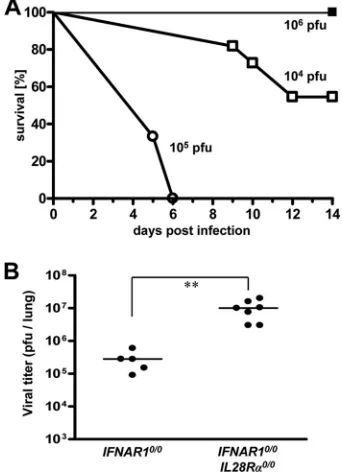
FIG. 1. Mice lacking functional receptors for both IFN-␣/ and IFN-exhibit high susceptibility toward influenza virus strain A/HH/ 05/2009 (H1N1). (A) Survival ofIFNAR10/0mice (filled squares;n⫽4) andIFNAR10/0IL28R␣0/0 double-knockout mice (open symbols; 104
PFU [n⫽11] and 105PFU [n⫽3]) after intranasal infection with the
indicated doses of virus. (B) Virus titers in lungs at 48 h following intranasal infection with 103PFU of virus. Combined data for two
independent experiments are shown. Each dot represents the data for one animal.ⴱⴱ,P⬍0.01.
on November 8, 2019 by guest
http://jvi.asm.org/
1A). To corroborate these findings, 48 h after infection with 103 PFU of virus per animal, the viral lung titers of both mouse lines
were determined. The average titers were 3⫻105PFU per lung
inIFNAR10/0mice and 107PFU per lung inIFNAR10/0IL28R␣0/0
mice (Fig. 1B). This difference was statistically significant, clearly
demonstrating that IFN-contributes to resistance against swine
origin influenza A virus.
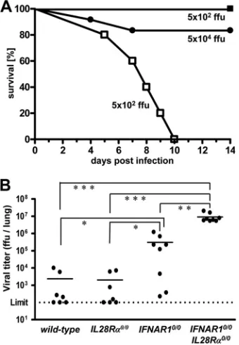
We next used influenza virus strain B/Lee/40 for challenge.
If infected with 5 ⫻ 102 FFU of B/Lee/40 per animal, all
IFNAR10/0IL28R␣0/0double-knockout mice developed severe
disease within 5 to 10 days, whereas allIFNAR10/0mice
sur-vived (Fig. 2A). If challenged with 100 times more virus (5⫻
104FFU), about 80% of the single-knockoutIFNAR10/0mice
still survived. Wild-type andIL28R␣0/0 mice were even more
resistant and survived the highest virus dose (5⫻ 105 FFU)
that we tested (data not shown). In contrast, conventional
C57BL/6 mice, which lack functional alleles of theMx1gene,
were highly susceptible toward B/Lee/40 (50% lethal dose
[LD50]⫽2⫻103FFU) (data not shown), demonstrating that
Mx1is a major factor contributing to resistance against
influ-enza B virusin vivo. Replication of B/Lee/40 in the lungs of the
various mouse lines was assessed at 72 h postinfection with 5⫻
104 FFU of virus. Titers in lungs of wild-type andIL28R␣0/0
mice were near or below the detection limit (Fig. 2B). In contrast, B/Lee/40 replicated to very high titers in lungs of
IFNAR10/0IL28R␣0/0 double-knockout mice. Viral titers in
lungs of IFNAR10/0 mice were nonuniform but, on average,
were about 20-fold reduced compared to the level for
double-knockout mice (Fig. 2B). Thus, IFN-clearly plays a
substan-tial role in host resistance toward influenza B virus.
We also assessed the contribution of IFN- to resistance
against respiratory syncytial virus (RSV). Replication of RSV is strain dependent. Four days after intranasal inoculation of
8⫻106FFU of virus, RSV titers reached about 105FFU per
lung in BALB/c mice, which are frequently used for RSV
research (27), and about 5⫻103FFU per lung in wild-type
C57BL/6 mice, irrespective of the presence (wild type) or
ab-sence (C57BL/6) of functional Mx1 alleles (Fig. 3). As was
previously reported (18),IFNAR1deficiency does not increase
susceptibility to RSV replication in C57BL/6 mice, whereas
STAT1deficiency does. In agreement with these findings, we
detected only low titers of RSV in the lungs of both wild-type
and IFNAR10/0 mice, while RSV titers in lungs of STAT10/0
mice were at least 50-fold higher than those in wild-type mice (Fig. 3). Virus control was not impaired by the lack of
func-tional receptors for IFN- (Fig. 3). Surprisingly, however,
IFNAR10/0IL28R␣0/0 double-knockout mice showed RSV
ti-ters of approximately 106FFU per lung (Fig. 3). These results
demonstrate that either IFN-- or IFN-␣/-mediated signals
are sufficient for RSV control. The absence of both, however, leads to a drastic increase in viral replication. Interestingly,
there was a significant difference in viral titers between
STAT1-deficient andIFNAR10/0IL28R␣0/0 double-deficient mice,
im-plicating possible STAT1-independent effects of IFN- or
IFN-␣/on RSV replication.
Human metapneumovirus (HMPV) does not replicate to high titers in lungs of standard C57BL/6 mice and does not induce substantial signs of illness in these animals
(unpub-lished data). After intranasal infection with 105 PFU of
HMPV, our single-knockout IFNAR10/0 mice also remained
healthy and showed no weight loss during the 9-day
observa-tion period (Fig. 4A). In contrast,IFNAR10/0IL28R␣0/0mice
experienced substantial respiratory disease and showed weight losses of up to 23% around day 6 postinfection, and in infre-quent cases, they had to be killed (Fig. 4A). Analysis of viral RNA in lungs revealed significantly elevated HMPV-RNA
[image:3.585.297.538.67.193.2]load inIFNAR10/0IL28R␣0/0double-knockout mice compared
FIG. 2. Mice lacking functional receptors for both IFN-␣/ and IFN-exhibit high susceptibility toward influenza virus strain B/Lee/ 40. (A) Survival ofIFNAR10/0mice (filled symbols; 5⫻102PFU [n⫽
7] and 5 ⫻ 104 PFU [n ⫽ 10]) and IFNAR10/0IL28R␣0/0 double-knockout mice (open squares;n⫽4) after intranasal infection with the indicated doses of virus. Combined data for two independent experi-ments are shown. (B) Virus titers in lungs at 72 h following intranasal infection with 5⫻104FFU of virus. Combined data for several
[image:3.585.79.249.70.315.2]inde-pendent experiments are shown. Each dot represents the data for one animal.ⴱ,P⬍0.05;ⴱⴱ,P⬍0.01;ⴱⴱⴱ,P⬍0.001.
FIG. 3. Mice lacking functional receptors for both IFN-␣/ and IFN-exhibit high susceptibility toward RSV. Virus titers in lungs at day 4 following intranasal infection with 8⫻106FFU of RSV.
Wild-type mice and C57BL/6 mice differ only by the presence or absence of functionalMx1alleles. Combined data for several independent exper-iments are shown. Each dot represents the data for one animal.ⴱⴱⴱ,
P⬍0.001.
on November 8, 2019 by guest
http://jvi.asm.org/
to the level for IFNAR10/0 single-knockout mice (Fig. 4B),
demonstrating that IFN- contributes to resistance against
HMPV.
To determine whether IFN-is also active against
patho-gens, which can enter the body through the respiratory tract but eventually replicate in cells of other organs, we per-formed infection experiments with Lassa fever virus. Groups
of IFNAR10/0 single-knockout and IFNAR10/0IL28R␣0/0
double-knockout mice were infected by the intranasal route
with either 3⫻ 105PFU (high dose) or 3⫻103PFU (low
dose) of Lassa fever virus strain AV. At various times postinfection, blood samples were examined for infectious virus. In addition, serum transaminase levels were moni-tored to assess liver damage. Peak titers of Lassa fever virus
(⬎105PFU per ml) were measured around day 8
postinfec-tion for both mouse lines. Importantly, we did not observe any significant differences in viral growth kinetics (Fig. 5) or
transaminase levels (data not shown) between IFNAR10/0
andIFNAR10/0IL28R␣0/0mice. Thus, Lassa fever virus
rep-lication was not influenced by IFN-, although in our
exper-imental setting the virus was forced to enter the host via the respiratory tract.
Lung epithelial cells preferentially express functional IFN- receptors.To identify the cell types in the lung that respond to
IFN-, we used an indirect assay which takes advantage of the
fact that expression of the mouse Mx1 gene is under tight
transcriptional control by type I and type III IFN (29). Since
IFNAR10/0mice cannot respond to type I IFN,Mx1expression
in such mice is solely dependent on IFN-(26). Thus, cells of
IFNAR10/0mice containing Mx1 protein are likely to express
functional IFN-receptor complexes. This assay is highly
re-liable, as Mx1 is a nuclear protein that forms aggregates with a characteristic appearance (29).
To make sure that IFN-is abundantly present in the lung,
IFNAR10/0mice were infected with the influenza A virus
mu-tant strain SC35M-⌬NS1, which is a known inducer of type I
and type III IFN in mouse lungs (26). At 20 h postinfection, the animals were sacrificed and lung sections were prepared for immunohistochemical analysis. Mx1 staining was readily
de-tected in lungs of IFNAR10/0 mice (Fig. 6A and C).
Mx1-positive cells were mainly clustered around bronchioles. From their locations and shapes, it seemed that most Mx1-positive cells are epithelial cells (Fig. 6C). In contrast, Mx1 staining in
lungs of SC35M-⌬NS1-infectedIFNAR10/0IL28R␣0/0mice was
very weak (Fig. 6B and D). In such animals, only a few cells, which were of unknown identity, expressed Mx1, but strik-ingly, lung epithelial cells showed no substantial Mx1 stain-ing (Fig. 6D).
The responsiveness of bronchiolar epithelial cells to IFN-
was confirmed for mice expressing a plasmid for mouse IFN-3
in the tibialis muscle (29). In such mice, which produce
sys-temic IFN-, Mx1 was also prominently expressed in
bronchio-lar epithelial cells. Mx1 expression in these cells depended on
functional IFN-receptors, as Mx1 was detected in wild-type
mice (data not shown) and IFNAR10/0 mice but not in
IL28R␣0/0mice (see Fig. S1 in the supplemental material). The
fact that lung epithelial cells abundantly express functional
IFN-receptor complexes readily explains our results
indicat-ing that IFNAR10/0IL28R␣0/0 mice are more susceptible to
several pneumotropic viruses thanIFNAR10/0 mice. This fact
[image:4.585.135.451.68.193.2]further suggests that Lassa fever virus may not be restricted by
FIG. 4. Mice lacking functional receptors for both IFN-␣/and IFN-exhibit high susceptibility toward HMPV. (A) Body weight changes of
IFNAR10/0(n⫽4) andIFNAR10/0IL28R␣0/0(n⫽5) mice intranasally infected with 105PFU of HMPV. Crosses indicate that animals had to be
killed due to severe symptoms. (B) HMPV-RNA load in lungs at day 4 following intranasal infection with 105PFU of HMPV was determined by
qRT-PCR. Combined data for two independent experiments are shown. Each dot represents the data for one animal.ⴱⴱⴱ,P⬍0.001.
FIG. 5. Lassa fever virus replication is not affected by IFN-.
IFNAR10/0andIFNAR10/0IL28R␣0/0mice were infected by the intra-nasal route with either 3⫻105PFU (high dose) or 3⫻103PFU (low
dose) of Lassa fever virus. Blood samples were taken at the indicated times postinfection and tested for presence of virus. Each group con-sisted of three mice, except theIFNAR10/0IL28R␣0/0low-dose group, which consisted of two mice.
on November 8, 2019 by guest
http://jvi.asm.org/
[image:4.585.60.266.519.659.2]IFN-, because this virus presumably uses other cells for initial replication in the respiratory tract before reaching the blood-stream.
Epithelial cells in the intestine express functional IFN- receptor complexes.We further determined whether epithelial cells of other organs, such as the intestine (which is prone to infection with important pathogenic viruses), might also carry
functional receptors for IFN-. Again, we employed the
above-described indirect assay in which expression of Mx1 is used to
identify IFN--responsive cells in mice expressing systemic
IFN-(29). Prominently stained cells were abundantly present
in various parts of the small intestine ofIFNAR10/0mice (Fig.
7A), whereas Mx1-positive cells were virtually absent from
small intestine sections of mock-treatedIFNAR10/0mice (data
not shown) or IFN-3-expressing IFNAR10/0IL28R␣0/0 mice
(Fig. 7C). Careful inspection revealed that the majority of
IFN--responsive cells in the intestines of IFNAR10/0 mice
lined the lumen. From their morphology, it appeared that these Mx1-positive cells are epithelial cells (Fig. 7B). Analysis of additional tissue samples from other parts of the gastroin-testinal tract showed that epithelial cells in the esophagus (Fig. 7D), stomach (Fig. 7E), small intestine (Fig. 7F), and colon
(Fig. 7G) of IFN-3-expressingIFNAR10/0mice were strongly
positive for Mx1. In the large intestine but not in the esopha-gus, stomach, or small intestine, a number of cells of unknown
identity also expressed Mx1 in the absence of IFN-(data not
shown).
To corroborate the epithelial cell specificity of the IFN re-sponse in infected mice, we performed additional experiments,
in whichIFNAR10/0mice were infected with Rift Valley fever
virus clone 13, which lacks the IFN-antagonistic NSs protein and therefore induces large amounts of type I and type III IFN
in mice (4).IFNAR10/0mice were killed at 48 h postinfection
when they showed signs of virus-induced disease and when serum IFN titers were reported to reach peak values (4). Un-der these conditions, large numbers of Mx1-positive epithelial cells were readily detected in the small intestine and other parts of the gastrointestinal tract (see Fig. S2 in the supple-mental material). Thus, virus-induced and plasmid-mediated
expression of IFN-inIFNAR10/0mice yielded similar results.
Taken together, these data demonstrate that epithelial cells
are the main targets of IFN-in all parts of the gastrointestinal
tract.
IFN-reduces replication of SARS-CoV in lungs and virus excretion in feces. Patients infected with SARS-CoV during the outbreak in 2003, besides developing severe respiratory symptoms, excreted infectious virus in their feces (6). Studies with SARS-CoV-related viruses of bats further support the view that these viruses can replicate in epithelial cells of both lung and gastrointestinal tract (23). Therefore, we hypothe-sized that experiments with SARS-CoV might reveal a possible
role for IFN- during viral infection of the gastrointestinal
tract.
Although replication of SARS-CoV in lungs ofIFNAR10/0
andSTAT10/0mice is substantially enhanced compared to the
level for wild-type mice (5, 15), a possible contribution of type III IFN has not previously been documented. To address this
issue, we infected mice with 106 PFU of SARS-CoV by the
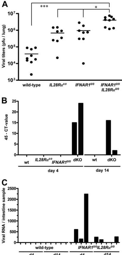
[image:5.585.112.472.70.326.2]intranasal route and determined viral titers in lungs at day 4 postinfection. We found that SARS-CoV titers in lungs of
FIG. 6. Lung epithelial cells express functional IFN-receptor complexes.IFNAR10/0 (A, C) andIFNAR10/0IL28R␣0/0(B, D) mice were intranasally infected with 5⫻104PFU of SC35M-⌬NS1, a potent inducer of type I and type III IFN (26). At 20 h postinfection, the lungs were
removed and stained for Mx1 by immunohistofluorescence. (A, B) Low magnification overview. Mx1-positive cells (stained nuclei) are mainly clustered around bronchioles inIFNAR10/0mice but mostly absent inIFNAR10/0IL28R␣0/0mice. (B, D) High magnification of bronchioles and surrounding tissue. Epithelial cells were prominently stained for Mx1 inIFNAR10/0but not inIFNAR10/0IL28R␣0/0mice.
on November 8, 2019 by guest
http://jvi.asm.org/
IFNAR10/0 andIL28R␣0/0 single-knockout mice were, on
av-erage, at least 10-fold higher than those in wild-type mice (Fig. 8A). In agreement with these observations, viral lung titers in
IFNAR10/0IL28R␣0/0 double-knockout mice were 10-fold
higher than those in single-knockout mice (Fig. 8A).
To determine whether virus excretion was affected by IFN-,
fecal samples of the various SARS-CoV-infected mice were an-alyzed for the presence of viral RNA on days 4 and 14
postinfec-tion. Neither wild-type norIL28R␣0/0orIFNAR10/0
single-knock-out mice contained detectable levels of viral RNA in their feces.
However, fecal samples from cages ofIFNAR10/0IL28R␣0/0
dou-ble-knockout mice tested positive on day 4 and day 14 postinfec-tion (Fig. 8B). When intestine samples of individual infected mice were analyzed for the presence of viral RNA by quantitative
RT-PCR (qRT-PCR), we observed that three of fiveIFNAR10/0
IL28R␣0/0mice were positive on days 4 and 14 postinfection (Fig.
8C). In contrast, none of the intestine samples of infected wild-type mice scored positive in this assay. These results demonstrate
that IFN-contributes to restriction of SARS-CoV in both
respi-ratory and gastrointestinal tracts.
DISCUSSION
Previous infection studies with knockout mice lacking func-tional receptors for type I IFN, type III IFN, or both clearly
indicated that IFN-contributes to resistance against influenza
A virus (26). A limitation of the published work was that highly adapted laboratory viruses had been used and that enhanced
virus susceptibility of IFN- receptor-deficient mice was
de-tected only if infection experiments were performed with at-tenuated virus mutants that induce a strong innate immune
response. Consequently, it remained unclear whether IFN-
also plays a role in the defense of wild-type influenza A virus. Here, we addressed this issue by employing a nonadapted primary isolate of the new pandemic swine-origin H1N1 influ-enza virus. This virus was unable to induce disease in Mx1-positive mice lacking either functional type I or functional type III IFN receptors. However, it replicated to high titers and induced disease in mice lacking both IFN receptor systems.
These results show that our earlier conclusion that IFN-plays
a role in host defense against influenza A virus (26) remains valid and also applies for primary virus isolates.
By using a panel of different pneumotropic viruses, we
man-aged to demonstrate that IFN-also has a beneficial effect
[image:6.585.81.500.70.371.2]during infections with other important pathogens, such as in-fluenza B virus, RSV, HMPV, and SARS coronavirus. All these viruses did not grow well and were mainly benign in single-knockout mice lacking functional receptors for either type I or type III IFN, but they replicated to high titers and were able to induce clinical symptoms in mice lacking both IFN
FIG. 7. Epithelial cells of the intestine express functional IFN-receptor complexes. The response toin vivoelectroporation of a plasmid encoding mouse IFN-3 was monitored by immunohistofluorescence staining for Mx1. The duodenum (section of the villi) ofIFNAR10/0mice is shown in low (A) and high (B) magnification. Prominent labeling of nuclear Mx1 is detected mainly in epithelial cells of the villi. (C) No such staining was observed inIFNAR10/0IL28R␣0/0mice, which cannot respond to IFN-. Strongly stained epithelial cells were detected in all regions of the gastrointestinal tract of IFN--treatedIFNAR10/0mice, including esophagus (D), stomach (E), small intestine (F), and colon (G).
on November 8, 2019 by guest
http://jvi.asm.org/
receptor systems. In agreement with these results, other re-searchers previously noted that type I IFN receptor-deficient mice exhibited a degree of resistance to RSV similar to that
observed for wild-type mice but that STAT1-deficient mice
were highly susceptible (18). The molecular basis for this difference remained unexplained at the time. Our new find-ings suggest that the type I IFN receptor-deficient mice remained resistant to RSV because the functionally
redun-dant IFN-system was still active in these mice. In
STAT1-deficient mice, in contrast, both the type I and the type III IFN systems are severely compromised, resulting in en-hanced virus susceptibility.
Interestingly, experiments in which we performed intranasal infections of mice with Lassa fever virus yielded no evidence
for a protective role for IFN- against this pathogen. We
believe that this result can be explained by our finding that
expression of functional IFN-receptors in the lung was
re-stricted to epithelial cells. As the strictly pneumotropic viruses discussed above preferentially use lung epithelial cells for pro-ductive replication, it is understandable that these viruses are
susceptible to IFN-. Lassa fever virus, on the other hand,
before becoming systemic, presumably infects other cell types in the respiratory tract that do not express or express only very
low levels of functional IFN-receptors. Consequently,
virus-induced IFN-cannot efficiently restrict initial replication of
Lassa fever virus in the respiratory tract. It should be noted
that once the virus has reached the bloodstream, IFN-is not
expected to afford any further protection, as previously re-ported for Rift Valley fever virus and Thogoto virus (26), presumably due to restricted expression of the type III IFN receptor.
One of the most exciting findings of our study is that the
protective function of IFN-was not restricted to the
respira-tory tract but also included the gastrointestinal tract. We ob-served that SARS-CoV-infected mice excreted virus in the feces only if these mice lacked both type I and type III IFN receptors. If either IFN system remained functional, no virus replication could be detected in the intestine of the infected mice. This finding can be explained by the results of our type III IFN receptor expression analysis, which showed that epi-thelial cells in all areas of the gastrointestinal tract readily
responded to IFN-. Thus, a picture emerges which suggests
that IFN-mainly serves to strengthen the antiviral defense of
mucosal surfaces that are potential entry sites for pathogenic viruses.
SinceIFNAR10/0 mice are highly sensitive toward many
vi-ruses (9), whereas mice lacking IFN-receptors do not show or
show only weak phenotypes in respect to viral infection (1, 26), it remains obvious that the type I IFN system certainly is the dominant system in terms of antiviral protection. However, our data clearly demonstrate that the presence of a functional type III IFN system does indeed provide beneficial and protective antiviral responses that can influence survival. Additionally, we
showed that IFN-also plays a previously unappreciated role
in preventing viral spread to and shedding from the gastroin-testinal tract. It will be both interesting and imperative to investigate which other mucosal surfaces and/or exposed body tissues are expressing functional type III IFN receptor
com-plexes and thus may contribute to IFN--mediated antiviral
responses. For example, previous studies yielded indirect (3) and direct (31) hints that the skin, which represents a body part that is heavily exposed to pathogens if physically injured, may
acquire virus resistance from IFN-.
The recently identified IFN genes of fish seem to represent components of an evolutionarily well conserved type III rather than type I IFN system (24). Although this view is not generally
accepted (32), it would indicate that IFN- is a primordial
antiviral cytokine that may serve basic functions. Our finding
[image:7.585.58.263.69.497.2]that IFN- of the mouse seems to selectively contribute to
FIG. 8. IFN-contributes to restriction of SARS-CoV replication in both lungs and intestinal tracts of mice. (A) Virus titers in lungs of various mouse lines at day 4 following intranasal infection with 106
PFU of SARS-CoV per animal. Combined data for two independent experiments are shown. Each dot represents the data for one animal. ⴱ,P⬍0.05;ⴱⴱⴱ,P⬍0.001. (B) At days 4 and 14 postinfection, RNA samples extracted from 20 droppings of cages harboring the different mouse lines were analyzed by qRT-PCR for SARS-CoV. (C) RNA samples from intestines of infected wild-type andIFNAR10/0IL28R␣0/0 double-knockout mice were analyzed by qRT-PCR for SARS-CoV. Viral RNA levels were calculated using appropriate standards.
on November 8, 2019 by guest
http://jvi.asm.org/
von SARS”) to C.D., FRSM to T.M., and the University of Louvain (FSR) to F.S.
REFERENCES
1.Ank, N., M. B. Iversen, C. Bartholdy, P. Staeheli, R. Hartmann, U. B. Jensen, F. Dagnaes-Hansen, A. R. Thomsen, Z. Chen, H. Haugen, K. Klucher, and S. R. Paludan.2008. An important role for type III interferon (IFN-lambda/ IL-28) in TLR-induced antiviral activity. J. Immunol.180:2474–2485. 2.Asper, M., T. Sternsdorf, M. Hass, C. Drosten, A. Rhode, H. Schmitz, and S.
Gunther. 2004. Inhibition of different Lassa virus strains by alpha and gamma interferons and comparison with a less pathogenic arenavirus. J. Vi-rol.78:3162–3169.
3.Bartlett, N. W., K. Buttigieg, S. V. Kotenko, and G. L. Smith.2005. Murine interferon lambdas (type III interferons) exhibit potent antiviral activity in vivo in a poxvirus infection model. J. Gen. Virol.86:1589–1596.
4.Bouloy, M., C. Janzen, P. Vialat, H. Khun, J. Pavlovic, M. Huerre, and O. Haller.2001. Genetic evidence for an interferon-antagonistic function of rift valley fever virus nonstructural protein NSs. J. Virol.75:1371–1377. 5.Cervantes-Barragan, L., R. Zust, F. Weber, M. Spiegel, K. S. Lang, S. Akira,
V. Thiel, and B. Ludewig.2007. Control of coronavirus infection through plasmacytoid dendritic-cell-derived type I interferon. Blood109:1131–1137. 6.Cheng, P. K., D. A. Wong, L. K. Tong, S. M. Ip, A. C. Lo, C. S. Lau, E. Y. Yeung, and W. W. Lim.2004. Viral shedding patterns of coronavirus in patients with probable severe acute respiratory syndrome. Lancet363:1699– 1700.
7.Childs, R. A., A. S. Palma, S. Wharton, T. Matrosovich, Y. Liu, W. Chai, M. A. Campanero-Rhodes, Y. Zhang, M. Eickmann, M. Kiso, A. Hay, M. Matrosovich, and T. Feizi.2009. Receptor-binding specificity of pandemic influenza A (H1N1) 2009 virus determined by carbohydrate microarray. Nat. Biotechnol.27:797–799.
8.Dare, R., S. Sanghavi, A. Bullotta, M. C. Keightley, K. S. George, R. M. Wadowsky, D. L. Paterson, K. R. McCurry, T. A. Reinhart, S. Husain, and C. R. Rinaldo.2007. Diagnosis of human metapneumovirus infection in immunosuppressed lung transplant recipients and children evaluated for pertussis. J. Clin. Microbiol.45:548–552.
9.Drosten, C., S. Gunther, W. Preiser, S. van der Werf, H. R. Brodt, S. Becker, H. Rabenau, M. Panning, L. Kolesnikova, R. A. Fouchier, A. Berger, A. M. Burguiere, J. Cinatl, M. Eickmann, N. Escriou, K. Grywna, S. Kramme, J. C. Manuguerra, S. Muller, V. Rickerts, M. Sturmer, S. Vieth, H. D. Klenk, A. D. Osterhaus, H. Schmitz, and H. W. Doerr.2003. Identification of a novel coronavirus in patients with severe acute respiratory syndrome. N. Engl. J. Med.348:1967–1976.
10.Dumoutier, L., A. Tounsi, T. Michiels, C. Sommereyns, S. V. Kotenko, and J. C. Renauld.2004. Role of the interleukin (IL)-28 receptor tyrosine resi-dues for antiviral and antiproliferative activity of IL-29/interferon-lambda 1: similarities with type I interferon signaling. J. Biol. Chem.279:32269–32274. 11.Durbin, J. E., R. Hackenmiller, M. C. Simon, and D. E. Levy.1996. Targeted disruption of the mouse Stat1 gene results in compromised innate immunity to viral disease. Cell84:443–450.
12.Gunther, S., P. Emmerich, T. Laue, O. Kuhle, M. Asper, A. Jung, T. Grew-ing, J. ter Meulen, and H. Schmitz.2000. Imported Lassa fever in Germany: molecular characterization of a new Lassa virus strain. Emerg. Infect. Dis.
6:466–476.
13.Haller, O., G. Kochs, and F. Weber.2006. The interferon response circuit: induction and suppression by pathogenic viruses. Virology344:119–130. 14.Haller, O., P. Staeheli, and G. Kochs.2009. Protective role of
interferon-induced Mx GTPases against influenza viruses. Rev. Sci. Tech.28:219–231.
ratory syncytial virus immunopathology. J. Immunol.174:7234–7241. 19.Kochs, G., I. Koerner, L. Thiel, S. Kothlow, B. Kaspers, N. Ruggli, A.
Summerfield, J. Pavlovic, J. Stech, and P. Staeheli.2007. Properties of H7N7 influenza A virus strain SC35M lacking interferon antagonist NS1 in mice and chickens. J. Gen. Virol.88:1403–1409.
20.Koerner, I., G. Kochs, U. Kalinke, S. Weiss, and P. Staeheli.2007. Protective role of beta interferon in host defense against influenza A virus. J. Virol.
81:2025–2030.
21.Kotenko, S. V., G. Gallagher, V. V. Baurin, A. Lewis-Antes, M. Shen, N. K. Shah, J. A. Langer, F. Sheikh, H. Dickensheets, and R. P. Donnelly.2003. IFN-lambdas mediate antiviral protection through a distinct class II cytokine receptor complex. Nat. Immunol.4:69–77.
22.Lasfar, A., A. Lewis-Antes, S. V. Smirnov, S. Anantha, W. Abushahba, B. Tian, K. Reuhl, H. Dickensheets, F. Sheikh, R. P. Donnelly, E. Raveche, and S. V. Kotenko.2006. Characterization of the mouse IFN-lambda ligand-receptor system: IFN-lambdas exhibit antitumor activity against B16 mela-noma. Cancer Res.66:4468–4477.
23.Lau, S. K., P. C. Woo, K. S. Li, Y. Huang, H. W. Tsoi, B. H. Wong, S. S. Wong, S. Y. Leung, K. H. Chan, and K. Y. Yuen.2005. Severe acute respi-ratory syndrome coronavirus-like virus in Chinese horseshoe bats. Proc. Natl. Acad. Sci. U. S. A.102:14040–14045.
24.Levraud, J. P., P. Boudinot, I. Colin, A. Benmansour, N. Peyrieras, P. Herbomel, and G. Lutfalla.2007. Identification of the zebrafish IFN recep-tor: implications for the origin of the vertebrate IFN system. J. Immunol.
178:4385–4394.
25.Meier, E., J. Fah, M. S. Grob, R. End, P. Staeheli, and O. Haller.1988. A family of interferon-induced Mx-related mRNAs encodes cytoplasmic and nuclear proteins in rat cells. J. Virol.62:2386–2393.
26.Mordstein, M., G. Kochs, L. Dumoutier, J. C. Renauld, S. R. Paludan, K. Klucher, and P. Staeheli.2008. Interferon-lambda contributes to innate immunity of mice against influenza A virus but not against hepatotropic viruses. PLoS Pathog.4:e1000151.
27.Peebles, R. S., Jr., and B. S. Graham.2005. Pathogenesis of respiratory syncytial virus infection in the murine model. Proc. Am. Thorac. Soc.2:110– 115.
28.Sheppard, P., W. Kindsvogel, W. Xu, K. Henderson, S. Schlutsmeyer, T. E. Whitmore, R. Kuestner, U. Garrigues, C. Birks, J. Roraback, C. Ostrander, D. Dong, J. Shin, S. Presnell, B. Fox, B. Haldeman, E. Cooper, D. Taft, T. Gilbert, F. J. Grant, M. Tackett, W. Krivan, G. McKnight, C. Clegg, D. Foster, and K. M. Klucher.2003. IL-28, IL-29 and their class II cytokine receptor IL-28R. Nat. Immunol.4:63–68.
29.Sommereyns, C., S. Paul, P. Staeheli, and T. Michiels.2008. IFN-lambda (IFN-lambda) is expressed in a tissue-dependent fashion and primarily acts on epithelial cells in vivo. PLoS Pathog.4:e1000017.
30.van Pesch, V., H. Lanaya, J. C. Renauld, and T. Michiels.2004. Character-ization of the murine alpha interferon gene family. J. Virol.78:8219–8228. 31.Witte, K., G. Gruetz, H. D. Volk, A. C. Looman, K. Asadullah, W. Sterry, R. Sabat, and K. Wolk.2009. Despite IFN-lambda receptor expression, blood immune cells, but not keratinocytes or melanocytes, have an impaired re-sponse to type III interferons: implications for therapeutic applications of these cytokines. Genes Immun.10:702–714.
32.Zhou, Z., O. J. Hamming, N. Ank, S. R. Paludan, A. L. Nielsen, and R. Hartmann.2007. Type III interferon (IFN) induces a type I IFN-like re-sponse in a restricted subset of cells through signaling pathways involving both the Jak-STAT pathway and the mitogen-activated protein kinases. J. Virol.81:7749–7758.