0022-538X/10/$12.00 doi:10.1128/JVI.02382-09
Copyright © 2010, American Society for Microbiology. All Rights Reserved.
Role of Human Immunodeficiency Virus Type 1 Integrase in
Uncoating of the Viral Core
䌤
Marisa S. Briones, Charles W. Dobard,† and Samson A. Chow*
Department of Molecular and Medical Pharmacology, Molecular Biology Institute and UCLA AIDS Institute, UCLA School of Medicine, Los Angeles, California 90095
Received 12 November 2009/Accepted 28 February 2010
After membrane fusion with a target cell, the core of human immunodeficiency virus type 1 (HIV-1) enters into the cytoplasm, where uncoating occurs. The cone-shaped core is composed of the viral capsid protein (CA), which disassembles during uncoating. The underlying factors and mechanisms governing uncoating are poorly understood. Several CA mutations can cause changes in core stability and a block at reverse transcription, demonstrating the requirement for optimal core stability during viral replication. HIV-1 integrase (IN) catalyzes the insertion of the viral cDNA into the host genome, and certain IN mutations are pleiotropic. Similar to some CA mutants, two IN mutants, one with a complete deletion of IN (NL-⌬IN) and the other with a Cys-to-Ser substitution (NL-C130S), were noninfectious, with a replication block at reverse transcription. Compared to the wild type (WT), the cytoplasmic CA levels of the IN mutants in infected cells were reduced, suggesting accelerated uncoating. The role of IN during uncoating was examined by isolating and character-izing cores from NL-⌬IN and NL-C130S. Both IN mutants could form functional cores, but the core yield and stability were decreased. Also, virion incorporation of cyclophilin A (CypA), a cellular peptidyl-prolyl isomer-ase that binds specifically to CA, was decreisomer-ased in the IN mutants. Cores isolated from WT virus depleted of CypA had an unstable-core phenotype, confirming a role of CypA in promoting optimal core stability. Taken together, our results indicate that IN is required during uncoating for maintaining CypA-CA interaction, which promotes optimal stability of the viral core.
Human immunodeficiency virus type 1 (HIV-1) is a member of the lentivirus subfamily of retroviruses. HIV-1 particles have a unique morphology, with the viral core adopting a conical shape. The viral core is formed by the capsid protein (CA; p24), with approximately 1,500 CA molecules forming oligo-mers via interactions with the N-terminal and C-terminal do-mains. The combination of hexamers and pentamers yields the mature CA lattice comprising the core as a fullerene cone (8, 25, 43). The core surrounds a viral ribonucleoprotein complex composed of the viral RNA genome and viral and cellular proteins (52). During an infection, the viral membrane fuses with the host cell plasma membrane and the viral core is released into the cytoplasm, where uncoating occurs and the CA lattice disassembles (18). Although the precise timing of uncoating is not known, studies indicate uncoating is an im-mediate-early event, as reverse transcription complexes iso-lated from infected cells contained minimal amounts of CA (18). Currently, a model of biphasic CA disassembly has been suggested to account for the initial CA release after entry and the presence of CA persisting near the nuclear periphery (15). Defects in core stability can lead to a block at other steps, including reverse transcription of the viral RNA by reverse transcriptase (RT) (19, 22, 31, 46, 54, 55, 59). Uncoating of the viral core and synthesis of the viral cDNA generate the
prein-tegration complex (PIC), a subviral structure whose composi-tion has been only partially characterized (52). The PIC is then transported into the nucleus, where the viral cDNA is inte-grated into the host chromosome by integrase (IN) (52).
Although uncoating is an important early event in the viral life cycle, little is known about the uncoating mechanism (4). Since CA surrounds the viral ribonucleoprotein complex, var-ious CA mutants have been studied to better understand the uncoating process, and certain mutations in CA result in de-creased infectivity and loss of reverse transcription products (19, 22, 31, 46, 54, 55, 59). Examining events upstream of reverse transcription indicates that uncoating of the cores of these CA mutants, as measured by core stability and yield, is defective (22, 59). HIV-1 core stability defects, as determined using anin vitrocore disassembly assay, fall into two categories, unstable and hyperstable, with both leading to a block in in-fectivity (22). Compared to the wild type (WT), a hyperstable CA mutant has an increased core yield and slower core disas-sembly, whereas an unstable CA mutant has a decreased core yield and more rapid core disassembly. These results indicate that core stability is a key determinant in the uncoating process and that changes in stability result in a block to infection (19, 22, 31, 46, 55, 59). The CA protein and core stability also play critical roles in the ability of HIV-1 to infect nondividing cells (66) and have also been suggested to be important for nuclear import of the PIC (15). Therefore, core stability defects may affect other steps in the viral life cycle beyond uncoating.
Host factors have also been shown to be important for un-coating, though they have not been fully identified and char-acterized (4, 5, 21). Certain host restriction factors, such as TRIM5␣, have been shown to target CA early after entry and to inhibit infection by accelerating uncoating, but the
underly-* Corresponding author. Mailing address: Molecular and Medical Pharmacology, 650 Charles E. Young Dr., CHS 23-133, Los Angeles, CA 90095. Phone: (310) 825-9600. Fax: (310) 825-6267. E-mail: schow @mednet.ucla.edu.
† Present address: CDC/NCHSTP/DHAP, 1600 Clifton Rd., NE, Bldg. 18, Rm. 2-104, MS G45, Atlanta, GA 30333.
䌤Published ahead of print on 10 March 2010.
5181
on November 8, 2019 by guest
http://jvi.asm.org/
ing process is not well understood (41, 42, 45, 50, 51, 63). Another host factor, cyclophilin A (CypA), binds CA in both the producer and target cells and has been suggested to par-ticipate in the proper uncoating of the viral core (7, 23, 24, 27, 48, 53, 56, 57). CypA is a ubiquitously expressed cytoplasmic peptidyl-prolyl isomerase (37, 48, 57) that recognizes an ex-posed loop on CA and catalyzes the isomerization of the pep-tide bond linking G89 and P90 in CA (6, 24). The CypA-CA interaction within the target cell specifically promotes infec-tivity, and disrupting this interaction leads to a decrease in infectivity in certain cell types (49, 58, 67). Though the specific mechanism governing the uncoating step has not been fully elucidated, the process of uncoating and the ma-jor viral and host factors involved in the uncoating step, such as CA and CypA, have been investigated as new drug targets (2, 32, 33, 68).
HIV-1 IN is processed from the Gag-Pol polyprotein and is important for many steps in the viral life cycle, including re-verse transcription, nuclear import of the PIC, and integration of the viral cDNA into the host genome (52). However, the role that IN plays in the process of uncoating has never been formally examined. We have previously shown that IN is im-portant during reverse transcription and have hypothesized that a physical interaction between IN and RT is necessary during viral replication (69). In particular, we found two non-infectious IN mutant viruses, one with a Cys-to-Ser substitu-tion at posisubstitu-tion 130 of IN (NL-C130S) (69) and the other with two stop codons at the beginning of the IN sequence
(NL-⌬IN), that have normal cell attachment and entry but are defective in synthesizing the early reverse transcription prod-ucts. Since these two IN mutants and the CA mutants defective in uncoating have similar phenotypes of loss of infectivity and loss of early reverse transcription products, we hypothesize that the IN mutation or deletion can affect uncoating, which then interferes with reverse transcription. To test this hy-pothesis, we purified HIV-1 cores from WT and IN mutant viruses through the use of a detergent-based sucrose gradi-ent and examined core yield and stability. We found that both the C130S substitution and lack of IN resulted in de-creased CypA incorporation, lower core yields, and faster CA disassembly than in the WT. These results indicate a requirement for IN during uncoating, and deletion or cer-tain mutations of IN can lead to core instability by affecting CypA-CA interactions.
MATERIALS AND METHODS
Cells and reagents.293T cells (GenHunter Corporation) were cultured in Dulbecco’s modified Eagle medium (DMEM) (Cellgro) containingL-glutamine, sodium pyruvate, and 4.5 g/liter glucose and supplemented with 10% fetal bovine serum (FBS), penicillin (100 IU/ml), and streptomycin (0.1 mg/ml). CEM-GFP cells, a human CD4⫹T-cell line, were grown in RPMI 1640 medium containing L-glutamine and supplemented with 10% FBS, 500g/ml G418, penicillin (100 IU/ml), and streptomycin (0.1 mg/ml). The sera from HIV-1-infected patients were purchased from The Scripps Research Institute. CEM-GFP cells from Jacques Corbeil, HeLa CD4⫹cells from Richard Axel, monoclonal antibody to HIV-1 p24 (AG3.0) from Jonathan Allen, HIV-1SF2p24 antiserum and a
non-nucleoside reverse transcriptase inhibitor (NNRTI), efavirenz, were provided through the AIDS Research and Reference Program, Division of AIDS, Na-tional Institute of Allergy and Infectious Diseases (NIAID), NIH. The polyclonal antibody to CypA was obtained from Cell Signaling Technology.
Preparation of mutant viral clones and viral stocks.The HIV-1 molecular clone NL4-3 was used to prepare stocks of WT virus and to generate stocks of
both viral mutants, NL-C130S and NL-⌬IN. The generation of NL-C130S was described previously (69). NL-⌬IN, which contains two stop codons at the 5⬘end of the IN coding sequence, was constructed using the “rolling-circle” PCR mutagenesis method (36) with pBSNL-AE, an IN-containing AgeI-EcoRI frag-ment (nucleotide positions 3486 to 5744) subclone of NL4-3, as the template. Briefly, primers that anneal 15 nucleotides (nt) upstream and downstream of the first and second codons of IN (TTTTTA) were designed with mismatched base pairing to create two stop codons, TAA and TGA. The two mutation primers for the PCR were AE1, which anneals to the antisense strand (5⬘-ATCAGGAAA GTACTATAATGAGATGGAATAGATAAG), and AE2, which anneals to the sense strand (5⬘-CTTATCTATTCCATCTCATTATAGTACTTTCCTG AT). (Nucleotide mutations are indicated in boldface, and the stop codons are underlined.) After PCR, the product was digested with DpnI, which cleaves only methylated recognition sites, to remove the parental WT DNA template. The digested PCR product was then purified using a QIAQuick PCR purification kit (Qiagen) and cloned. To construct the mutant proviral clone, the AgeI-EcoRI DNA fragment of the mutant subclone was purified and ligated with pNL4-3 previously digested with AgeI and EcoRI. The presence of mutations in both the subclone and the full-length construct was verified by DNA sequencing. To generate proviral constructs that did not express HIV-1 envelope, we used pdenv(wt), which has enhanced green fluorescent protein fused to firefly lucif-erase (EGFPLuc) in place of HIV-1 envelope (38). The SalI-BamHI fragment of pdenv(wt) spanning the envelope region was purified and ligated with both pNL-C130S and pNL-⌬IN, previously digested with SalI and BamHI. The insertion resulted in an increase in plasmid size and yielded a new EcoRI site. The presence of the insert was verified by restriction digestion and gel electrophoresis.
Viral stocks were prepared by transient transfection of 293T cells at 60 to 70% confluence in 75-cm2flasks, using Polyfect transfection reagent (Qiagen) and 8 g of plasmid DNA (62). For pseudotyped viruses, cotransfections were done with the envelope-minus proviral constructs and pMD.G, which expresses the envelope glycoprotein G of vesicular stomatitis virus (VSV-G). To generate HIV-1 depleted of CypA, cyclosporine (CsA) (Sigma-Aldrich) was added at a final concentration of 10M during transfection (7). Up to 48 h after transfec-tion or cotransfectransfec-tions, culture supernatants were harvested and clarified by passing them through a cellulose acetate membrane with a pore size of 0.45m (Corning). Virions were concentrated through a 20% (wt/vol) sucrose cushion by ultracentrifugation at 100,000⫻gat 4°C for 3 h and resuspended in viral buffer (VB) (100 mM NaCl, 10 mM Tris-HCl, pH 7.4). Viral titers were quantified using a p24 enzyme-linked immunosorbent assay (ELISA) (Perkin Elmer) and stored at⫺80°C until they were used.
Infection of CEM-GFP cells.One hundred nanograms of p24-equivalent viral stock was treated with 10 mM MgCl2and 2 U/ml DNase I (Amersham
Bio-sciences) at 37°C for 1 h and used to infect 2⫻106cells in 1.5 ml of RPMI 1640
containingL-glutamine and supplemented with 10% FBS, 100 mg/ml G418, 2 mg/ml Polybrene (Sigma), penicillin (100 IU/ml), and streptomycin (0.1 mg/ml) for 4 h at 37°C. For negative controls, virus was heat inactivated at 85°C, and mock infections contained culture medium alone. The cells were pelleted by centrifugation at 1,000⫻gand washed twice with 5 ml of phosphate-buffered saline (PBS). The cells were then resuspended in 5 ml RPMI 1640 containing L-glutamine and supplemented with 10% FBS, 100 mg/ml G418, 2 mg/ml Poly-brene (Sigma), penicillin (100 IU/ml), and streptomycin (0.1 mg/ml) and incu-bated in six-well plates at 37°C. At 16 h postinfection, the cells were collected by centrifugation at 300⫻gand washed twice with 5 ml PBS. The cell pellet was resuspended in 200l PBS, and total DNA was extracted using the DNeasy tissue kit (Qiagen) following the manufacturer’s protocol, including the optional RNase A treatment. Quantitation of viral DNA was done with primers M667 (5⬘-GCTAACTAGGGAACCCACTG) and AA55 (5⬘-CTGCTAGAGATTTTC CACACTGAC), which amplify the early reverse-transcription product, minus-strand strong-stop DNA (⫺sssDNA) (29, 69). The QuantiTect SYBR green PCR Kit (Qiagen) was used according to the manufacturer’s manual, except that volumes were adjusted to 25l. The assay was carried out using a Bio-Rad iCycler in a 96-well format. Plasmid DNA of NL4-3 linearized by EcoRI was amplified in parallel as HIV-1 DNA standards. The program conditions con-sisted of 95°C for 15 min, followed by 40 cycles of 95°C for 15 s, 60°C for 30 s, and 72°C for 30 s. All reactions were done in duplicate, and copy numbers were determined as the average of the two reactions for each sample.
Isolation of viral cores.Concentrated virions were subjected to ultracentrifu-gation at 100,000⫻gat 4°C for 16 to 18 h through a 10.5-ml continuous sucrose gradient (20, 22, 30), with all sucrose layers dissolved in VB, in a polyallomer tube with dimensions of 9/16 in. (diameter) by 3.5 in. (length). Starting from the top, the tube contained a 0.25-ml 7.5% sucrose layer, a 0.25-ml 15% sucrose layer with or without 1% Triton X-100, a 9-ml 30 to 70% continuous sucrose density
on November 8, 2019 by guest
http://jvi.asm.org/
gradient, and finally, a 1-ml 85% sucrose layer. Concentrated virions were ap-plied to the top of the gradient, with the 7.5% sucrose layer being used to prevent exposure of the virions to detergent prior to ultracentrifugation. After ultracen-trifugation, 1-ml fractions were collected from the top of the tube and analyzed directly for CA content by p24 ELISA. The density of each fraction was deter-mined by weighing an aliquot of the fraction using an analytical balance (Mettler Toledo).
Gradient profiles of WT and IN mutant viruses were examined by Western blot analysis. A 0.5-ml aliquot was removed from each gradient fraction, diluted in a total volume of 5 ml VB, and subjected to ultracentrifugation (100,000⫻g at 4°C for 20 min). The supernatant fractions were removed, and the pellets were resuspended in VB. The resuspended fractions were used for both Western blot analysis and the endogenous reverse transcription assay (see below). Western blotting was performed by separating samples on a 10% SDS-polyacrylamide gel and then transferring them to a nitrocellulose membrane. Gradient profile mem-branes were probed with human anti-HIV serum. For determining core yields, 100l of each fraction was used to measure CA content by p24 ELISA. The core yields were calculated by dividing the amounts of CA in peak core fractions, based on density, by the total CA content in the gradient (20, 22, 30).
ERT assay.The resuspended gradient fractions described above were also subjected to an endogenous reverse-transcription (ERT) assay to determine reverse transcriptase activity (29). For each reaction, 5-l aliquots from each fraction were incubated in a total volume of 20l of RT buffer, which contained 50 mM Tris-HCl, pH 7.5, 50 mM KCl, 1 mM Tris (2-carboxyethyl) phosphine (TCEP) (Pierce), 8 mM MgCl2, and 20 M deoxynucleotide triphosphates
(dNTPs). As specificity controls, the assay was performed either in the absence of dNTPs or with 7.5M efavirenz. For reaction mixtures containing the RT inhibitor, efavirenz was added to aliquots of each fraction, with pretreatment incubation at 37°C for up to 3 h prior to the addition of RT buffer. Samples and control reaction mixtures were then incubated at 37°C for 16 h to allow reverse transcription to occur, followed by heat inactivation at 95°C for 7 min and storage at 4°C. Five microliters of each reaction mixture was then subjected to real-time PCR to quantify⫺sssDNA, following the protocol described above. The results are given as the copy number measured for each fraction. All reactions were done in duplicate, and copy numbers were determined as the average of the two reactions for each fraction.
In vitrocore disassembly assay.Core stability was measured using anin vitro core disassembly assay modified from previous reports (15, 20, 22, 30). Four hundred-microliter aliquots of gradient fractions were pooled within a density range of 1.20 to 1.26 g/ml. The pooled fraction was then divided into six equal samples and diluted up to 1 ml in VB. The diluted core samples were incubated at 4°C or 37°C for up to 2 h. Following incubation, the samples were subjected to ultracentrifugation at 100,000⫻gat 4°C for 20 min. The supernatant fractions were removed, and the pellets were resuspended in an SDS reducing buffer (62.5 mM Tris-HCl, pH 6.8, 25% glycerol, 2% SDS, 0.01% bromophenol blue, 5%
-mercaptoethanol) and separated on a 10% SDS-polyacrylamide gel, followed by transfer to a nitrocellulose membrane. For Western blot analysis, membranes were probed with anti-CA monoclonal antibody or human anti-HIV serum. To quantify CA disassembly, exposed X-ray films were imaged with ChemImager 4400 (Alpha Inotech Corporation), and band intensity was measured using ImageQuant Software (GE Healthcare) with background levels subtracted. The validity of the disassembly assay was confirmed by testing under identical reac-tion condireac-tions a known CA hyperstable mutant (E128A/R132A) that exhibits a hyperstable core phenotype with slow CA disassembly (data not shown) (22).
Infection of HeLa CD4ⴙcells and cytoplasmic CA degradation.Six thousand nanograms of p24-equivalent WT or IN mutant viruses was used to infect 6⫻106
HeLa CD4⫹cells, which were seeded 24 h before infection. The cells were incubated with viruses at 4°C for 30 min to facilitate viral attachment, and then the cells were shifted to 37°C for 6 h. The cells were washed three times with ice-cold PBS and detached with 1 ml pronase (7 mg/ml in DMEM) at 4°C for 5 min. The cells were then washed once with DMEM containing 10% FBS and twice with PBS, resuspended in 2.5 ml lysis buffer (10 mM Tris-HCl [pH 8.0], 10 mM KCl, 1 mM EDTA, and one Complete protease inhibitor tablet [Roche]), and placed on ice for 15 min. The cells were lysed in a 7-ml Dounce homogenizer with 15 strokes of pestle B. Nuclei and cellular debris were discarded after centrifugation at 2,000⫻gfor 3 min. Whole lysates were standardized for total protein concentration, and CA contents were determined by Western blot anal-ysis using the polyclonal anti-CA antibody as the probe. To confirm that similar amounts of whole lysate were analyzed, we measured the cytoplasmic CypA level using the polyclonal anti-CypA antibody.
RESULTS
HIV-1 IN mutants are replication defective, with a loss of early reverse transcription products.Mutations in IN can have pleiotropic effects on viral replication, affecting not only inte-gration, but also virion morphology (10), reverse transcription (28, 64, 69), and nuclear import (14, 52), as well as later steps, including polyprotein processing, assembly, and maturation (10, 44). We have previously reported that NL-C130S is non-infectious and unable to form reverse-transcription products, though the virion-associated RT is functional (69). Similarly, a virus lacking IN (S-IN) that contains a stop codon prior to the IN sequence has also been described previously to be nonin-fectious with a defect in reverse transcription (64).
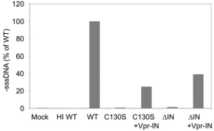
[image:3.585.316.525.69.197.2]Since IN is synthesized and packaged into the immature virion as part of the Gag-Pol polyprotein, many of the alter-ations in the virion morphology and defects at steps other than integration can be caused by the effects of IN mutations on the Gag-Pol precursor protein (10, 44). To confirm that the ob-served defect in reverse transcription was indeed a direct effect of the IN mutation or deletion, we examined whether the reverse transcription block could be rescued by incorporating WT IN into virions intrans using a Vpr-IN fusion construct (35, 64). At 16 h after the infection of CEM-GFP cells, total nucleic acid was extracted and ⫺sssDNA was analyzed by real-time PCR. Mock-infected cells and cells infected with heat-inactivated (HI) virus were used as negative controls (Fig. 1). Consistent with published results (64, 69), both IN mutants exhibited a loss of early reverse transcription products com-pared to the WT, with levels measuring less than 1% for NL-C130S and 1.5% for NL-⌬IN (Fig. 1). However, supplying IN intranswas able to rescue the loss of early reverse tran-scription products for both NL-C130S and NL-⌬IN and re-stored the⫺sssDNA levels to 25.0% and 39.2% of WT, re-spectively (Fig. 1). In addition to reverse transcription, using
FIG. 1.trans incorporation of IN rescues the reverse transcription defect of the IN mutants. 293T cells were transfected with pNL-C130S or pNL-⌬IN or cotransfected with C130S and pLR2P-Vpr-IN, or pNL-⌬IN and pLR2P-Vpr-IN. WT NL4-3 was used as a positive control. Two days posttransfection, viruses were collected from the culture media and equal amounts of p24-equivalent of each virus were used to infect CEM-GFP cells. Quantitative analysis of viral cDNA synthesis was done by real-time PCR. The amount of early reverse-transcribed viral DNA present in infected cells was measured 16 h after infection with WT, NL-C130S, NL-C130S plus Vpr-IN, NL-⌬IN, or NL-⌬IN plus Vpr-IN viruses. Control infections with culture medium alone (Mock) and cells infected with heat-inactivated WT virus (HI) were used as negative con-trols. The values are⫺sssDNA copy numbers and are expressed as the average percentages of WT virus from two experiments.
on November 8, 2019 by guest
http://jvi.asm.org/
the CEM-GFP reporter cell assay (26), we found thattrans
incorporation of WT IN was able to rescue infectivity of NL-C130S (data not shown). The ability to rescue reverse tran-scription and infectivity of an HIV lacking IN through thetrans
addition of WT IN has also been reported (64). The ability of
thetrans-incorporated IN to partially rescue the reverse
tran-scription defect of NL-C130S and NL-⌬IN indicated that the impairment is due at least in part to the absence or mutation of IN and is not an indirect result of the IN mutation or deletion of the Gag-Pol polyprotein.
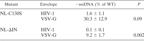
Pseudotyping with VSV-G rescues reverse-transcription de-fects for both IN mutant viruses.As previously reported, both cell attachment and entry for NL-C130S (69) and S-IN, an-other virus lacking IN (64), are similar to those of the WT, suggesting that the infectivity defect occurred soon after cell entry. Some early postentry blocks can be bypassed upon pseudotyping HIV-1 with envelope proteins of other viruses (3, 13). Pseudotyping with envelope glycoprotein G from VSV alters the pathway of virus entry, targeting it to a low-pH endosomal compartment (3). The infectivity of certain CA mutants that are noninfectious and with a specific defect in core stability can be rescued by pseudotyping them with VSV-G (9). Targeting the CA mutants to the endosomal path-way was reported to enhance infectivity by relocalizing the reverse transcription complexes or PICs to the appropriate site within the host cell cytoplasm to bypass the uncoating defect and continue with the replication cycle (9). We pseudotyped WT virus and both IN mutants with VSV-G by cotransfection with proviral clones that are env⫺ and a plasmid expressing VSV-G. Total nucleic acid was extracted from cells infected with the pseudotyped viruses at 16 h postinfection, and rescue was measured by real-time PCR of⫺sssDNA. Compared to the WT, pseudotyping with VSV-G rescued⫺sssDNA synthe-sis for both IN mutants, with 18.5- and 107.3-fold increases for NL-C130S and NL-⌬IN, respectively (Table 1). By altering the entry pathway, both IN mutants were able to undergo reverse transcription in target cells. The result suggested that the re-verse transcription defect of the IN mutants is not due to an alteration in particle formation or composition but, rather, is a result of aberrant uncoating.
Isolation of HIV-1 cores from WT and IN mutant viruses.As described above, a replication block occurring after entry but prior to reverse transcription has also been reported for certain mutations in CA (22, 40, 46, 54, 55). These CA mutants display
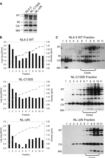
a defect in uncoating with altered core stability and yield. Because both the CA mutants and the IN mutants described here share a phenotype of a block at reverse transcription, we sought to examine the role of HIV-1 IN during uncoating by isolating cores from the WT, NL-C130S, and NL-⌬IN and examining their core yields and stabilities. We first confirmed, using Western blot analysis, that both IN mutants had protein packaging and processing similar to those of the WT (Fig. 2A), which is consistent with published results (64, 69). For isolating viral cores, previous studies have employed protocols using different concentrations of sucrose and nonionic detergents to separate the cores based on density (1, 5, 8, 19, 20, 22, 30, 55, 59, 60). In this study, virions were exposed to 1% Triton X-100 to remove the viral envelope, and intact cores (densities, 1.20 to 1.26 g/ml) were separated from intact virions (densities, 1.16 to 1.18 g/ml) and from non-particle-associated proteins using a continuous sucrose gradient (20–22). In the absence of detergent, the majority of viral proteins from all three viruses were contained within the top fractions of the gradient, coin-ciding with the expected density of intact virions (data not shown). The lipid bilayer of the virion was removed through the addition of 1% Triton X-100 in the 15% sucrose layer of the gradient, resulting in a change in the density of the particles consistent with that of viral cores (Fig. 2B) and causing a shift of the core-associated viral proteins (IN, RT, and CA) to the lower fractions of the gradient (Fig. 2C). The density and p24 content of each fraction were determined directly from the gradient frac-tions (Fig. 2B). Aliquots of each fraction were then subjected to ultracentrifugation, and the protein composition of the pellet was analyzed by Western blot analysis (Fig. 2C). In the presence of 1% Triton X-100, both IN mutants produced gradient profiles similar to that of the WT, with the majority of core-associated viral proteins contained within the lower fractions of the gradient (Fig. 2C). The density measurement and p24 content also indi-cated that these lower fractions contained viral cores (Fig. 2B). Thus, the gradient profiles for both IN mutants further confirmed that the C130S IN substitution or lack of IN does not affect core formation or the packaging and processing of the major known viral proteins comprising the core.
Isolated cores are functional in catalyzing endogenous re-verse transcription. To examine if the isolated cores were functional, we measured the activities of cores using an ERT assay. This assay examines the ability of RT associated with the core to perform reverse transcription by using the endogenous template (viral RNA) and primer (tRNA3Lys) (29). After 16 h at
37°C in the presence of 20M dNTPs, gradient fractions from all three viruses were capable of generating⫺sssDNA in the ERT reaction (Fig. 3), with fractions corresponding to viral cores exhibiting peak values. When the total⫺sssDNA synthe-sis per nanogram p24 within the core fractions was quantified, cores from all three viruses exhibited similar total⫺sssDNA synthesis, with levels reaching 5.5⫻105, 1.5⫻106, and 4.2⫻105
[image:4.585.43.282.90.158.2]copies/ng p24 for the WT, NL-C130S, and NL-⌬IN, respectively. These levels of ERT activity are consistent with published reports, indicating that viral cDNA synthesis is detectable upon virion exposure to detergent when assaying for endogenous reverse transcription (29). The synthesis of⫺sssDNA was blocked in the presence of efavirenz or in the absence of dNTPs (data not shown), indicating that the PCR product indeed resulted from reverse transcriptase. These results indicated that cores isolated
TABLE 1. Pseudotyping with VSV-G partially restores reverse transcription of IN mutantsa
Mutant Envelope ⫺sssDNA (% of WT) P
NL-C130S HIV-1 1.6⫾1.1
VSV-G 30.3⫾12.9 0.09
NL-⌬IN HIV-1 0.1⫾0.1
VSV-G 9.2⫾1.7 0.002
aCEM-GFP cells were infected with viruses with or without VSV envelope at
a multiplicity of infection (MOI) of 0.03 to 0.05. At 16 h postinfection, total nucleic acids were extracted and⫺sssDNA was measured by real-time PCR with primers specific to the R-U5 region (69). The⫺sssDNA synthesis of IN mutants was expressed as a percentage of that of the WT control, which encoded WT IN, and pseudotyped with VSV-G. The results are the averages of duplicates of at least 3 independent experiments.
on November 8, 2019 by guest
http://jvi.asm.org/
FIG. 2. Isolation of viral cores from WT HIV-1 and IN mutant viruses. (A) Western blot of concentrated viruses. WT and IN mutant viruses prepared by transient transfections of 293T cells were concentrated over a 20% sucrose cushion, and the viral titer was measured by p24 ELISA. Fifty nanograms of p24-equivalent concentrated virus was separated on a 10% SDS-polyacrylamide gel, and viral protein expression was examined by Western blot analysis. The blots were probed with human anti-HIV serum. (B) Graphic depiction of the p24 contents obtained by p24 ELISA (bars) and the densities of the sucrose gradient fractions (lines) for all three viruses. (C) Western blots of gradient fractions after ultracentrifu-gation. Each blot was probed with human anti-HIV serum. Fractions containing cores are indicated. In panels A and C, viral proteins are labeled as reverse transcriptase (RT), integrase (IN), capsid (CA), and matrix (MA).
on November 8, 2019 by guest
http://jvi.asm.org/
from all three viruses contained viral RNA, tRNA primer, and RT and were functional in carrying out reverse transcription un-der thein vitroconditions.
IN mutants have poor core yields.Defects in uncoating can affect core yield, as has been reported for CA mutants defec-tive at uncoating (22, 59). We therefore measured the core yields for both IN mutants. The core yields were determined by calculating the percentages of CA in core fractions compared to the total amount of CA in the entire gradient (22). For WT, the core yield obtained was 14.1⫾1.9%, which is similar to that reported previously (22). For NL-C130S and NL-⌬IN, the yields were 7.0⫾0.4% and 9.0⫾0.3%, respectively. The core yields from both NL-C130S and NL-⌬IN were significantly lower (P⬍0.05) than that of the WT. These results indicated that the IN C130S substitution or lack of IN can decrease the core yield. Although the core yields for both IN mutants were decreased, the gradient profiles were not detectably altered (Fig. 2C), suggesting that decreases in core yield alone are not the sole cause of loss of infectivity for these IN mutants.
IN mutants have altered core disassembly kinetics. In ad-dition to assessment of core yield, we determined core stability using anin vitrocore disassembly assay (20, 22). Uncoating is accelerated at higher temperatures, and this assay capitalizes upon this temperature-sensitive release of CA from the core to measure core stability (20, 22). Sucrose gradient fractions con-taining cores were pooled and placed at either 4°C or 37°C for 0, 60, or 120 min. Upon ultracentrifugation, intact cores sedi-mented as a pellet while disassembled particles remained in the supernatant fraction. The pelleted samples were then sub-jected to Western blot analysis and were probed with either human anti-HIV serum or anti-CA antibodies to monitor core disassembly. At 4°C, CA was retained in the pellets from all three viruses, indicating that the isolated cores remained stable for at least 120 min (Fig. 4A). At 37°C, cores isolated from both NL-C130S and NL-⌬IN disassembled more rapidly than WT cores, as illustrated by the loss of CA from the pellet fraction over time (Fig. 4A and B). These results demonstrated that the absence of IN or the C130S substitution causes an instability of the viral core, which leads to an accelerated ki-netics of core disassembly.
IN mutants have accelerated CA degradation in the cyto-plasm of infected cells. To determine if the accelerated CA disassembly of the IN mutants observedin vitroalso occurred
in vivo, we analyzed the total cytoplasmic CA content in the
cellular lysate of infected HeLa CD4⫹cells (12). The change in CA degradation in the whole-cell lysate has been previously used to examine acceleration of uncoating in the presence of restriction factors, including TRIM5␣ and TRIM-Cyp (12). HeLa CD4⫹cells were infected with the WT, NL-C130S, or NL-⌬IN for 6 h at 37°C. Nuclei and cellular debris were re-moved by centrifugation, and the cytoplasmic CA content was determined by Western blot analysis using the polyclonal anti-CA antibody (Fig. 4C). Compared to the WT, the CA con-tent in the lysate extracted from cells infected by either NL-C130S or NL-⌬IN was decreased. At 6 h postinfection, the cytoplasmic CA levels of NL-C130S and NL-⌬IN were 79.3% and 16.1%, respectively, of that of the WT virus. The reduced cytoplasmic CA content suggested that uncoating of both IN mutant virusesin vivowas accelerated compared to the WT, resulting in an increase in CA degradation during infection. Therefore, the reduced cytoplasmic CA contents of the IN mutantsin vivoare consistent with the unstable-core pheno-type observed using thein vitrocore disassembly assay.
[image:6.585.84.243.66.394.2]IN mutants have decreased CypA incorporation, and WT virus depleted of CypA has an unstable core. We examined virion incorporation of CypA to determine if the mutation or lack of IN resulted in a change in viral recognition by host factors. Fifty nanograms of p24-gequivalent concentrated virus was separated on a 10% SDS-polyacrylamide gel, and CypA incorporation was determined by Western blot analysis using polyclonal anti-CypA and monoclonal anti-CA antibodies as probes (Fig. 5). Compared to the WT, both IN mutant viruses had a decrease in CypA incorporation, with a 2.1-fold decrease for NL-C130S and a 15.4-fold decrease for NL-⌬IN. These results indicate that the C130S substitution or lack of IN con-tributed to a change in CypA recognition, possibly via CA. Due to the ability of trans-incorporated IN to restore the reverse transcription defects of both IN mutants (Fig. 1), we examined whether CypA incorporation could be similarly restored. We
FIG. 3. Viral cDNA synthesis of gradient fractions from WT and IN mutant viruses. Fraction samples from WT (A) and both IN mutant viruses, NL-C130S (B) and NL-⌬IN (C), were subjected to an endog-enous reverse-transcription assay. After 16 h, each sample was ana-lyzed for the early reverse transcription product using real-time PCR with primers specific for⫺sssDNA (gray bars). As a control, efavirenz was added to the reaction mixture to a final concentration of 7.5M (black bars). The results are expressed as copy numbers per microliter for each fraction with the indicated densities and are representative of at least three independent experiments for each virus.
on November 8, 2019 by guest
http://jvi.asm.org/
found that the CypA levels of both IN mutants increased when IN was incorporated intrans (data not shown), further con-firming a role for IN in maintaining CypA-CA interaction.
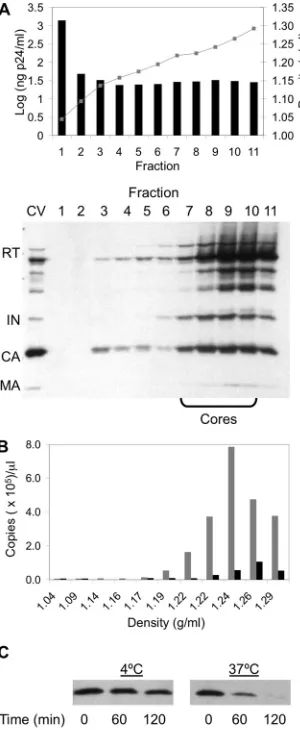
Based on the decrease in CypA incorporation in the IN mutant viruses and the resultant defects in uncoating, we sought to examine a functional role in uncoating for CypA by generating WT virions depleted of CypA through the addition of 10M CsA during transfection (7). Compared to a
non-CsA-treated control, CypA was efficiently depleted from the WT virions by the CsA treatment during transfection (data not shown). Concentrated HIV-1 generated in the presence of CsA was loaded onto a continuous sucrose gradient containing 1% Triton X-100. After the gradient fractions were subjected to ultracentrifugation, Western blot analysis of the pellet showed that the presence of core-associated viral proteins was similar to that of the non-CsA-treated control (Fig. 6A and WT in Fig. 2). Different gradient fractions were also examined by the ERT assay, and core fractions were competent for reverse transcription, indicating that the viral proteins and both the viral template and primer were not defective (Fig. 6B). In addition, the amounts of total ⫺sssDNA synthesis within the core fractions were 7.1⫻105copies/ng p24, which
is similar to the WT level (5.5⫻105copies/ng p24). This result
further confirmed that the CypA-depleted samples had no defects in the endogenous reverse transcription machinery. The core yield was also measured as described above, and the yield obtained from a CypA-depleted sample (8.64%⫾0.52%) was significantly lower (P ⬍ 0.05) than that of the WT. In addition to yield, core stability was measured using thein vitro
core disassembly assay, and CypA-depleted cores disassembled faster than a non-CsA-treated control (Fig. 6C). The kinetics of the CypA-depleted cores was similar to those of the IN mutants, with 38% and 67% CA disassembly at 60 min and 120 min, respectively. To determine if CypA depletion and the IN mutation or deletion resulted in an additive effect on uncoat-ing, NL-C130S virus was generated in the presence of 10M CsA, and cores were isolated and examined as described above. We found that both the yield and the disassembly ki-netics of the isolated cores from the mutant virus were not altered by CsA treatment (data not shown). This result indi-cated that the effects of CsA treatment and the IN mutation on core stability are not additive, and they therefore likely share the same mechanism in disrupting core stability. Taken together, the results suggested that CypA functions to promote core stability during uncoating and that the C130S IN mutation or deletion accelerates uncoating by decreasing CypA incorporation.
DISCUSSION
The uncoating step of the HIV-1 life cycle is a crucial but poorly understood process (4). The majority of insights into this early replication event have been obtained through
muta-FIG. 4. CA disassembly of WT and IN mutant virusesin vitroand
[image:7.585.329.513.68.147.2]in vivo. (A) Western blots illustrating disassembly of CA from the viral core. Gradient fractions corresponding to viral cores were pooled and placed at either 4°C or 37°C for 0, 60, or 120 min. Samples were then subjected to ultracentrifugation at 100,000⫻gfor 20 min at 4°C. The pellets were resuspended in 2% SDS sample buffer for Western blot analysis, and the blots were probed with a monoclonal CA anti-body. The blots were analyzed by chemiluminescence of the horserad-ish peroxidase (HRP)-conjugated secondary antibody and are repre-sentative of at least 3 independent experiments. (B) Disassembly kinetics of viral cores at 37°C. The Western blots shown in panel A were scanned, and ImageQuant TL software (GE Healthcare) was used to quantify band intensity. Percent disassembly was determined by using the value at time zero as the total p24 amount. The results are the mean values⫾standard errors of the mean (SEM) of at least three independent experiments for WT (Œ), NL-C130S (f), and NL-⌬IN (}). (C) CA degradation in the cytoplasmic cell lysates of infected cells. HeLa CD4⫹cells were infected with either WT, NL-C130S, or NL-⌬IN virus for 6 h at 37°C. The CA contents in cytoplasmic lysates were examined by Western blot analysis, and the blots were probed with polyclonal anti-CA antibodies (top). The cytoplasmic CypA con-tent, probed with polyclonal anti-CypA antibody, served as a specificity control and was used to normalize the amount of lysate loaded onto the gel (bottom). The results are representative of two independent experiments.
FIG. 5. Decrease in CypA incorporation into IN mutant virus. WT and IN mutant viruses prepared by transient transfections of 293T cells were concentrated over a 20% sucrose cushion, and the viral titer was measured by p24 ELISA. Fifty nanograms of p24-equivalent concen-trated virus was separated on a 10% SDS-polyacrylamide gel, and Western blot analysis was carried out using a polyclonal anti-CypA antibody to measure viral incorporation of CypA (top) and a mono-clonal antibody to CA (bottom).
on November 8, 2019 by guest
http://jvi.asm.org/
[image:7.585.82.239.70.380.2]tional analysis of the CA protein, which has indicated that the uncoating step is a temporally regulated process and that changes in core stability are detrimental to replication (22, 31, 40, 54, 55, 59). In this study, we examined the role of HIV-1 IN during the uncoating of the viral core. Our results indicate that IN is required for the optimal stability of the viral core, with the C130S substitution or lack of IN detrimentally affecting both the core yield and the rate of disassembly, leading to a
block in infection. The IN requirement during the uncoating step is manifested through CypA, a host factor that recognizes and binds CA. Our data show that the IN mutations affect CypA-CA interactions, which are required for optimal core stability.
Our results indicate a novel function for HIV-1 IN, namely, to promote optimal stability of the viral core. Mutational stud-ies of HIV-1 IN have demonstrated the pleiotropic effects of IN on different stages of the viral life cycle (10, 14, 17, 28, 44, 62, 64, 69), but to our knowledge, a requirement for IN during uncoating has not been previously reported. By isolating and characterizing IN mutant cores, we found decreases in core yield and core stability, which are consistent with the unstable-core phenotype described previously with certain CA mutants (22). Also, by measuring the cytoplasmic CA content in the infected cells, we found that CA degradation of the IN mutants is increased, which is consistent with the poor core yield and stability observedin vitro. Our results suggest that the block to reverse transcription and infectivity for these IN mutants is due to changes in core stability. The compositions of the viral cores of these IN mutants are similar to that of the WT core, and thus, one likely explanation for the loss of reverse transcription products in target cells is that the CA disassembly of the IN mutant viral cores is defective, preventing efficient proviral DNA synthesisin vivo. Previous reports have suggested that altered CA disassembly kinetics may prematurely expose the viral genome and proteins to damaging cellular factors or re-sult in a loss of required viral components prior to completion of reverse transcription (22, 51). By pseudotyping with VSV-G, we were able to rescue⫺sssDNA synthesis for both IN mu-tants in target cells, further supporting the idea that one of the blocks to infectivity for these mutants is at uncoating and can be bypassed by targeting to a different entry pathway. These results are consistent with the ability of VSV-G pseudotyping to rescue a noninfectious CA mutant (S178A) that displays an unstable core phenotype (9). In addition to its role in uncoat-ing, HIV-1 IN physically interacts with RT (28, 62, 69), and we have shown previously that IN can stimulate the initiation and elongation modes of reverse transcriptionin vitroby enhancing the processivity of RT (16). Therefore, in light of the present result, we propose that HIV-1 IN can affect reverse transcrip-tion either directly, by interacting with RT, or indirectly, by altering viral core stability.
[image:8.585.86.236.69.434.2]Host factors participate in different stages of viral replica-tion, both positively and negatively regulating infection. Nu-merous cellular factors have been identified that are required for efficient HIV-1 replication, highlighting the reliance of the virus on the host cell machinery to lead to a productive infec-tion (11). At the uncoating step, CypA and TRIM5␣are two host factors that target the incoming viral core, regulating infection in a species-specific manner (37, 48, 53, 57). The CypA-CA interaction is important for replication, and disrup-tion of this interacdisrup-tion by either mutadisrup-tions in CA or CsA treatment results in a block to infection (7, 27, 49). Based on the defects observed for core yield and CA disassembly for our IN mutants, we sought to examine possible changes in host factor recognition by examining CypA incorporation into the virion. By Western blot analysis, both IN mutant viruses had decreased levels of CypA incorporation compared to the WT. In addition, the CypA level could be restored by incorporating
FIG. 6. Depletion of CypA from HIV-1 results in an unstable-core phenotype. (A) Isolation of viral cores from WT virus depleted of CypA. (Top) Graphic depiction of the p24 contents obtained by p24 ELISA (bars) and the densities of the sucrose gradient fractions (gray lines). (Bottom) Western blot of gradient fractions after ultracentrif-ugation that was probed with human anti-HIV serum. Fifty nanograms of p24-equivalent concentrated virus (CV) was added as a size marker for viral proteins. The other symbols have the same meaning as in Fig. 2. (B) Viral cDNA synthesis from gradient fractions. The ERT assay was used to measure⫺sssDNA by real-time PCR in the absence (gray bars) or presence (black bars) of 7.5M efavirenz. The results are expressed as the copy number per microliter for each fraction with the indicated densities. (C)In vitrocore disassembly assay. Isolated cores were pooled and placed at either 4°C or 37°C for up to 120 min. Samples were subjected to ultracentrifugation, and the CA contents of the pellet fractions were determined by Western blot analysis by prob-ing with monoclonal anti-CA antibody. In all panels, the results are representative of at least three independent experiments.
on November 8, 2019 by guest
http://jvi.asm.org/
IN in trans into the IN mutant viruses. These observations indicate that C130S substitution or lack of IN alters the ability of CypA to recognize and bind CA. The loss of CypA incor-poration into the IN mutant viruses in the producer cell pre-sumably persists in the target cell, and a decrease in CypA recognition of the viral core results in defective reverse tran-scription and a block in replication.
CypA is a CA-binding cellular factor that either can be incorporated into the HIV-1 virion during assembly or can recognize the incoming viral core during infection of a target cell. Based on the observation that blocking CypA-CA inter-actions in the target cell by either CsA treatment or CA mu-tations leads to a block at reverse transcription and a decrease in infectivity, the requirement for CypA in HIV-1 replication has been attributed solely to CypA incorporation within the target cell (27, 49). Previous studies have shown that CypA-depleted cores have normal protein processing and packaging and are able to synthesize reverse transcription products using the endogenous RNA template and primer (7, 23, 49, 61). Furthermore, based on the ability of CA to sediment upon detergent treatment, CypA-depleted cores have been reported to possess the same stability as WT cores (61). Due to the association of unstable cores and a loss of CypA in our mu-tants, we reexamined a possible role for CypA in core stability using the core disassembly assay. Consistent with previous re-ports (7, 23, 49, 61), we found that the CypA-depleted cores had a composition and ability to carry out reverse transcription similar to those of non-CsA-treated cores. However, both the yield and the stability of CypA-depleted cores were decreased compared to those of WT cores. These results suggest a func-tional role for CypA-CA interactions during uncoating by pro-moting optimal core stability. CypA-CA interactions within the target cell are important in promoting infectivity (34), and recently, a mechanistic role for this interaction during uncoat-ing was proposed to modulate CA disassembly by either sta-bilizing or destasta-bilizing the viral core (49). Our results are consistent with this report, indicating that the CypA-CA inter-action is important for core stability and that changes in CypA-CA recognition result in changes in the kinetics of CA disassembly.
Many reports have demonstrated that CA is the viral deter-minant for host factor recognition of the viral core (21, 39, 40, 47, 51, 58, 65) and that certain mutations in CA can specifically alter CypA recognition (7, 21, 23, 27, 49, 55, 58, 61), but this is the first report that absence or certain mutations of IN can lead to changes in CypA recognition. How does IN mediate a change in host factor recognition of CA? Our data suggest a possible interaction within the viral core between IN and CA that leads to changes in CypA recognition. We speculate that this interaction may be required to promote the proper con-formation or substrate availability of the core to allow CypA to recognize and bind CA. Blocking the interaction between CypA and CA within the target cell leads to defects in infec-tivity and may cause changes in CA disassembly (34). Our data support this model, as the IN mutants have decreased CypA recognition and accelerated kinetics of CA disassembly. In addition to IN mutants, the unstable-core phenotype can be mimicked by depleting CypA from WT virus, confirming that CypA-CA interactions are important for optimal core stability. Future work will be required to identify possible interactions
between IN and CA and to understand how changes in IN can lead to modulation of recognition by CA-binding molecules.
In summary, our data reveal a novel role for IN during uncoating by promoting optimal stability of the viral core. In addition, our results indicate that CypA contributes to core stability and provide additional evidence to support the finding that CypA modulates CA disassembly (34). These findings suggest that complex interactions between viral and host fac-tors exist to promote optimal stability of the viral core, leading to a productive infection. Further work in characterizing these interactions will provide new insights into the uncoating step of the HIV-1 replication cycle.
ACKNOWLEDGMENTS
We thank members of the Chow laboratory for critically reading the manuscript and Hong Wu for the use of the Bio-Rad iCycler. The pdenv(wt) construct was a gift from Jerome Zack at UCLA.
The ELISA for p24 was carried out by the Center for AIDS Re-search Virology Core Facility, which is supported by National Insti-tutes of Health award AI28697, the UCLA AIDS Institute, and the UCLA Council of Bioscience Resources. This work was supported by NIH grants CA68859 and AI077386 to S.A.C. and a Supplementary Grant for Minority Students to C.W.D. M.S.B. was partly supported by NIH grants, Research Training in Pharmacological Sciences (RTPS) 5T32GM008652, and a MARC Predoctoral Fellowship (F31GM083356).
REFERENCES
1.Accola, M. A., A. Ohagen, and H. G. Gottlinger.2000. Isolation of human immunodeficiency virus type 1 cores: retention of Vpr in the absence of p6(gag). J. Virol.74:6198–6202.
2.Adamson, C. S., K. Salzwedel, and E. O. Freed.2009. Virus maturation as a new HIV-1 therapeutic target. Exp. Opin. Ther. Targets13:895–908. 3.Aiken, C.1997. Pseudotyping human immunodeficiency virus type 1 (HIV-1)
by the glycoprotein of vesicular stomatitis virus targets HIV-1 entry to an endocytic pathway and suppresses both the requirement for Nef and the sensitivity to cyclosporin A. J. Virol.71:5871–5877.
4.Aiken, C.2006. Viral and cellular factors that regulate HIV-1 uncoating. Curr. Opin. HIV AIDS1:194–199.
5.Auewarakul, P., P. Wacharapornin, S. Srichatrapimuk, S. Chutipongtanate, and P. Puthavathana.2005. Uncoating of HIV-1 requires cellular activation. Virology337:93–101.
6.Bosco, D. A., E. Z. Eisenmesser, S. Pochapsky, W. I. Sundquist, and D. Kern. 2002. Catalysis of cis/trans isomerization in native HIV-1 capsid by human cyclophilin A. Proc. Natl. Acad. Sci. U. S. A.99:5247–5252.
7.Braaten, D., E. K. Franke, and J. Luban.1996. Cyclophilin A is required for an early step in the life cycle of human immunodeficiency virus type 1 before the initiation of reverse transcription. J. Virol.70:3551–3560.
8.Briggs, J. A., T. Wilk, R. Welker, H. G. Krausslich, and S. D. Fuller.2003. Structural organization of authentic, mature HIV-1 virions and cores. EMBO J.22:1707–1715.
9.Brun, S., M. Solignat, B. Gay, E. Bernard, L. Chaloin, D. Fenard, C. Devaux, N. Chazal, and L. Briant.2008. VSV-G pseudotyping rescues HIV-1 CA mutations that impair core assembly or stability. Retrovirology5:57. 10.Bukovsky, A., and H. Gottlinger.1996. Lack of integrase can markedly affect
human immunodeficiency virus type 1 particle production in the presence of an active viral protease. J. Virol.70:6820–6825.
11.Bushman, F. D., N. Malani, J. Fernandes, I. D’Orso, G. Cagney, T. L. Diamond, H. Zhou, D. J. Hazuda, A. S. Espeseth, R. Konig, S. Bandyo-padhyay, T. Ideker, S. P. Goff, N. J. Krogan, A. D. Frankel, J. A. Young, and S. K. Chanda.2009. Host cell factors in HIV replication: meta-analysis of genome-wide studies. PLoS Pathog.5:e1000437.
12.Chatterji, U., M. D. Bobardt, P. Gaskill, D. Sheeter, H. Fox, and P. A. Gallay. 2006. Trim5alpha accelerates degradation of cytosolic capsid associated with productive HIV-1 entry. J. Biol. Chem.281:37025–37033.
13.Chazal, N., G. Singer, C. Aiken, M. L. Hammarskjold, and D. Rekosh.2001. Human immunodeficiency virus type 1 particles pseudotyped with envelope proteins that fuse at low pH no longer require Nef for optimal infectivity. J. Virol.75:4014–4018.
14.Depienne, C., A. Mousnier, H. Leh, E. Le Rouzic, D. Dormont, S. Benichou, and C. Dargemont.2001. Characterization of the nuclear import pathway for HIV-1 integrase. J. Biol. Chem.276:18102–18107.
15.Dismuke, D. J., and C. Aiken.2006. Evidence for a functional link between uncoating of the human immunodeficiency virus type 1 core and nuclear import of the viral preintegration complex. J. Virol.80:3712–3720.
on November 8, 2019 by guest
http://jvi.asm.org/
16.Dobard, C. W., M. S. Briones, and S. A. Chow.2007. Molecular mechanisms by which human immunodeficiency virus type 1 integrase stimulates the early steps of reverse transcription. J. Virol.81:10037–10046.
17.Engelman, A., G. Englund, J. M. Orenstein, M. A. Martin, and R. Craigie. 1995. Multiple effects of mutations in human immunodeficiency virus type 1 integrase on viral replication. J. Virol.69:2729–2736.
18.Fassati, A., and S. P. Goff.2001. Characterization of intracellular reverse transcription complexes of human immunodeficiency virus type 1. J. Virol. 75:3626–3635.
19.Fitzon, T., B. Leschonsky, K. Bieler, C. Paulus, J. Schroder, H. Wolf, and R. Wagner.2000. Proline residues in the HIV-1 NH2-terminal capsid domain: structure determinants for proper core assembly and subsequent steps of early replication. Virology268:294–307.
20.Forshey, B. M., and C. Aiken.2003. Disassembly of human immunodefi-ciency virus type 1 cores in vitro reveals association of Nef with the subviral ribonucleoprotein complex. J. Virol.77:4409–4414.
21.Forshey, B. M., J. Shi, and C. Aiken.2005. Structural requirements for recognition of the human immunodeficiency virus type 1 core during host restriction in owl monkey cells. J. Virol.79:869–875.
22.Forshey, B. M., U. von Schwedler, W. I. Sundquist, and C. Aiken.2002. Formation of a human immunodeficiency virus type 1 core of optimal sta-bility is crucial for viral replication. J. Virol.76:5667–5677.
23.Franke, E. K., H. E. Yuan, and J. Luban.1994. Specific incorporation of cyclophilin A into HIV-1 virions. Nature372:359–362.
24.Gamble, T. R., F. F. Vajdos, S. Yoo, D. K. Worthylake, M. Houseweart, W. I. Sundquist, and C. P. Hill.1996. Crystal structure of human cyclophilin A bound to the amino-terminal domain of HIV-1 capsid. Cell87:1285–1294. 25.Ganser-Pornillos, B. K., A. Cheng, and M. Yeager.2007. Structure of
full-length HIV-1 CA: a model for the mature capsid lattice. Cell131:70–79. 26.Gervaix, A., D. West, L. M. Leoni, D. D. Richman, F. Wong-Staal, and J.
Corbeil.1997. A new reporter cell line to monitor HIV infection and drug susceptibility in vitro. Proc. Natl. Acad. Sci. U. S. A.94:4653–4658. 27.Hatziioannou, T., D. Perez-Caballero, S. Cowan, and P. D. Bieniasz.2005.
Cyclophilin interactions with incoming human immunodeficiency virus type 1 capsids with opposing effects on infectivity in human cells. J. Virol.79:176–183. 28.Hehl, E. A., P. Joshi, G. V. Kalpana, and V. R. Prasad.2004. Interaction between human immunodeficiency virus type 1 reverse transcriptase and integrase proteins. J. Virol.78:5056–5067.
29.Khan, M., M. Garcia-Barrio, and M. D. Powell.2001. Restoration of wild-type infectivity to human immunodeficiency virus wild-type 1 strains lacking nef by intravirion reverse transcription. J. Virol.75:12081–12087.
30.Kotov, A., J. Zhou, P. Flicker, and C. Aiken.1999. Association of Nef with the human immunodeficiency virus type 1 core. J. Virol.73:8824–8830. 31.Leschonsky, B., C. Ludwig, K. Bieler, and R. Wagner.2007. Capsid stability
and replication of human immunodeficiency virus type 1 are influenced critically by charge and size of Gag residue 183. J. Gen. Virol.88:207–216. 32.Li, J., Z. Tan, S. Tang, I. Hewlett, R. Pang, M. He, S. He, B. Tian, K. Chen, and M. Yang.2009. Discovery of dual inhibitors targeting both HIV-1 capsid and human cyclophilin A to inhibit the assembly and uncoating of the viral capsid. Bioorg. Med. Chem.17:3177–3188.
33.Li, J., S. Tang, I. Hewlett, and M. Yang.2007. HIV-1 capsid protein and cyclophilin a as new targets for anti-AIDS therapeutic agents. Infect. Disord. Drug Targets7:238–244.
34.Li, Y., A. K. Kar, and J. Sodroski.2009. Target cell type-dependent modu-lation of human immunodeficiency virus (HIV-1) capsid disassembly by cyclophilin A. J. Virol.83:10951–10962.
35.Liu, H., X. Wu, H. Xiao, J. A. Conway, and J. C. Kappes.1997. Incorporation of functional human immunodeficiency virus type 1 integrase into virions independent of the Gag-Pol precursor protein. J. Virol.71:7704–7710. 36.Lizardi, P. M., X. Huang, Z. Zhu, P. Bray-Ward, D. C. Thomas, and D. C.
Ward.1998. Mutation detection and single-molecule counting using isother-mal rolling-circle amplification. Nat. Genet.19:225–232.
37.Luban, J.2007. Cyclophilin A, TRIM5, and resistance to human immuno-deficiency virus type 1 infection. J. Virol.81:1054–1061.
38.Marsden, M. D., and J. A. Zack.2007. Human immunodeficiency virus bearing a disrupted central DNA flap is pathogenic in vivo. J. Virol.81:6146– 6150.
39.Matsuoka, S., E. Dam, D. Lecossier, F. Clavel, and A. J. Hance.2009. Modulation of HIV-1 infectivity and cyclophilin A-dependence by Gag se-quence and target cell type. Retrovirology6:21.
40.Owens, C. M., P. C. Yang, H. Gottlinger, and J. Sodroski.2003. Human and simian immunodeficiency virus capsid proteins are major viral determinants of early, postentry replication blocks in simian cells. J. Virol.77:726–731. 41.Perez-Caballero, D., T. Hatziioannou, F. Zhang, S. Cowan, and P. D.
Bien-iasz.2005. Restriction of human immunodeficiency virus type 1 by TRIM-CypA occurs with rapid kinetics and independently of cytoplasmic bodies, ubiquitin, and proteasome activity. J. Virol.79:15567–15572.
42.Perron, M. J., M. Stremlau, M. Lee, H. Javanbakht, B. Song, and J. So-droski.2007. The human TRIM5alpha restriction factor mediates acceler-ated uncoating of the N-tropic murine leukemia virus capsid. J. Virol.81: 2138–2148.
43.Pornillos, O., B. K. Ganser-Pornillos, B. N. Kelly, Y. Hua, F. G. Whitby,
C. D. Stout, W. I. Sundquist, C. P. Hill, and M. Yeager.2009. X-ray struc-tures of the hexameric building block of the HIV capsid. Cell137:1282–1292. 44.Quillent, C., A. M. Borman, S. Paulous, C. Dauguet, and F. Clavel.1996. Extensive regions of pol are required for efficient human immunodeficiency virus polyprotein processing and particle maturation. Virology219:29–36. 45.Sayah, D. M., E. Sokolskaja, L. Berthoux, and J. Luban.2004. Cyclophilin A
retrotransposition into TRIM5 explains owl monkey resistance to HIV-1. Nature430:569–573.
46.Scholz, I., B. Arvidson, D. Huseby, and E. Barklis.2005. Virus particle core defects caused by mutations in the human immunodeficiency virus capsid N-terminal domain. J. Virol.79:1470–1479.
47.Shi, J., and C. Aiken.2006. Saturation of TRIM5 alpha-mediated restriction of HIV-1 infection depends on the stability of the incoming viral capsid. Virology350:493–500.
48.Sokolskaja, E., and J. Luban.2006. Cyclophilin, TRIM5, and innate immu-nity to HIV-1. Curr. Opin. Microbiol.9:404–408.
49.Sokolskaja, E., D. M. Sayah, and J. Luban.2004. Target cell cyclophilin A modulates human immunodeficiency virus type 1 infectivity. J. Virol.78: 12800–12808.
50.Stremlau, M., C. M. Owens, M. J. Perron, M. Kiessling, P. Autissier, and J. Sodroski.2004. The cytoplasmic body component TRIM5alpha restricts HIV-1 infection in Old World monkeys. Nature427:848–853.
51.Stremlau, M., M. Perron, M. Lee, Y. Li, B. Song, H. Javanbakht, F. Diaz-Griffero, D. J. Anderson, W. I. Sundquist, and J. Sodroski. 2006. Specific recognition and accelerated uncoating of retroviral capsids by the TRIM5alpha restriction factor. Proc. Natl. Acad. Sci. U. S. A.103:5514–5519.
52.Suzuki, Y., and R. Craigie.2007. The road to chromatin—nuclear entry of retroviruses. Nat. Rev. Microbiol.5:187–196.
53.Takeuchi, H., and T. Matano.2008. Host factors involved in resistance to retroviral infection. Microbiol. Immunol.52:318–325.
54.Tang, S., T. Murakami, B. E. Agresta, S. Campbell, E. O. Freed, and J. G. Levin.2001. Human immunodeficiency virus type 1 N-terminal capsid mu-tants that exhibit aberrant core morphology and are blocked in initiation of reverse transcription in infected cells. J. Virol.75:9357–9366.
55.Tang, S., T. Murakami, N. Cheng, A. C. Steven, E. O. Freed, and J. G. Levin. 2003. Human immunodeficiency virus type 1 N-terminal capsid mutants containing cores with abnormally high levels of capsid protein and virtually no reverse transcriptase. J. Virol.77:12592–12602.
56.Thali, M., A. Bukovsky, E. Kondo, B. Rosenwirth, C. T. Walsh, J. Sodroski, and H. G. Gottlinger.1994. Functional association of cyclophilin A with HIV-1 virions. Nature372:363–365.
57.Towers, G. J.2007. The control of viral infection by tripartite motif proteins and cyclophilin A. Retrovirology4:40.
58.Towers, G. J., T. Hatziioannou, S. Cowan, S. P. Goff, J. Luban, and P. D. Bieniasz.2003. Cyclophilin A modulates the sensitivity of HIV-1 to host restriction factors. Nat. Med.9:1138–1143.
59.Wacharapornin, P., D. Lauhakirti, and P. Auewarakul.2007. The effect of capsid mutations on HIV-1 uncoating. Virology358:48–54.
60.Welker, R., H. Hohenberg, U. Tessmer, C. Huckhagel, and H. G. Krausslich. 2000. Biochemical and structural analysis of isolated mature cores of human immunodeficiency virus type 1. J. Virol.74:1168–1177.
61.Wiegers, K., G. Rutter, U. Schubert, M. Grattinger, and H. G. Krausslich. 1999. Cyclophilin A incorporation is not required for human immunodefi-ciency virus type 1 particle maturation and does not destabilize the mature capsid. Virology257:261–274.
62.Wilkinson, T. A., K. Januszyk, M. L. Phillips, S. S. Tekeste, M. Zhang, J. T. Miller, S. F. Le Grice, R. T. Clubb, and S. A. Chow.2009. Identifying and characterizing a functional HIV-1 reverse transcriptase-binding site on inte-grase. J. Biol. Chem.284:7931–7939.
63.Wu, X., J. L. Anderson, E. M. Campbell, A. M. Joseph, and T. J. Hope.2006. Proteasome inhibitors uncouple rhesus TRIM5alpha restriction of HIV-1 re-verse transcription and infection. Proc. Natl. Acad. Sci. U. S. A.103:7465–7470. 64.Wu, X., H. Liu, H. Xiao, J. A. Conway, E. Hehl, G. V. Kalpana, V. Prasad, and J. C. Kappes.1999. Human immunodeficiency virus type 1 integrase protein promotes reverse transcription through specific interactions with the nucleoprotein reverse transcription complex. J. Virol.73:2126–2135. 65.Yamashita, M., and M. Emerman.2009. Cellular restriction targeting viral
capsids perturbs human immunodeficiency virus type 1 infection of nondi-viding cells. J. Virol.83:9835–9843.
66.Yamashita, M., O. Perez, T. J. Hope, and M. Emerman.2007. Evidence for direct involvement of the capsid protein in HIV infection of nondividing cells. PLoS Pathog.3:1502–1510.
67.Yin, L., D. Braaten, and J. Luban.1998. Human immunodeficiency virus type 1 replication is modulated by host cyclophilin A expression levels. J. Virol.72:6430–6436.
68.Zhang, J., X. Liu, and E. De Clercq.2009. Capsid (CA) protein as a novel drug target: recent progress in the research of HIV-1 CA inhibitors. Mini Rev. Med. Chem.9:510–518.
69.Zhu, K., C. Dobard, and S. A. Chow.2004. Requirement for integrase during reverse transcription of human immunodeficiency virus type 1 and the effect of cysteine mutations of integrase on its interactions with reverse transcrip-tase. J. Virol.78:5045–5055.