from a Hyperthermophilic Archaeal Lipothrixvirus
N. Peixeiro,a,e* J. Keller,bB. Collinet,b* N. Leulliot,b* V. Campanacci,cD. Cortez,aC. Cambillau,cK. R. Nitta,d* R. Vincentelli,c P. Forterre,aD. Prangishvili,aG. Sezonov,a,eH. van Tilbeurghb
Institut Pasteur, Unité Biologie Moléculaire du Gène chez les Extrémophiles, Département de Microbiologie, Paris, Francea
; Institut de Biochimie et de Biophysique Moléculaire et Cellulaire, Université Paris-Sud, UMR8619-CNRS, Orsay, Franceb
; Architecture et Fonction des Macromolécules Biologiques, CNRS, and Université d’Aix-Marseille, UMR 7257, Case 932, d’Aix-Marseille, Francec
; Institut du Biologie de Développement de Marseille Luminy, IBDML, UMR 6216, CNRS, Marseille, Franced
; Université Pierre et Marie Curie, UMR 7138 “Systématique, Adaptation, Evolution,” Paris, Francee
The structural and functional analysis of the protein AvtR encoded by
Acidianus
filamentous virus 6 (AFV6), which infects the
archaeal genus
Acidianus
, revealed its unusual structure and involvement in transcriptional regulation of several viral genes. The
crystal structure of AvtR (100 amino acids) at 2.6-Å resolution shows that it is constituted of a repeated ribbon-helix-helix
(RHH) motif, which is found in a large family of bacterial transcriptional regulators. The known RHH proteins form dimers that
interact with DNA using their ribbon to create a central

-sheet. The repeated RHH motifs of AvtR superpose well on such
dimers, but its central sheet contains an extra strand, suggesting either conformational changes or a different mode of DNA
binding. Systematic evolution of ligands by exponential enrichment (SELEX) experiments combined with systematic mutational
and computational analysis of the predicted site revealed 8 potential AvtR targets in the AFV6 genome. Two of these targets were
studied in detail, and the complex role of AvtR in the transcriptional regulation of viral genes was established. Repressing
tran-scription from its own gene,
gp29
, AvtR can also act as an activator of another gene,
gp30
. Its binding sites are distant from both
genes’ TATA boxes, and the mechanism of AvtR-dependent regulation appears to include protein oligomerization starting from
the protein’s initial binding sites. Many RHH transcriptional regulators of archaeal viruses could share this regulatory
mechanism.
V
iruses infecting
Archaea
, one of the three domains of life, have
been studied for more than 30 years. During that time, more
than 50 archaeal viruses have been described, 32 of which infect
hyperthermophilic archaea of the kingdom
Crenarchaeota
.
Spe-cific studies in different laboratories have revealed that these
cre-narchaeal viruses possess unique morphologies, distinct from
those of viruses infecting bacteria and eukaryotes (
1
). Consistent
with their exceptional morphotypes, more than 90% of the viral
genes have no homologues in the sequences available in public
databases (
2
).
In silico
analysis has revealed the diversity of transcriptional
regulators encoded by different archaeal viruses (
2
). The most
prevalent structural motifs found in these proteins are the
helix-turn-helix (HTH) and the ribbon-helix-helix (RHH). Proteins
with the latter domain are encoded by nearly all crenarchaeal
vi-ruses (
2
) and some euryarchaeal viruses (
3
). In addition, some
archaeal viruses encode proteins with looped-hinged-helix and Zn
finger domains (
2
). The abundance of genes coding for proteins
belonging to the RHH family in the genomes of
Crenarchaea
and
their viruses could underline the important role of these proteins
in both host and viral gene transcription regulation (
4
). Despite
their abundance in archaeal virus genomes and crucial role in the
regulation of viral genome expression, only one archaeoviral
tran-scription factor, protein SvtR encoded by the rudivirus SIRV1, has
so far been studied in detail (
5
). The nuclear magnetic resonance
(NMR) structure of the protein revealed a typical RHH fold. The
protein was found to form a dimer and bind DNA with its

-sheet
face. Two regions within the SIRV1 genome were pinpointed as
the SvtR binding sites; the protein was found to act as a repressor
of its own gene as well as the gene for the viral structural protein
gp30 (ORF1070) (
5
).
The RHH proteins are well-known transcriptional regulators
present in the bacterial and archaeal domains and their respective
viruses, where this DNA binding motif appears to be a common
structural scaffold (
6
). Remarkably, RHH proteins seem to be
ab-sent in eukaryotes (
6
). Although more than 4,000 RHH proteins
have currently been predicted by
in silico
analysis, only a few have
been studied experimentally (
6
). Since they are widely distributed
in these forms of life and because of their predictable importance
for the regulation of cellular processes, there has been over the last
years a dramatic increase in structural studies of proteins bearing
the RHH domain (
6
).
The structures of 21 RHH proteins are presently available (
5
,
7
–
26
). Among them, the methionine repressor MetJ (
24
), the
reg-ulator of plasmid copy number CopG (
11
,
27
–
29
), and the
bacte-Received29 May 2012Accepted3 October 2012
Published ahead of print10 October 2012
Address correspondence to G. Sezonov, guennadi.sezonov@pasteur.fr, or H. van Tilbeurgh, herman.van-tilbeurgh@u-psud.fr.
* Present address: N. Peixeiro, Commissariat à la Énergie Atomique et aux Énergies Alternatives, iBiTec-S, Service de Biologie Intégrative et Génétique Moléculaire, Gif-sur-Yvette, France; B. Collinet, UFR 927 des Sciences de la Vie, Université Pierre et Marie Curie, Paris, France; N. Leulliot, Laboratoire de Cristallographie et RMN Biologiques, UMR CNRS 8015, Université Paris Descartes, Sorbonne Paris Cité, Faculté des Sciences Pharmaceutiques et Biologiques, Paris, France; K. R. Nitta, Department of Biosciences and Nutrition, Karolinska Institutet, Stockholm, Sweden.
N.P. and J.K. contributed equally to this article.
Copyright © 2013, American Society for Microbiology. All Rights Reserved.
doi:10.1128/JVI.01306-12
on November 7, 2019 by guest
http://jvi.asm.org/
riophage P22 Mnt and Arc repressors (
21
,
30
) could be considered
the prototypical RHH proteins. The RHH motif consists of an
N-terminal

-strand and two
␣
-helices connected by a short turn.
RHH proteins form functional intertwined dimers through

-sheet formation of the N-terminal strands. These dimers
pres-ent their cpres-entral two-stranded antiparallel

-sheet to the major
groove of cognate DNA, and residues of this sheet are involved in
specific DNA base interactions. The second helix of the motif is
engaged in nonspecific phosphate backbone interactions. RHH
proteins contain either a single RHH domain (as in Arc and
CopG) or a RHH repeat (as in TraY), or they can be combined
with additional domains (as in NikR, PutA, and ParD). All RHH
proteins bind DNA as higher oligomers and contact inverted or
tandem repeats within operators.
In this study, we were focused on a 11.98-kDa putative DNA
binding protein encoded by
Acidianus
filamentous virus 6
(AFV6_gp29), which we term AvtR (
Acidianus
virus
transcrip-tional regulator). This protein is highly conserved in all members
of the
Betalipothrixvirus
genus of the
Lipothrixviridae
family
(AFV3, AFV6, AFV7, AFV8, AFV9, and SIFV) (
31
). All these
vi-ruses contain double-stranded DNA (dsDNA) genomes of around
40 kb, and
in silico
analysis revealed a high level of conservation of
their organization (
31
). Sequence analysis of AvtR shows clearly
that it has an N-terminal CopG-like RHH motif, but no structural
predictions could be made for the C-terminal half of the protein.
In order to study proteins involved in crenarchaeal virus
replica-tion, we set out to determine the crystal structure of AvtR and to
characterize its targets, functions, and mechanism of
transcrip-tional regulation The detailed characterization of this new
ar-chaeal regulator will allow a better understanding of the
mecha-nisms of transcription regulation that participate in the control of
the virus infection cycle in
Archaea
.
MATERIALS AND METHODS
Cloning, expression, and purification.The coding sequence of AvtR was amplified by PCR using cDNA as a template. The cDNA was cloned in a derived pET9 plasmid with a His tag at the C terminus. Expression was done at 37°C using theEscherichia coliRosetta(DE3)/pLysS strain grown in 2⫻YT medium (Bio 101 Inc.). When the cell culture reached an optical density at 600 nm (OD600) of 0.8, induction at 37°C was performed for 3 h with 0.5 mM IPTG (isopropyl--D-thiogalactopyranoside) (Sigma). Cells were harvested by centrifugation and suspended in buffer A (20 mM Na citrate [pH 5.6], 200 mM NaCl, 5 mM-mercaptoethanol). Cell lysis was completed by sonication, and the lysate was heated for 20 min at 50°C before centrifugation at 20,000⫻gfor 20 min. The soluble fraction was loaded on an Ni-nitrilotriacetic acid (Ni-NTA) column (Qiagen Inc.) equilibrated with buffer A. The protein was eluted with imidazole and subsequently loaded on a heparin column (GE Healthcare) equilibrated with buffer A=(20 mM Na citrate [pH 5.6], 50 mM NaCl, 10 mM -mer-captoethanol). Elution was performed using a gradient between buffer A= and buffer B (20 mM Na citrate, 1 M NaCl, 10 mM-mercaptoethanol). AvtR was eluted at about 0.7 M NaCl. The His tag was not proteolytically removed before the crystallogenesis experiments. Selenomethionine (SeMet)-substituted AvtR was produced in autoinducible medium at 37°C during 24 h. The medium composition (per liter) was as follows: glycerol, 10 g; glucose, 1 g; lactose, 1 g; KH2PO4, 13.6 g; (NH4)2SO4, 6.6 g; Na2HPO4· 2H2O, 14.2 g; MgSO4· 7H2O, 800 mg; CaCl2· 2H2O, 100 mg; thiamine, 4 mg, FeSO4· 7H2O, 5 mg; ZnSO4· 7H2O, 0.8 mg; MnSO4, 0.8 mg; CuSO4· 5H2O, 0.08 mg; NaMoO4, 0.4 mg; H3BO3, 1 mg; KI, 0.2 mg; amino acids (L-leucine,L-isoleucine,L-valine,L-tryptophan,L -phenylala-nine,L-lysine,L-alanine,L-arginine,L-aspartic acid, glycine,L-asparagine,
L-proline,L-serine,L-threonine,L-histidine,L-glutamine, andL-glutamic
acid), 200 mg; and selenomethionine, 125 mg. The SeMet-substituted protein was purified as the native protein. The protein sample was judged homogeneous as checked by SDS-PAGE, and the purity was estimated on gel to 98%. After purification, 7 mg and 4 mg of pure protein per liter of culture were obtained for the native protein and the SeMet-substituted protein, respectively.
Structure resolution.Crystals of SeMet-substituted AvtR were grown from a 1:1-l mixture of protein (8 mg/ml) with 22 to 27% polyethylene glycol 4000 (PEG4000)– 0.1 M HEPES (pH 7.5)–5 to 10% isopropanol, using the hanging-drop vapor diffusion method at 23°C. Crystals were soaked in a mixture of mother liquor and 30% glycerol before flash freez-ing at 100 K. X-ray diffraction data were collected from a crystal of the SeMet-substituted AvtR on beamline ID29 (ESRF) at the wavelength of the Se K edge. The crystals belong to the P21space group with four copies per asymmetric unit, corresponding to a 49.7% solvent content. Data were collected at a resolution of 2.6 Å and processed with the programs MOSFLM (32) and SCALA (33) for merging and scaling. The structure was solved using the SAD method using diffraction data collected at 2.6-Å resolution from SeMet-substituted crystals. Eight selenium atom sites were found with the program PHENIX.HYSS (34) These sites were used for phasing with the program SHARP (35). After solvent flattening with the program RESOLVE (36), the quality of the electron density map al-lowed automated construction of 80% of the model. The missing residues were built by hand using the O molecular graphics program (37), and the model was refined with REFMAC (38).
Systematic evolution of ligands by exponential enrichment (SELEX) assay.A 56-bp oligonucleotide in which the 16 central bases are com-pletely degenerate [5=-CTCAGGGTCGACTTCAGCG(N16)CAACCAGT CGTCGACCAGC-3=] was used. The double-stranded oligonucleotide DNA pool was amplified by PCR using primers 5=-CTCAGGGTCGACT TCAGCG-3=and 5=-GCTGGTCGACGACTGGTTG-3=. PCR amplifica-tion was performed with GoTaq Flexi DNA polymerase (Promega). The PCR products were mixed with AvtR protein in the binding buffer and were incubated at room temperature for 10 min [10 mM Tris-HCl (pH 7.5), 50 mM NaCl, 1 mM MgCl2, 0.5 mM dithiothreitol (DTT), 0.5 mM EDTA, 4% glycerol, 5g/ml poly(dI-dC)]. Ni-Sepharose 6 FF resin (GE Healthcare) equilibrated in binding buffer was then added, and the mix-ture was incubated for an additional 20 min. After washing of nonspecific binding oligonucleotides with binding buffer without poly(dI-dC), bead-protein-DNA complexes were dissolved in Milli-Q water. Retrieved oli-gonucleotides were amplified by PCR, and the PCR products were used as a DNA pool for the next round of selection. This process was repeated seven times. Selected DNA pools were sequenced by 454 sequencing, and sequences of the central regions of DNA pools were used for further anal-ysis.
Oligonucleotides. Oligonucleotides were purchased from Proligo (Sigma-Aldrich) or obtained by PCR using Phusion high-fidelity DNA polymerase (Finnzymes) as indicated. Double-stranded DNA was ob-tained by annealing the corresponding single-strand nucleotides follow-ing standard techniques. T4 polynucleotide kinase (New England Bio-Labs) was used for32P radiolabeling of oligonucleotides used in gel shift experiments.
Single-nucleotide polymorphisms: PAGE-EMSA experiments. PAGE-electrophoretic mobility shift assay (EMSA) analyses were per-formed using oligonucleotides with specific point mutations on the ATT GTGGTACCACCTT sequence provided by the SELEX. Sixteen different double-stranded 40-bp oligonucleotides with single-nucleotide polymor-phisms were constructed as shown inFig. 2B. [5= -AAAAAAAAAAAAA-(mutated SELEX)-GGGGGGGGGGGG-3=]. Using the mobility of the wild-type sequence as a control, 1.5 pmol of each mutated fragment was incubated with 2l of 50% (vol/vol) glycerin and 0.5g of salmon sperm in a total of 20l of transcription buffer (TB) (50 mM Tris-HCl [pH 8.0], 75 mM KCl, 25 mM MgCl2, and 1 mM dithiothreitol) for 15 min at 68°C with various amounts of purified AvtR as indicated in the figure legends. Five microliters of 5⫻RB loading dye (10 mM Tris-HCl [pH 7.5], 1 mM
on November 7, 2019 by guest
http://jvi.asm.org/
EDTA, 20% glycerol, 100g/ml bovine serum albumin [BSA], 1 mg/ml xylene-cyanol) was added. Twenty microliters of the DNA-protein mix-tures was deposited in a nondenaturing 8% 37.5:1 acrylamide-bisacryl-amide gel. PAGE was run in TBE buffer (89 mM Tris-borate, 2 mM Na EDTA, pH 8.3). After migration, the gel was vacuum dried, exposed with Amersham Biosciences Hyperfilm-MP, and developed with a Kodak X-OMAT 2000 processor. The procedure of vacuum drying, exposure, and development was repeated for all PAGE experiments described below.
In silicosearch for AvtR DNA binding sites.A PERL script was writ-ten, where a weighted matrix was used to search for similar sequences in a given genome. The matrix was built assigning different weights to differ-ent nucleotides. The script analyzed both strands of the AFV6 genome using an 18-nucleotide (nt) sliding window. The sequences located within the hypothetical promoter regions, ranging from position⫺150 to⫹50 of each gene, were considered for further analysis. Sequences with less than 75% of the matrix maximum score were not considered to eliminate a large number of false positives.
DNA binding activity of AvtR: PAGE-EMSA experiments.Eight 39-bp double-stranded DNA fragments (s1 through s8) encompassing the identified 16-bp binding sites were obtained using the corresponding sin-gle-stranded oligonucleotides. An unspecific 40-bp double-stranded DNA fragment (5=-CTTATCATTTCATGGATAAGAGGTTCCATGAAA CGCATGG-3=) was used as a negative control. To obtain the labeled dou-ble-stranded fragment, one strand of each doudou-ble-stranded fragment was 32
P radiolabeled, followed by annealing with the corresponding reverse oligonucleotide. A 1.5-pmol portion of each double-stranded labeled fragment (⬃75 ng) was incubated with 2l of 50% (vol/vol) glycerin and 0.5g of salmon sperm in a total of 20l of transcription buffer (TB) for 15 min at 68°C with increasing amounts of AvtR (from 0 to 500 ng). Five microliters of 5⫻RB loading dye (10 mM Tris-HCl [pH 7.5], 1 mM EDTA, 20% glycerol, 100g/ml BSA, 1 mg/ml xylene-cyanol) was added, and the sample was analyzed in a nondenaturing 8% 37.5:1 acrylamide-bisacrylamide gel in TBE buffer. Samples were run for 2 h at 200 V. Gels were vacuum dried, exposed, and developed as described above.
Primer extension analysis.A primer extension protocol was used to define the promoter regions and to precisely determine the transcription start sites of genesgp29andgp30. A 289-bp DNA fragment covering the shared promoter region between genesgp29andgp30(positions 15550 to 15839) was generated by PCR and cloned into a pDrive (Qiagen) vector. RNA was produced by incubating 100 ng of the corresponding linear fragment with 6.58 nMSulfolobus solfataricusRNA polymerase, 2.94 nM TATA binding protein (TBP), and 2.86 nM transcription factor B (TFB). Reactions were performed at 70°C for 30 min with 0.2 mM nucleoside triphosphates (NTPs) in 50l transcription buffer (50 mM Tris-HCl [pH 8.0], 75 mM KCl, 25 mM MgCl2, and 1 mM DTT). To obtain the gene gp30transcription product, AvtR was added to the reaction mixture to a final concentration of 208M. The RNA product was annealed to a ra-diolabeled reverse primer, and either genegp29or gp30transcription products were amplified into cDNA in a reverse transcription reaction. The reverse reactions were stopped by addition of 20l of 50% formam-ide loading dye, and 20l of the denatured sample was analyzed on a 10% denaturing polyacrylamide gel in TBE buffer. The 5=termini of the tran-scripts were mapped by comparison of the primer extension product with a sequencing ladder generated with a Thermo Sequenase cycle sequencing kit (USB Corporation) in a separate reaction with the same primer. Gels were fixed in 10% acetic acid–10% ethanol before vacuum drying, expo-sure, and development.
IVT.A crenarchaeon-specificin vitrotranscription (IVT) system was used to test the influence of the purified protein AvtR on transcription from the promoters Pgp29and Pgp30, where theSulfolobus solfataricus RNA polymerase and transcription factors TBP and TFB were obtained as described previously (39). A fragment covering the intergenic promoter region between the genes (positions 15550 to 15839) was generated by PCR from genomic AFV6 DNA and cloned directly into a pDrive (Qiagen) cloning vector by T/A cloning. TheSulfolobus shibatae virus
SSV1 promoter T6 (42) was used as a control in IVT experiments, as previously described (40).In vitrotranscription reactions were performed using 100 ng of the corresponding plasmid DNA in the presence of 0.2 mM NTPs, 6.58 nMSulfolobus solfataricusRNA polymerase, 2.94 nM TBP, 2.86 nM TFB, and increasing amounts of AvtR (seeFig. 5). The IVT reactions were performed for 30 min at 70°C in 50l transcription buffer (50 mM Tris-HCl [pH 8.0], 75 mM KCl, 25 mM MgCl2, and 1 mM DTT). Reactions were stopped by the addition of 250l of NEW buffer (10 mM Tris-HCl [pH 8.0], 750 mM NaCl, 10 mM EDTA, 0.5% SDS, and 40 mg/ml glycogen). The newly synthesizedin vitroRNA was isolated by a phenol-chloroform extraction, followed by ethanol precipitation. Tran-scription products were detected by primer extension using sequence-specific primers for the previously described viral promoter templates (41,
42). After addition of 20l of 50% formamide loading dye, 20l of the denatured sample was analyzed on a 10% denaturing polyacrylamide gel in TBE buffer. Gels were fixed in 10% acetic acid–10% ethanol before vacuum drying, exposure, and development.
Oligomerization assays: DNase I footprinting.DNase I footprinting experiments were performed on a 170-bp fragment covering positions 15616 to 15786 of the AFV6 genomic sequence. The two strands of radio-actively labeled DNA were generated by PCR in separate reactions by using, in each reaction, one radiolabeled primer. The DNA templates were incubated, in separate reactions, with AvtR in 50l transcription buffer for 15 min at 48°C. Samples were then treated for 5 min with 1 U of DNase I (Roche). Reactions were stopped by addition of 200l of stop solution (10 mM Tris [pH 8.0], 750 mM NaCl, 10 mM EDTA, 0.5% SDS, 0.04 g/l glycogen). DNA fragments were isolated by ethanol precipitation. After addition of 10l of 50% formamide loading dye, 5l of the dena-tured samples was analyzed on a denaturing 6% 29:1 acrylamide-bisacryl-amide sequencing gel. Samples were run for 2 h at 60 W in TBE buffer. Gels were fixed in 10% acetic acid–10% ethanol before vacuum drying, exposure, and development.
Oligomerization assays: PAGE-EMSA experiments.The mobility of a 289-bp radiolabeled DNA fragment (positions 15550 to 15839) contain-ing the shared promoter region between genesgp29andgp30was assessed by PAGE-EMSA experiments in the presence of increasing amounts of AvtR. The labeled DNA was generated by PCR by using a previously 32P-radiolabeled forward primer. A 1.5-pmol portion of the
double-stranded labeled fragment (⬃75 ng) was incubated with 2l of 50% (vol/vol) glycerin and 0.5g of salmon sperm in a total of 20l of tran-scription buffer (TB) for 15 min at 68°C with increasing amounts of AvtR (from 0 to 500 ng). Five microliters of 5⫻RB loading dye (10 mM Tris-HCl [pH 7.5], 1 mM EDTA, 20% glycerol, 100g/ml BSA, 1 mg/ml xylene-cyanol) was added, and the sample was analyzed in a nondenatur-ing 8% 37.5:1 acrylamide-bisacrylamide gel in TBE buffer. Samples were run for 4 h at 200 V. Gels were vacuum dried, exposed, and developed as described above.
RESULTS
Overall structure of AvtR.
Sequence analysis of AvtR suggested
the presence of an N-terminal RHH motif most closely related to
the CopG protein. CopG contains 45 residues and forms
inter-twined homodimers. No homologous sequence could be detected
for the C-terminal half of AvtR, but fold prediction programs
suggested that this part would be structured. We therefore
ex-pressed and purified the intact protein. AvtR crystallized in the P2
1space group with 4 copies in the asymmetric unit. The structure
was solved at 2.6-Å resolution by SAD phasing using
selenome-thionine-labeled protein. The statistics on data collection and
re-finement are provided in
Table 1
. AvtR is made of two very similar
RHH motifs, related by a pseudo-2-fold symmetry (
Fig. 1A
).

1
␣
1
␣
2 forms the first motif (RHH1, residues 1 to 36) and

3
␣
3
␣
4 the second (RHH2, residues 60 to 100) (
Fig. 1F
). The
linker between the first and the second motifs (24 residues)
on November 7, 2019 by guest
http://jvi.asm.org/
tains a strand (

2) that aligns with

1 and

3 to form the central
three-strand antiparallel

-sheet (
Fig. 1A
). The quasi-2-fold
sym-metry axis is at the center of the central

-ribbon. The two motifs
RHH1 and RHH2 superpose with a root mean square deviation
(RMSD) of 2.66 Å for 30 aligned C
␣
positions. The main
differ-ence between the two motifs resides in the length of the

-strands
(

3 is 1 residue longer than

1) and the length of the linker
be-tween the strand and the helix (the

3-
␣
3 linker contains 5
resi-dues and the

1-
␣
1 linker only 1). Consequently, the

1 strand
superposes on the extended

3-
␣
3 linker rather than on strand

3
itself (
Fig. 1B
). The two motifs share only 13% sequence identity
based on the structural alignment.
The RHH motif of AvtR.
Ribbon-helix-helix (RHH) DNA
binding proteins can use different strategies and oligomeric states
for the recognition of their target sites. The structure of the N
terminus of AvtR is clearly classified as an RHH motif, which
characterizes a large family of transcription factors. Interestingly,
the majority of these RHH transcription factors are small proteins
(about 45 amino acids), covering one RHH motif, and the
func-tional unit that binds to DNA is created through
homodimeriza-tion. A search for structural analogues of intact AvtR yielded a
good match with the Arc repressor dimer (Protein Data Bank
[PDB] code 1myk, RMSD of 2.07 Å for 76 aligned C
␣
positions).
However, the RHH1 motif from AvtR matches best with the CopG
monomer (RMSD of 0.79 Å on 35 aligned C
␣
positions) (
Fig. 1C
).
The best alignment for the RHH2 motif was also obtained with a
CopG monomer (RMSD of 2.62 Å for 38 aligned C
␣
positions)
(
Fig. 1C
). CopG shares 40% sequence identity with AvtR RHH1
but only 11% with AvtR RHH2. Overall, the structures of the
CopG dimer and the AvtR monomer also superpose well (RMSD
of 2.42 Å) (
Fig. 1C
): helices from AvtR and CopG occupy identical
positions, and the length of the linkers between helices is the same.
The

-ribbon of the CopG dimer superposes on the

1 and

3
strands from AvtR. However, the CopG

-strands are longer than
those of AvtR.
Identification of an AvtR consensus binding site.
The
struc-tural analysis of AvtR clearly established that it belongs to the
RHH superfamily of DNA binding proteins. As members of
this family are most often characterized as or predicted to be
transcriptional regulators (
6
), we decided to confirm the DNA
binding properties of AvtR and to identify its targets in the
AVF6 genome.
In order to define the hypothetical consensus DNA binding site
for AvtR, we used a random binding site selection by the SELEX
approach (
43
,
44
; K. R. Nitta, E. Jacox, R. Vincentelli, A. Jolma, D.
Sobral, A. Mistral, A. Ohtsuka, A. Kubo, C. Cambillau, Y. Satou, J.
Taipale, and P. Lemaire, submitted for publication). The selection
of putative AvtR binding sites was achieved using the His-tagged
version of the protein (immobilized on Ni
2⫹chelating affinity
resin) and a pool of double-stranded DNA oligonucleotides with a
central core of 16 random nucleotides (see Materials and
Meth-ods). PCR pools from selection rounds 5, 6, and 7 were sequenced
by 454 sequencing. The sequence analysis was performed using
Bioprospector (
45
). The consensus binding site was extracted
us-ing the Weblogo program (
46
) and is represented in
Fig. 2A
.
The random binding site selected by SELEX revealed the
pres-ence of a conserved central 10-bp palindromic sequpres-ence, GTGGT/
ACCAC. Nonetheless, this sequence could not be found in the
AFV6 genome. To identify the conserved positions of the AvtR
consensus binding site obtained by SELEX, we carried out a
sys-tematic mutational analysis of its 16-bp sequence. Out of the 16
analyzed nucleotides, 9 were essential for AvtR binding (
Fig. 2B
).
This information allowed us to refine the data from the SELEX
analysis and to better identify the putative consensus AvtR
bind-ing site as ATnnTnnTAnnACnTT. Usbind-ing this site, we were able to
screen the AFV6 genome to identify all genes belonging to the
putative AvtR regulon.
In silico
search for AvtR targets.
One way to identify the
hy-pothetical regulon of a predicted transcriptional regulator is to
perform
in silico
genomic screenings of the protein’s consensus
binding site. To identify the putative targets of AvtR, we created a
specific PERL script to search for binding sequences in the AFV6
genome using a 16-nucleotide sliding window. The script uses a
weighted matrix built by assigning different weights to different
nucleotides based on both the data issued from the SELEX
exper-iments and the results of mutational analysis. Only the sequences
located within the hypothetical promoter regions, positions
⫺
150
to
⫹
50 of each gene, and with more than 75% of the matrix
max-imum score were considered. Eight sites with scores over the
cut-off (s1 to s8) were retained for further analysis (
Table 2
and
Fig.
3A
). The eight genes found downstream of the hypothetical
tar-gets of AvtR are listed in
Table 2
.
All predicted binding sites are generally well conserved in the
promoter regions of the corresponding homologous genes in the
Betalipothrixvirus
genus of the
Lipothrixviridae
family (
Table 3
).
While sites s1 and s2 are present virtually in all
betalipothrixvi-ruses, except AFV7 and AFV9, respectively, other sites, such as s3,
s5, s6, s7, and s8, are detected in about half of the members. Site s4,
on the other hand, is found only in AFV6 and AFV3. The putative
target genes are also well conserved in this viral family. The only
exception is gene
gp66
, which is absent from the genomes of AFV7
and SIFV and appears to be duplicated in AFV3. In the genome of
the distant relative deltalipothrix virus AFV2, three of the target
associated genes,
gp14
,
gp26
, and
gp29
, are also present, even
[image:4.585.40.287.76.255.2]though none of the identified target sites was found in this virus
(
Table 3
).
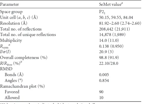
TABLE 1Statistics on data collection and structure refinement
Parameter SeMet valuea
Space group P21
Unit cell (a,b,c) (Å) 50.15, 59.55, 84.04 Resolution (Å) 81.92–2.60 (2.74–2.60) Total no. of reflections 208,442 (21,911) Total no. of unique reflections 14,878 (1,989) Multiplicity 14.0 (11.0) Rsym
a 0.138 (0.950)
I/(I) 20.9 (3)
Overall completeness (%) 98.8 (91.9) R/Rfree(%)
b 22.10/28.0
RMSD
Bonds (Å) 0.005
Angles (°) 0.854
Ramachandran plot (%)
Favored 90
Allowed 10
aValues in parentheses indicate the highest resolution shell. b
Rsym⫽ ⌺h⌺i|Ihi⫺ 具Ih典|/⌺h⌺iIhi, whereIhiis theith observation of the reflectionhand 具Ih典is the mean intensity of reflectionh.
c
R⫽ ⌺||Fo|⫺|Fc||/|Fo|.Rfreewas calculated with a small fraction (5%) of randomly selected reflections.
on November 7, 2019 by guest
http://jvi.asm.org/
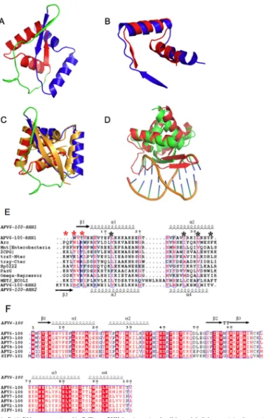
FIG 1Structure of AvtR. (A) Ribbon presentation of AvtR. The two RHH domains are in red and blue, and the linker containing the extra-strand is in green. (B) Superposition of the two RHH domains of AvtR. (C) Superposition of AvtR (red, blue, and green) and CopG dimer (gold) (PDB code 1b01). (D) Superposition of the AvtR dimer (red) onto a CopG dimer (green) in complex with a DNA half site (sticks and gold ribbon). (E) Sequence alignments of RHH domains with secondary structure elements as extracted from the AvtR crystal structure. The black asterisks show the conserved hydrophobic positions, and the red asterisks indicate the three amino acids making sequence-specific nucleotide base contacts. (Panels E and F were generated by Espript.) (F) Sequence alignment of AvtR orthologues in lipothrixviruses, with secondary structure elements as extracted from the AvtR crystal structure.
on November 7, 2019 by guest
http://jvi.asm.org/
[image:5.585.101.490.63.658.2]In vitro
confirmation of AvtR binding sites.
To verify our
in
silico
predictions and the biological significance of the eight
puta-tive targets, we assessed the binding of AvtR to these targets
in
vitro
. All predicted sites were synthesized as double-stranded
39-bp oligonucleotides containing the target-specific 16-bp
se-quence. The results of the electrophoretic mobility shift assays for
the oligonucleotides representing these sites are presented in
Fig.
3B
. Six of the eight tested fragments were specifically recognized
by AvtR, confirming the results of the
in silico
analysis. No binding
was observed for sites s4 and s8 under the conditions used. The
biological significance of these two sites remains unclear. These
two cases could represent false-positive predictions, although it
cannot be excluded that AvtR requires additional factors to
recog-nize these sites.
A visual inspection of the EMSA results obtained for the eight
analyzed sites clearly showed that site s7 is one of the strongest
targets for AvtR (
Fig. 3B
). Site s7 is located in the intergenic region
shared between genes
gp29
and
gp30
, which also contains site s6.
The two sites are separated by only 43 nucleotides. Interestingly,
the
gp30
gene is predicted to code for another putative RHH
pro-tein that could also be involved in the gene transcription
regula-tion in AFV6. Also, the presence of a binding site of AvtR
up-stream of its own gene (
gp29
) suggests that the expression of AvtR
is autoregulated. The genetic configuration of this shared
pro-moter region could be a good model to confirm the biological role
[image:6.585.102.482.72.359.2]FIG 2The consensus target site for AvtR and its mutational analysis. (A) SELEX-based prediction of the hypothetical consensus DNA binding site for AvtR. 454 sequencing was done on the PCR pools from selection rounds 5, 6, and 7, the sequence was analyzed using Bioprospector (45), and the consensus binding site was extracted using the Weblogo program. The height of each base is proportional to the percentage of its occurrence in the SELEX experiment at each position. The binding site selected by SELEX shows a central 10-bp palindromic sequence, GTGGT/ACCAC. This sequence could not be found in the AFV6 genome. (B) Mutational analysis of the SELEX predicted AvtR binding sequence. To identify the conserved positions of the AvtR consensus binding site obtained by SELEX, the importance of each nucleotide in the predicted 16-bp binding site was tested by systematic single-nucleotide substitutions. Transversion mutations were performed for each position individually. The same amount of each of the corresponding double-stranded labeled fragments (1.5 pmol) was tested by PAGE-EMSA in the presence of 104M AvtR and an excess of nonspecific competitor DNA. Under these conditions, the binding of AvtR to the consensus site is specific. Nucleotides 1, 2, 5, 8, 9, 12, 13, 15, and 16 (gray boxes) were considered to be essential for the site recognition, as mutations in those sites decreased AvtR binding. The remaining substitutions did not alter the DNA binding activity. With this information, the putative consensus AvtR binding site is now defined as ATnnTnnTAnnACnTT.
TABLE 2Eight putative AvtR targets on the AFV6 genomea
Site
Closest
gene Sequence
Position
Start End
s1 gp11 TTTATGGTATCACATT 5128 5141 s2 gp13 AATGTAGCAGAACAAA 6088 6103 s3 gp14 TTTGTGATAGTACATT 6142 6157 s4 gp16 TATGTTCTACTACAAT 7209 7224 s5 gp26 TTTGTGTTACTACATT 14203 14218 s6 gp29 TTTGTAGTGCAATATT 15677 15692 s7 gp30 AATGTGCTATCATAAA 15721 15736 s8 gp66 AAGGTAGAACAAAAAA 39328 39343 aOnly the sites located within the hypothetical promoter region, ranging from position
⫺150 to⫹50, of each gene were considered. Those that had a score of greater than 75% of the matrix maximum value were retained as biologically significant. The start and the end positions within the AFV6 genome sequence are indicated (accession number NC_010152).
on November 7, 2019 by guest
http://jvi.asm.org/
[image:6.585.40.287.567.679.2]of AvtR. Therefore, we decided to use the region s6/s7 for further
characterization of AvtR.
Primer extension analysis of the
gp29
/
gp30
promoter region.
The transcriptional regulation of the
gp29
/
gp30
genes appears to
be complex, as two AvtR binding sites are present in this region.
To better understand the transcription regulation of these genes,
we decided to determine the positions of the corresponding
tran-scription initiation points by a primer extension analysis. The
re-sults of this experiment are presented in
Fig. 4
. In the absence of
AvtR, the
gp30
gene is weakly transcribed. Interestingly, a strong
transcription signal was observed in the presence of AvtR in the
reaction mixture. This observation of the activation role of AvtR
was further confirmed by
in vitro
transcriptional analysis (see next
section). The accurate identification of the transcription start site
revealed the presence of the typical archaeal conserved sequence
elements, the archaeal TATA box and the BRE element, for both
genes (
Fig. 4B
). Even though these sites corresponded to typical
archaeal promoters, we were unable to demonstrate the presence
of a GTC motif, which is characteristic of the genomes of the
rudiviruses SIRV1 and SIRV2 infecting members of the genus
Sul-folobus
(
41
).
The analysis of the transcription initiation mapping data also
highlights that
gp29
and
gp30
transcripts overlap by 45
nucleo-tides. This could point to a common regulation of their promoters
by AvtR. One of the AvtR binding sites is situated 5 nucleotides
downstream of the TATA box of
gp29
, whereas the second one is at
a distance of 19 nucleotides downstream of the TATA box of
gp30
.
On this DNA fragment the AvtR binding sites do not overlap the
promoter regions, raising the question of the mechanism of the
presumed AvtR transcriptional regulation.
In vitro
functional analysis of AvtR.
The transcription factors
of the RHH superfamily are known to be able to regulate both
positively and negatively the transcription of the genes under their
control (
6
). The position of the AvtR binding sites in the promoter
regions did not give clear indications about the putative role of
AvtR in the transcription of
gp29
/
gp30
. The transcription of these
genes was studied using a crenarchaeon-specific
in vitro
transcrip-tion system. The transcriptranscrip-tion activities for these genes were
com-pared in the absence and in the presence of increasing amounts of
AvtR. As shown in
Fig. 5
, AvtR performs a strong repression of the
promoter activity of its own gene,
gp29
. The effect is very sharp; in
the presence of 100 ng or more of AvtR in solution (417
M), the
FIG 3Specific binding of AvtR to the predicted binding sites in the AVF6 genome. (A) AFV6 genome and positions of the predicted AvtR binding sites in the vicinity of viral genes. Sites from s1 to s8 are indicated by vertical lines. The corresponding genes are indicated by arrows. (B) Specific binding of AvtR to six of the eight predicted sites. Short dsDNA oligonucleotides were used to test the AvtR binding. They correspond to the predicted sites s1 to s8 described in Materials and Methods. The PAGE-EMSA tests were performed in the presence of increasing concentrations of AvtR from 0 to 417M. Binding to allin silico-predicted sites except s4 and s8 is specific. No binding of AvtR is observed with a heterologous DNA fragment of 40 bp. All PAGE-EMSA experiments were performed in the presence of an excess of nonspecific competitor DNA.
on November 7, 2019 by guest
http://jvi.asm.org/
[image:7.585.100.485.63.409.2]transcription from the
gp29
promoter is totally inhibited. This
result clearly shows that AvtR acts as a repressor of the
transcrip-tion of its own gene. The influence of AvtR on the transcriptranscrip-tion
efficiency of the
gp30
gene is more complex. Even though in the
absence of AvtR, this gene is weakly transcribed (
Fig. 5B
), in the
presence of a low concentration of AvtR (208
M), transcription
from
gp30
is strongly activated. Unexpectedly, at higher
concen-trations of the regulator, the activation effect disappears
progres-sively. If the activation effect obtained for the transcription of gene
gp30
at a relatively low AvtR concentration most probably reflects
the situation
in vivo
, the subsequent repression seen with higher
protein concentrations could either have biological significance or
be an
in vitro
artifact. No influence of AvtR on transcription when
using a control heterologous promoter,
T6
from SSV1, has been
detected.
In conclusion, at the lowest tested concentration, AvtR’s
func-tion in transcripfunc-tion regulafunc-tion is clearly target dependent. The
protein acts as a transcription repressor for P
gp29
but is able to
activate the transcription from the promoter P
gp30
. Interestingly,
at higher concentrations AvtR continues to represses
transcrip-tion from its own gene (
gp29
) and no longer activates P
gp30
.
Oligomerization of AvtR on a DNA template.
To better
char-acterize the regulation of transcription exerted by AvtR, we
per-formed a DNase I footprint assay on the DNA region directly
involved in the interaction between the protein and the promoters
of genes
gp29
and
gp30
under its control.
As seen in
Fig. 6A
, large protected regions were identified on
both DNA strands using increasing concentrations of the protein.
AvtR protects from DNase I digestion a region of about 100 nt
situated between the TATA boxes of genes
gp29
and
gp30
. Being a
relatively small protein of only 11.98 kDa, AvtR could protect a
DNA fragment of that size only if several subunits of the protein
form an oligomer tightly protecting the DNA target. The initiation
of this oligomerization is certainly dependent on the presence of
the primary binding sites, s6 and s7. Despite the fact that high
concentrations of AvtR in the probe appear to be required in order
to achieve this oligomerization, indicating that this process is not
very efficient
in vitro
, this process still appears to be specific. Even
at the highest concentration of AvtR used, the regions following
both TATA boxes remain unprotected (lower parts of gels A1 and
A2 in
Fig. 6
). It remains unclear why the oligomerization does not
include the loci containing the TATA boxes.
To independently verify the occurrence of the oligomerization
of AvtR on its target DNA, the migration of a 289-bp DNA
frag-ment encompassing the shared promoter region between genes
gp29
and
gp30
was analyzed by PAGE-EMSA in the presence of
increasing concentrations of AvtR. We observed a gradual
de-crease of the fragment’s mobility in the presence of increasing
concentrations of AvtR (
Fig. 6B
). This result confirms the initial
oligomerization hypothesis, which is further presented in
Discus-sion and schematically summarized in
Fig. 6C
.
DISCUSSION
The genomes of archaeal viruses are remarkably rich in RHH
pro-teins. A recent
in silico
analysis of 10 crenarchaeal virus genomes
revealed the presence of at least 18 putative RHH regulators in the
analyzed genomes (
2
). Their role in the regulation of the virus
infectious cycle remains largely unknown. The only examples of
well-studied RHHs in an archaeal context are protein ORF56,
[image:8.585.43.284.77.628.2]en-coded by the
Sulfolobus islandicus
plasmid pRN1 (
22
,
25
,
47
–
49
),
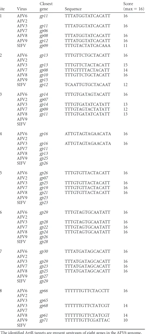
TABLE 3List of putative AvtR targets in theLipothrixviridaefamilya
Site Virus
Closest
gene Sequence
Score (max⫽16) s1 AFV6 gp11 TTTATGGTATCACATT 16
AFV2
AFV3 gp11 TTTATGGTATCACATT 16 AFV7 gp06
AFV8 gp08 TTTATGGTATCACATT 16 AFV9 gp12 TTTATGGTATCACATT 16 SIFV gp09 TTTGTACTATCACAAA 11
s2 AFV6 gp13 TTTGTTCTGCTACATT 16 AFV2
AFV3 gp13 TTTGTTCTACTACATT 15 AFV7 gp08 TTTGTTTTACTACATT 14 AFV8 gp10 TTTGTTCTGCTACATT 16 AFV9 gp15
SIFV gp12 TCAATTGTGCTACAAT 12
s3 AFV6 gp14 TTTGTGATAGTACATT 16 AFV2 gp07
AFV3 gp14 TTTGTGATATCATATT 13 AFV7 gp09 TTTGTAGTACTATATT 12 AFV8 gp11 TTTGTGATATCATATT 13 AFV9
SIFV
s4 AFV6 gp16 ATTGTAGTAGAACATA 16 AFV2
AFV3 gp16 ATTGTAGTAGAACATA 16 AFV7 gp11
AFV8 gp13 AFV9 gp25 SIFV gp26
s5 AFV6 gp26 TTTGTGTTACTACATT 16 AFV2 gp07
AFV3 gp25 TTTGTGTTACTACATT 16 AFV7 gp19 TTTGTGTTACTACATT 16 AFV8 gp21 TTTGTGTTACTACATT 16 AFV9 gp23
SIFV gp23
s6 AFV6 gp29 TTTGTAGTGCAATATT 16 AFV2
AFV3 gp28 TTTGTAGTGCAATATT 16 AFV7 gp22 TTTGTAGTGCAATATT 16 AFV8 gp24 TTTGTAGTGCAATATT 16 AFV9 gp26
SIFV gp28
s7 AFV6 gp30 TTTATGATAGCACATT 16 AFV2
AFV3 gp29 TTTATGATAGCACATT 16 AFV7 gp23 TTTATGATAGCACATT 16 AFV8 gp25 TTTATGATAGCACATT 16 AFV9 gp27
SIFV gp29
s8 AFV6 gp66 TTTTTTGTTCTACCTT 16 AFV2
AFV3 gp65
AFV3 gp68 TTTTTTGTTCTATCGT 14 AFV7
AFV8 gp61 TTTTTTGTTCTATCGT 14 AFV9 gp71 TTTTTTGTTCGATTAC 10 SIFV
a
The identified AvtR targets are present upstream of eight genes in the AFV6 genome. All these genes are conserved and shared in theBetalipothrixvirus genus of the Lipothrixviridaefamily (AFV3, AFV6, AFV7, AFV8, AFV9, and SIFV). The only exception is genegp66, which is absent from the genomes of AFV7 and SIFV and appears to be duplicated in AFV3. Some of these genes, such asgp14,gp26, andgp29, also could be found in the genome of a more distant relative, the detalipothrixvirus AFV2.
on November 7, 2019 by guest
http://jvi.asm.org/
the regulator SvtR, encoded by the rudivirus SIRV (
5
), and the
recently described protein E73, encoded by the virus SSV-RH
(
22
). ORF56 regulates the plasmid copy number, SvtR is involved
in the regulation of the lytic cycle of the rudiviruses, and E73 has
been suggested to target elements of the host genome. They exist
as dimers in solution and bind to their cognate DNAs as pairs of
dimers, without any apparent cooperativity.
In this study, we comprehensively characterized the structure,
function, and DNA targets of AvtR (
Acidianus
virus transcription
regulator), an RHH protein encoded by the virus AFV6, infecting
the hyperthermophilic crenarchaeon
Acidianus.
In the absence of
their cognate DNAs, RHH proteins form symmetric dimers with a
2-fold symmetry axis centered on the two-stranded antiparallel

-sheet. In contrast with other well-characterized RHH proteins,
the AvtR protein is composed of two tandem RHH motifs that are
connected by a linker that aligns with strands

1 and

3 to form a
central three-stranded antiparallel

-sheet. Orthologues of AvtR
are present and well conserved in several members of the
Lipothri-xviridae
viral family (
Fig. 1F
). The RHHs of AvtR are arranged as
a pseudosymmetric dimer, superposable to dimeric RHH
pro-teins. The TraY protein from the
E. coli
episome F is also an RHH
tandem repeat (
50
), but its structure is unknown. The MetJ (
24
),
Arc (
21
),
⍀
(
18
), and CopG (
11
,
27
–
29
) RHH proteins all bind to
their DNA recognition sites as dimers of dimers.
Sequence comparison shows that RHH motifs have no
abso-lutely conserved amino acid positions but that they share certain
sequence features: (i) the

-ribbons present a pattern of
alternat-ing hydrophilic/hydrophobic side chains, (ii) a G-X-T/S/N motif
is conserved in the loop between helix
␣
1 and helix
␣
2 (except in
MetJ), and (iii) four positions in helix
␣
1 and
␣
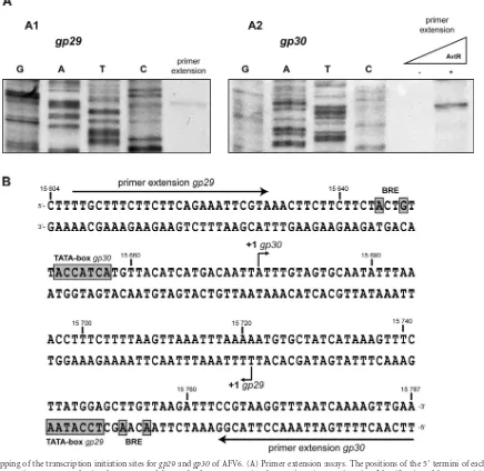
2 are conserved as
FIG 4Mapping of the transcription initiation sites forgp29andgp30of AFV6. (A) Primer extension assays. The positions of the 5=termini of eachin vitro generated transcript were mapped using the sequence of the 289-bp fragment covering thegp29/gp30intergenic region. Identification of the⫹1 position for the gp29(A1) and for thegp30(A2) genes is shown. The transcript for thegp30gene was obtained in the presence of AvtR. (B) Detailed map of transcription signals present in thegp29/gp30region of AFV6. Only 183 out of 289 nucleotides are represented. The positions of the transcription initiations points (⫹1), the BRE sites, and the TATA boxes for both genes,gp29andgp30, are indicated, as well as the positions of primers used for the extension and sequencing reactions.
on November 7, 2019 by guest
http://jvi.asm.org/
[image:9.585.73.509.71.495.2]hydrophobic (black asterisks in
Fig. 1E
). The pattern of
hydro-philic/hydrophobic residues along the

-strand, a hallmark of
RHH proteins, is present in AvtR along

1 (RHH1) and along the
linker between strand

3 and helix
␣
2 (RHH2). Although this
linker does not form a regular

-strand, it is an integral part of the
ribbon of the RHH motif (
Fig. 1C
). As observed in structures of
RHH-DNA oligonucleotide complexes, the central

-sheet of the
dimer specifically recognizes bases from the major groove of the
DNA target sequence: three amino acid side chains from each
strand (corresponding to Arg4, Thr6, and Thr8 in the CopG/DNA
structure [red asterisks in
Fig. 1D
]) point into the major DNA
groove and make crucial sequence-specific nucleotide contacts.
Amino acid residues located at these positions define the DNA
binding specificity of the RHH regulators. Although there is no
strict conserved sequence pattern, a basic residue (Arg or Lys) is
always present at position 1 or 3. Based on the position of this basic
residue, RHH proteins can be separated into two classes (
10
).
Class I proteins have a basic residue at the start of the

-strand
(CopG, ParD, ParG, and MetJ) whereas class II proteins have a
basic residue at the end (Arc, omega, and Mnt repressors). RHH
proteins usually bind DNA as dimers contacting two or more
inverted or tandem repeats within their operators and form
higher-order oligomers (
6
,
11
,
27
,
51
,
52
). It should be noted that the two
strands of the central

-sheet in RHH dimers do not interact
sym-metrically with the cognate DNA. The arrangement of the central

-sheet in AvtR is different from that observed for other RHH
proteins: the

1 strand (RHH1) is much shorter than the
homol-ogous strands of other RHH members and does not contain basic
residues, while strand

3 (RHH2) contains arginine residues at its
N and C termini. However, the main difference from canonical
RHH structures resides in the presence of a third strand in the
central sheet in AvtR. Superposition of the structure of the AvtR
dimer onto that of the CopG-DNA complex shows that it cannot
bind to its cognate DNA in the same way, because the third strand
clashes with the phosphate backbone (
Fig. 1D
). In order to bind its
DNA recognition site, AvtR must either rearrange the linker
strand to relieve the clash or use a noncanonical DNA binding
mode.
All RHH proteins studied in detail so far are involved in
tran-scription regulation. They recognize specific DNA binding sites
and influence the efficiency of transcription initiation. The AvtR
binding site as obtained by the SELEX methodology was not found
in the sequence of the AFV6 genome. A systematic mutational
analysis of the conserved nucleotides allowed us to predict
in silico
8 putative AvtR binding sites in the AFV6 genome. At least 6 of
these sites could be
in vivo
targets for AvtR, as specific binding of
the protein to these targets was demonstrated
in vitro
. The other
two remaining sites either could have been wrongly predicted or
might require additional factors to be recognized by AvtR.
The identified binding sites are generally well conserved in the
Lipothrixviridae
viral family (
Table 3
). Viruses AFV6 and AFV3
appear to be the closest relatives in this family, as all identified sites
are always present in both genomes. In the more distantly related
deltalipothrixvirus AFV2, none of the target sites was found, even
though three of the identified target-associated genes were present
in its genome sequence. Most of the genes under AvtR control are
also well conserved throughout this family and could play an
im-portant role in these virus cycles. For example, some of these genes
code for structural proteins conserved among all
betalipothrixvi-ruses (
gp11
and
gp66
)
.
Also, the protein encoded by gene
gp16
(annotated as a DNA binding protein) is present in several
cren-archaeal viruses, such as betalipothrixviruses, the rudiviruses, the
icosahedral virus STIV1, the unclassified STIV2, the bicaudavirus
ATV, and the gammalipothrixvirus AFV1 (
53
). For the remaining
genes, no predictions could be made for the proteins they code for,
and the complete biological scenario behind the regulation of
AvtR in the AVF6 viral cycle remains unclear.
One of the regions recognized by AvtR in the AFV6 genome is
particularly interesting in its genetic organization and complexity
of its transcription regulation. This region includes the
overlap-ping promoters of genes
gp29
(AvtR’s own gene) and
gp30
, coding
for another hypothetical RHH protein. We showed here that AvtR
strongly represses transcription from the promoter of its own
gene,
gp29
, but activates the promoter of
gp30
. The position of the
AvtR binding sites in the studied intergenic region raises the
ques-tion about the mechanism of regulaques-tion. The primary AvtR
bind-ing sites do not overlap the promoters of
gp29
and
gp30
. DNase I
footprint analysis and mobility shift assays performed with DNA
fragments carrying both binding sites provide strong indications
that AvtR is capable of oligomerization on the DNA template,
FIG 5AvtR regulates transcription from the promoters of thegp29andgp30 genes. The involvement of AvtR in the transcription regulation ofgp29and gp30was demonstrated using a host reconstitutedin vitrotranscription system (IVT). All IVT reactions were performed in the presence of limiting amounts of transcription factors TBP and TFB. (A and B) Gene-specific transcription assays were performed on the promoters of AFV6 genesgp29(A) andgp30(B). The concentrations of AvtR in each reaction mixture are indicated inM. Bottom, schematic representation of the positions of the primers used for the IVT reactions forgp29andgp30and of the respective transcription start sites. AvtR strongly represses the transcription of its own gene,gp29, and represses that of thegp30promoter regulation. (C) To demonstrate that the AvtR activ-ity is target specific, the IVT reaction was performed under the same condi-tions as for panels A and B using a T6 promoter of the unrelated virus SSV1.
on November 7, 2019 by guest
http://jvi.asm.org/
[image:10.585.43.284.65.363.2]most likely starting from the primary sites s6 and s7 and
subse-quently covering the entire region between the TATA boxes of
gp29
and
gp30
. An analogous situation is described for the
tran-scriptional repressor CopG (
28
). Initial binding of the CopG
dimer to its specific site takes place through the major groove of
DNA. At higher repressor concentrations, binding of CopG to the
primary site may promote successive binding to secondary sites in
the operator (
27
). We propose a similar mechanism to explain
how a small protein such as AvtR could protect the long intergenic
gp29
/
gp30
region. Other archaeal regulatory proteins with
differ-ent DNA binding motifs, such as TrpY (
54
) and FL11 (
55
), are also
capable of binding to control regions as multiple copies in a
con-FIG 6DNase I footprinting analysis of the regions protected by AvtR in thegp29andgp30promoters. (A) DNase I footprinting assays performed on a 170-bp fragment corresponding to thegp29/gp30intergenic region. Leading and lagging DNA strands were analyzed separately (A1 and A2, respectively), and the concentrations of AvtR present prior to the addition of DNase I are indicated on the upper part of each gel. The gray boxes represent the identified DNA binding sites of AvtR, and the arrows indicate the transcription start points and their directions. The AvtR oligomerization starts from both of its binding sites and extends upstream and downstream to a total of 97 nucleotides. (B) Migration of a 289-bp DNA fragment containing the promoter region between genesgp29andgp30, analyzed by PAGE-EMSA. A gradual decrease in the mobility of the fragment is seen in the presence of increasing concentrations of AvtR, confirming the oligomerization hypothesis. (C) Proposed model of regulation by AvtR. The shared promoter region of genesgp29andgp30contains two binding sites for AvtR. When present at a low concentration, AvtR binds primarily to site s7. This binding activates transcription from genegp30, probably by inducing conformational changes on the promoter DNA (shown by wavy line). At higher concentrations, AvtR occupies both sites s6 and s7. Oligomerization of AvtR will then cover both promoter’s TATA boxes and impair the formation of the transcription initiation complex, repressing transcription from both genesgp29andgp30.
on November 7, 2019 by guest
http://jvi.asm.org/
[image:11.585.77.511.65.540.2]centration-dependent manner. In this case, the oligomerization of
AvtR appears to be guided by short degenerate secondary sites. A
careful analysis of the sequence located between the two initial
binding sites, s6 and s7, revealed the presence of several short
repeats (TTTAA) that could play this role.
A hypothetical model integrating all observed events is
pre-sented in
Fig. 6C
. At low concentrations, AvtR binds only to the
strongest site, s7, situated 62 nucleotides downstream of the
TATA box of
gp30
. We presume that this binding causes the
acti-vation of
gp30
transcription. At this stage, no exact mechanism for
this effect can be proposed. One hypothesis could be that this
binding induces some conformational changes in the promoter
region. At higher concentrations, AvtR will also bind to the weaker
second site, s6, and the oligomerization starting from both sites s7
and s6 could cover the
gp29/gp30
intergenic region, reach the
TATA boxes, and then directly interact with the transcription
ini-tiation complexes. The biological significance of this unusual
mechanism of regulation remains unclear. One could suppose
that the intracellular concentration of AvtR is a crucial parameter
of the system that plays a role of the “molecular timer” activating
and repressing viral genes depending on the stage of the virus
development cycle.
To conclude, AvtR represents the second example of an RHH
protein encoded by archaeal viruses, and it is distinguished from
other RHH proteins by some unusual structural and regulatory
properties. The relatively high number of genes under AvtR
con-trol clearly indicates the importance of this regulator in the AFV6
infection cycle.
ACKNOWLEDGMENTS
This work was supported by a VIRAR grant (NT05-2_41674) from the Agence Nationale de Recherche. N.P. was supported by the EU Marie Curie research training network SOLAR (grant MRTN-CT-2006-033499).
We thank Chloé Danioux for help and useful discussions.
REFERENCES
1.Pina M, Bize A, Forterre P, Prangishvili D.2011. The archeoviruses. FEMS Microbiol. Rev.35:1035–1054.
2.Prangishvili D, Garrett RA, Koonin EV.2006. Evolutionary genomics of archaeal viruses: unique viral genomes in the third domain of life. Virus Res.117:52– 67.
3.Geslin C, Gaillard M, Flament D, Rouault K, Le Romancer M, Prieur D, Erauso G. 2007. Analysis of the first genome of a hyperthermophilic marine virus-like particle, PAV1, isolated fromPyrococcus abyssi.J. Bac-teriol.189:4510 – 4519.
4.Krupovic M, White MF, Forterre P, Prangishvili D. 2012. Postcards from the edge: structural genomics of archaeal viruses. Adv. Virus Res.
82:33– 62.
5.Guilliere F, Peixeiro N, Kessler A, Raynal B, Desnoues N, Keller J, Delepierre M, Prangishvili D, Sezonov G, Guijarro JI.2009. Structure, function, and targets of the transcriptional regulator SvtR from the hyper-thermophilic archaeal virus SIRV1. J. Biol. Chem.284:22222–22237. 6.Schreiter ER, Drennan CL.2007. Ribbon-helix-helix transcription
fac-tors: variations on a theme. Nat. Rev. Microbiol.5:710 –720.
7.Burgering MJ, Boelens R, Gilbert DE, Breg JN, Knight KL, Sauer RT, Kaptein R.1994. Solution structure of dimeric Mnt repressor (1-76). Biochemistry33:15036 –15045.
8.Chopra N, Agarwal S, Verma S, Bhatnagar S, Bhatnagar R. 2011.
Modeling of the structure and interactions of theB. anthracisantitoxin, MoxX: deletion mutant studies highlight its modular structure and re-pressor function. J. Comp. Aid. Mol. Design25:275–291.
9.Gallo M, Ferrari E, Eliseo T, Amata I, Pertinhez T, Katsuyama A, Paci M, Farah C, Spisni A, Cicero D.2010. A new member of the ribbon-helix-helix transcription factor superfamily from the plant pathogen Xan-thomonas axonopodispv.citri. J. Struct. Biol.170:21–31.
10. Golovanov AP, Barilla D, Golovanova M, Hayes F, Lian LY.2003. ParG, a protein required for active partition of bacterial plasmids, has a dimeric ribbon-helix-helix structure. Mol. Microbiol.50:1141–1153.
11. Gomis-Ruth FX, Sola M, Acebo P, Parraga A, Guasch A, Eritja R,
Gonzalez A, Espinosa M, del Solar G, Coll M.1998. The structure of plasmid-encoded transcriptional repressor CopG unliganded and bound to its operator. EMBO J.17:7404 –7415.
12. Huang L, Yin P, Zhu X, Zhang Y, Ye K.2011. Crystal structure and centromere binding of the plasmid segregation protein ParB from pCXC100. Nucleic Acids Res.39:2954 –2968.
13. Larson JD, Jenkins JL, Schuermann JP, Zhou Y, Becker DF, Tanner JJ.
2006. Crystal structures of the DNA-binding domain ofEscherichia coli proline utilization A flavoprotein and analysis of the role of Lys9 in DNA recognition. Protein Sci.15:2630 –2641.
14. Li G-Y, Zhang Y, Inouye M, Ikura M.2008. Structural mechanism of transcriptional autorepression of theEscherichia coliRelB/RelE antitoxin/ toxin module. J. Mol. Biol.380:107–119.
15. Lu J, den Dulk-Ras A, Hooykaas P, Glover M.2009. Agrobacterium tumefaciensVirC2 enhances T-DNA transfer and virulence through its C-terminal ribbon-helix-helix DNA-binding fold. Proc. Natl. Acad. Sci. U. S. A.106:9643–9648.
16. Madl T, Van Melderen L, Mine N, Respondek M, Oberer M, Keller W, Khatai L, Zangger K.2006. Structural basis for nucleic acid and toxin recognition of the bacterial antitoxin CcdA. J. Mol. Biol.364:170 –185. 17. Mattison K, Wilbur JS, So M, Brennan RG.2006. Structure of FitAB
fromNeisseria gonorrhoeaebound to DNA reveals a tetramer of toxin-antitoxin heterodimers containing pin domains and ribbon-helix-helix motifs. J. Biol. Chem.281:37942–37951.
18. Murayama K, Orth P, de la Hoz AB, Alonso JC, Saenger W. 2001.
Crystal structure of omega transcriptional repressor encoded by Strepto-coccus pyogenesplasmid pSM19035 at 1.5 A resolution. J. Mol. Biol.314: 789 –796.
19. Popescu A, Karpay A, Israel DA, Peek RM, Jr, Krezel AM. 2005.
Helicobacter pyloriprotein HP0222 belongs to Arc/MetJ family of tran-scriptional regulators. Proteins59:303–311.
20. Pryor E, Waligora E, Xu B, Dellos-Nolan S, Wozniak D, Hollis T.2012. The transcription factor AmrZ utilizes multiple DNA binding modes to recognize activator and repressor sequences ofPseudomonas aeruginosa virulence genes. PLoS Pathog. 8:e1002648. doi:10.1371/journal. ppat.1002648.
21. Raumann BE, Rould MA, Pabo CO, Sauer RT.1994. DNA recognition
by beta-sheets in the Arc repressor-operator crystal structure. Nature367: 754 –757.
22. Schlenker C, Goel A, Tripet BP, Menon S, Willi T, Dlakic M, Young MJ, Lawrence CM, Copie V.2012. Structural studies of E73 from a hyper-thermophilic archaeal virus identify the “RH3” domain, an elaborated ribbon-helix-helix motif involved in DNA recognition. Biochemistry51: 2899 –2910.
23. Schreiter ER, Wang SC, Zamble DB, Drennan CL.2006. NikR-operator complex structure and the mechanism of repressor activation by metal ions. Proc. Natl. Acad. Sci. U. S. A.103:13676 –13681.
24. Somers WS, Phillips SE.1992. Crystal structure of the met repressor-operator complex at 2.8 A resolution reveals DNA recognition by beta-strands. Nature359:387–393.
25. Weininger U, Zeeb M, Neumann P, Löw C, Stubbs M, Lipps G, Balbach J.2009. Structure-based stability analysis of an extremely stable dimeric DNA binding protein fromSulfolobus islandicus.Biochemistry48:10030 – 10037.
26. Wong J, Lu J, Edwards R, Frost L, Glover M.2011. Structural basis of cooperative DNA recognition by the plasmid conjugation factor, TraM. Nucleic Acids Res.39:6775– 6788.
27. Costa M, Sola M, del Solar G, Eritja R, Hernandez-Arriaga AM, Espi-nosa M, Gomis-Ruth FX, Coll M.2001. Plasmid transcriptional repres-sor CopG oligomerises to render helical superstructures unbound and in complexes with oligonucleotides. J. Mol. Biol.310:403– 417.
28. del Solar G, Hernandez-Arriaga AM, Gomis-Ruth FX, Coll M, Espinosa M.2002. A genetically economical family of plasmid-encoded transcrip-tional repressors involved in control of plasmid copy number. J. Bacteriol.
184:4943– 4951.
29. Hernandez-Arriaga AM, Rubio-Lepe TS, Espinosa M, del Solar G.2009. Repressor CopG prevents access of RNA polymerase to promoter and actively dissociates open complexes. Nucleic Acids Res.37:4799 – 4811. 30. He YY, McNally T, Manfield I, Navratil O, Old IG, Phillips SE,
on November 7, 2019 by guest
http://jvi.asm.org/
Girons I, Stockley PG.1992. Probing met repressor-operator recognition in solution. Nature359:431– 433.
31. Vestergaard G, Aramayo R, Basta T, Haring M, Peng X, Brugger K, Chen L, Rachel R, Boisset N, Garrett RA, Prangishvili D.2008. Structure of the acidianus filamentous virus 3 and comparative genomics of related archaeal lipothrixviruses. J. Virol.82:371–381.
32. Leslie AGW.1992. Joint CCP4 and EACMB Newsl. Protein Crystallogr., no. 26.
33. Evans P.2006. Scaling and assessment of data quality. Acta Crystallogr. D Biol. Crystallogr.62:72– 82.
34. Grosse-Kunstleve RW, Adams PD.2003. Substructure search procedures for macromolecular structures. Acta Crystallogr. D Biol. Crystallogr.59: 1966 –1973.
35. Bricogne G, Vonrhein C, Flensburg C, Schiltz M, Paciorek W.2003. Generation, representation and flow of phase information in structure determination: recent developments in and around SHARP 2.0. Acta Crystallogr. D Biol. Crystallogr.59:2023–2030.
36. Terwilliger TC.2000. Maximum-likelihood density modification. Acta Crystallogr. D Biol. Crystallogr.56:965–972.
37. Jones TA, Kjeldgaard M.1991. Improved methods for building protein models in electron density maps and the location of errors in these models. Acta Crystallogr. A47:110 –119.
38. Murshudov GN, Vagin AA, Dodson EJ.1997. Refinement of macromo-lecular structures by the maximum-likelihood method. Acta Crystallogr. D Biol. Crystallogr.53:240 –255.
39. Abella M, Rodriguez S, Paytubi S, Campoy S, White MF, Barbe J.2007. TheSulfolobus solfataricusradA paralogue sso0777 is DNA damage induc-ible and positively regulated by the Sta1 protein. Nucleic Acids Res.35: 6788 – 6797.
40. Kessler A, Sezonov G, Guijarro JI, Desnoues N, Rose T, Delepierre M, Bell SD, Prangishvili D.2006. A novel archaeal regulatory protein, Sta1, activates transcription from viral promoters. Nucleic Acids Res.34:4837– 4845.
41. Kessler A, Brinkman AB, van der Oost J, Prangishvili D.2004. Tran-scription of the rod-shaped viruses SIRV1 and SIRV2 of the hyperthermo-philic archaeonSulfolobus.J. Bacteriol.186:7745–7753.
42. Qureshi SA, Bell SD, Jackson SP.1997. Factor requirements for tran-scription in the ArchaeonSulfolobus shibatae.EMBO J.16:2927–2936. 43. Jolma A, Kivioja T, Toivonen J, Cheng L, Wei G, Enge M, Taipale M,
Vaquerizas JM, Yan J, Sillanpaa MJ, Bonke M, Palin K, Talukder S,
Hughes TR, Luscombe NM, Ukkonen E, Taipale J.2010. Multiplexed
massively parallel SELEX for characterization of human transcription fac-tor binding specificities. Genome Res.20