ACKNOWLEDGEMENT
First and foremost I thank the LORD ALMIGHTY for providing me this
opportunity and granting me the capability to proceed successfully.My sincere thanks and deep sense of gratitude to Dr.(Capt) S.Gokulanathan B.Sc., M.D.S., Dean, Vivekanandha dental college for women, for permitting me to pursue this Dissertation.
I am deeply grateful to Dr. N.Balan M.D.S., Principal, Vivekanandha dental college for women for his kind permission, encouragement and for providing me with all the facilities needed to complete this work.
I respect and thank, Dr. V. Mahesh Mathian M.D.S., Professor and Head, Department of Pedodontics and Preventive Dentistry, Vivekanandha dental college for women for his constant encouragement and expert guidance without whom this submission wouldn’t have been feasible.
With submissive ambition, I aspire to register my gratitude to my respected mentor and guide Dr. M. Gawthaman M.D.S., Professor, Department of Pedodontics and Preventive Dentistry, Vivekanandha dental college for women for his inspiring guidance, invaluable counsel and encouragement throughout the course of the study.
I am thankful to the faculty members of our department for helping me in completing this work Dr.S.Vinodh MDS, Reader and Dr.M.Manoharan MDS, Senior Lecturer for their support and encouragement in this work.
I acknowledge my batch mates Dr.S.S.Gaytry and Dr.A.Kiruthiga Revathy for their support and encouragement.
My heartfelt gratitude to my family members and to my husband Dr.R.Ajay MDS who made possible my tomorrow by sacrificing their yesterday and for their evergreen love and support for which I will be eternally indebted.
.
CONTENTS
CONTENTS
S.NO TITLE PAGE NO
1 INTRODUCTION 1
2 AIMS AND OBJECTIVES 10
3 REVIEW OF LITERATURE 11
4 MATERIALS AND METHODS 23
5 RESULTS 45
6 DISCUSSION 57
7 CONCLUSION 68
8 REFERENCES 69
9 ANNEXURES 79
CONTENTS
LIST OF FIGURES
FIGURE
NO
CONTENTS PAGE NO
1 a: Self etch adhesive composite
b: Self adhesive flowable composite
32
32
2 Samples mounted in acrylic resin 33
3 Occlusal grinding of teeth 33
4 a: Metal split mould
b: Twisted ligature wire
c: Twisted wire placed in composite resin
d: Restoration after the removal of metal split mould
34
34
34
34
5 SAF applied on to the prepared tooth surface 35
6 Samples immersed in water 35
7 Occlusal load was applied 36
8 a,b: Testing of TBS in universal testing machine 36
9 a: Samples for group B1, µ-L
b: Samples for group B2, µ-NL
37
37
10 Class V cavity 38
11 Thermocycling unit 38
12 a,b: Samples immersed in basic fuschin dye 39
13 a,b,c,d: Samples washed and dried after removal from the dye 40,41
14 a, b : Labio lingually sectioned samples 42,43
15 a : Sample without microleakage under stereomicroscope
b : Sample with microleakage under stereomicroscope
44
CONTENTS
LIST OF TABLES
TABLE NO
CONTENTS PAGE
NO
1 Tensile bond strength for Self-etch adhesive system [SEA] 45
2 Tensile bond strength for Self adhering flowable system [SAF] 47
3 Tensile bond strength for Self-etch adhesive system [SEA] and Self adhering flowable composite [SAF] sub-groups with cyclic loading
48
4 Tensile bond strength for Self-etch adhesive system [SEA] and Self adhering flowable composite [SAF] sub-groups without cyclic loading
50
5 Within Sub-Group IB-Self etch adhesive composite resin [SEA] 51
6 Chi-Square Tests 52
7 Within Sub-Group IIB-Self adhesive flowable composite [SAF] 53
8 Chi-Square Tests 53
9 Sub-groups IB1 & IIB1 – With Loading [L] 54
10 Chi-Square Tests 55
11 Sub-groups IB2 & IIB2 – Without Loading [NL] 55
[image:11.612.106.548.119.662.2]CONTENTS
LIST OF GRAPHS
GRAPH
NO
CONTENTS PAGE NO
1 Mean value for SEA-TBS-NL & SEA-TBS-L 46
2 Mean value for SAF-TBS-NL & SAF-TBS-L 47
3 Mean value for SAF-TBS-L & SEA-TBS-L 49
ABBREVIATIONS
S.No. ABBREVIATIONS EXPANSIONS
1. BIS-GMA Bisphenol A-Glycidyl methacrylate
2. HEMA Hydroxy ethyl methacrylate
3. TEGDMA Triethylene glycol dimethacrylate
4. BIS-EMA Bisphenol A ethoxylate dimethacrylate
5. UDMA Urethane dimethacrylate
6. GPDM Glycerol-phosphate dimethacrylate
7. SAF Self-adhering flowable composite resin
8. SEA Self-etch adhesive flowable composite resin
9. TBS Tensile bond strength
10. NL Without cyclic loading
11. L With cyclic loading
12. µ Micro-leakage
13. SEM Scanning electron microscope
14. N Newton
15. MO Mesio-occlusal
INTRODUCTION
1 Adhesion is a complex set of physical, chemical, and mechanical process that
allow the attachment and binding of one substance to another dissimilar substrate.
According to the American Society for Testing and Materials (ASTM; specification D
907), adhesion is defined as “the state in which two surfaces are held together by
interfacial forces which may consist of valence forces or interlocking forces or both”.
An adhesive is a viscous fluid material which joins two substrates together and
solidifies, and henceforth is able to transfer a load from one surface to another. Four
different mechanisms of adhesion have been described in the literature. Firstly,
mechanical adhesion, that is, interlocking of the adhesive with surface irregularities of
the substrate. Second is the adsorption adhesion which is chemical bonding between
the adhesive and the substrate. The forces involved may be primary or secondary
valence forces. Third is the diffusion adhesion which means interlocking between
unstable molecules, such as the adhesion of two polymers through diffusion of
polymer chain. Fourth mechanism is the electrostatic adhesion in which an electrical
double layer at the interface of a metal with a polymer that is part of the total bonding
mechanism. [1]
The trend of adhesion in dentistry commenced at the mid 1960s with the
advent of the first commercial resin monomer BIS-GMA [Bisphenol A-Glycidyl
methacrylate] invented by Bowen
Bonding of resins to tooth structure may be a result of four probable
mechanisms:
1. Mechanical: Penetration of resin and formation of resin tags within the tooth
INTRODUCTION
2 2. Diffusion: Precipitation of substances on the tooth surfaces to which resin
monomers can bond mechanically or chemically.
3. Adsorption: Chemical bonding to the inorganic component or organic collagen
components of tooth structure.
4. A combination of the all the above mechanisms.
To obtain good adhesion, an intimate surface contact should exist between the
adhesive and the adherend (tooth surface). Furthermore, the surface tension of the
adhesive must be lower than the surface energy of tooth structure. [2]
The goal and functions of a dental bonding adhesive system are to provide
strong durable bond resisting separation of an adherend substrate from a restorative or
cementing material so that there exists optimum retention, distribution of stress along
bonded interfaces, better color stability, and sealing the interface between dentin
and/or enamel and the bonded material ensuring no or minimal microleakage. Hence,
an adhesive system should assure decreased risk of post-operative sensitivity,
marginal leakage, and secondary caries. [1]
Modern adhesive dentistry offers certain significant advantages as it allows
hard tissue conservation, which in turn paves way for effective and efficient
restoration. The foundation for modern adhesive dentistry was laid in 1955, when
Buonocore reported that acid etching created the ability to produce clean, high energy,
roughened tooth surfaces capable of establishing a micro-mechanical retentive
interface with resin-based luting and restorative materials. Presently, acid etching is
one of the most effective ways to achieve restoration retention and it ensured a sealed
interfacial joint at restoration margins. Acid etching has markedly expanded the use of
resin-based restorative materials as it provided a intricate, strong, durable bond
INTRODUCTION
3 evolved through a number of generations with changes in their chemistries,
mechanisms of bonding, number of bottles, methods of application, and clinical
effectiveness. [3]
Etching and bonding mechanisms differ for enamel and dentin due to its
physical and chemical differences. Enamel has little potential for bonding by
micromechanical retention because the surface appears smooth. Phosphoric acid is the
most commonly used acid to etch the tooth surface as described by Buonocore. On
acid-treatment, the ultra-structure of the enamel surface is modified considerably.
Phosphoric acid solution is difficult to control when applied to the smooth surface of
enamel. Some acid inevitably contacts other areas of tooth, which are not intended to
be etched. Development of acidified gels alleviated this problem and provided a
confined etched tooth surface. The acidified gel contains 37% phosphoric acid in
viscous aqueous gel, which shall be applied onto the enamel surface in
well-controlled manner in the desired area. [4]
The pattern of etching enamel can be variable. Silverstone in 1975 identified 3
basic types of etching patterns. Most commonly [type 1] the enamel prism cores are
removed preferentially and the prism peripheries remain intact. The type 2 etching
pattern is the exact converse of type 1, where the cores are being left intact and the
prism peripheries are removed preferentially. The type 3 etching pattern appears with
resemblance of both type 1 and type 2 along with less distinct areas where the pattern
of etching does not correlate to the morphology of enamel prism.
Bonding mechanism to enamel involves the resin to penetrate into the
relatively porous surface of the enamel, which has been etched to form an interlocking
mechanically. It was thought many years ago that a similar pattern of bonding
INTRODUCTION
4 of the exposed dentinal surface to expose the patent dentinal tubules into which the
resin penetrated to form resin tags. Potential damaging effect on exposed dentine by
phosphoric acid etching was the hurdle in achieving bond. This was considered as an
established fact that phosphoric acid-etching in contact with vital dentine caused
irritation and/or irreversible pathological damage to the pulp. Etching dentine opens
dentinal tubules and encourages dentinal fluid flow. Adhesive resin monomers are
relatively hydrophobic. The moisture content of the dentinal surface due to the
dentinal fluid flow is likely to make bonding more difficult.
A major problem recognized was the inability of the hydrophobic adhesive
resin monomers to wet the moist dentinal surfaces and hindrance to close enough
adaptation of adhesive resin to dentinal surfaces to meet bonding criteria. Drying of
dentine as an important step in the bonding procedure was the earliest attempts to
achieve bonding. Currently, drying procedures lead to desiccation of dentine and tries
to overcome the moisture problem by the use of primers and solvents. Moisture
content of dentin before applying adhesives may have effect on their bond efficacy.
However, the effect of moist or dried dentin on the efficacy of bond strength of
adhesives in dental clinical procedures has not been fully documented. [5] Nevertheless, it is also recognized that in anaesthetized teeth, dentinal tubular fluid
flow is negligible due to the vasoconstrictor effect of the local anaesthetic solution.
The dentinal tubule openings occupy only about 5% of the cut dentine surface at the
dentino-enamel junction. This increases to nearly 20% in deep dentine. Therefore, it
was suggested that when the adhesive monomer penetrates into dentinal tubules for
the formation of resin tags, the relatively small proportion of the area being utilized
would limit the bonding effectiveness to dentine. In the 1970s and 1980s, dentine
INTRODUCTION
5 achieve bonding to dentine through the formation of chemical links between
adhesives and chemical moieties in the dentine surface. These events soon led to a
burgeoning growth in the manufacture of different adhesive monomers and bonding
techniques, into the beginning of the twenty-first century. Concepts in adhesive
dentistry have been changing continuously during the past decades, and steadily
gained pivotal importance.
Current strategies in adhesive dentistry involve two distinct methods. Firstly,
the total-etch bonding technique, which is characterized by the complexity of its
components and of the bonding procedure. Simultaneous etching of enamel and
dentin is the basis for the total-etch technique, which leads to hybridization at
resin-dentine interface by a molecular level mixture of adhesive polymers and dentinal hard
tissues. [6]
In etch-and-rinse adhesives, an initial etching step is compulsorily followed by
a rinsing phase. This etching step de-mineralizes dentin and removes the smear layer
and smear plugs to achieve a micro-porous surface for enhanced bonding capacity.
Originally, etch-and-rinse systems typically consisted of three separate application
steps: (a) conditioning / etching; (b) priming; and (c) adhesive resin application. An
adhesive system following this sequence of clinical procedure is called a three-step
etch-and-rinse adhesive. A two-step etch-and-rinse adhesive has been designed as an
attempt of simplification, which combines the priming and bonding steps into one. In
three-step etch-and-rinse adhesives, the priming step should ensure sufficient wetting
of the exposed collagen fibrils and remove remaining water aiding in adhesive resin
infiltration. HEMA [Hydroxy ethyl methacrylate] or TEGDMA [triethylene glycol
INTRODUCTION
6 added in primer solutions. Both three- and two-step etch-and-rinse adhesives possess
similar mechanism of adhesion. [7]
Nowadays, efforts are being made to simplify and shorten bonding procedures
while retaining the effectiveness of dentin adhesives. [8] The second strategy in
adhesive dentistry is the self-etching systems. Self-etch adhesives do not require a
separate “etch-and-rinse” phase because of its acidic monomer content, which
simultaneously condition and prime the tooth surface when applied. Hence, the smear
layer, which has dissolved and de-mineralized products are not rinsed away.
However, they are incorporated in the adhesive resin. Self-etching adhesive systems
were developed to eliminate dentist variables and minimize chair-side time. [9] A two-step and a one-step self-etch adhesive systems exist just like the
etch-and rinse adhesives. Two-step self-etch systems consisted of an acidic primer
application, followed by an adhesive resin. Recently, “all-in-one” adhesives or
one-step self-etch adhesives have been evolved that combine etching, priming and
conditioning in one solution. Since, single-bottle systems are highly hydrophilic
adhesive monomers, which means they are permeable to water movement, it was
concluded that they were the best promising adhesive approach. [10] Solvent-free self-etch adhesive in which the solvent contained in all other adhesive systems having
been removed was introduced later. [11] This type of adhesive has created a possibility
of an interactive ionic bond between the tooth minerals and the resins of the bonding
agent without the use of water, acetone, or alcohol. [11,12]
When compared with etch-and-rinse adhesives, self-etch adhesives have many
advantages. Omitting the rinsing phase in etch-and-rinse adhesives and thus reducing
the chair-side time. Conditioning, rinsing and drying steps are eliminated, which is
INTRODUCTION
7 mechanism of bonding to dehydrated demineralized dentin has been eliminated, as it
avoids rinsing and drying. Collagen network collapse is prevented, as monomers
infiltrate concomitantly as they get demineralize. Rewetting of dentin by dentinal
fluid from the tubules is prevented, as the smear layer and smear plugs are not
removed. Hence, potential post-operative sensitivity has been reduced. [13]
Prolonged mouth opening and maintaining the operatory field moisture free
during composite restoration procedures in young children is a challenge to every
pediatric dentist. Conventional, total-etch or self-etch adhesive systems are time
consuming and hectic procedures in pediatric care. However, the introduction of a
novel self-adhesive flowable composite dramatically decreased the chairside time.
Limited studies have been conducted in comparing the bond strength efficacy of
conventional systems and self-adhesive flowable composite resins.
Currently, Constic DMG, a novel self-adhesive flowable composite, has been
evolved that eliminates both etching and bonding steps in composite restoration and
associated time expenditure when treating young children in pediatric dental care.
[14,15] They possess a superior esthetic properties and low viscosity, which makes them
easier to place and more self-adaptable than conventional composite resins. [16] Self-adhesive flowable composite resin hybrids the merits of Self-adhesive and restorative
technologies in single product. Hence, it is a direct composite resin restorative
material that has an adhesive resin together with a flowable composite resin with
BIS-GMA as a principal resin matrix. [17] The bonding technology of Constic DMG self
adhesive flowable resin is unclear and necessitates future investigations on these
perspectives. Another self-adhesive flowable composite, Vertise-Flow [Kerr] that uses
glycerol-phosphate dimethacrylate (GPDM) to etch enamel/dentin, and a hydrophilic
INTRODUCTION
8 not only bonds micromechanically between the polymerized monomers of the
self-adhering flowable composite resin and the collagen fibers and smear layer of dentin
but also chemically between the phosphate groups of a GPDM monomer and the
hydroxyapatite of tooth structure. [17,19]
Effective replacement of tooth structure is the major goal of restorative
treatment. The interface must resist dimensional changes in order to prevent
deterioration of the seal between adhesive resin and dentin. The restoration may
deteriorate eventually due to chemical, thermal, and mechanical load stresses even
when the polymerization shrinkage is controlled. It is mandatory to formulate a
methodology using different stresses, since rapid evolution of adhesive resins does not
allow for long-term clinical trial evaluations. Therefore, it has been advocated to
apply in-vitro methodologies using different types of stresses to simulate the aging of
restorations. These stressing strategies could hasten deterioration of the
dentin/adhesive interface, and enable better evaluation of adhesive materials exposed
to stresses in a similar pattern existing in the oral environment. [20]
Differences existing in the coefficient of thermal expansion between
enamel/dentin and adhesives or restorative resins might induce degradation of the
tooth-restoration interface. Thermal cycling test in composite resin restorations is
frequently employed in laboratory studies evaluating micro-leakage in order to
simulate changing intraoral temperature conditions. [21]
Teeth are subjected to occlusal stresses when they come in contact in both
centric and eccentric positions. [22] Compressive stresses arise on the tooth aspect being bent, while tensile stresses are generated simultaneously on the opposite tooth
aspect. Exactly a same scenario occurs in cervically restored teeth when subjected to
INTRODUCTION
9 margin. [23] Thus, in terms of leakage, the marginal integrity of resin composite restorations is highly affected by cyclic loading.
An in-vitro simulation of masticatory loads is necessary to measure the effect
of cyclic loading on micro-leakage at the resin–dentin interface. [20] The most
frequently used laboratory tests to study the mechanisms that may minimize, or
eliminate, the leakage around composite restorations are microleakage tests using
dye-penetration technique. A microleakage test is a useful method in the investigation
of resin composite restorations though the clinical relevance of the tests does not
always correlate appropriately with the clinical situation. [24] Moreover, the
consequences of both thermal cycling and cyclic loading on the micro-leakage of
self-adhesive flowable composite resin used in cervical composite restorations have yet to
be completely analyzed.
Since there is a paucity of information regarding self adhesive flowable
composite, the present study was conducted to test tensile bond strength and
AIM & OBJECTIVES
10
Aim:
Aim of this in-vitro study was to compare the bond strength of self-adhesive
flowable [SAF] composite with that of self-etch adhesive [SEA] systems. Also, to
evaluate the longevity of the composite restoration seal in response to mechanical
stresses, in terms of micro-leakage.
Objectives:
1. To measure and compare the tensile bond strength of self-etch adhesive-composite
system and self-adhesive flowable composite regarding to dentin.
2. To determine whether or not cyclic loading influences the bond strength of the
materials compared.
3. To assess and compare the degree of micro-leakage between self-etch
adhesive-composite system and self-adhesive flowable adhesive-composite by using dye penetration
technique.
4. To determine whether or not artificial ageing like occlusal cyclic loading
influence micro-leakage.
5. To determine the efficacy of self adhesive flowable composite in terms of bond
REVIEW OF LITERATURE
11
Nelson et al [25] [1952] experimented the fluid exchange at the margins of
dental restorations. Temperature changes of teeth and restorations in the mouth cause
a fluid exchange between the teeth and restorations made of gutta-percha, zinc oxide
and eugenol cement, silicate cement, zinc phosphate cement, amalgam, cast gold,
gold foil and acrylic resin. This marginal percolation is caused in part by a difference
in the coefficients of thermal expansion of the tooth and the restoration and by
thermal expansion of the fluid occupying the crevice between the tooth and the
restoration. In light of the present concepts of the mechanisms of dental caries,
marginal percolation may be an explanation for the recurrence o f caries at the
margins of some restorations. Further observation of the influence of marginal
percolation as a factor in the efficiency of the direct filling resin as a permanent
filling material is required.
Kidd et al [26] [1978] studied the cavity sealing ability of composite
restorations subjected to thermal stress and concluded that thermal percolation may
not be of clinical significance in relation to composite restorations.
Luscher et al [27] [1978] investigated the microleakage and marginal
adaptation of composite resin restorations in class II cavities. It has been shown that
the use of a sealant offered no advantage with respect to the retentive strength of a
composite resin restoration. However, for the improvement of marginal seal and
adaptation, the results indicate that enamel etching and the application of a sealant, in
conjunction with use of a cavity geometry which reduces shrinkage strain, are
absolute necessities.
Fusayama et al [28] [1979] investigated the non-pressure adhesion of clearfil
REVIEW OF LITERATURE
12 both enamel and dentin as well as to carious dentin and showed strong adhesion to all
substrated which were tested. Additionally they found that acid etching further
increased the adhesion to dentin
Harper et al [29] [1980] studied the in-vivo measurements of thermal diffusion
through restorations of various materials. Temperature diffusion was highest through
amalgam restorations and slowest through unfilled resin restorations. The rate of
diffusion through composite resins and silicate cement fell between the extremes.
Bases of both zinc phosphate cement and zinc oxide-eugenol cement reduced the rate
of temperature change on the floor of the cavity beneath amalgam restorations. The
temperature change was generally less under the 1 mm base than the 0.5 mm base.
Bases used beneath the nonmetallic restorations did not reduce the magnitude of
temperature change on the cavity floor.
Crim and Mattingly [30] [1981] evaluated two methods such asthermocycling
and constant temperature immersion in basic fuschin dye to assess the marginal
microleakage. The results of this study showed significant differences between these
two methods and concluded that thermocycling was effective in testing
microleakage because it simulates oral conditions.
Quist [31] [1983] evaluated the effect of mastication on marginal adaptation of
composite restorations in vivo. The results showed that functional mastication has a
major influence on the marginal adaptation of composite restorations in the oral
environment and must therefore be considered in the planning of future leakage
experiments.
Lee and Eakle [32] [1984] studied the possible role of tensile stress in the
etiology of cervical erosive lesions of teeth. It was proposed that when occlusion is
REVIEW OF LITERATURE
13 bending disrupt the chemical bonds of the crystalline structures of enamel and dentin.
Small molecules may enter between the crystals and prevent the reestablishment of
the chemical bonds. As a result, the disrupted tooth structure is more susceptible to
loss through dissolution and abrasion and results in the development of the typically
wedge-shaped lesions.
Crim et al [33] [1985] compared the effectiveness of four thermocycling
techniques, with basic fuchsin dye and 45Ca radio-isotope used as the tracers. This
investigation revealed no significant difference among the four thermocycling
techniques. The use of a dye or an isotope was equally effective and penetrated the
tooth/restoration interface to a similar degree. The extent of tracer penetration
appeared to be independent of the dwell time in the thermal baths. They also
suggested that all procedures involving thermal changes were more potent in
demonstrating leakage than the non-cycled method.
Darbyshire et al [34] [1988] evaluated the microleakage in Class II composite
restorations bonded to dentin using thermal and load cycling. All restorations
exhibited microleakage which was unaffected by load cycling. Both the dentin
bonding agent and the glass ionomer cement liner significantly reduced microleakage
independently. When glass ionomer cement liner was present, the additional presence
of the dentin bonding agent did not provide a statistically significant additional
reduction of microleakage.
Munksgaard and Irie [35] [1988] studied theeffect of load-cycling on bond strength between composite fillings and dentin established by Gluma and various
resins like Silux, P-30, or Concise in cylindrical dentin cavities. They concluded that
fillings made of Silux, P-30, or Concise exhibited margins without stain upon axial
REVIEW OF LITERATURE
14
Meerbeek et al [23] [1993] compared the clinical effectiveness of two dentin
adhesives, Clearfil New Bond and Scotchbond-2, was evaluated in two different
cavity designs. It was concluded that the adhesion of current dental materials to
dentin is more promising. Although the clinical effectiveness of present dentin
bonding agents has not achieved the efficacy of enamel bonding agents, a trend of
improvement is apparent with specific products. Nevertheless, more longitudinal in
vivo studies are necessary to support their effectiveness. Clinical marginal adaptation
is directly influenced by the type of composite resin used.
Van Meerbeek et al [36] [1993] comparatively examined the ultra-structure of
the resin-dentin inter-diffusion zone through scanning and transmission electron
microscopes. It was concluded that diffusion of resin monomers into the decalcified
dentin surface layer diminishes with depth, probably resulting in resin encapsulation
of remaining hydroxyapatite crystals at the base layer of the interdiffusion zone. The
acidic dentin pretreatment probably caused de-naturation of superficial collagen
fibrils.
Nakabayashi and Saimi [37] [1996] described the bonding mechanism of the
adhesives to the intact dentin. It has been reported that the presence of a smear layer
on dentinal substrates can compromise bonding. Typically, smear layers are removed
by acidic agents that selectively extract calcium salts from dentin surfaces to leave a
collagen-rich substrate. Hybrid layers identified under microscopic examination
demonstrated resistance to both hydrochloric acid and sodium hypochlorite
treatments, suggesting that the hybrid layer was not defective, and that bonding was
REVIEW OF LITERATURE
15
Christensen [38] [2001] reviewed the four categories of dentin-bonding agents
available, viz, three and two component materials of the total-etch concept and two
and single component materials of the self-etching primer concept. He concluded that
The “self-etching” primer concept has proved itself both scientifically and clinically.
The concept reduces clinical steps, can be placed inexpensively, provides adequate
bonding to dentin and enamel and, most importantly, ensures the patient’s
postoperative comfort.
Eliades et al [10] [2001] studied the diverse supply of one-bottle adhesive
monomers in the resin-dentin inter-dispersion zone. It is beyond any doubt that the
clinical use of single-bottle adhesives, especially when applied in a single-layer,
simplifies and shortens restorative treatment. However, monomer separation as found
in the present study along with the previously documented moisture sensitivity of
these adhesives preclude any extrapolation of original adhesives preclude any
extrapolation of original adhesive film properties to the clinical analog. A
re-assessment of the design of single-bottle adhesives aiming to the development of
products with structural homogeneity of the polymer network formed in the resin
infiltrated dentin may substantially improve clinical performance.
Meerbeek et al [39] [2001] provided an overview of commercial adhesives
recently used and classified according to their adhesive approach towards enamel and
dentin. Some critical steps in the rather technique-sensitive bonding procedure are
discussed in detail. Finally, bonding effectiveness of a selected group of adhesives is
presented in terms of micro-tensile bond strength to enamel and dentin and by
REVIEW OF LITERATURE
16 restoration, in conclusion, has many advantages over conventional non-adhesive
restorative techniques except that it cannot yet be realized in a simple way.
Perdigao and Swift [40] [2002] published the fundamental concepts of enamel
and dentin adhesion. They described the four basic four mechanisms of adhesion to
substrates. They are mechanical, adsorption, diffusion, and electrostatic adhesive
mechanisms. However, the first three mechanisms holds true for adhesion to happen
with the tooth structure. Various generations of dental adhesives were described
according to the bonding strategies used.
Swift [3] [2002] reviewed the dentin and enamel adhesives. He described the
current strategies for bonding composite resin materials to tooth structure. The two
main methods described were the total-etch and self-etch adhesives. Either of the
strategies had their own advantages and disadvantages. It was concluded that there
was a market shift towards the self-etch adhesives due to its less post-operative
sensitivity. However, there were no proven clinical performance studies regarding
these materials.
Pazinatto et al [24] [2003] studied the effect of the number of thermocycles on
microleakage of resin composite restorations and concluded that there is no direct
relation between the use of the thermocycling test and an increase of microleakage of
resin composite restorations. The number of thermocycles did not increase
microleakage.
Bedron-de-Casro et al [20] [2004] studied the result of thermal and
mechanical load cycling on nanoleakage of class II composite resin restorations and
REVIEW OF LITERATURE
17 nanoleakage. In addition they also stated that simulation of the oral condition might
be crucial to better evaluate and understand the performance of adhesive materials.
Yoshida et al [12] [2004] comparatively evaluated the adhesive performance of
functional monomers. Mild self-etch adhesives demineralize dentin only partially,
leaving hydroxyapatite around collagen within a submicron hybrid layer. They
hypothesized that this residual hydroxyapatite may serve as a receptor for chemical
interaction with the functional monomer and, subsequently, contribute to adhesive
performance in addition to micro-mechanical hybridization. Therefore, chemically
characterized the adhesive interaction of three functional monomers with synthetic
hydroxyapatite, using x-ray photoelectron spectroscopy and atomic absorption
spectrophotometry. They further characterized their interaction with dentin
ultra-morphologically, using transmission electron microscopy. The monomer
10-methacryloxydecyl di-hydrogen phosphate (10-MDP) readily adhered to
hydroxyapatite. This bond appeared very stable, as confirmed by the low dissolution
rate of its calcium salt in water. The bonding potential of 4-methacryloxyethyl
trimellitic acid (4-META) was substantially lower. The monomer
2-methacryloxyethyl phenyl hydrogen phosphate (phenyl-P) and its bond to
hydroxyapatite did not appear to be hydrolytically stable. Besides self-etching dentin,
specific functional monomers have additional chemical bonding efficacy that is
expected to contribute to their adhesive potential to tooth tissue.
Stalin et al [13] [2005] evaluated tensile bond strength, fracture mode and
microleakage of fifth and sixth generation adhesive systems in primary dentition and
concluded that concerning the single step application with similar efficacy, the
REVIEW OF LITERATURE
18
Dhanyakumar et al [8] [2006] compared the micro-shear bond strength of
adhesive resins to dentin at crown versus dentin at pulpal floor and concluded that
micro-shear bond strength was higher to coronal dentin when compared to dentin at
floor of pulp chamber.
Mitsui et al [21] [2006] evaluated the influence of different thermal and
mechanical cycling protocols on microtensile bond strength to cervical dentin
margins of Class II restorations using two total-etch adhesives and one self etching
primer. The effectiveness of the cycling protocols showed different behaviors
according to the adhesive system evaluated. They concluded that the higher the
amount of thermal/mechanical cycles, the greater the number of mixed failures and
the lower the percentage of adhesive failures.
McCabe and Walls [4] [2008] reviewed about various adhesive strategies
available in the literature. It was concluded thatcontamination of an etched dentinal /
enamel surface with saliva or gingival crevicular fluid would prevent bonding and
facilitate microleakage as a sequel. Furthermore, they are di-methacrylate-based
adhesives and would undergo apparent shrinkage during polymerization. Whilst the
luting space ought to be relatively small, in most instances, the C factor is very large
as the only free surface is the restoration margins. This might not be a problem for
extra-coronal restorations. It is however a problem with inlays where considerable
tensile load strain continues to be applied on the remaining tooth structure.
Mandava et al [6] [2009] compared the TBS of total-etch adhesives and
Self-etch adhesives with single and multiple consecutive applications and concluded that
the application of multiple coats of total-etch adhesive improves bond strength to
REVIEW OF LITERATURE
19
Miyasaka and Okamura [16] [2009] studied the dimensions of conventional
and flowable composite resins by means of a laser dislodgment sensor and concluded
that the polymerization shrinkage was less and compressive strength was higher for
the conventional composite resins than flowable composite. In lights of the results
obtained in this study, the newly developed displacement meter with a laser
displacement sensor proved to be effective for in-depth investigation of the
polymerization shrinkage of dental composites.
Vichi et al [35] [2009] evaluated a six-month follow-up period of the clinical
outcome of restorations performed with a new self-adhering flowable composite
resin. It was concluded that all the evaluated restorations remained in place and in
acceptable conditions over the 6-month follow-up period. No post-operative
sensitivity was recorded at any evaluation.
Ameri et al [22] [2010] illustrated the consequence of load cycling on
nanoleakage of butt joint, occlusal enamel margins with bevelling in Class V
composite resin restorations, and concluded that load cycling influences nanoleakage
of margins of Class V composite restorations, but enamel margin configuration does
not affect nanoleakage. Since enamel margin configuration does not affect
nanoleakage, there is no need for enamel beveling of shallow Class V cavity
preparations for composite restorations at present
REVIEW OF LITERATURE
20
Hedge et al [9] [2012] carried out a estimation of new total etching and self
etching adhesive system interfaces with dentin morphologically. The adaptation of
self-etch adhesives to the resin- dentin interface was good without voids or separation
of phases; showing a thin, continuous hybrid layer. It was concluded that such an
adaptation of resin with dentin tissue, as in the case of self-etch adhesives, was
required for better treatment outcome to reduce postoperative sensitivity.
Rengo C et al [41] [2012] evaluated the influence of preliminary phosphoric
acid etching on the microleakage of a adhering flowable composite and a
self-etch adhesive used in combination with the proprietary flowable composite. They
concluded that the early sealing ability of the self-adhering flowable composite and
the self-etch adhesive in Class V restorations did not significantly advantage from
selective enamel etching and added that preliminary phosphoric acid etching of
dentine negatively exaggerated the quality of the seal when using the adhesive-free
flowable composite.
Sadeghi [14] [2012] studied an in-vitro microleakage of class V cavities
restored with a new self-adhesive flowable composite resin and different flowable
materials and concluded that the application of self-adhesive flowable composite
provided better occlusal marginal sealing.
Bektas et al [17] [2013] evaluated the micro-shear bond strength and
microleakage of a self-adhering flowable composite and arrived at a conclusion that
REVIEW OF LITERATURE
21
Fu et al [42] [2013] studied the bonding act of a recently developed step-less
all-in-one system on dentin and concluded that there are significant differences in the
bond durability between the newly developed step-less 1-step system, self-adhering
light-cured flowable composite resin and the 2-step systems.
Swathi et al [43] [2014] evaluated the effect of single and multiple consecutive
applications of all-in-one adhesive on tensile bond strength to dentin and concluded
that the bond strength with two consecutive applications of all-in-one self etch
adhesive was significantly higher than with a single application, but application of
further coatings caused a decrease in bond strength..
Malik et al [5] [2014] determined the tensile bond strength of total etch and
self etch adhesives on air dried and moist dentin substrates and post operative
sensitivity after one week, under clinical conditions and concluded that the
post-operative sensitivity after one week was found to be maximum in total-etch bonding
agent, moist dentin group.
Koliniotou-Koumpia et al [11] [2014] compared the shear bond strength of a
solvent free adhesive and current adhesive systems. It was concluded that elimination
of the solvent from a self-etch adhesive system may lead to decrease of infiltration of
the adhesive components into the dental tissue’s microstructures, debility of hybrid
zone formation and eventually to a decrease of the bond strength to the dentin.
Abo El Naga et al [44] [2015] evaluated the act of a self-adhesive flowable
composite and two self-etching adhesive systems, when subjected to cyclic loading
REVIEW OF LITERATURE
22 self-adhesive flowable composite provided superior sealing ability. Although two
MATERIALS AND METHODS
23 Materials used in the study:
The following materials and equipments were used in the study
➢ Filtek Z350 XT flowable restorative resin – A2 shade (3M ESPE)
Product Description
3M ESPE Filtek Z350 XT universal restorative is a visible light-activated
composite designed for use in anterior and posterior restorations. All shades are
radiopaque. A dental adhesive, such as those manufactured by 3M ESPE, is used to
permanently bond the restoration to the tooth structure. The restorative is available in
a wide variety of Dentin, Body, Enamel and Translucent shades. It is packaged in
syringes and single-dose capsules.
Indications for Use
Filtek Z350 XT restorative is indicated for use in:
• Direct anterior and posterior restorations (including occlusal surfaces)
• Core build-ups
• Splinting
• Indirect restorations (including inlays, onlays and veneers)
Composition
The resin contains bis-GMA, UDMA, TEGDMA, and bis-EMA resins. The
fillers are a combination of agglomerated/aggregated 20 nm silica filler,
non-agglomerated/non-aggregated 4 to 11 nm zirconia filler, and aggregated zirconia/silica
cluster filler (comprised of 20 nm silica and 4 to 11 nm zirconia particles). The
Dentin, Enamel and Body (DEB) 3 shades have an average cluster particle size of 0.6
MATERIALS AND METHODS
24 0.6 to 20 microns. The inorganic filler loading is about 72.5% by weight (55.6% by
volume) for the translucent shades and 78.5% by weight (63.3% by volume) for all
other shades.
➢ Constic – self adhesive flowable resin- A2 shade (DMG)
Product description
Constic is a self-etching, self-adhesive, radiopaque, flowable composite which
is light-cured. The composite is immediately ready for use because the preparatory
steps of etching, priming and bonding are contained in Constic.
Indications
• Small restorations of class I.
• Cavity liner for class I and II.
• Fissure sealing.
• Repairs of composite restorations.
• Modifications to temporaries and long-term temporaries.
• Blocking out and filling of undercuts in cavities.
• Small occlusal primary tooth cavities.
Contra-indications
• Do not use Constic in the case of allergies to any of the components or in the
event of contact allergies.
• Application of the material is contraindicated if dry isolation or the
recommended application technique is not possible.
• Do not apply to the exposed pulp.
MATERIALS AND METHODS
25 Composition
Barium glass in a Bis-GMA-based matrix from dental resins. Pigments, additives
and catalyst. Filler content: 65 wt.-% = 38 vol.-%. The range of variation of the
inorganic filler particles is between 0.02 and 2.3 μm.
The other materials and equipments are as follows:
➢ Single bond – Universal – adhesive (3M ESPE)
➢ Self cure acrylic resin
➢ Micromotor with diamond disc
➢ Light curing unit.
➢ Metal split mold -5mm height and 4 mm diameter.
➢ Applicator tip
➢ Luer lock tip syringe
➢ 26 gauge ligature wire
➢ Basic fuschin dye
➢ Dental stone
➢ Universal testing machine [Lloyd LRX Plus II universal testing machine -
Ametek, Inc.,USA; Model UNITEK-94100; FIE Pvt. Ltd]
➢ Stereomicroscope - X40 magnification [Olympus]
➢ Thermocycling unit [RTD dental products, France]
Materials to be compared:
I. Self-etch adhesive [SEA] system - Filtek Z350 XT, 3M ESPE [Figure 1a]
II. Self-adhesive flowable [SAF] composite resin - Constic, DMG [Figure
MATERIALS AND METHODS
26 Sample size calculation:
Single Mean - Hypothesis testing - one population mean
Standard Deviation = 1.5, Sample mean = 3.95, Population mean = 4.5
Alpha Error(%) = 5, Power (%) = 80, sided = 2, Effect Size = .4533
Number needed (n)= 30
Using the above formula , sample size of 30 was obtained. Henceforth 30
sample was selected for each group.
Sample collection:
A total of 120 intact non-carious human maxillary premolars [n=120]
extracted for orthodontic treatment were collected and cleaned, explored, and
decontaminated. Exclusion criteria included carious teeth, teeth with extrinsic and
intrinsic stains, enamel hypoplasia and fractures.
Criteria to be evaluated: [Groups]
Group A1: Tensile bond strength with cyclic loading [TBS-L].
Group A2: Tensile bond strength with no cyclic loading [TBS-NL].
Group B1: Micro-leakage with cyclic loading [µ-L].
MATERIALS AND METHODS
27 Grouping of the samples:
Teeth were subjected to random division into two main groups i.e., tensile
bond strength group [TBS, n=60] and micro-leakage group [µ, n=60]. Under each
MATERIALS AND METHODS
28 I. PREPARATION OF SAMPLES FOR TENSILE BOND STRENGTH
TESTING:
n=60 samples were mounted in self-cure acrylic resin [Figure 2]. The occlusal
surface were ground using a water-cooled diamond disc mounted on a slow speed
micro-motor hand piece until all occlusal enamel is removed [Figure 3]. This resulted
in the exposure of flat dentin surface, with enamel at periphery. The exposed dentin
surface on the occlusal surface were hand polished to 600 grit on a series of silicon
carbide papers under running water for 30 s in order to create a standardized smear
layer.[43]
Restoration phase for TBS testing:
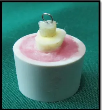
1. Self-etch adhesive system [Filtek Z350 XT, 3M ESPE]:
Among the mounted samples , 30 samples were restored by self etch adhesive
system. The flat dentinal surface were treated with 3M single bond universal adhesive
and light cured for 20s. A hollow metal split mould with 5 mm height and 4mm
diameter [Figure 4a] was held on adhesive treated surface of specimens and then
composite resin of thickness 2mm were placed inside the mould and were condensed.
A 26-gauge ligature wire [Figure 4b] was twisted at one end, and a loop was formed
at other end.
Twisted end was placed inside the 2 mm of uncured composite resin and were
light-cured for 20 s [Figure 4c]. Another 2 mm of composite resin was placed and
cured. Later 1mm of composite resin was placed and cured incrementally. Following
MATERIALS AND METHODS
29 2. Self-adhesive flowable composite [Constic, DMG]:
Remaining 30 samples were restored using self adhesive flowable composite.
Using the same hollow metal split mould, SAF of 2mm thickness was directly placed
onto the prepared tooth surface with the aid of the Luer-Lock-Tip by pressing the
syringe [Figure 5] and massaged for 25 s using the brush. A 26-gauge ligature wire
was twisted at one end, and a loop was formed at other end. Twisted end was placed
inside the 2 mm of uncured composite resin and were light-cured for 20 s. Another 2
mm of SAF composite resin was placed and cured. Later 1mm of composite resin was
placed and cured incrementally. Following complete curing the metal mould was
split and removed.
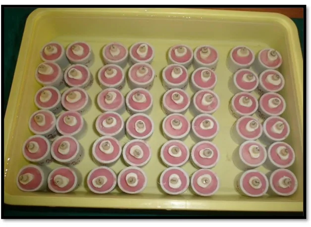
Loading and testing for TBS:
Load cycling protocols and TBS testing were performed at Department of
Manufacturing Engineering, Annamalai University, Chidambaram. All the samples
were stored in water for 24 hours [Figure 6]. For TBS-L, an occlusal load of 90N for
5000 cycles at a rate of 1 Hz was applied to the flat resin composite buildups [Figure
7] using a 5 mm diameter spherical stainless plunger which was attached to cyclic
loading machine [Lloyd LRX Plus II universal testing machine - Ametek, Inc.,USA].
[44] This rate equals to the average cycles of mastication of 0.8-1 seconds. The loop
end was then engaged to the hook of servo-controlled universal testing machine
[Model UNITEK-94100; FIE Pvt. Ltd] [Figure 8a] and pulled for measurement of
MATERIALS AND METHODS
30 II. PREPARATION OF SAMPLES FOR MICROLEAKAGE TESTING:
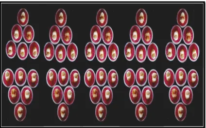
Among 60 samples, 30 samples for group B1, µ-L [Figure 9a] and remaining
30 samples for group B2, µ-NL [Figure 9b]. For all samples, class V cavity were
prepared with rounded outlines of 4 mm width, 2 mm height, and 2mm depth with
No. 330 bur in airotor hand piece. [Figure 10]
Restoration phase for microleakage testing:
1. Self-etch adhesive system [Filtek Z350 XT, 3M ESPE]:
The flat dentinal surfaces were treated with 3M single bond universal adhesive
and light cured for 20 s. On adhesive treated surface of specimens, composite resin
were placed and cured incrementally for 20 s.
2. Self-adhesive flowable composite [Constic, DMG]:
SAF of were directly placed onto the prepared tooth surface with the aid of the
Luer-Lock-Tip by pressing the syringe and massaged for 25 s using the brush
followed by light-curing for 20 s.
Thermocycling phase:
Thermocycling was performed at Central Institute of Plastics Engineering and
Technology, Ministry of Chemicals and Fertilizers, Govt. of India, Guindy, Chennai.
After immersing in water for 24 hours, the samples were thermocycled at 5-55°C for
200 cycles with a dwell time of 30 s and a temperature changing time of 3 min in
MATERIALS AND METHODS
31 dental wax and two coatings of nail varnish were applied on the teeth except for
restorations and their margins. [13]
Occlusal loading phase:
For µ-L, occlusal load of 90N for 5000 cycles at a rate of 1Hz was applied. [44]
This rate equals to the average cycles of mastication of 0.8-1 seconds.
Testing for Microleakage:
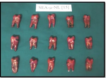
Samples were immersed in 2% aq.solution of basic fuschin dye for 24 hours at
room temperature [Figure 12a,12b]. After removal from the dye, the samples were
washed, dried [Figure 13a,13b, 13c,13d]. and sectioned labio-lingually through the
middle of the restoration using a diamond disk in an air-motor hand piece [Figure
14a,14b]. Each section was examined using a stereo-microscope at X40 magnification
to assess dye penetration at the margins of the restorations in the department of Oral
Pathology, Vivekanandha Dental College for Women, Tiruchengode. The degree of
microleakage [Figure 15a, 15b] was evaluated and scored as follows: [17]
Score Criteria
0 No dye penetration.
1 Dye penetration within ½ of occlusal or gingival wall.
2 Dye penetration extending to the end of occlusal or gingival walls.
3 Dye penetration through the gingival or occlusal wall to 1/
3 of axial wall.
4 Dye penetration through the gingival or occlusal wall to 2/3 of axial wall.
MATERIALS AND METHODS
[image:45.595.161.469.170.378.2]32 MATERIALS USED
Figure 1a: Self etch adhesive composite
[image:45.595.160.468.476.686.2]MATERIALS AND METHODS
33 Figure 2: Samples mounted in acrylic resin
[image:46.595.109.534.154.418.2]
Figure 3: Occlusal grinding of teeth
MATERIALS AND METHODS
[image:47.595.339.504.455.634.2]34 Figure 4a: Metal split mould Figure 4b: Twisted ligature wire
Figure 4c: Twisted wire placed in composite resin
Figure 4d: Restoration after the removal of metal split mould
MATERIALS AND METHODS
[image:48.595.157.471.450.680.2]35 Figure 5: SAF applied onto the prepared tooth surface
Figure 6: Samples immersed in water
MATERIALS AND METHODS
[image:49.595.192.435.149.362.2]36 Figure 7: Occlusal load was applied
Figure 8: Testing of TBS in universal testing machine
Figure 8a Figure 8b
MATERIALS AND METHODS
[image:50.595.167.462.142.372.2]37 Figure 9a : Samples for group B1, µ-L
[image:50.595.164.464.453.664.2]MATERIALS AND METHODS
[image:51.595.240.388.164.384.2]38 Figure 10: Class V cavity
[image:51.595.134.495.449.684.2]MATERIALS AND METHODS
[image:52.595.132.496.157.383.2]39 Figure 12: Samples immersed in basic fuschin dye
[image:52.595.130.498.449.684.2]Figure 12a
MATERIALS AND METHODS
[image:53.595.139.489.157.393.2]40 Figure 13: Samples washed and dried after removal from the dye
[image:53.595.138.491.459.718.2]Figure 13a
MATERIALS AND METHODS
[image:54.595.135.495.128.374.2]41 Figure 13c
Figure 13d
[image:54.595.141.488.434.684.2]MATERIALS AND METHODS
[image:55.595.152.536.196.669.2]42
Figure 14: Labio lingually sectioned samples
MATERIALS AND METHODS
[image:56.595.149.537.153.625.2]43 Figure 14b
MATERIALS AND METHODS
44 Figure 15a : Sample without microleakage under stereomicroscope
Figure 15b : Sample with microleakage under stereomicroscope
RESULTS
45 After testing the tensile bond strength of the groups, the data were subjected to
t-test for equality of means and 95% confidence interval of the differences
[P<0.05] statistical analysis to identify the significant groups. Chi-square test was
used to find a statistical difference in microleakage of two groups.
[image:58.595.110.523.470.682.2]I. TENSILE BOND STRENGTH:
Table 1 and Graph 1:
The mean, standard deviation and t- value for sub-group IA with cyclic
loading and without cyclic loading [n=15 each] are presented in Table-1. The mean
and standard deviation for the subgroups IA1 [SEA-TBS-L] and IA2 [SEA-TBS-NL]
are 4.78±1.02 MPa and 10.31±1.46 MPa respectively. Graph-1 describes the mean of
the two sub-groups IA1 and IA2.
Table-1: Tensile bond strength for Self-etch adhesive system [SEA]:
Sub-Groups IA N Mean Std.
Deviation t- value P- Value
Sub-group IA1: With cyclic
loading
[SEA-TBS-L]
15 4.783 1.024
10.162 0.000
Sub-group IA2: Without cyclic
loading
[SEA-TBS-NL]
RESULTS
46
[image:59.595.108.538.103.359.2]Graph 1: Mean value for SEA-TBS-NL & SEA-TBS-L
Table 2 and Graph 2:
The mean, standard deviation and t- value for sub-group IIA with cyclic
loading and without cyclic loading [n=15 each] are presented in Table-2. The mean
and standard deviation for the subgroups IIA1 L] and IIA2
[SAF-TBS-NL] are 11.33±1.09 MPa and 13.82±1.20 MPa respectively. Graph -2 describes the
mean of the two sub-groups IIA1 and IIA2.
10.3119
4.783
0 2 4 6 8 10 12
Mean
SEA Without loading Tensile
SEA With loading Tensile SEA-TBS-NL
RESULTS
[image:60.595.112.519.127.344.2]47
Table-2: Tensile bond strength for Self Adhering flowable system [SAF]:
Sub-Groups IIA N Mean Std.
Deviation t- value P- Value
Sub-group IIA1: With cyclic
loading
[SAF-TBS-L]
15 11.3303 1.0934
6.570 0.000
Sub-group IIA2: Without cyclic
loading
[SAF-TBS-NL]
15 13.8243 1.2096
Graph 2 : Mean value for SAF-TBS-NL & SAF-TBS-L
13.8243 11.3303 0 2 4 6 8 10 12 14 16 Mean
SAF Without loading Tensile
SAF With loading Tensile SAF-TBS-NL
RESULTS
48
Table 3 and Graph 3 :
The mean, standard deviation and t- value comparing sub-groups IA1 and
IIA1 with cyclic loading [n=15 each] are presented in Table-3. The mean and standard
deviation for the subgroups IA1 [SEA-TBS-L] and IIA1 [SAF-TBS-L] are 4.78±1.02
MPa and 11.33±1.09 MPa respectively. Graph -3 describes the mean of the two
[image:61.595.110.521.339.549.2]sub-groups IA1 and IIA1.
Table-3: Tensile bond strength for Self-etch adhesive system [SEA] and Self
adhering flowable composite [SAF] sub-groups with cyclic loading:
Sub-Groups N Mean Std.
Deviation t- value P- Value
Sub-group IA1: With cyclic
loading
[SEA-TBS-L]
15 4.783 1.024
14.437 0.000 Sub-group IIA1: With cyclic
loading
[SAF-TBS-L]
RESULTS
49
[image:62.595.109.544.107.355.2]Graph -3: Mean value for SAF-TBS-L & SEA-TBS-L
Table 4 and Graph 4:
The mean, standard deviation and t- value comparing sub-groups IA2 and
IIA2 without cyclic loading [n=15 each] are presented in Table-4. The mean and
standard deviation for the subgroups IA2 [SEA-TBS-NL] and IIA2 [SAF-TBS-NL]
are 10.31±1.46 MPa and 13.82±1.20 MPa respectively. Graph -4 describes the mean
of the two sub-groups IA2 and IIA2.
11.3303
4.783
0 2 4 6 8 10 12
Mean
SAF With loading Tensile
SEA With loading Tensile SAF-TBS-L
RESULTS
[image:63.595.108.543.408.669.2]50
Table-4: Tensile bond strength for Self-etch adhesive system [SEA] and Self
adhering flowable composite [SAF] sub-groups without cyclic loading:
Sub-Groups N Mean Std.
Deviation t- value P- Value
Sub-group IA2: Without cyclic
loading
[SEA-TBS-NL]
15 10.3119 1.4675
5.481 0.000 Sub-group IIA2: Without cyclic
loading
[SAF-TBS-NL]
15 13.8243 1.2096
Graph 4: Mean value for SAF-TBS-NL & SEA-TBS-NL
When the mean and standard deviation of the tensile bond strength values
compared within the groups, either in Group I [SEA] with [L] or without [NL] the
application of cyclic loads or in Group II [SAF] with [L] or without [NL] the
13.8243 10.3119 0 2 4 6 8 10 12 14 16
Mean SAF Without loading Tensile
SEA Without loading Tensile SAF-TBS-NL
RESULTS
51 application of cyclic loads, a statistically significant difference [P<0.05] existed
between the sub-groups compared.
II. MICRO-LEAKAGE:
A. Comparison within sub-groups - With loading [L] & Without loading [NL]:
Table 5:
The number and percentage of specimens from each sub-group [IB1 & IB2] with
[image:64.595.103.530.340.596.2]corresponding micro-leakage scores are presented in table-5.
Table-5: Within Sub-Group IB-Self etch adhesive composite resin [SEA]:
Microleakage
scores
Sub-group IB
Total [%] Sub-group IB1-With
cyclic loading [%]
[SEA-µ-L]
Subgroup IB2
-Without cyclic
loading [%]
[SEA-µ-NL]
0 2 [13.3] 2 [6.7]
1 10 [66.7] 10 [33.3]
2 3 [20] 3 [10]
3 3 [20] 3 [10]
4 9 [60] 9 [30]
5 3 [20] 3 [10]
RESULTS
52
Table 6:
The Chi-square test to find a statistical difference in micro-leakage within
Sub-Group IB is given in table-6. The obtained P value [P=0.000] indicates there is
statistically significant difference between the subgroups with and without cyclic
loading.
Table-6: Chi-Square Tests:
Value d.f. Asymp. Sig. (2-sided)
Pearson Chi-Square 24.000 4 0.000
Likelihood Ratio 22.006 4 0.000
Linear-by-Linear
Association 11.546 1 0.001
N of Valid Cases 30
Table 7:
The number and percentage of specimens from each sub-group [IIB1 & IIB2] with
corresponding micro-leakage scores are presented in table-7
RESULTS
53 Table-7: Within Sub-Group IIB-Self adhesive flowable composite [SAF]:
Microleakage
scores
Sub-group IIB
Total [%] Sub-group IIB1-With
cyclic loading [%]
[SAF-µ-L]
Sub-group IIB2
Without cyclic
loading [%]
[SAF-µ-NL]
0 5 [33.3] 5 [16.7]
1 10 [66.7] 10 [33.3]
2
3 5 [33.3] 5 [16.7]
4 10 [66.7] 10 [33.3]
5
Total 15 [100] 15 [100] 30 [100]
Table 8:
The Chi-square test to find a statistical difference in micro-leakage within
Sub-Group IIB is given in table-8. The obtained P value [P=0.001] indicates there is
statistically significant difference between the subgroups with and without cyclic
[image:66.595.101.531.125.381.2]loading.