0022-538X/11/$12.00 doi:10.1128/JVI.05296-11
Copyright © 2011, American Society for Microbiology. All Rights Reserved.
Human T-Cell Lymphotropic Virus Type 3 (HTLV-3)- and
HTLV-4-Derived Antisense Transcripts Encode Proteins
with Similar Tax-Inhibiting Functions but Distinct
Subcellular Localization
䌤
E
´ milie Larocque,
1† Marile
`ne Halin,
1† Se
´bastien Landry,
1‡ Susan J. Marriott,
3William M. Switzer,
4and Benoit Barbeau
1,2*
De´partement des Sciences Biologiques1and Centre de Recherche BioMed,2Universite´ du Que´bec a` Montre´al, 2080 Saint-Urbain,
Montre´al, Que´bec, Canada H2X 3X8; Department of Molecular Virology and Microbiology, Baylor College of Medicine, Houston, Texas 770303; and Laboratory Branch, Division of HIV/AIDS Prevention, National Center for HIV,
AIDS, Viral Hepatitis, STD, and TB Prevention, Centers for Disease Control and Prevention, Atlanta, Georgia 303334
Received 2 June 2011/Accepted 6 September 2011
The human T-cell lymphotropic virus (HTLV) retrovirus family is composed of the well-known HTLV type 1 (HTLV-1) and HTLV-2 and the most recently discovered HTLV-3 and HTLV-4. Like other retroviruses, HTLV-1 and HTLV-2 gene expression has been thought to be orchestrated through a single transcript. However, recent reports have demonstrated the unique potential of both HTLV-1 and HTLV-2 to produce an antisense transcript. Furthermore, these unexpected and newly identified transcripts lead to the synthesis of viral proteins termed HBZ (HTLV-1 basic leucine zipper) and APH-2 (antisense protein of HTLV-2), respec-tively. As potential open reading frames are present on the antisense strand of HTLV-3 and HTLV-4, we tested whetherin vitroantisense transcription occurred in these viruses and whether these transcripts had a coding potential. Using HTLV-3 and HTLV-4 proviral DNA constructs, antisense transcripts were detected by reverse transcriptase PCR. These transcripts are spliced and polyadenylated and initiate at multiple sites from the 3ⴕ long terminal repeat (LTR). The resulting proteins, termed APH-3 and APH-4, are devoid of a typical basic leucine zipper domain but contain basic amino acid-rich regions. Confocal microscopy and Western blotting experiments demonstrated a nucleus-restricted pattern for APH-4, while APH-3 was localized both in the cytoplasm and in the nucleus. Both proteins showed partial colocalization with nucleoli and HBZ-associated structures. Finally, both proteins inhibited Tax1- and Tax3-mediated HTLV-1 and HTLV-3 LTR activation. These results further demonstrate that retroviral antisense transcription is not exclusive to HTLV-1 and HTLV-2 and that APH-3 and APH-4 could impact HTLV-3 and HTLV-4 replication.
Human T-cell lymphotropic viruses (HTLVs) are human deltaretroviruses that are part of the primate T-cell lympho-tropic virus (PTLV) group that also includes simian T-cell lymphotropic viruses (STLVs). Most research has been con-ducted on the two first identified members of this family, i.e., HTLV type 1 (HTLV-1), the first retrovirus to be isolated in humans (34, 37, 38, 51), and HTLV-2. HTLV-1 has a signifi-cant impact on human health, as this virus is the etiological agent of adult T-cell leukemia/lymphoma (ATLL) and HTLV-1-associated myelopathy (HAM)/tropical spastic paraparesis (TSP). Unlike HTLV-1, HTLV-2 has been linked to HAM-like pathologies but not to leukemia, although patients infected with HTLV-2 demonstrate a higher lymphocyte count than noninfected patients (6). Recently, two new HTLVs, termed HTLV-3 and HTLV-4, have been isolated, the former being
closely related to STLV type 3 (STLV-3) (10, 11, 50). Cur-rently, these viruses have been identified in a relatively small number of persons from Africa, and no diseases, such as large granuloma leukemia (16, 46), have yet been associated with these viruses.
The discovery of these two retroviruses led to a series of recent studies for further characterization. These studies have demonstrated that HTLV-3 and HTLV-4 share a similar genomic organization to HTLV-1 and HTLV-2, and weak but reproducible cross-reactivities were observed in serologic as-says employing HTLV-1 and HTLV-2 antigens (10, 11, 41, 42, 50). Further studies focusing on the Tax3 protein of HTLV-3 have found that its intracellular localization, its domains (such as the PDZ domain binding motif), and its transactivation activity are similar to those of HTLV-1 Tax (10, 14). A recent study also provided evidence that the HTLV-3 genome, when reconstituted, produces infectious particles (13). Interestingly, like HTLV-1, the existence of a potential open reading frame (ORF) called HBZ, for HTLV-1 basic leucine zipper (bZIP), which could be produced from the antisense strand, has been suggested for both new human viruses (10, 13, 42, 43).
Previous studies have shed light on the existence of this HBZ protein encoded by the antisense strand of the HTLV-1 ge-nome (18). Typically, two HBZ isoforms, one of which is more
* Corresponding author. Mailing address: De´partement des Sci-ences Biologiques, Universite´ du Que´bec a` Montre´al, Room SB-R860, 2080 Saint-Urbain Montre´al, Que´bec, Canada H2X 3X8. Phone: (514) 987-3000, ext. 4576. Fax: (514) 987-4647. E-mail: barbeau.benoit @uqam.ca.
‡ Present address: Laboratory of Genetics, The Salk Institute for Biological Studies, La Jolla, CA.
† Authors have contributed equally to this work. 䌤Published ahead of print on 14 September 2011.
12673
on November 7, 2019 by guest
http://jvi.asm.org/
abundant and depends on a spliced transcript, are produced (12, 35, 40). Both HBZ isoforms block Tax-induced and basal HTLV-1 transcription and interact with several Jun family members, rendering some of them inactive through degrada-tion or by possible sequestradegrada-tion in transcripdegrada-tionally inactive nuclear bodies (7, 23, 29, 45). However, subsequent studies have demonstrated that HBZ interacts with and activates JunD, thereby augmenting gene expression of the human tel-omerase reverse transcriptase (hTERT) component through this transcription factor (22, 25, 45). Other transcription fac-tors such as NF-B and MafG are additional targets of HBZ, likely contributing to the disturbance of gene expression in HTLV-1-infected cells (39, 53). A number of reports have also demonstrated that HBZ is expressed in cells from ATLL pa-tients, therefore implicating this viral protein in the develop-ment of ATLL, in part through its hyperproliferative action on T cells (2, 4, 31, 32, 40, 47).
Antisense transcription has also been suggested in other retroviruses like HIV-1 (2, 8, 9, 27, 33, 36, 44, 48). We have recently demonstrated that antisense transcription could be detected in HTLV-2 and that the encoded antisense protein of HTLV-2 (APH-2) shares a Tax-inhibiting activity like HBZ (21). On the basis of these former results and given that pre-vious sequence analysis studies have predicted the possible existence of antisense transcription in HTLV-3 and HTLV-4, we have investigated the presence of antisense transcripts in these new viruses and their potential coding capacity. Our results indicate that both viruses produce a spliced and poly-adenylated antisense transcript. The encoded proteins show distinct localization, with the HTLV-3 antisense protein being both nuclear and cytoplasmic and the HTLV-4 counterpart being almost exclusively contained in the nucleus. However, both antisense proteins inhibited Tax-mediated HTLV long terminal repeat (LTR) activation. These important results in-dicate the potentially essential role of antisense proteins in retroviral replication.
MATERIALS AND METHODS
Cell lines and antibodies.The 293T and COS-7 cell lines were maintained in Dulbecco modified Eagle medium (DMEM) supplemented with 10% fetal bo-vine serum (FBS) (PAA Laboratories Inc., Toronto, Canada). The T-cell line Jurkat E6.1 was maintained in RPMI 1640 medium supplemented with 10% FBS. The anti-Myc antibody (9E10) and mouse anti-glyceraldehyde-3-phosphate dehydrogenase (anti-GAPDH) antibody were purchased from Santa Cruz Bio-technology Inc. (Santa Cruz, CA). The enhance chemiluminescence (ECL) sheep anti-mouse IgG antibody coupled to horseradish peroxidase was obtained from GE Healthcare Inc. (Buckinghamshire United Kingdom), and the goat anti-mouse IgG antibody coupled to Alexa Fluor 488 (A11001) was obtained from Invitrogen Canada Inc. (Burlington, Canada). The anti-Lap2 antibody (L6043) was from Sigma-Aldrich (Oakville, Canada). Anti-HTLV-1 Tax
poly-clonal antibodies were kindly provided by Jean-Michel Mesnard (Universite´
Montpellier I, Montpellier, France) (3).
Plasmids. Both HTLV-3 (2026ND) and HTLV-4 (pUC-HT4v2) proviral
DNAs were cloned in pUC18. The pHTLV-3⌬EcoRV construct was derived
from the HTLV-3 (2026ND) proviral DNA clone by EcoRV/XhoI digestion,
thereby deleting 4,359 bp of the 5⬘end of the proviral sequence. The
pHTLV-3-as-luc vector was derived from pHTLV-3⌬EcoRV by cloning the luciferase
gene from pGL3-basic in frame in the second exon of APH-3. Briefly, PacI and
BstZ171 restriction sites were added to pHTLV-3⌬EcoRV with a Phusion
site-directed mutagenesis kit using reverse primer 5⬘-ATAGTATACTGCAATC
CCAGGAACTG-3⬘(BstZ171 restriction site in bold) and forward primer 5⬘-A
TATTAATTAATGTCTCCGGGGCTAGG-3⬘(PacI restriction site in bold). The
luciferase gene was amplified using reverse primer 5⬘-ATATTAATTAAGGAAG
ACGCCAAAAACATAAAGAA-3⬘(PacI restriction site in bold) and forward
primer 5⬘-ATAGTATACTACCACATTTGTAGAGGTTTTAG-3⬘(BstZ171
re-striction site in bold). Luciferase amplicons were ligated into the pHTLV-3
⌬EcoRV-digested vector, resulting in pHTLV-3-as-luc. The pHTLV-4⌬SacI
construct was produced by digesting HTLV-4 (pUC-HT4v2) proviral DNA with
SacI/HindIII, thereby deleting 3,213 bp of the 5⬘end of the proviral sequence.
The pHTLV-4-as-luc vector was obtained by cloning the luciferase gene in frame into the second exon of APH-4 at position 7037 (sense transcript) of
pHTLV-4⌬6560 (construct containing the last 2,183 bp of the HTLV-4 genome in the
pUC18 vector) by amplifying the luciferase gene with reverse primer 5⬘-ATAG
TATACAGAGGAGATGCCTGGTA-3⬘(BstZ171 restriction site in bold) and
forward primer 5⬘-ATATTAATTAATGGTGTTGAGACCTTCTTTG-3⬘ (PacI
restriction site in bold). The pHTLV-1 luc vector contains the HTLV-1 LTR 3⬘
region cloned into the XhoI/HindIII sites of the pGL2-basic vector (Promega) (19). The Tax1-expressing vector has been previously described (30). The pCMV-Tax3 vector contains the Tax3-coding region under the control of the
cytomegalovirus (CMV) promoter region in pcDNA3.1Zeo(⫹). The
pMy-cAPH-3 and pMycAPH-4 expression vectors were generated by PCR amplifica-tion of the coding regions of APH-3 and APH-4 using primers that contain the
sequence of a Myc tag at their 5⬘ends. Briefly, APH-3 was amplified using
reverse primer 5⬘-ATGGAACAAAAACTCATCTCAGAAGAGGATCTGATGGC
TCGATCCCGAAGCGG-3⬘(Myc tag in bold) and forward primer 5⬘-ATATC
TAGATTATAACAGATCTGCTACCTCCTGTAG-3⬘(XbaI restriction site in
bold). APH-4 was amplified using reverse primer 5⬘-ATGGAACAAAAACTCAT
CTCAGAAGAGGATCTGATGGACACTCGAGAATTTTTTAGGGG-3⬘ (Myc
tag in bold) and forward primer 5⬘-ATATCTAGATTATAATAACTCCGCCAA
TACACCCAAC-3⬘ (XbaI restriction site in bold). Amplified products were
digested by XbaI and ligated into pcDNA3.1Zeo(⫹) digested with XbaI/EcoRV.
The pAPH-3 and green fluorescent protein (GFP) fusion (pAPH-3–GFP) construct and pAPH-4–GFP were generated by amplifying APH-3 using
forward primer 5⬘-ATAGAATTCATGGCTCGATCCCGAAGCGG-3⬘(EcoRI
re-striction site in bold) and reverse primer 5⬘-ATAACCGGTGCTAACAGATCTGC
TACCTCCTGTAG-3⬘(AgeI restriction site in bold) and APH-4 using forward
primer 5⬘-ATAGAATTCATGGACACTCGAGAATTTTTTAGGGG-3⬘ (EcoRI
restriction site in bold) and reverse primer 5⬘-ATAACCGGTGCTAATAACTC
CGCCAATACACCCAACA-3⬘(AgeI restriction site in bold). Amplified
prod-ucts were ligated into EcoRI/AgeI-digested peGFP-N1. To minimize synthesis of free GFP, the methionine initiation codon from the GFP reporter gene was mutated to a leucine (TTG) using the Phusion site-directed mutagenesis kit for both the APH-3–GFP and APH-4–GFP constructs. The forward and reverse
primers for APH-3–GFP were 5⬘-GCAGATCTGTTATTGGTGAGCAAGG-3⬘
(mutated nucleotide in bold) and 5⬘-TACCTCCTGTAGCAGGAGGCTAT-3⬘.
As for APH-4–GFP, the forward and reverse primers were 5⬘-CGGAGTTATT
ATTGGTGAGCAAGGG-3⬘(mutated nucleotide in bold) and 5⬘-CCAATAC
ACCCAACAGGTCCC-3⬘. The HBZ and monomeric red fluorescent protein
(mRFP) fusion (HBZ-mRFP) construct was generated by amplifying the HBZ
SP1 cDNA with forward primer 5⬘-ATAAGCTTATGGCGGCCTCAGG-3⬘
(HindIII restriction site in bold) and reverse primer 5⬘-ATGAATTCTTGCAAC
CACATCGCCT-3⬘(EcoRI restriction site in bold). The amplified products were
then digested and ligated into pcDNA3.1Zeo(⫹)-mRFP (kindly provided by
Matthew Weitzman, The Salk Institute, La Jolla, CA). The Nucleolin-DsRed expression vector has been previously described and expresses a chimeric form of
nucleolin (20). The pRcActin-LacZ vector contains the-galactosidase (-Gal)
gene under the control of the-actin promoter. An expression vector encoding
the unspliced HBZ isoform tagged with the Myc epitope was obtained from Jean-Michel Mesnard (45). The HBZ exon 1 sequence (unspliced HBZ) in this vector was modified for HBZ SP1 exon 1 by reverse PCR using forward and
reverse primers 5⬘-ATGGCGGCCTCAGGGCTGTTTCGATGCTTGCCTG-3⬘
and 5⬘-GGGGATCCACTAGTCCAGTGTG-3⬘. The resulting vector was
termed pHBZ-SP1-Myc. From this plasmid, pAPH-3Myc and pAPH-4Myc ex-pression vectors were generated by excising the HBZ cDNA with EcoRI and HindIII restriction enzymes and cloning APH-3 and APH-4 cDNAs amplified with EcoRI- and HindIII-containing primers.
RT-PCR.Total RNA was extracted with the TRIzol reagent (Invitrogen) from
293T and Jurkat cells at 48 h posttransfection. Poly(A)⫹RNA was purified from
total RNA using an Oligotex mRNA minikit (Qiagen, Mississauga, Canada) according to the manufacturer’s instructions. Reverse transcriptase PCR (RT-PCR) analyses were conducted using an oligo(dT) primer (Invitrogen). Briefly,
total RNA (5g) was mixed with 1l of 10M oligo(dT) primer. The RNA-RT
primer mix was heated at 70°C and incubated for 2 h at 42°C in the presence of
1⫻avian myeloblastosis virus (AMV) reaction buffer, 1 mM deoxynucleoside
triphosphates, 10 U of SUPERase䡠In RNase inhibitor (Ambion), and 15 U of
AMV reverse transcriptase (USB). cDNAs synthesized from 293T cells were
PCR amplified with reverse primer LTR-HTLV-3as1 (5⬘-CAAGCCTCGCTGC
12674 LAROCQUE ET AL. J. VIROL.
on November 7, 2019 by guest
http://jvi.asm.org/
TGACAGC-3⬘) and forward primer env-HTLV-3-s8 (5⬘-GACGCCCTGGCCC
CAACAG-3⬘) to amplify the antisense transcript of HTLV-3 and reverse primer
LTR-HTLV-4as2 (5⬘-CGGCGGCGTCTCAACTGATTG-3⬘) and forward
primer env-HTLV-4s1 (5⬘-ACGAGTCCCCCCATATGTCCAAA-3⬘) to amplify
the antisense transcript of HTLV-4. PCR conditions were as follows: a first step of denaturation at 94°C for 5 min, followed by 35 cycles of denaturation (94°C for 1 min), annealing (60°C for 1 min), and extension (72°C for 1 min) and a final extension at 72°C for 5 min. The synthesized cDNAs from transfected Jurkat cells were PCR amplified under the same conditions used for transfected 293T cells with reverse primer LTR-HTLV-3as1 (see above) and forward primer
env-HTLV-3-s3 (5⬘-CAGCATCGCAGTCAGCCCTA-3⬘) for detection of the
antisense transcript of HTLV-3 and reverse primer LTR-HTLV-4as2 (see
above) and forward primer env-HTLV-4as5 (5⬘-GGTCCATGCACTTCCGTTG
TTGATGACG-3⬘) for detection of the antisense transcript of HTLV-4.
5ⴕand 3ⴕRACE analyses.Total RNA was isolated from 293T cells at 48 h posttransfection. Initiation sites of APH-3 and APH-4 transcripts were then determined using a FirstChoice RNA ligase-mediated–rapid amplification of cDNA end (RLM-RACE) kit (Ambion) according to the manufacturer’s
instruc-tions. cDNAs were synthesized in the presence of the supplied 5⬘RACE adapter.
PCR amplification was performed using 5⬘RACE outer and inner primers and
different primers derived from the ORF sequences: H3race-em-1 (5⬘-GCCATT
CCCCTGAAGCATGTC-3⬘) and H3race-em-2 (5⬘-GCTCCTCTGCAGTCAAC
ACCG-3⬘) for the HTLV-3 antisense transcript and 5RACEH4-1 (5⬘-GTCTG
TTCCAAGCCAGCTGATAACCGAAAT-3⬘) and 5RACEH4-2 (5⬘-CTCCTA
AGTTATGGTTACATTCCTCCTCCAG3⬘) for the HTLV-4 antisense
transcript. Amplified products were then cloned in the pDrive vector and
se-quenced. To identify the poly(A) signal, cDNAs were synthesized from poly(A)⫹
RNA isolated from transfected 293T cells in the presence of the supplied 3⬘
RACE adapter. PCR amplification was performed using 3⬘RACE outer and
inner primers and a primer derived from the sequence downstream of the ORFs:
23-2 (5⬘-GGAGAGGAACCACACTGGATCAT-3⬘) for the HTLV-3 antisense
transcript and 23-5 (5⬘-GAGTCAGGACATGCTCTAGGTCT-3⬘) for the
HTLV-4 antisense transcript. Amplified products were cloned into SmaI-di-gested pBluescript KS and sequenced. All PCRs were performed under the conditions in the manufacturer’s instructions.
Transfection and promoter studies.293T cells (4⫻105) were transfected with
10g luciferase-expressing DNA constructs using the calcium phosphate
proto-col as previously described (17) or using the Lipofectamine reagent (Invitrogen) according to the manufacturer’s instructions. Jurkat cells were transfected by
electroporation with 15g luciferase-expressing DNA constructs using a Gene
Pulser Xcell system (960F, 250 V; Bio-Rad, Hercules, CA). Transfected 293T
cells were lysed at 48 h posttransfection, and luciferase activity was determined with an MLX microplate luminometer (Dynex Technologies) as previously de-scribed (5). Each sample was cotransfected with pRcActin-LacZ for
normaliza-tion of transfecnormaliza-tion efficiency.-Galactosidase activities were measured using a
Galacto-Light kit (Applied Biosystems, Bedford, MS) according to the manu-facturer’s protocol. Luciferase activity is presented as normalized relative light
units (RLU/-Gal) and represents the calculated mean⫾standard deviation of
three transfected samples. For Jurkat cell transfection, cells were resuspended at
2.5⫻107
/ml of complete medium, transfected in bulk, and separated at 16 h
posttransfection into various treatment groups at a density of 1⫻105cells/well
(100l) in 96-well plates. Cells were left untreated or treated with phorbol
12-myristate 13-acetate (PMA; Sigma-Aldrich, Oakville, Canada) at 20 ng/ml,
phytohemagglutinin (PHA-P; Sigma-Aldrich) at 3g/ml, ionomycin
(Sigma-Aldrich) at 1M, bis-peroxovanadium (bpV[pic]; Alexis Corporation, Lausen,
Switzerland) at 15M, forskolin (Sigma-Aldrich) at 10M, and tumor necrosis
factor alpha (TNF-␣; Sigma-Aldrich) at 20 ng/ml. Luciferase activity was
mon-itored at 8 h poststimulation.
Confocal microscopy.COS-7 cells were seeded in 6-well plates containing a 1.5-mm-thick coverslip for 24 h and then transfected using the Lipofectamine reagent (Invitrogen) according to the manufacturer’s instructions. Jurkat cells were microporated with an MP-100 device (Digital Bio, Montreal, Canada) with
1.5⫻105
cells and 1g of expression vectors at 1,350 V with 1 pulse for 30 ms.
For Jurkat cells, 1 h before cells were fixed, Jurkat cells were seeded on a coverslip treated with polylysine. At 36 h posttransfection, cells were washed with phosphate-buffered saline (PBS), fixed with 4% paraformaldehyde for 15 min, and permeabilized with 0.1% Triton X-100 for 5 min at room temperature. Cells were then washed three times with PBS and incubated with the anti-Myc 9E10 antibody (dilution, 1:400) for 1 h at room temperature. Samples were washed three times with PBS, incubated with goat anti-mouse IgG coupled to Alexa Fluor 488 for 45 min at room temperature, and washed again with PBS. Cells
were incubated in a 2.5-g/ml propidium iodide (PI) solution. Coverslips were
then mounted in a drop of ProLong antifade reagent (Invitrogen). For live-cell
imaging, cells were cultured on a Lab-Tek chambered cover glass (Thermo Fisher Scientific, Rochester, NY) for 24 h and then transfected using the Lipo-fectamine reagent (Invitrogen) according to the manufacturer’s instructions. Cells were observed at 36 h and 48 h posttransfection with an MRC-1024ES confocal laser scanning microscope (Bio-Rad, Hercules, CA).
Western blot (WB) analysis.Total extracts were prepared as previously de-scribed (49). Cytoplasmic and nuclear extracts from transfected COS-7 and 293T cells were prepared using NE-PER nuclear and cytoplasmic extraction reagents (Thermo Fisher Scientific) according to the manufacturer’s instructions. Equal quantities of extracts were run on a 10% SDS-polyacrylamide gel and transferred to polyvinylidene difluoride membranes (Millipore, Mississauga, Canada). The blot was next blocked in PBS–5% milk and incubated with either anti-Myc 9E10 (1:1,000), anti-HTLV-1 Tax (1:100), or anti-GAPDH (1:1,000) antibodies. After several washes, signals were revealed by adding horseradish peroxidase-conju-gated sheep anti-mouse IgG antibodies (dilution, 1:10,000), followed by incuba-tion with BM chemiluminescence blotting substrate (horseradish peroxidase [POD]; Roche Diagnostic, Indianapolis, IN). Membranes were exposed on Am-ersham hyperfilms ECL (AmAm-ersham Bioscience, Buckinghamshire, United King-dom). To detect Tax, a SignalBoost immunoreaction enhancer kit was used to amplify the signal (EMD Bioscience, San Diego, CA).
RESULTS
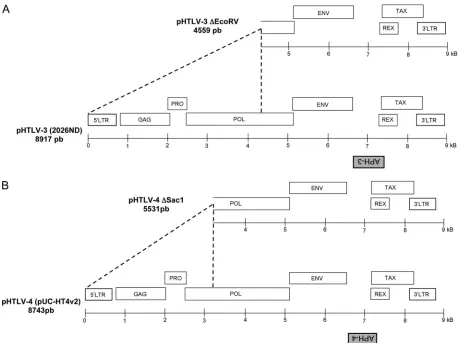
Detection of spliced antisense transcripts from HTLV-3 and HTLV-4.Previous studies have predicted the presence of an ORF in the antisense strand of HTLV-3 and HTLV-4 (10, 13, 42, 43). Since antisense proteins have been shown to exist in HTLV-1 and HTLV-2, we asked whether the newly discovered HTLV-3 and HTLV-4 retroviruses express similar antisense proteins. Initialin silicoanalysis of HTLV-3 (strains 2026NB and Lobak) and HTLV-4 proviral DNAs revealed the exis-tence of ORFs in the antisense strand between thetaxandenv
genes (Fig. 1A and B) (42, 43). Both HTLV-3 strains presented an homology of 93% at the amino acid level for this ORF. We also identified the presence of a shorter antisense ORF in the proviral DNA of the Pyl 43 HTLV-3 strain, which contains a 366-nucleotide (nt) deletion in the pX region, leading to re-moval of more than 50% of the predicted carboxyl end of the antisense ORF (10). These ORFs were termed APH-3 (anti-sense protein of HTLV-3) and APH-4. The encoded proteins are predicted to be 221 and 158 amino acids in length for HTLV-3 and HTLV-4, respectively.
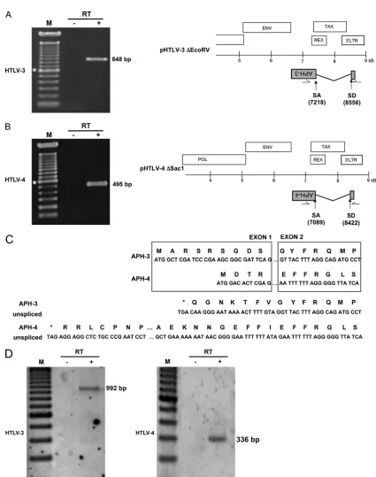
To demonstrate the existence of antisense transcripts in these retroviruses, we first performed RT-PCR analyses. The 5⬘ LTR regions of the proviral DNA constructs were first deleted to minimize interference from sense transcription (Fig. 1). The resulting pHTLV-3 ⌬EcoRV and pHTLV-4 ⌬SacI constructs were transfected into 293T and Jurkat cells, and RNA extracted from these cells was analyzed by RT-PCR using forward and reverse primers derived from the 3⬘ LTR and APH-3/APH-4 regions, respectively (Fig. 2). The choice and position of primers were based on the previously described splicing pattern of the antisense HBZ transcript of HTLV-1 (12, 35, 40). The results of RT-PCR analyses revealed the presence of antisense transcripts, which were successfully am-plified using different sets of primers (Fig. 2A, B, and D and data not shown). In addition, the size of these transcripts supported the occurrence of splicing. The HTLV-3 antisense transcript splice donor (SD) and splice acceptor (SA) were positioned at nt 8556 and 7218, respectively, while the corre-sponding HTLV-4 antisense transcript SD and SA sites were at nt 8422 and 7089, respectively. The positions of the splice sites and lengths of the intronic regions were well conserved
on November 7, 2019 by guest
http://jvi.asm.org/
tween HTLV-3 and HTLV-4. Similar to the HTLV-1 HBZ transcript, the splicing ofAPH-3andAPH-4transcripts led to the addition of an N-terminal amino acid sequence containing a methionine initiation codon derived from exon 1 (in the 3⬘ LTR) to the corresponding ORF sequence (Fig. 2C). For APH-3, 9 amino acids were added from exon 1, while for APH-4, 4 amino acids were added from exon 1. Interestingly, if the unspliced mRNA could encode a different APH isoform in both HTLV-3 and HTLV-4, the resulting APH isoform would initiate downstream of the splice acceptor site, leading to an isoform shorter than the protein encoded by the spliced transcripts, unlike HBZ. Thus, these results demonstrate that both viruses are capable of producing a spliced antisense script with the potential to encode a protein. Unspliced tran-scripts were not analyzed in these experiments.
APH-3andAPH-4transcripts have multiple initiation sites and are polyadenylated.To more precisely characterizeAPH-3
andAPH-4transcripts, we performed 5⬘RACE analyses (Fig. 3). Total RNA was isolated from 293T cells transfected with versions from which proviral DNA was deleted (for HTLV-3, nt 4731 to 8918; for HTLV-4, nt 4873 to 8742) and that con-tained the 3⬘LTR and presumed antisense ORF. On the basis of our 5⬘ RACE analyses, four different initiation sites were
detected for theAPH-3transcripts (Fig. 3A), while as many as seven initiation sites located at the end of the 3⬘ LTR were detected forAPH-4transcripts (Fig. 3B).
We next wanted to characterize the 3⬘end of both antisense transcripts (Fig. 3C and D). Again, versions of HTLV-3 and HTLV-4 from which proviral DNA was deleted were trans-fected into 293T cells and poly(A)⫹RNA was used to identify the 3⬘end of the transcripts by 3⬘RACE analysis. As depicted in Fig. 3C and D, both transcripts were cleaved and polyade-nylated at a single site between a consensus poly(A) signal and a GU-rich sequence, which is often found in proximity to the poly(A) tail addition site. Interestingly, although the sequence of the dinucleotide targeted for cleavage was different for the two retroviruses (AG for HTLV-3 and CC for HTLV-4), they occurred at an equal distance (19 nt) from the poly(A) signal. On the basis of the overall position of transcription initiation sites and poly(A) addition sites, the sizes of the APH-3and
APH-4 transcripts were predicted to be 2.5 kb and 2.4 kb, respectively.
[image:4.585.61.529.67.410.2]Cellular localization of APH-3 and APH-4.Comparison of the predicted APH-3 and APH-4 sequences to the published HBZ and APH-2 sequences (Fig. 4) revealed that APH-3 and APH-4 do not contain a classical basic leucine zipper (bZIP)
FIG. 1. Position of antisense ORF in HTLV-3 and HTLV-4 genomes. The HTLV-3 2026ND (A) and HTLV-4 HT4v2 (B) molecular clones and known genes are depicted. The positions of the putative APH-3 and APH-4 ORFs from the antisense strand are indicated below each proviral DNA. Versions of these vectors with 5⬘deletions, termed pHTLV-3⌬EcoRV and pHTLV-4⌬SacI, are also presented. The size of the proviral DNA (full-length version or version with 5⬘-end deletion) is indicated for each retrovirus.
12676 LAROCQUE ET AL. J. VIROL.
on November 7, 2019 by guest
http://jvi.asm.org/
FIG. 2. Detection of spliced HTLV-3 and HTLV-4 antisense transcripts. 293T cells were transfected with either pHTLV-3⌬EcoRV (A) or pHTLV-4⌬SacI (B). RNA was extracted and analyzed by RT-PCR for the presence of antisense transcripts using the primer combinations LTR-HTLV-3as1/env-HTLV-3s8 (A) or LTR-HTLV-4as2/env-HTLV-4s1 (B). Spliced transcripts for each proviral DNA are depicted, and the positions of the SD and SA sites are indicated below (nucleotidic positioning from sense strand). Lanes:⫺, negative control (no RT added in RT step before PCR); M, 100-bp marker (*, 600 bp);⫹, test lane. (C) Amino acid sequence deduced from splicedAPH-3andAPH-4RNA next to the splice junction. The amino acid sequence is shown above each nucleotide sequence. The nucleotide and amino acid sequences predicted for unspliced transcripts are provided for comparison. Asterisks, in-frame stop codons. (D) Jurkat cells were transfected with pHTLV-3⌬EcoRV or pHTLV-4⌬SacI, and RNA was extracted and analyzed by RT-PCR as described above using the primer combination LTR-HTLV-3as1/env-HTLV-3s3 (left) or LTR-HTLV-4as2/env-HTLV-4as5 (right).
on November 7, 2019 by guest
http://jvi.asm.org/
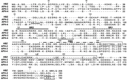
motif. Basic-rich (BR) regions were, however, found at posi-tions similar to the position of the HBZ BR region (24) and could function as nuclear localization signals (NLSs). In addi-tion, LXXLL and LXXLL-like motifs, known to be responsible for the interaction of HBZ with p300/CBP (15), were identified in the predicted APH-2, APH-3, and APH-4 amino acid se-quences. These motifs were, however, positioned in different regions than equivalent motifs in HBZ. Following amino acid comparison between all four antisense proteins, we found that APH-3 and APH-4 presented 35.8% and 33.6% homology with HBZ, respectively, showing an important level of differences between these proteins. Interestingly, APH-3 and APH-4 ap-peared to be more closely related to APH-2, showing 50.6% and 71% homology, respectively.
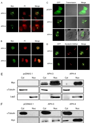
We next determined the subcellular localization of APH-3 and APH-4 using expression vectors for both proteins tagged with a Myc epitope at their amino ends. COS-7 and Jurkat cells were transfected with APH-3 and APH-4 expression vectors and analyzed by confocal microscopy. In these analyses, al-though APH-3 mainly localized in the nucleus, detectable cy-toplasmic staining was also observed (Fig. 5A and B). Similar
staining was also noted in transfected 293T cells (data not shown). Of note, a cytoplasmic aggregate was usually present in cells transfected with the expression vector of APH-3 and likely results from overexpression. In COS-7 cells transfected with the Myc-tagged APH-4 expression vector, APH-4 was in the nucleus, similar to HBZ. Confocal microscopy was also used to examine cells transfected with C-terminus-tagged APH expression vectors and showed similar results (data not shown), indicating that the added tag and its position likely did not contribute to the subcellular distribution of APH-3 and APH-4.
To further analyze the subcellular localization of APH-3 and APH-4, live-cell imaging experiments were conducted in COS-7 cells transiently transfected with either the APH-3– GFP or APH-4–GFP expression vector. As illustrated in Fig. 5C, the APH-3–GFP fusion protein was observed in the nu-cleus and, to a lesser extent, in the cytoplasm at 48 h posttrans-fection. In contrast, the APH-4–GFP fusion protein exclusively localized to the nucleus. GFP itself, when expressed in COS-7 cells, demonstrated more diffuse signals different from the distribution of our fusion proteins. Hence, the subcellular
lo-FIG. 3. Identification of transcription initiation sites and poly(A) addition sites forAPH-3andAPH-4transcripts. Cultured 293T cells were transfected with versions of either pHTLV-3 (nt 4731 to 8918 containing the 3⬘LTR and the antisense ORF) (A and C) or pHTLV-4 (nt 4873 to 8742 containing the 3⬘LTR and the antisense ORF) (B and D) from which proviral DNA was deleted. RNA was extracted at 48 h posttransfection and analyzed by 5⬘or 3⬘RLM-RACE. (A and B) The antisense nucleotide sequence of the 3⬘LTR is depicted, and initiation sites are shown by arrows. (C and D) The positions of theAPH-3andAPH-4mRNAs (grey boxes) and of the 3⬘poly(A) tails are shown. In the sequence below, the position of the cleavage site and the sequences of the poly(A) signal and GU-rich elements are shown. Lanes:⫹, test lane;⫺, PCR amplification with no prior RT step; M, 100-bp marker (*, 600 bp).
12678 LAROCQUE ET AL. J. VIROL.
on November 7, 2019 by guest
http://jvi.asm.org/
[image:6.585.40.509.61.398.2]calization of these fusion proteins was similar to that of the Myc tag constructs. Transfection experiments in 293T cells demonstrated similar results (data not shown). To determine whether APH-3 and APH-4 had a nucleolar localization (Fig. 5D), we cotransfected APH-3–GFP or APH-4–GFP expres-sion vectors with a Nucleolin-DsRed expresexpres-sion vector in COS-7 cells. As shown in Fig. 5D, APH-3 and APH-4 indeed showed partial nucleolar localization at 48 h posttransfection. To further confirm their subcellular distribution, WB anal-yses were performed on nuclear and cytoplasmic extracts pre-pared from 293T and COS-7 cells transfected with the Myc-tagged HBZ, APH-3, or APH-4 expression vector (Fig. 5E and F). APH-3 migrated at the expected size (28 kDa), while APH-4 migrated at a lower molecular mass, i.e., 18 kDa, than the predicted size of 22 kDa. In previous reports (1, 12), HBZ has also been shown to migrate differently from its predicted size of 27 kDa. As expected, both APH-4 and HBZ were mainly present in nuclear extracts, with a minor signal in cy-toplasmic extracts. On the other hand, APH-3 was present in both extracts, although it was present at higher levels in the nucleus. Interestingly, a higher-molecular-mass signal was ap-parent mostly in 293T cells. Together, these results suggest that HBZ, APH-3, and APH-4 all localize primarily to the nucleus. The localization of APH-4 was very similar to that of HBZ. The localization of APH-3, while primarily in the nucleus, showed a signal in the cytoplasm.
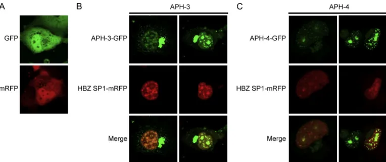
Partial colocalization between HBZ and APH-3 and APH-4.
As APH-3 and APH-4 present similarities with HBZ, cellular colocalization of these proteins was assessed. COS-7 cells were
transiently cotransfected with APH-3–GFP or APH-4–GFP expression vectors along with an HBZ SP1-mRFP expression vector and analyzed at 48 h posttransfection (Fig. 6B and C). The majority of the punctuated structures specific to APH-3 and APH-4 fusion proteins colocalized with HBZ SP1-mRFP in the nucleus, although certain speckled structures did not. As expected, the cytoplasmic signals observed in APH-3–GFP-expressing cells did not colocalize with HBZ (Fig. 6B). As a control, free GFP and free mRFP expressed in COS-7 cells appeared as homogeneous signals different from those seen with the antisense GFP fusion proteins (Fig. 6A).
Promoter studies on both HTLV-3 and HTLV-4 antisense transcripts.To characterizeAPH-3and APH-4 gene expres-sion, promoter activity was analyzed in Jurkat cells using con-structs in which a luciferase reporter gene along with a poly(A) signal was inserted in frame with the amino acid sequence present in exon 2 of both APH-3 and APH-4 (Fig. 7A). Jurkat cells were subsequently transfected and then either left un-treated or stimulated with a series of known T-cell activators. Interestingly, under unstimulated conditions, luciferase activity was lower for the HTLV-3 LTR construct (9.22⫾0.92 RLU) than the HTLV-4 LTR construct (232.69⫾30.24 RLU). Both reporters responded similarly to the stimulators and tended to be most responsive to the addition of the protein tyrosine phosphatase (PTP) inhibitor bpV[pic], a strong T-cell-activat-ing agent (Fig. 7B).
Tax-driven HTLV LTR activation is inhibited by APH-3 and APH-4 expression and is dose dependent.Former studies
dem-FIG. 4. Predicted amino acid sequences of APH-3 and APH-4. The amino acid sequences of APH-3 and APH-4 were deduced from the resulting antisense transcripts produced by the 2026ND (HTLV-3) and HT4v2 (HTLV-4) strains, respectively. Both sequences were compared to the amino acid sequence of the HBZ-SP1 isoform and to the previously reported sequence of the HTLV-2-derived APH-2 antisense protein. The described BR2, BR1, and DNA-binding domain (DBD) are indicated above the HBZ sequence, while the LZ domain is displayed in a box. LXXLL and LXXLL-like motifs are underlined in both APH-3 and APH-4 amino acid sequences.
on November 7, 2019 by guest
http://jvi.asm.org/
[image:7.585.78.506.71.339.2]FIG. 5. APH-3 and APH-4 demonstrate different cellular localizations. (A and B) pMycAPH-3 and pMycAPH-4 expression vectors were transfected into COS-7 (A) or Jurkat (B) cells. At 36 h posttransfection, cells were fixed and stained as described in Materials and Methods. Cells were mounted in ProLong antifade reagent in the presence of propidium iodide. Images are representative of those for the entire population of transfected cells. Panels on the right show the merging of both Myc (green) and PI (red) signals. Samples were observed with a Bio-Rad laser scanning confocal microscope (MRC-1024ES) with a⫻60 objective under oil immersion and with a numerical aperture of 1.4. Images were acquired with the LaserSharp software. (C) The parental peGFP-N1, pAPH-3–GFP, and pAPH-4–GFP expression vectors were transfected into COS-7 cells. At 36 h posttransfection, cells were visualized by confocal microscopy. Panels on the left show the GFP signal, the middle panels show phase-contrast, and the panels on the right show merging of both the phase-contrast and GFP signals. (D) pAPH-3–GFP and pAPH-4–GFP expression vectors were cotransfected into COS-7 cells with pNucleolin-dsRed. At 48 h posttransfection, cells were observed by confocal microscopy. Panels on the left show the GFP signal, the middle panels show the Nucleolin-dsRed signal, and the panels on the right show merged GFP and Nucleolin-dsRed signals. Samples were maintained in supplemented DMEM during observation. For both panels C and D, a Bio-Rad laser scanning confocal microscope (MRC-1024ES) was used to visualize positive cells with a⫻20 objective under water immersion and with a numerical aperture of 0.75. (E) Cell lysates (50g) from 293T cells transfected with pHBZ-Myc, pMycAPH-3, or pMycAPH-4 were separated into cytoplasmic (Cyt) and nuclear (Nuc) fractions, subjected to electrophoresis on a 12% bis-Tris gel, and analyzed by Western blotting with anti-Myc, anti-␣-tubulin, and anti-Lap-2 antibodies. Cells transfected with the pcDNA3.1/Myc parental vector served as a negative control (pCDNA3.1). (F) Cell lysates (50g) from COS-7 cells transfected with pHBZ-Myc, pMycAPH-3, or pMycAPH-4 were separated into cytoplasmic (Cyt) and nuclear (Nuc) fractions, subjected to electrophoresis on a 12% bis-Tris gel, and analyzed by Western blotting with anti-Myc and anti-␣-tubulin antibodies.
12680 LAROCQUE ET AL. J. VIROL.
on November 7, 2019 by guest
http://jvi.asm.org/
onstrated that HBZ inhibits Tax-mediated activation of HTLV-1 LTR-driven sense expression (12, 18). In addition, this inhibition is mediated at least partially by direct interac-tion with CREB-2 through its leucine zipper domain. As APH-3 and APH-4 do not contain a typical leucine zipper domain, we asked whether they would affect Tax-mediated HTLV-1 LTR activation (Fig. 8A). HBZ, APH-3, or APH-4 expression vectors were transfected in 293T cells along with pHTLV-1 Luc and a Tax1 expression vector. Results indicated that both APH-3 and APH-4 could block HTLV-1 LTR trans-activation by Tax1 as potently as HBZ. The analysis further demonstrated that higher levels of the APH-3 or APH-4 ex-pression vectors led to more pronounced inhibition of Tax1-mediated HTLV-1 LTR transactivation. To verify that repres-sion of the HTLV-1 LTR was not due to reduced Tax1 expression mediated by inhibition of the CMV promoter in the Tax expression vector or variability in the expression of the antisense proteins, lysates from transfected cells were exam-ined by Western blotting. Variations in Tax1 levels were ob-served between samples but could not account for the signifi-cant effect of APH-3 and APH-4 on Tax-mediated LTR activation. The intensity of the GAPDH signal was also com-parable.
We next wanted to determine whether the three antisense proteins could inhibit the Tax protein from another HTLV and also on its own promoter. Hence, expression vectors for APH-3, APH-4, and HBZ were transfected into 293T cells along with the expression vector for Tax3 and pHTLV-1 Luc or pHTLV-3 Luc (Fig. 8B and C). Our data demonstrated that HBZ, APH-3, and APH-4 blocked Tax3 activation of both the HTLV-1 and HTLV-3 LTRs.
These results therefore indicated that, although APH-3 and APH-4 lack a prototypical bZIP domain, both proteins can suppress the activation of the HTLV-1 LTR and HTLV-3 LTR mediated by either Tax1 or Tax3 and could therefore play a similar role in HTLV-3 and HTLV-4 replication.
DISCUSSION
HTLV-3 and HTLV-4 retroviruses have recently been iden-tified in primate hunters from Cameroon, who showed no signs of illness, though thorough medical exams were not performed (11, 50). Studies of the viral genomes demonstrated that a number of retroviral genes in these novel HTLVs are shared with the distantly related HTLV-1 and HTLV-2 (10, 14, 50). Since evidence indicated that the newly discovered antisense-encoded HBZ gene is important in both HTLV-1 replication and ATLL development (4, 31, 32), the goal of this study was to determine whether similar antisense transcripts existed in these new viruses and to assess the possible functional rele-vance of their encoded proteins.
RT-PCR analyses and RACE experiments conclusively in-dicated that the antisense transcripts of HTLV-3 and HTLV-4 are spliced and polyadenylated. The splicing pattern was sim-ilar to that of HBZ with a simsim-ilar intronic size. Although more analyses are needed, current results argue for a low abundance of unspliced transcripts and therefore suggest that no other isoform could be produced from the antisense transcript. In addition, unlike HBZ, translation of APH-3 and APH-4 from such unspliced transcripts would result in the deletion of 15 and 24 amino acids from the NH2end, respectively, compared to the lengths of their own isoforms derived from spliced mRNAs. Consensus poly(A) signal and GU-rich sequences were also identified in proximity to the poly(A) addition site for both HTLV-3 and HTLV-4 transcripts, reminiscent of the findings for the HBZ transcript. In fact, the position of the poly(A) addition site forAPH-3andAPH-4RNA is equivalent to that of theHBZgene and likely reveals specific constraints associated with antisense transcription.
[image:9.585.100.482.68.229.2]Analysis of the amino acid compositions of both APH-3 and APH-4 and their comparison to HBZ demonstrated significant differences. First, analysis of APH-3 and APH-4 amino acid sequences did not predict a typical bZIP domain, a marked difference from HBZ. However, this region is conserved in the
FIG. 6. Partial colocalization of APH-3 and APH-4 with HBZ. (A) Parental vectors peGFP-N1 and pcDNA3.1Zeo(⫹)-mRFP were transfected into COS-7 cells and visualized by confocal microscopy. (B and C) pAPH-3–GFP (B) and pAPH-4–GFP (C) expression vectors were cotransfected with HBZ SP1-mRFP into COS-7 cells. At 48 h posttransfection, cells were observed by confocal microscopy. Panels on the top show the GFP signal, middle panels show the mRFP signal, and panels at the bottom show merging of both GFP and mRFP signals. Samples were maintained in supplemented DMEM during observation. A Bio-Rad laser scanning confocal microscope (MRC-1024ES) was used to visualize positive cells with a⫻20 objective under water immersion and with a numerical aperture of 0.75.
on November 7, 2019 by guest
http://jvi.asm.org/
predicted antisense ORF from STLV-3 strains and in two of three HTLV-3 strains, suggesting a possible role (42). In ad-dition, basic regions were identified and LXXLL and LXXLL-like motifs (known to be responsible for binding p300/CBP) were also observed, but their positions were different from the position for HBZ. Confocal and WB experiments further in-dicated that APH-3 and APH-4 are both nuclear (at least partly for APH-3). The NLS sequence, which mediates nuclear targeting, remains to be identified for APH-3 and APH-4 but might involve regions similar to those responsible for nuclear localization of HBZ. Furthermore, like HBZ, both APH-3 and APH-4 colocalized with the nucleolus. Another interesting ob-servation was the cytoplasmic localization of APH-3 observed in 293T, COS-7, and Jurkat cells. This observation might indi-cate the existence of a nuclear export signal in APH-3 or an imperfect NLS. Of note, a higher-molecular-weight signal was observed in our WB analysis in APH-3-expressing cells. This raises the possibility that nuclear localization for APH-3 (and possibly for HBZ and APH-4) could be modulated by post-translational modifications, such as phosphorylation or sumoy-lation. Interestingly, sumoylation has been shown to affect the subcellular localization of the HTLV-1 Tax (26). Further
ex-periments will be required to determine if this is the case for other HTLV-derived antisense proteins.
Studies of APH-3 and APH-4 promoters using a luciferase reporter gene showed that their activity was stimulated by known T-cell activators, albeit weakly. Similar results have been obtained with the HBZ promoter (12). It might be ex-pected that, as for HBZ expression, Tax expression would induce their expression via equivalent Tax-responsive element (TRE) sequences within the U3 region of the LTR (28, 52). Current experiments are addressing this possibility.
The HTLV-1 LTR has previously been shown to be acti-vated by Tax3 (10). Our analyses revealed that both APH-3 and APH-4 could inhibit Tax1- and Tax3-mediated HTLV-1 LTR and HTLV-3 LTR activation. These results highlight the possibility, as we have previously demonstrated, that the leu-cine zipper domain is not the only amino acid segment respon-sible for Tax inhibition by HBZ (15). Hence, the mechanism of Tax inhibition for other HTLVs is likely different from that of HBZ inhibition and might be influenced by differences in the subcellular distribution of different antisense proteins. Alter-natively, although the corresponding leucine zipper domains of APH-3 and APH-4 do not share a typical consensus sequence,
FIG. 7. Antisense expression is upregulated in Jurkat T cells after activation. (A) The firefly luciferase reporter gene was inserted in frame with exon 2 of both APH-3 and APH-4 along with a poly(A) signal cassette at the 3⬘end to generate pHTLV-3-as-luc and pHTLV-4-as-luc. (B) Both luciferase-encoding constructs were transfected into Jurkat T cells and were then stimulated for 8 h with PHA, PMA, ionomycin, bpV[pic], forskolin, TNF-␣, or different combinations of these agents. Luciferase activity was measured from cell lysates of three independently stimulated samples, and the average fold induction (⫾standard deviation) is presented, where unstimulated cells are assigned a value of 1. These results are representative of four independent experiments.
12682 LAROCQUE ET AL. J. VIROL.
on November 7, 2019 by guest
http://jvi.asm.org/
they are predicted to form a leucine zipper-like coiled-coil domain that could mediate interactions with cellular proteins and be required for APH-3 and APH-4 Tax-inhibiting func-tion. It should nonetheless be underscored that differences in the distribution of HBZ, APH-3, and APH-4 could affect their ability to modulate cellular gene expression and might alter their capacity to modify cellular proliferation and/or transfor-mation.
Our data demonstrate that antisense transcription is a
com-mon mode of expression in HTLVs and, likely, STLV family members since sequence analysis similarly demonstrates po-tential APH-coding regions (data not shown). Our former study confirmed that a spliced antisense transcript and en-coded protein were produced from HTLV-2 (21), a virus which has not been related to any hematological malignancies. Inter-estingly, the APH-2 produced from this transcript does not contain a consensus bZIP domain and does not colocalize to the nucleolus (21). Furthermore, APH-2 could block Tax2
FIG. 8. APH-3 and APH-4 repress Tax1- and Tax3-dependent transactivation of the HTLV-1 LTR. (A) 293T cells were transiently cotrans-fected with pHTLV-1 luc and pCMVTax1 together with 0.2g or 0.4g pHBZ-SP1-Myc, pMycAPH-3, pMycAPH-4, or the empty vector pcDNA3.1 and pRcActin-lacZ. Cells were lysed at 48 h posttransfection. These results represent those from three independent experiments. (Bottom set of panels) Cell lysates (25g) were prepared from these transfections and analyzed by Western blotting using mouse anti-Myc (upper), anti-Tax serum (middle), and mouse anti-GAPDH (bottom). (B) 293T cells were transiently transfected with pHTLV-1 luc and pCMVTax3 together with pHBZ-SP1-Myc, pMycAPH-3, pMycAPH-4, or the empty vector pcDNA3.1 and pRcActin-lacZ. (C) 293T cells were transiently cotransfected with pHTLV-3 luc and pCMVTax3 along with pHBZ-SP1-Myc, pMycAPH-3, pMycAPH-4, or the empty vector pcDNA3.1 and pRcActin-lacZ. The presented luciferase activities are averages of three independent transfection experiments and are depicted as the average normalized luciferase activity⫾standard deviation. These results represent those from three independent experiments.
on November 7, 2019 by guest
http://jvi.asm.org/
activation of the HTLV-2 LTR. The similarities and differ-ences between these retroviral antisense transcripts and the subcellular localization and function of their encoded proteins highlight the importance of the antisense-encoded HTLV genes in viral replication. Further studies are required to de-termine the exact mechanism of action of these proteins. Fur-thermore, a link between HBZ and ATLL development has been suggested (4, 31, 32). It is not currently known if HTLV-3 and HTLV-4 are associated with human diseases, and future studies will help to assess this possibility as well as the possible association of viral proteins (including APH-3 and APH-4) with the disease process. Finally, it will be exciting to deter-mine if other human and nonhuman retroviruses also produce antisense transcripts with a coding capacity.
ACKNOWLEDGMENTS
This work was supported by a grant to B.B. from the Cancer Re-search Society (CRS) Inc. M.H. was supported by an institutional Hydro-Quebec scholarship, and S.L. held a CIHR Ph.D. scholarship. B.B. holds a Canada Research Chair in human retrovirology (Tier 2). We are thankful to David Derse for providing us with both HTLV-3 (2026ND) and HTLV-4 (pUC-HT4v2) plasmid DNAs. We also thank Denis Flipo for excellent technical support with confocal microscopy experiments.
Use of trade names is for identification only and does not imply endorsement by the U.S. Department of Health and Human Services, the Public Health Service, or the Centers for Disease Control and Prevention. The findings and conclusions in this report are those of the authors and do not necessarily represent the views of the Centers for Disease Control and Prevention.
The authors declare no competing financial interests.
REFERENCES
1.Arnold, J., et al.2006. Enhancement of infectivity and persistence in vivo by
HBZ, a natural antisense coded protein of HTLV-1. Blood107:3976–3982.
2.Arnold, J., B. Zimmerman, M. Li, M. D. Lairmore, and P. L. Green.2008. Human T-cell leukemia virus type-1 antisense-encoded gene, Hbz, promotes
T lymphocyte proliferation. Blood112:3788–3797.
3.Arpin-Andre, C., and J. M. Mesnard.2007. The PDZ domain-binding motif of the human T cell leukemia virus type 1 tax protein induces mislocalization
of the tumor suppressor hScrib in T cells. J. Biol. Chem.282:33132–33141.
4.Barbeau, B., and J. M. Mesnard.2007. Does the HBZ gene represent a new potential target for the treatment of adult T-cell leukemia? Int. Rev.
Immu-nol.26:283–304.
5.Barbeau, B., G. A. Robichaud, J.-F. Fortin, and M. J. Tremblay.2001. Negative regulation of the NFAT1 factor by CD45: implication in HIV-1
LTR activation. J. Immunol.167:2700–2713.
6.Bartman, M. T., et al.2008. Long-term increases in lymphocytes and
plate-lets in human T-lymphotropic virus type II infection. Blood112:3995–4002.
7.Basbous, J., et al.2003. The HBZ factor of human T-cell leukemia virus type I dimerizes with transcription factors JunB and c-Jun and modulates their
transcriptional activity. J. Biol. Chem.278:43620–43627.
8.Briquet, S., J. Richardson, C. Vanhee-Brossollet, and C. Vaquero.2001. Natural antisense transcripts are detected in different cell lines and tissues of
cats infected with feline immunodeficiency virus. Gene267:157–164.
9.Briquet, S., and C. Vaquero.2002. Immunolocalization studies of an
anti-sense protein in HIV-1-infected cells and viral particles. Virology292:177–
184.
10.Calattini, S., et al.2006. Human T-cell lymphotropic virus type 3: complete nucleotide sequence and characterization of the human tax3 protein. J.
Vi-rol.80:9876–9888.
11.Calattini, S., et al.2005. Discovery of a new human T-cell lymphotropic virus
(HTLV-3) in Central Africa. Retrovirology2:30.
12.Cavanagh, M. H., et al.2006. HTLV-I antisense transcripts initiating in the
3⬘LTR are alternatively spliced and polyadenylated. Retrovirology3:15.
13.Chevalier, S. A., et al.2008. Construction and characterization of a human
T-cell lymphotropic virus type 3 infectious molecular clone. J. Virol.82:
6747–6752.
14.Chevalier, S. A., et al.2006. The tax protein from the primate T-cell lym-photropic virus type 3 is expressed in vivo and is functionally related to
HTLV-1 Tax rather than HTLV-2 Tax. Oncogene25:4470–4482.
15.Clerc, I., et al.2008. An interaction between the human T cell leukemia virus type 1 basic leucine zipper factor (HBZ) and the KIX domain of p300/CBP
contributes to the down-regulation of tax-dependent viral transcription by
HBZ. J. Biol. Chem.283:23903–23913.
16.Duong, Y. T., et al.2008. Short communication: absence of evidence of HTLV-3 and HTLV-4 in patients with large granular lymphocyte (LGL)
leukemia. AIDS Res. Hum. Retroviruses24:1503–1505.
17.Fortin, J. F., R. Cantin, G. Lamontagne, and M. Tremblay.1997. Host-derived ICAM-1 glycoproteins incorporated on human immunodeficiency virus type 1 are biologically active and enhance viral infectivity. J. Virol.
71:3588–3596.
18.Gaudray, G., et al.2002. The complementary strand of the human T-cell leukemia virus type 1 RNA genome encodes a bZIP transcription factor that
down-regulates viral transcription. J. Virol.76:12813–12822.
19.Geleziunas, R., et al.1998. Human T-cell leukemia virus type 1 Tax induction of NF-kappaB involves activation of the IkappaB kinase alpha (IKKalpha)
and IKKbeta cellular kinases. Mol. Cell. Biol.18:5157–5165.
20.Gomez Corredor, A., and D. Archambault.2009. The bovine immunodefi-ciency virus Rev protein: identification of a novel lentiviral bipartite nuclear
localization signal harboring an atypical spacer sequence. J. Virol.83:12842–
12853.
21.Halin, M., et al.2009. Human T-cell leukemia virus type 2 produces a spliced antisense transcript encoding a protein that lacks a classic bZIP domain but
still inhibits Tax2-mediated transcription. Blood114:2427–2438.
22.Hivin, P., C. Arpin-Andre, I. Clerc, B. Barbeau, and J. M. Mesnard.2006. A modified version of a Fos-associated cluster in HBZ affects Jun
transcrip-tional potency. Nucleic Acids Res.34:2761–2772.
23.Hivin, P., et al.2007. The HBZ-SP1 isoform of human T-cell leukemia virus type I represses JunB activity by sequestration into nuclear bodies.
Retrovi-rology4:14.
24.Hivin, P., et al.2005. Nuclear localization of HTLV-I bZIP factor (HBZ) is
mediated by three distinct motifs. J. Cell Sci.118:1355–1362.
25.Kuhlmann, A. S., et al.2007. HTLV-1 HBZ cooperates with JunD to en-hance transcription of the human telomerase reverse transcriptase gene
(hTERT). Retrovirology4:92.
26.Lamsoul, I., et al.2005. Exclusive ubiquitination and sumoylation on over-lapping lysine residues mediate NF-kappaB activation by the human T-cell
leukemia virus tax oncoprotein. Mol. Cell. Biol.25:10391–10406.
27.Landry, S., et al.2007. Detection, characterization and regulation of
anti-sense transcripts in HIV-1. Retrovirology4:71.
28.Landry, S., et al.2009. Upregulation of human T-cell leukemia virus type 1
antisense transcription by the viral Tax protein. J. Virol.83:2048–2054.
29.Matsumoto, J., T. Ohshima, O. Isono, and K. Shimotohno.2005. HTLV-1 HBZ suppresses AP-1 activity by impairing both the DNA-binding ability
and the stability of c-Jun protein. Oncogene24:1001–1010.
30.Matsumoto, K., et al.1997. Human T-cell leukemia virus type 1 Tax protein
transforms rat fibroblasts via two distinct pathways. J. Virol.71:4445–4451.
31.Matsuoka, M., and K. T. Jeang.2007. Human T-cell leukaemia virus type 1
(HTLV-1) infectivity and cellular transformation. Nat. Rev. Cancer7:270–
280.
32.Mesnard, J. M., B. Barbeau, and C. Devaux.2006. HBZ, a new important
player in the mystery of adult T-cell leukemia. Blood108:3979–3982.
33.Michael, N. L., L. D’Arcy, P. K. Ehrenberg, and R. R. Redfield.1994. Naturally occurring genotypes of the human immunodeficiency virus type 1 long terminal repeat display a wide range of basal and Tat-induced
tran-scriptional activities. J. Virol.68:3163–3174.
34.Miyoshi, I., et al.1981. Type C virus particles in a cord T cell line derived by co-cultivating normal human cord leukocytes and human leukaemic T cells.
Nature294:770.
35.Murata, K., et al.2006. A novel alternative splicing isoform of human T-cell leukemia virus type 1 bZIP factor (HBZ-SI) targets distinct subnuclear
localization. J. Virol.80:2495–2505.
36.Peeters, A., P. F. Lambert, and N. J. Deacon.1996. A fourth Sp1 site in the human immunodeficiency virus type 1 long terminal repeat is essential for
negative-sense transcription. J. Virol.70:6665–6672.
37.Poiesz, B., et al.1980. Detection and isolation of type C retrovirus particles from fresh and cultured lymphocytes of a patient with cutaneous T-cell
lymphoma. Proc. Natl. Acad. Sci. U. S. A.77:7415–7419.
38.Poiesz, B., F. W. Ruscetti, M. S. Reitz, V. S. Kalyanaraman, and R. C. Gallo.
1981. Isolation of a new type C retrovirus (HTLV) in primary uncultured
cells of a patient with Se´zary T-cell leukemia. Nature294:268–271.
39.Reinke, A. W., G. Grigoryan, and A. E. Keating. Identification of bZIP interaction partners of viral proteins HBZ, MEQ, BZLF1, and K-bZIP using
coiled-coil arrays. Biochemistry49:1985–1997.
40.Satou, Y., J. I. Yasunaga, M. Yoshida, and M. Matsuoka.2006. HTLV-I basic leucine zipper factor gene mRNA supports proliferation of adult T cell
leukemia cells. Proc. Natl. Acad. Sci. U. S. A.103:720–725.
41.Switzer, W. M., et al.2006. Serologic testing for human T-lymphotropic
virus-3 and -4. Transfusion46:1647–1648.
42.Switzer, W. M., et al.2006. Ancient origin and molecular features of the novel human T-lymphotropic virus type 3 revealed by complete genome
analysis. J. Virol.80:7427–7438.
43.Switzer, W. M., et al.2009. Ancient, independent evolution and distinct
12684 LAROCQUE ET AL. J. VIROL.
on November 7, 2019 by guest
http://jvi.asm.org/
molecular features of the novel human T-lymphotropic virus type 4.
Retro-virology6:9.
44.Tagieva, N. E., and C. Vaquero.1997. Expression of naturally occurring antisense RNA inhibits human immunodeficiency virus type 1 heterologous
strain replication. J. Gen. Virol.78(Pt 10):2503–2511.
45.Thebault, S., J. Basbous, P. Hivin, C. Devaux, and J. M. Mesnard.2004. HBZ interacts with JunD and stimulates its transcriptional activity. FEBS
Lett.562:165–170.
46.Thomas, A., et al.2010. LGL leukemia and HTLV. AIDS Res. Hum.
Ret-roviruses26:33–40.
47.Usui, T., et al.2008. Characteristic expression of HTLV-1 basic zipper factor
(HBZ) transcripts in HTLV-1 provirus-positive cells. Retrovirology5:34.
48.Vanhee-Brossollet, C., et al.1995. A natural antisense RNA derived from the HIV-1 env gene encodes a protein which is recognized by circulating
anti-bodies of HIV⫹individuals. Virology206:196–202.
49.Vargas, A., et al.2009. Syncytin-2 plays an important role in the fusion of
human trophoblast cells. J. Mol. Biol.392:301–318.
50.Wolfe, N. D., et al. 2005. Emergence of unique primate T-lymphotropic viruses among central African bushmeat hunters. Proc. Natl. Acad. Sci.
U. S. A.102:7994–7999.
51.Yoshida, M., I. Miyoshi, and Y. Hinuma.1982. Isolation and characterization of retrovirus from cell lines of human adult T-cell leukemia and its
implica-tion in the disease. Proc. Natl. Acad. Sci. U. S. A.79:2031–2035.
52.Yoshida, M., Y. Satou, J. I. Yasunaga, J. I. Fujisawa, and M. Matsuoka.
2008. Transcriptional control of spliced and unspliced human T-cell
leuke-mia virus type 1 bZIP factor (HBZ) gene. J. Virol.82:9359–9368.
53.Zhao, T., et al. 2009. Human T-cell leukemia virus type 1 bZIP factor
selectively suppresses the classical pathway of NF-kappaB. Blood113:2755–
2764.