“FUNCTIONAL OUTCOME OF LOCKING PLATES IN
PROXIMAL HUMERAL FRACTURES: BASED ON AGE
AND FRACTURE PATTERN”
Dissertation Submitted to
THE TAMILNADU DR.MGR MEDICAL UNIVERSITY,
CHENNAI - 600032.
In partial fulfillment of the regulations for the
Award of the Degree of
M.S. (ORTHOPAEDIC SURGERY)
BRANCH –II
GOVERNMENT KILPAUK MEDICAL COLLEGE
CHENNAI - 600 010
CERTIFICATE
This is to certify that the dissertation entitled “FUNCTIONAL
OUTCOME OF LOCKING PLATES IN PROXIMAL HUMERAL
FRACTURES: BASED ON AGE AND FRACTURE PATTERN” is a
bonafide work done by Dr.M.RATHNA KUMAR, M.S
ORTHOPAEDIC SURGERY BRANCH-II at Government Kilpauk
Medical College, Chennai-600010, to be submitted to The Tamil Nadu
Dr.M.G.R Medical University, Chennai in partial fulfillment of the
university rules and regulations for the award of M.S.Degree Branch-II
Orthopaedic Surgery, under my supervision and guidance during the
period from May 2015 to May 2018.
Prof.Dr.S.Veera Kumar, Prof.Dr.S.Senthil Kumar,
M.S.Ortho, M.S.Ortho, D.Ortho,
Professor of Orthopaedics, Professor and HOD,
Department of Orthopaedics, Department of Orthopaedics,
Govt.Kilpauk Medical College, Govt.Kilpauk Medical College,
Chennai-600010. Chennai-600010.
Prof.Dr.P.VASANTHA MANI, MD, DGO. MNAMS. DCPSY, MBA,
DEAN,
DECLARATION
I solemnly declare that this dissertation “FUNCTIONAL
OUTCOME OF LOCKING PLATES IN PROXIMAL HUMERAL
FRACTURES: BASED ON AGE AND FRACTURE PATTERN” is a
bonafide work done by me at Govt. Kilpauk Medical College and
Hospital, Chennai-10 during the period from May 2015 to May 2018
under the guidance and supervision of my unit Chief
Prof.Dr.S.VEERA KUMAR, M.S.Ortho, Professor of Orthopaedic
surgery, Govt. Kilpauk Medical College and Hospital, Chennai-10.
This dissertation is submitted to “THE TAMILNADU DR MGR
MEDICAL UNIVERSITY”, Chennai in partial fulfillment of the
University regulations for the award of degree of M.S. BRANCH II
ORTHOPAEDIC SURGERY.
Place :
CERTIFICATE - II
This is to certify that this dissertation work titled “FUNCTIONAL
OUTCOME OF LOCKING PLATES IN PROXIMAL HUMERAL
FRACTURES : BASED ON AGE AND FRACTURE PATTERN” of
the candidate Dr.M.RATHNA KUMAR with Registration Number
221512158 for the award of M.S degree in the branch of ORTHOPAEDIC
SURGERY. I personally verified the urkund.com website for the purpose of
plagiarism check. I found that the uploaded thesis file contains from
introduction to conclusion pages and result shows 5 percentage of plagiarism
in this dissertation.
ACKNOWLEDGEMENT
I express my utmost gratitude to Prof.Dr.P.VASANTHA MANI, MD, DGO. MNAMS, DCPSY, MBA, Dean, Government Kilpauk Medical College, Chennai, for providing me an opportunity to conduct
this study and for permitting me to use the college and hospital facilities
for my study to the full extent.
I would like to express my sincere thanks and gratitude to my
beloved chief, Prof.Dr.S.Veera Kumar, M.S. Ortho, Professor of Orthopaedics, Department of Orthopaedics, Govt. Kilpauk Medical
College, Chennai -10, for allowing me to choose this topic and his
valuable suggestions and guidance to make this study a successful one.
I would like to express my gratitude and reverence to my beloved
Head of the Department, Prof.Dr.S.Senthil Kumar, M.S.Ortho,
D.Ortho, Professor of Orthopaedics, for his excellent guidance and encouragement during this study.
I wish to express my sincere gratitude and heartfelt thanks to
I am deeply indebted to my beloved Assistant Professors
Dr.G.Mohan, M.S.Ortho, Mch Ortho, DNB. Ortho, MNAMS, Dr.S.Makesh Ram, M.S. Ortho, D.Ortho, DNB.Ortho, Dr.S.Prabakar, M.S.Ortho., D.Ortho. DNB Ortho, Dr.A.Anand, M.S.Ortho., Dr.M.Arun Mozhi Rajan, M.S.Ortho, Dr.R.Prabhakar Singh M.S.Ortho, Dr.R.Karu Shanmuga Karthikeyan, M.S.Ortho, Dr.R.Manoj Kumar M.S.Ortho, for their valuable advice and support.
I wish to express my thanks to postgraduate colleagues, staff
members, and theatre staff for the help they have rendered.
I thank the Lord All Mighty and I am eternally grateful to my
parents for their unfaltering support.
Lastly, I thank all the patients who wholeheartedly consented for
the surgery for the betterment of science without whom this study would
CONTENTS
SL.NO TITLE PAGE NO
PART - A
1 INTRODUCTION 1
2 REVIEW OF LITERATURE 2
3 ANATOMY OF SHOULDER 4
4 BIOMECHANICS 15
5 CLASSIFICATION 18
6 MECHANISM OF INJURY 21
7 INVESTIGATIONS 23
8 PRINCIPLES OF MANAGEMENT 26
9 COMPLICATIONS 35
10 IMPLANT PROFILE 39
11 EVALUATION OF OUTCOME 41
PART – B
12 PREAMBLE 46
13 AIM OF THE STUDY 47
14 MATERIALS AND METHODS 48
15 PROCEDURE AND POST OPERATIVE
PROTOCOL
58
16 CASE ILLUSTRATIONS 66
17 DISCUSSION 75
18. CONCLUSION 84
19 BIBLIOGRAPHY
PART - A
INTRODUCTION
With progressively increasing incidence of Road Traffic Accidents,
Assault and Occupational industrial accidents, occurrence of multiple
fractures has become common. They form the major epidemic of modern
era.
Proximal humerus account for 4-5% of all fractures.1 Out of all
humerus fractures it accounts for nearly 45%. In the Elderly patients it is
the third most common fracture following fractures around proximal part
of Hip and Colle’s fracture. The increasing incidence is attributed to
osteoporosis in aged population and higher velocity injuries in young
patients. Various authors have suggested non operative treatment for
proximal humerus fractures in elderly patients. But following
conservative management complications like pain, joint stiffness,
decreased function and muscle power have been documented in more
percentage of patients29. 15% to 20% of displaced proximal humerus
fractures may need surgical fixation for better results. The incidence of
complications from operative treatment varies between 11% and 50%.
Over the last three decades, various modalities of fixation have
evolved for the proximal humerus fractures. Of these Proximal Humerus
locking plate is the implant of choice now for treatment of displaced
fractures. Stable anatomical fixation can be achieved through surgery
REVIEW OF LITERATURE
In ancient times, physicians advised bandaging and rest for closed
proximal humerus fractures, and open fractures were usually considered
fatal.4 In 460 BC Hippocrates first documented a proximal humerus
fracture and he treated it with traction which aided in bone healing.
However till the end of 19th century, less was known about this fracture.
In 1896 Kocher, to improve the diagnosis and treatment devised
an anatomic classification of proximal Humerus4. This Classification
didn’t gain significance as it was not descriptive enough and lacked
consistency. Pean in 1893 described the first prosthetic arthroplasty for
the shoulder joint.
Codman in 1934 devised a classification system based on
epiphyseal lines that divided the proximal humerus into 4 parts. In 1970,
Neer included anatomical, biomechanical, and treatment principles and
expanded the classification on a 4 part concept.31 Upto 1950, treatment
modalities have included manipulation, traction and casting with an
emphasis on early functional range of motion.1
A systematic approach to surgical fixation of proximal humerus
was described first by Lambotte and Lane.1The concept of minimum
artery” was first described by Neer.8 In 1970, the AO group published its
“Manual of internal fixation”. A three dimensional anatomically adjusted
PHILOS plate provides a locking system for internal fixation of proximal
ANATOMY OF THE SHOULDER
The Humerus ossifies from one primary center and seven
secondary centers. The primary ossification center appears in the middle
of the humeral diaphysis during eighth week of development.1,6 The upper
part ossifies from 3 secondary centers. One Center for Head, one center
for Greater tubercle and one center for Lesser tubercle. These three
centers fuse and form the epiphysis. The Epiphysis then fuses with the
diaphysis during the 20th year. The Epiphyseal line at the lowest margin
of the head is the growing end of the bone.
The Lower end of Humerus ossifies from four centers and form
two epiphyses. One center for Capitellum, Lateral Flange of Trochlea (1st
Year). One Center for Trochlea medial flange (ninth year) and one center
for Lateral Epicondyle of Humerus at twelfth year. These three centers
fuse during the fourteenth year and form an epiphysis which inturn fuses
with the diaphysis by 16 years. The ossification Center for Medial
Epicondyle appears by 4-6 years, form a separate epiphysis which in turn
RELEVANT ANATOMY
To achieve a good reduction and correct alignment, it is important
to understand the anatomy of shoulder joint. The Humerus is the largest
and the longest bone of the upper limb. It has wide expanded upper part, a
shaft and a lower end.
The upper end of Humerus consists of following parts.6
1) The Head
2) The Anatomical neck
3) The Greater tuberosity
4) The Lesser tuberosity
5) The Surgical neck
6) Intertubercular sulcus
7) Proximal Humeral Shaft
The Head
The head forms about one-third of a sphere and is larger in size
than the glenoid cavity.6The head is directed medially, backwards and
upwards. The diameter of curvature of the head is about 46mm on an
The Humeral head articulation is in retroversion on an average of
about 20o. The inclination of humeral head in relation to the shaft
averages 130 degrees.
The Anatomical Neck
Anatomical Neck is the line which separates the articular surface of
the head from the rest of the proximal humerus. The articular capsule of
the shoulder joint attaches to the anatomical neck.
The Greater Tuberosity
The greater tuberosity is an elevation that forms the lateral aspect
of the proximal humerus.6 The posterior part has three impressions, to
which Supra spinatus, Infra spinatus, Teres minor attaches. The lateral
surface of the greater tuberosity is convex and is covered by the Deltoid
The Lesser Tuberosity
The Lesser tuberosity is an elevation present on the anterior aspect
of the proximal humerus. It is directed medially and forward and the
multipennate subscapularis muscle gets inserted to it.
The Surgical Neck
It is the constriction present between the tuberosities and shaft of
humerus.
The Intertubercular Sulcus (Bicipital Groove)
The sulcus separates the lesser tubercle from the anterior part of the
greater tubercle. Its contents are:
1) Long head of biceps
2) Branch of the anterior humeral circumflex artery.
Its upper part is covered with a thin layer of cartilage, lined by
synovial membrane which prolongs from the synovial membrane
of the shoulder joint. Its lower portion gives attachment to the
ANATOMY OF THE ANTERIOR PORTION OF THE
SHOULDER
Glenoid
The Glenoid is a convex, shallow structure and is like an inverted
“comma” shaped structure. It is approximately one third to one fourth of
the surface area of the humeral head.1,6It articulates with the head of the
humerus and the glenoidal labrum and capsule gets attached with it.
Gleno humeral joint
The Shoulder joint is a synovial joint and is a Ball and Socket
variety. It is formed by the articulation of the scapula and the head of the
humerus. Hence it is also known as Gleno-humeral Articulation. Shoulder
joint has the greatest range of motion than any other joint in the body.
The Shoulder joint is weak structurally because it has a small and
shallow glenoid cavity that holds the humeral head in place. The Humeral
head’s size is four times larger than the size of the glenoid cavity.6 This
arrangement allows greater range of motion.
There are various factors that maintain the stability of the
1. The Rotator Cuff (Musculotendinous Cuff)
2. The Coracoacromial arch which act as the second socket for
the humeral head
3. Glenoid Labrum-It helps in deepening the glenoid fossa.
Additional stability for the shoulder joint is also provided by other
structures such as Long head of Biceps, Long head of Triceps, Pectoral
muscles and atmospheric pressure.
The Static stabilizers of the shoulder joint are as follows:1
1. Fibrous Capsule
2. Coracohumeral Ligament
3. Glenohumeral Ligament
4. Transverse humeral Ligament
The Shoulder joint has also dynamic stabilizers which are
1.Musculotendinous Cuff or Rotator Cuff
2.Deltoid
3.Trapezius
4.Serratus Anterior
5 .Lattissimus dorsi
6.Rhomboids
7.Levator scapulae
The factors that maintain the dynamic stability of the Shoulder
Joint are,6
1. Optimum Retrotorsion of the humeral head in relation to the
humeral Shaft
2. Normal retrotilt of the glenoid articular surface in relation to
Rotator cuff
The tendons that gets attached to the greater and lesser tuberosity
form the rotator cuff6. They are:
1) Supra spinatus
2) Infra spinatus
3) Teres minor
4) Subscapularis
The orientation of the rotator cuff attachment to the humerus is
important to understand the displacement of tuberosities in proximal
Surgical Anatomy
The muscles of the Rotator cuff are attached to the greater and
lesser tuberosities of the humerus. It is important to know the direction of
pull of fibers to understand the displacement of fractured tuberosity
fragments. In case of fracture of greater tuberosity, the fragment will be
pulled superiorly and posteriorly because of the pull of supraspinatus and
teres minor.7,13In case of fractures of Lesser tuberosities, the fragment
will be pulled anteriorly and medially by the subscapularis muscle.
During closed reductions of the fractures, the long head of biceps
act as a block for reduction and it is a landmark to identify the rotator
tuberosity causes displacement of fracture of the shaft at the surgical neck
of humerus.
The Pectoralis major which gets inserted into the lip of inter
tubercular sulcus causes displacement of the fracture medially as seen in
surgical neck of humerus fractures.
Neurovascular structures like Axillary artery and Brachial plexus
are just medial to the coracoid process. Precautions should always be
taken to avoid injury to this structures while coracoid is osteotomised for
better exposure. Axillary nerve leaves the posterior wall of axilla and
penetrates the quadrangular space, it then winds around the humerus and
enters the deltoid muscle posteriorly about seven centimeters from the tip
of acromion process.1,6So care should be taken to avoid injury to the
nerve during dissection of the deltoid.
VASCULAR ANATOMY
Proximal humerus gets its blood supply from the following
vessels:8,9
1) Ascending branch of anterior humeral circumflex artery
(main supply)
2) Branch from the posterior humeral circumflex artery
3) Vessels from the tuberosities
The major blood supply to the head comes from Anterior
Circumflex Humeral artery which is a branch of third division of Axillary
artery. Arcuate artery is a continuation of the ascending branch of
Anterior Circumflex Humeral artery. This artery supplies a large portion
of the head of the humerus. It enters the humerus in the area of the
Intertubercular sulcus.
Posterior Circumflex Humeral artery enters from the posteromedial
aspect of proximal humerus. Blood supply to proximal humerus is also
contributed by Vessels of the greater tuberosity, Lesser tuberosity,
Vessels entering through Rotator cuff insertion and Metaphyseal
vessels.8,9
The articular necrosis of humeral head incidence is high in fracture
involving anatomical neck, four part fracture with dislocation. The
incidence of articular necrosis of humeral head is relatively low in
fractures impacted in valgus position, articular segment with
BIOMECHANICS
Of all the joints in the body, the glenohumeral joint has greatest
range of motion and has the least stability. The Shoulder joint is not
located exactly either in the coronal or sagittal plane. The head of the
humerus is in retroversion of about 30o to 40o to articulate with the
glenoid.6 The average Radius of curvature of the adult humeral head is 44
mm.
The humeral head at any given point has only 25% to 30%
articulation with the glenoid cavity. The area of the contact was increased
by the presence of glenoidal labrum. Majority of the proximal humerus
fractures occurs as are sult of indirect force such as fall with an
outstretched arm. Injury caused by a direct blow to shoulder are relatively
less. The fracture pattern was determined by the muscular pull of adjacent
tendon attachments on humeral fracture fragments.13
Displacements:
Greater tuberosity fragment Posterosuperiorly (by supraspinatus and
Infraspinatus)
Lesser tuberosity fragment Medially (by Subscapularis)
In case of three part fractures, if the greater tuberosity remains
attached to the head, the articular surface faces anteriorly and faces
posteriorly when the lesser tuberosity remains attached to the head.
Based on the bone quality,the patients can be divided into groups.
In group I, the patients are young, with either minimally displaced
fractures or more comminution. These group of individuals are suitable
for rigid fixation because of good quality bone. In group II patients, the
bone quality is osteoporotic because of older age, have most often
displaced fragments than impacted. Fixation in these group of patients
will be a challenging one because of the poor bone quality.
Greater tuberosity - attached to the head
CLASSIFICATION
The most commonly used classification system for Proximal
Humerus Fractures is Neer’s classification.31 The Classification is based
on:
Number of fragments
Displacement of fragments
The criteria for displacement of fracture fragments are 45 degree
The other classification system available for proximal Humerus
fractures are
1. Kocher’s Classification
2. Watson –Jones Classification
MECHANISM OF INJURY
Fractures in younger adults and adolescents are produced by Road
Traffic Accidents, fall from height, injuries sustained during sports or
gunshot injuries. In elderly patients the fractures result mostly from low
velocity injuries because of the osteoporotic nature of the bone. The risk
of fracture is increased in people with familial history of osteoporotic
fractures, low bone mineral density, frequent falls and impaired balance.
Middle aged patients with preexisting co-morbid conditions or
physiologically older because of use of tobacco, alcohol or drug may
sustain fracture even from a low velocity injury. In females an early
menopause is a cause for fractures.
When an impact on a shoulder occurs, a proximal humerus
fracture occurs by a combination of external forces and intrinsic forces
and bone quality of the proximal humerus. These forces determines the
initial fracture pattern and the displacement of the fractures. Displaced
fractures are likely to occur in patients with multiple medical
comorbidities and patients with advanced osteoporosis.
Proximal humerus fractures occur either by a direct impact onto the
shoulder or by an indirect forces transmitted to the shoulder, such as fall
may be a pathologic fracture from metastatic tumour deposits or a
primary bone tumour. Occult fracture may cause persistent pain after
significant injury, which may be detectable by Ultrasound or Magnetic
Resonance Imaging (MRI).
Codman, described that fracture may occur due to increased
rotation of the arm especially in abduction, when the humeral head gets
locked against the acromion producing a pivotal position, facilitating a
fracture. Proximal humerus fracture dislocation are also reported to have
occur in cases of Electric shock or Convulsive episodes, where they may
INVESTIGATIONS
History
A detailed history should be elicited from the patient including
patient’s health, handedness, occupation and details of the injury.
Patient’s general health condition should be understood, whether he or
she has osteoporosis or metabolic disorders and is of critical importance,
as this factors predict the outcome of surgery in these patients.
Imaging
Adequate radiographic evaluation is essential for accurate fracture
classification and treatment decision. Most of the shoulder injuries are
missed with radiographs taken in the plane of the body rather than in the
plane of the Scapula. To avoid this limitation, a Trauma series
Radiographs with 3 views was introduced. The radiographic views which
are useful in making the diagnosis are,1,2
1) True Anteroposterior (AP) view of shoulder
2) Axillary view (or) Velpeau axillary view
True anteroposterior view of shoulder:
For true AP view, the posterior aspect of the affected shoulder is
placed against the X-ray plate and the contralateral shoulder is rotated out
approximately 400.This view facilitates the visualization of the shoulder
joint without any bony superimposition.
B
Axillary view
The arm is held in abduction of 70oto 90o, X-ray plate is placed
above the patient’s shoulder. The X-ray beam is directed from inferior to
superior. This view helps in assessing the degree of tuberosity
displacement, articular surface of glenoid and relationship of humeral
head to the glenoid.
Lateral “Y” view of the scapula
The anterior aspect of the injured shoulder is placed against the
X-ray plate and the contralateral shoulder is rotated out approximately
40o.The X-ray tube is directed from posterior to anterior along the spine
of scapula. In this view the scapula appears “Y” shaped with Glenoid in
the center, the upperlimbs of the “Y” was formed by coracoid and
acromion and the vertical limb was formed by the scapular body.
Computed Tomography
3-Dimentional reconstructive computed tomography are useful in
evaluation of intraarticular fractures and aids in the preoperative planning
and to assess the degree and nature of damage to articular surface. 11
PRINCIPLES OF MANAGEMENT
The factors which determine the type of management and
influencing the prognosis includes
1) Fracture Related:
Fracture pattern and number of fractured fragments
Displacement of greater and lesser tuberosities
Presence of posteromedial spike in articular fragment
Degree of comminution
2) Other Factors:
Bone Quality(Degree Of Osteoporosis)
Extent of soft tissue injuries
Associated neurovascular injuries
Presence of multiple trauma
Magnitude of joint involvements
Chance of Avascular Necrosis of the head of humerus is increased
with increased number of fragments and dislocation of the articular
segment. The presence of posteromedial spike in the articular fragment
osteoporosis and the extent of soft tissue injury will decide the implant to
be used.
So the objectives of treatment of proximal humerus fractures in
adults are :
1) To obtain and maintain satisfactory reduction
2) To treat the associated injuries
3) To regain the functional range of movements of shoulder
joint.
METHODS OF TREATMENT
The main goal of treatment is to make the patient return to day to
day activities as early as possible and to achieve a near normal
function.The variable treatment options for treating an adult with a
fracture proximal humerus are:
1) Conservative management
2) Surgical management
1. Closed reduction and fixation with K-wires / cancellous
screw
2. Trans osseous suturing
3. Tension band wiring
5. ORIF with plate and screws
a. Locking compression plate
b. Blade Plate
c. Conventional AO plate
6. Replacement surgery
Conservative management
In conservative management, fracture is immobilized in a sling or
'U' slab for 2-3 weeks. After 2 weeks, passive range of motion is allowed
in case of one part fractures and impacted fractures.17 It can be delayed
upto 6-10 weeks in displaced four part fractures. Repeated and forced
attempts at closed reduction may produce complications like fracture
displacement, angulation or neurovascular injury.
The reduction is done by holding one hand anteriorly on the
fracture site followed by forceful flexion of the arm combined with
adduction. This is done to disimpact the posterior impaction and to relax
the pectoralis major muscle. The proximal humeral shaft next to the
fracture site is there by manipulated posteriorly and laterally.26
Watson and Jones described a reduction technique, that is done by
hyperabduction and traction.26To treat three part and four part fractures
incidence of pain, malunion and avascular necrosis is found to be high.
Conservative management may be indicated in :
1) Elderly patients with co-morbid illness
2) One part fractures
3) Impacted fractures
Surgical management
In recent times surgical fixation of the fracture of proximal
humerus is made safe and possible by better understanding of the fracture
configuration and pattern, good knowledge of the implant profile,
minimal soft tissue handling technique and preoperative antibiotics. The
goals of operative treatment includes :
a) To achieve anatomical alignment (restoration of tuberosity
anatomy)
b) To provide Stable fixation
c) To provide early functional rehabilitation of upper limb
Indications for surgery:1,2
Fracture fragments with more than 1cm displacement
Angulation more than 45o between the fracture fragments
Two part, Three part displaced fractures
Fracture dislocation
The choice of surgical approach is decided depending upon the
fracture pattern. The surgical approach may be an extended Deltopectoral
approach or Deltoid splitting approach.
Preoperative planning:
When surgical management is indicated or decided for a patient
proper preoperative evaluation and planning should be performed. This
includes standard history taking, proper examination of the patient with
a possible shoulder injury. Neurovascular assessment and full
radiological examination of the patient should be done.
Trauma series X-ray evaluation should be done to assess the
fracture pattern that aids in proper preoperative planning. The axillary
view is useful in assessing head splitting fractures. It helps to visualize
any posterior displacement of the greater tuberosity and to assess the
relationship between the articular surfaces. Computerised Tomography
scans with 3D reconstruction helps in providing good details about the
fracture pattern,provides a three dimensional details about the fracture
Trans Osseous Suture fixation15
Trans Osseous fixation can be done in two part surgical neck
fractures and three part fractures. Four or five number sized
nonabsorbable sutures are used for fixation. Drill holes are created in the
humerus to secure anatomic reduction of the greater tuberosity fragments
and the sutures are passed through supraspinatus tendon.
Percutaneous Pinning 19,21
It can be used after closed reduction of the fractures when they are
unstable. It avoids further damage to the soft tissue envelope and blood
supply to the humeral head. The procedure is technically challenging and
it is not a good choice for patients with mental problems or substance
abuse problem. The complications include loss of fixation and pin tract
Proximal Humerus Nailing 2,3
Proximal Humerus Nailing provides a better fixation than
percutaneous pinning but not better than locking plate fixation. The
advantage of using intramedullary nails includes preservation of soft
tissues around the fracture, advantages from biomechanical properties of
a nail. Disadvantages is, insertion of intramedullary nail damages the
rotator cuff and hence the patient has postoperative pain. Recently newer
nail design with polyaxial screws have been introduced which provides
more stability than earlier designs.
Replacement Surgery 5,22
Indications for the replacement surgery in proximal humerus
fractures
1) Fracture in Anatomical neck
2) Head splitting fractures involving more than 40% of the articular
3) 4 part fracture with complete dislocation of articular surface.
4) Nonreconstructable tuberosity fractures.
The options available are:
1) Shoulder Hemiarthroplasty
2) Total Replacement Arthroplasty
ORIF with Plate Osteosynthesis 1,2,10
Open Reduction and Internal Fixation has gained popularity
because closed reduction and external fixation were unable to correct the
deformity and unable to maintain the reduction sufficiently. The aim of
Open Reduction and Internal Fixation is to achieve anatomical alignment
and provide a stable fixation. Various plates are used for the proximal
humerus fracture fixation.
1. Conventional clover leaf plate
2. AO ‘T’ plate
3. Angled blade plate
ORIF with plate osteosynthesis has the following advantages:
Provides stable fixation of the fracture with anatomical restoration
of the tuberosity.
Early mobilization of the joint.
Because of the anatomical restoration of the tuberosities, future
revision arthroplasty will be technically easy and functional output
COMPLICATIONS
Following operative treatment for proximal humerus fracture, the
complications are relatively less common because of better surgical
techniques and improved implants.
Complications of fractures:
i) Malunion
ii) Nonunion
iii) Associated vascular / nerve injury
Complications of operative treatment(1,2) :
i) Refractory Post traumatic shoulder stiffness
ii) Incomplete reduction (Restoration of tuberosity)
iii) Osteonecrosis of humeral head
iv) Secondary loss of fixation
v) Unstable fixation
vi) Heterotropic bone Formation
vii) Hardware problems
viii) Infections
Malunion :1,2,14
Malunion in proximal humerus fracture is not rare. It occurs in a
failed closed reduction or failed ORIF where the normal anatomic
relationships between the humeral head, shaft, tuberosities and
significant functional disability. Malunion of the tuberosity will
compromise the range of movements. Superior displacement which
encroaches upon the subacromial space will cause pain, weakness and
result in a mechanical block to overhead elevation of the arm.
Posterior displacement of the tuberosities results in limitation of
external rotation because of abutment with the posterior glenoid.
Thorough preoperative counseling is essential to discuss reasonable
expectations because the functional results are often modest.
Nonunion (2,18)
In general nonunion rate in proximal humerus is relatively less
because of the cancellous bone and its vascularity. In proximal humerus
fractures, nonunion occurs mostly in elderly patients with osteoporosis. It
may occur in upto 23% in surgical neck fractures of elderly patients.
Factors found to be associated with nonunion are soft tissue interposition,
inadequate fixation, hanging arm casts and associated comorbid
conditions like Diabetes Mellitus, alcoholism and smoking.
Neuro vascular injuries 1,2
Neurovascular injuries are rare in proximal humerus fractures but
in some cases iatrogenic injuries may occur. Axillary nerve and Anterior
Misplaced pins, excessive dissection and mobilization of medial fragment
are among the most common causes.
Stiffness 1,18
It is one of the most common side effects of proximal humerus
fractures. The factors found to be associated with shoulder stiffness are
severity of injury at the time of presentation, duration of immobilisation,
articular surface malunion and the compliance of the patient for
rehabilitation after treatment. Prolonged immobilisation causes scaring
between tissue planes and leads to stiffness. It can be avoided by stable
fixation and early mobilization.
Hardware problems 1,12
Problems with hardware are usually associated with other
complications such as nonunion and neurovascular problems. Hardware
related complications occurs depending upon the implant used. Incidence
of infections is found high with K wire fixation. Impingement syndrome
occurs with improperly placed plate during fixation. These problems are
sometimes related to inappropriate use of rigid devices in poor bone.
Infections 1,2
Infection is relatively less common in the shoulder joint owing to
infection is common in percutaneous pinning of fractures and K wire
fixations. In the shoulder arthroplasty, “propionibacterium” is the
frequent cause of infection nowadays. It causes persistent pain with
stiffness of the shoulder.
Instability
True instability is rare after fracture fixation. But subluxation can
occur due to haemarthrosis, Deltoid atony, Rotator cuff dysfunction.
Osteonecrosis 1,2
The incidence of osteonecrosis is proportional to the complexity of
the proximal humeral fractures, handling of soft tissues during surgery.
With proper understanding of the anatomy of humeral head and proper
preoperative planning these complications can be reduced. During
surgery, screw penetration of the humeral head causes chondrolysis of the
humeral head and lead to glenohumeral arthritis.
Osteonecrosis can occur either as complete humeral head collapse
or as a partial involvement of the humeral head with or without articular
collapse. In presence of severe pain and functional loss prosthetic
IMPLANT PROFILE
Proximal part
The proximal part of the proximal humerus locking plate is
anatomically shaped according to the shape of the humeral head and
neck.
It accommodates following screw
4 mm cortical / cancellous locking screws
Distal Part
It has the shaft holes. It accommodates the following screws
a) 4 mm locking screws (cortical / cancellous)
b) 4 mm cancellous screws
c) 3.5 mm cortical screws
It is available in
a) pure titanium
b) stainless steel
Stainless steel implants are produced from the implant quality
316L. It contains
Iron – 62.5%
Chromium – 14.5%
Nickel – 2.8%
Locking Screws
Self tapping screws
Available from 20mm to 60 mm size
PROXIMAL HUMERUS LOCKING PLATE
2.5mm Drill Bit
3.5mm Screw Driver
Depth gauge
3.5 mmTap
Screws
4mm Cancellous locking
screws
4mm Cortical locking screws
4mm Cancellous screws
EVALUATION OF OUTCOME
The functional outcome of fracture fixation was evaluated using
Constant Murley scoring system.34This system takes into account the
following parameters:
1. Pain – 15 (points)
2. Activities of Daily Living – 20
3. Range of Movements – 40
4. Strength – 25
Total –100
A. Pain
None 15
Mild 10
Moderate 5
B. Activities of Daily Living
Activity level Points
Full work 4
Full recreation 4
Unaffected sleep 2
Arm Positioning Points
Upto waist 2
Upto xiphoid 4
Upto neck 6
Upto top of head 8
C. Range of Movements
Forward Elevation: Points
0 – 300 0
310 – 600 2
610 – 900 4
910 – 1200 6
1210 – 1500 8
1510 – 1800 10
Lateral Elevation: Points
0 – 300 0
310 – 600 2
610 – 900 4
910 – 1200 6
1200 – 1500 8
External Rotation: Points
Hand behind head, Elbow held forward 2
Hand behind head, Elbow held back 2
Hand on top of head, Elbow held forward 2
Hand on top of head, Elbow held back 2
Full elevation from on top of head 2
Internal Rotation: Points
Dorsum of hand to lateral thigh area 2
Dorsum of hand to buttock 2
Dorsum of hand to Lumbo Sacral Junction 4
Dorsum of hand to waist (L3) 6
Dorsum of hand to D12 Vertebra 8
D. Strength
1st Pull 5
2nd Pull 5
3rd Pull 5
4th Pull 5
5th Pull 5
Total score: A + B + C + D(71)
Final Outcome
Results Score
Poor 0 – 55
Moderate 56 – 70
Good 71 – 85
Excellent 86 – 100
The other scoring systems used for functional evaluation of
proximal humerus fractures are:
a) DASH Scoring
b) Neer’s Scoring
PART B
PREAMBLE
Proximal humerus fractures account for 2 to 4% of all upper limb
fractures. The treatment modality depends upon many factors like
mechanism of injury, patient’s health and activity level, bone quality and
initial fracture pattern. Fracture that are undisplaced or minimally
displaced are treated conservatively and fractures that are displaced
require surgical treatment. Seventy five percent of proximal humerus
fractures occurs in older patients aged more than 60.1,10 The mechanism
injury in this patients are a Low energy trauma. The risk factors
associated with fractures in older age group are osteoporotic bone quality,
associated medical comorbid conditions, complex fracture pattern
because of poor bone quality. Most of the fractures were treated
conservatively or by semi rigid fixation. Recently with the increased use
of proximal humerus locking plate the functional outcome of fixation of
these fractures has improved a lot.
This series has 22 cases of proximal humerus fractures, all were
fixed with proximal humerus locking plate. The outcome was analyzed
by the pain, range of movements, ADL and stability (bony union) using
AIM OF THE STUDY
• To evaluate the efficacy of a proximal humeral locking plates in
proximal humerus fractures.
• To Analyze the outcome based on patients age (<60 years and > 60
MATERIALS AND METHODS
This is a prospective study of 22 proximal humerus fracture cases
treated surgically with Proximal Humerus locking plate fixation. The
functional outcome of patients with proximal humerus fracture treated by
locking plate fixation was evaluated in our study. In our study we also
evaluate any significant difference in the functional outcome in patients
with age less than 60 and more than 60 and between fracture types A,B,C
classified using AO classification. The patients were explained clearly
about the treatment modalities, post operative and rehabilitation protocols
and consent was obtained for surgical procedure.
Study Design
Prospective Study
Study Period
February 2016 to September 2017
Study Population
22 cases were randomly selected with proximal humerus fractures
admitted in Govt. Kilpauk Medical College.
Study Centre
Inclusion criteria
• Closed Proximal Humerus Fractures (AO/Unifocal, Bifocal,
Intraarticular).
• Acute Injuries
• Age >18 years
• Isolated Proximal Humeral Fractures
Exclusion criteria
• Age < 18 years
• Compound Fractures
• Head Splitting Fractures(>40% articular surface)
• Pathological Fractures
• Patients with Primary or Metastatic tumours
• Fractures with Nonunion.
All the cases were analyzed under the following factors
1. Age Distribution
2. Gender Distribution
3. Side of Injury
4. Mode of Injury
5. AO Classification
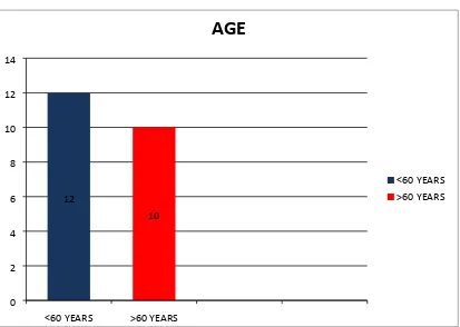
I. AGE DISTRIBUTION
TABLE - 1
S.NO AGE NO PERCENTAGE
1 <60 12 54.54
2 >60 10 45.45
12
10
<60 YEARS >60 YEARS 0
2 4 6 8 10 12 14
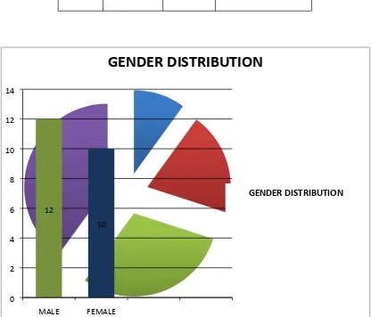
[image:59.595.103.516.356.650.2]II. GENDER DISTRIBUTION
TABLE - 2
S.NO SEX NO PERCENTAGE
1 MALE 12 54.54
2 FEMALE 10 45.45
12
10
MALE FEMALE 0
2 4 6 8 10 12 14
Column2
GENDERION DISTRIBUTION
GENDER DISTRIBUTION
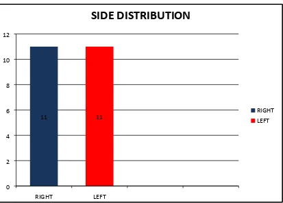
[image:60.595.101.517.312.668.2]III. SIDE DISTRIBUTION
TABLE - 3
SL.NO SIDE NO OF
CASES
PERCENTAGE
1 RIGHT 11 50
2 LEFT 11 50
11 11
RIGHT LEFT 0
2 4 6 8 10 12
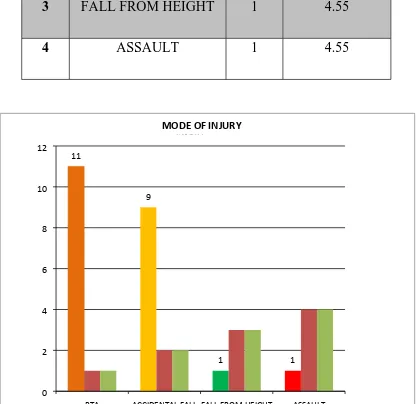
[image:61.595.103.516.391.687.2]IV. MODE OF INJURY
TABLE - 4
S.NO MODE OF INJURY NO PERCENTAGE
1 RTA 11 50
2 ACCIDENTAL FALL 9 40.9
3 FALL FROM HEIGHT 1 4.55
4 ASSAULT 1 4.55
11
9
1 1
RTA ACCIDENTAL FALL FALL FROM HEIGHT ASSAULT
0 2 4 6 8 10 12
MODE OF INJURY
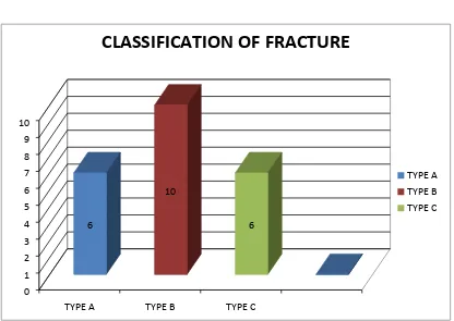
[image:62.595.102.518.327.731.2]V. CLASSIFICATION OF FRACTURE
TABLE - 5
S.NO TYPE NO PERCENTAGE
1 A 6 27.27
2 B 10 45.45
3 C 6 27.27
6
10
6
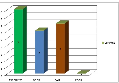
VI. CONSTANT MURLEY SCORE DISTRIBUTION
TABLE - 6
SL.NO OUTCOME NO OF CASES PERCENTAGE
1 EXCELLENT 9 40.9
2 GOOD 6 27.27
3 FAIR 7 31.82
4 POOR 0 0
9
6
7
0
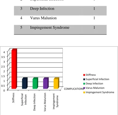
[image:64.595.104.517.439.728.2]VII. COMPLICATIONS
TABLE - 7
S.NO COMPLICATIONS NOS
1 Stiffness 4
2 Superficial Infection 1
3 Deep Infection 1
4 Varus Malunion 1
5 Impingement Syndrome 1
[image:65.595.102.509.272.667.2]Management of Complications
Four patients had shoulder stiffness which was managed through
vigorous physiotherapy and rehabilitation protocols. One patient had
Superficial Infection and found to be a stitch abscess on the 12th
postoperative period. It was managed with suture removal and with IV
Antibiotics. One patient presented with deep infection at second
postoperative follow up. It was managed with wound wash and
antibiotics.
One patient had Varus malunion. It was evident only radiologically
and the patient had no functional impairment. One patient had
Impingement Syndrome, the patient had pain over 120o degrees of
abduction. The patient is on follow up and we have planned for removal
of the plate after union. None of the patients had nonunion and all cases
PROCEDURE AND POST OPERATIVE PROTOCOL
All the patients were received in the casualty department and were
resuscitated. Any associated major injuries were treated accordingly at
first and after the general condition of the patient improved radiographs
were taken. The fractures were reduced and “U” slab immobilization was
given. The cases that met the inclusion criteria mentioned in our study
were operated. Cases were taken for elective fixation after doing all
investigation work up and after obtaining anaesthesia fitness and were
operated within 1 week of injury.
Preoperative Preparation
Informed Consent
Injured shoulder, axilla were prepared
Injection Cefotaxime 1 gram given intravenously 1 hour
before surgery after proper administration of test dose
Injection Xylocaine test dose was given.
Choice of Anaesthesia
Out of 22 cases, 14 cases were operated under Regional
Anaesthesia (Interscalene / Supraclavicular block) and remaining 8 cases
Positioning of the patient
All patients were positioned supine on the operating table with a
sand bag placed in the Interscapular Region.2It helps to push the affected
side forward and to open up the front of the joint. Head end of the
operating table is elevated 30o to 45o to reduce bleeding. It also allows the
blood to drain away from the operative field.
Surgical Approach
All twenty two patients were operated with standard Deltopectoral
approach.
Surgical Procedure
At first, the Deltopectoral groove was located and the incision was
made from the coracoid process proximally and extended distally along
the deltopectoral groove to the deltoid insertion for approximately
15cm.32 In obese patients, deltopectoral groove can be located by
abducting and externally rotating the shoulder. Deep fascia over the
cephalic vein was opened and the vein lies between the internervous
plane of Deltoid and Pectoralis major. The interval was developed, the
deltoid was retracted laterally along with cephalic vein and the Pectoralis
major was retracted medially. Any damage and ligation of the cephalic
Since most of the feeder vessels enter from the deltoid side, the
cephalic vein is retracted along with the deltoid muscle, but can also be
retracted medially when this facilitates fracture visualization and
reduction. The leading edge of the coracoacromial ligament may be
resected in order to have superior visualization and to mobilize the
fracture fragments. The tendon of pectoralis major may be released from
its attachment to decrease its deforming force on the proximal shaft of
humerus, which may complicate reduction of fragments.
Wide exposure is possible by transecting the muscle orginating
from the coracoid process. Sometimes it may be necessary to transect the
origin of the pectoralis minor muscle. Usually the insertion of the anterior
fibres of the deltoid is erased subperiosteally which gives a better
exposure of the operative field. The anterior circumflex vessels lie in the
middle of the wound, just superior to the pectoralis major muscle.
Adequate care was taken to avoid damaging the vessel.
Biceps tendon was used as a landmark between the two
tuberosities. The capsule of the shoulder joint was never opened. The
reduction of articular surface was indirect using the image intensifier.1,12
Soft tissue attachments to the fracture fragments were carefully preserved
to prevent devascularisation of the fracture fragments. Kirshner wires (K
Absorbable suture materials (Ethibond 5) were used to restore the greater
tuberosity anatomy. Plate was placed at least 5-8mm inferior to upper end
of greater tuberosity to avoid subaromial impingement and 2-4mm lateral
to the bicipital groove, ensuring that a sufficient gap was maintained
between the plate and biceps tendon.
After reduction of fracture fragments, reduction was held with
Kirschner Wires, plate is placed and holes are drilled in the humeral head
using the drill sleeve. Screws are applied in different directions in the
head for better stability and holding of the head fragment. The Calcar
screw is then applied. Then the shaft screws were applied. 3.5 mm
cortical non-locking screws were applied first in the shaft for better
approximation of the plate to the bone. Finally, screw positions and
1. Draping of the patient
2. Skin incision and retraction of cephalic vein
4. Positioning of the Plate
5. Reduction checked with image intensifier
POST OPERATIVE PROTOCOL
Following surgery, the operated arm was immobilized in a
shoulder sling. The drain tube was removed on the second postoperative
day. Suture removal was done on 12th postoperative day.
The factors which determine the timing of shoulder rehabilitation,
Stability of fixation
Bone Quality
Fracture pattern
Patient’s compliance
Postoperative rehabilitation is important to achieve a optimal
functional outcome. Adequate and stable fixation allows early
rehabilitation and functional recovery. Hughes and Neer devised a three
phased rehabilitation protocol.33The application of this protocol is
variable and it depends upon factors such as stability of fixation, fracture
pattern, and patient’s compliance towards rehabilitation. Elbow, wrist and
fingers active ROM exercises were started immediately after surgery.
PHASE 133
This phase of rehabilitation is started early in the postoperative
1. Elbow flexion and extension
2. Shoulder pendulum exercises
3. Supine external rotation with a stick
4. Assisted forward elevation and pulley exercises
PHASE 2
In this phase early active, resistive and stretching exercises are
started. The first exercise to start is supine active forward elevation. This
is done by 3 sets of 10-15 repetitions during each session. The patient is
then trained to place the hands behind the head to achieve abduction and
external rotation. This is followed by stretching for forward elevation.
PHASE 3
In this phase resistive strengthening exercises are started at three
months postoperative period. Arm is stretched higher on top of the wall.
Prone stretching for forward elevation is also started. Lifting light weight
objects is started after three months. Lifting weights is gradually
increased from one pound with one pound increments to a limit of 5
pounds.
Standard radiological evaluation done periodically (3,6 and 12
weeks post operatively). Then again at 6thand 12th month following
CASE ILLUSTRATIONS
CASE – 1
PRE OP IMMEDIATE POST OP
CASE - 2
PRE OP IMMEDIATE POST OP
CLINICAL PICTURES
CASE - 2
CASE - 3
PRE OP IMMEDIATE POST OP
1.5 YEARS FOLLOW UP
CLINICAL PICTURES
CASE - 4
PRE OP IMMEDIATE POST OP
DISCUSSION
Recent studies shows that the incidence of proximal humerus
fractures have increased to 7% of all fractures and 80% of all humerus
fractures.1Variable treatment options are available for treating proximal
humerus fractures. The treatment option depends upon many factors such
as fracture pattern, patient’s age, quality of the bone, patient’s functional
demand and associated comorbid conditions and patient’s general
wellbeing.
Many studies conducted in the past support nonoperative
management of undisplaced proximal humerus fractures. The indications
for nonoperative treatment patients with undisplaced or minimally
displaced fractures, valgus impacted fractures,17 patients not medically fit
for surgery and elderly patients with low functional demand. But
prospective studies conducted in the past reveals marked functional
impairment may occur in the setting of fracture treated non operatively
and this patients are reported to have chronic pain at the affected arm.
The outcome predictors which determine the results of treatment of
a proximal fractures are Age of the patient and AO/OTA classification of
fractures. Surgical treatment of proximal fractures are advised for
demand. The main aim of the surgical fixation is to achieve anatomical
reduction and stable fixation of the tuberosities, restore the rotator cuff
mechanism and to give a functional outcome which is near normal to the
preinjury status of the patient.
Open Reduction and Internal Fixation is the frequently used
method of surgical treatment. It allows direct visualization of the fracture
fragments and facilitates the direct reduction and aids in achieving
anatomical reduction. It also helps in proper positioning of the implant.
However dissection done during surgery may jeopardize the fracture
biologically and may interfere with fracture healing and increases the risk
of Avascular Necrosis of the humeral head.1 Therefore careful and
judiciary surgical dissection is advised during surgery.
Over the past five decades, fixation with compression plates and
screws has been the standard treatment modality. Satisfactory healing
rates and functional outcome after conventional plate and screw fixation
have been reported in several studies especially in young individuals. But
high rates of postoperative fracture displacement and varus collapse has
been reported with conventional compression plate and screw fixation.1
This is found especially in elder patients owing to the osteoporotic nature
Locking plates are precontoured and vary in terms of number of
proximal locking screws and their arrangement and also vary with ability
to place screws at different angles with regards to the plate.1,20Locking
plates allows angular stability between screws and plates. Constructs
using locking plates are biomechanically superior in strength and more
resilient than constructs using nonlocking plates/screws, blade plates and
Intramedullay nails. They help to prevent postoperative displacement and
varus collapse of the fractures.
Osterhoff et al in a recent study emphasized about the use of calcar
screws in the prevention of secondary loss of reduction. Calcar screws are
applied tangentially to the medial curvature of the surgical neck of
humerus. Previous studies report that use of calcar screws have
complications like axillary nerve damage, screw cut out and avascular
necrosis of humeral head especially when done percutaneously as in
minimally invasive technique.
Loss of reduction and varus malunion results in short lever arm of
rotator cuff and subacromial impingement because of decreased acromio
humeral distance. In this study, they concluded that the placement of
calcar screws prevents secondary loss of reduction by providing
In our study, we have analyzed twenty two cases of proximal
humerus fractures which were treated surgically using Proximal Humerus
Locking plates in Govt. Kilpauk Medical College Hospital.
Out of 22 cases, 12 cases were males and 10 cases were females. In
a study conducted by Hawkins and Bell involving 15 patients and in a
study conducted by Kristiansen et al involving 565 patients with proximal
humerus fractures shows female preponderance. This is attributed to
advanced osteoporosis in elderly women.
The average age of the patients in our study is 52 years and this
corresponds with reports of studies conducted by Hawkins, Bell and Gurr,
Cornell CN, Pagnani M J and Flatow et al. Also in our study we studied
the functional outcome of proximal humerus fractures based on patient’s
age.Patients who are less than 60 years and patients with more than 60
years age are divided into two groups and their outcome studied.
SEX
Frequency Percent Valid Percent Cumulative
Percent
M 12 54.5 54.5 54.5
F 10 45.5 45.5 100
In Recent studies it has been stated that in elderly patients aged
more than 65 years the incidence of proximal humerus fractures was
found to be more than 10% which ranks third next to proximal femur and
distal radius fractures respectively. It is also stated that the incidence of
fractures increases by 40 % every 5 years beyond the age of 40 for
females and 60 years for males.
In our study, we had statistical significance in the constant scores
between patients with age less than 60 and in patients with age more than
60 and this results were concurrent with study conducted by Agarwal
et al. The mean functional scores in younger age group is 86.75 and
mean value in elderly patients is 67.10 and the statistical significant value
is (p-0.00). This may be due to osteoporotic bone quality, initial fracture
pattern, associated comorbid conditions and patients poor compliance in
post operative physiotherapy and rehabilitation protocols. Hence proper
preoperative counseling should be given regarding the variable results
and high surgical caution should be excised.
Mann-Whitney Test
Ranks
Age N Mean Mean Rank p value
< 60 yrs 12 86.7500 16.50
> 60 yrs 10 67.1000 5.50 .000
SCORE
Total 22
In our study, the most common mechanism of injury is Road
Traffic Accidents followed by accidental fall sustained at ground level.
Other mode of injuries like direct injury caused by assault and fall from
height were also present in our studies. Unlikely to the high incidence of
proximal humerus fractures in the elderly patients we had 12 cases under
60 years owing to the nature of high energy trauma sustained by Road
Traffic Accidents.
INJURY
Frequency Percent Valid Percent
Cumulative Percent
ACC FALL 9 40.9 40.9 40.9
ASSAULT 1 4.5 4.5 45.5
FALL FROM
HEIGHT 1 4.5 4.5 50.0
RTA 11 50.0 50.0 100.0
Valid
In our study, the fractures are classified using AO/OTA
classification of proximal humerus fractures. There were six patients in
Type A fractures, 10 with Type B, 6 in Type C fractures. In our study, we
found that there is statistical significance between the mean functional
constant scores of the patients within Type A,B,C with a p value of 0.048.
CLASSIFICATION
Frequency Percent Valid Percent Cumulative Percent
A 6 27.3 27.3 27.3
B 10 45.5 45.5 72.7
C 6 27.3 27.3 100.0
Valid
Total 22 100.0 100.0
Kruskal-Wallis Test
Ranks CLASSIFICATION
RECODED
N Mean Mean
Rank
p value
A 6 85.1667 15.33
B 10 78.9000 12.30 .048
C 6 68.6667 6.33
SCORE