DISTRIBUTION, ANTIMICROBIAL SUSCEPTIBILITY PATTERN AND DETECTION OF BIOFILM FORMATION IN ENTEROCOCCUS SPECIES ISOLATED FROM VARIOUS CLINICAL SPECIMENS, WITH
PHENOTYPIC AND MOLECULAR CHARACTERISATION OF VANCOMYCIN RESISTANT ENTEROCOCCUS
Dissertation submitted for
M.D. MICROBIOLOGY BRANCH – IV DEGREE EXAMINATION
THE TAMILNADU DR.M.G.R. MEDICAL UNIVERSITY CHENNAI – 600 032
TAMILNADU
CERTIFICATE
This is to certify that this dissertation entitled “DISTRIBUTION, ANTIMICROBIAL SUSCEPTIBILITY PATTERN AND DETECTION OF
BIOFILM FORMATION IN ENTEROCOCCUS SPECIES ISOLATED FROM
VARIOUS CLINICAL SPECIMENS, WITH PHENOTYPIC AND MOLECULAR
CHARACTERISATION OF VANCOMYCIN RESISTANT ENTEROCOCCUS”
is the bonafide original work done by Dr.R.VENNILA, Post graduate in Microbiology, during the period of April 2016 to March 2017 under the guidance of Prof.Dr.UMADEVI.U.M.D., Professor of Microbiology, Institute of Microbiology, Madras Medical College and Rajiv Gandhi Government General Hospital, Chennai-600003, in partial fulfilment of the regulations of The Tamil Nadu Dr. M.G.R. Medical University for the award of M.D Degree in Microbiology (Branch IV) held in May 2018.
Dr.R.NARAYANA BABU, MD., DCH Dr. ROSY VENNILA., M.D.,
Dean, Director,
Madras Medical College & Institute of Microbiology, Rajiv Gandhi Government Madras Medical College &
General Hospital, Rajiv Gandhi Government
Chennai – 600003 General Hospital,
DECLARATION
I, Dr.R.Vennila, Post Graduate, Institute of Microbiology, Madras Medical college, solemnly declare that the dissertation titled “DISTRIBUTION, ANTIMICROBIAL SUSCEPTIBILITY PATTERN AND DETECTION OF
BIOFILM FORMATION IN ENTEROCOCCUS SPECIES ISOLATED FROM
VARIOUS CLINICAL SPECIMENS, WITH PHENOTYPIC AND MOLECULAR
CHARACTERISATION OF VANCOMYCIN RESISTANT ENTEROCOCCUS”
is the bonafide work done by me at the Institute of Microbiology, Madras Medical College, Chennai, under the guidance and supervision of Dr.UMADEVI.U.MD., Professor, Institute of Microbiology, Madras Medical College, Chennai-600 003. The dissertation is submitted to The Tamil Nadu Dr. M.G.R. Medical University, Chennai in partial fulfilment of the University regulations for the award of degree of M.D. Branch IV Microbiology examinations to be held in May 2018.
Place: Chennai,
Date: Dr. R.VENNILA
Signature of the Guide Prof. Dr.UMADEVI.U MD.,
Professor,
Institute of Microbiology,
ACKNOWLEDGEMENT
I wish to express my sincere thanks to the respected Dean Dr.R.NARAYANA BABU, MD, DCH, Madras Medical College & RGGGH,
Chennai for permitting me to use the resources of this Institution for my study.
I express my sincere thanks to Dr.ROSY VENNILA M.D., Director, Institute of Microbiology, Madras Medical College & RGGGH, Chennai.
I also express my thanks and gratitude to our former Professor Dr. MANGALA ADISESH M.D., for her guidance and support.
I feel indebted to be under the guidance of Dr.U.UMADEVI. M.D., Professor, Institute of Microbiology, for suggesting the topic for my dissertation and for her valuable advice, guidance in preparing and compilation of my work. She is a source of inspiration in my endeavors.
I extend my sincere thanks to our Professors Dr. S.THASNEEM BANU M.D., Dr.R.VANAJA M.D., Dr.C.P.RAMANI, for their support, guidance and valuable advices.
I express my sincere thanks to our Assistant Professors Dr. R.DEEPA M.D., Dr.N.RATHNAPRIYA M.D., Dr.K.USHAKRISHNAN M.D., Dr.K.G.VENKATESH M.D., Dr.N.LAKSHMIPRIYA M.D.,D.C.H., Dr.C.S.SRIPRIYA M.D., and Dr. DAVID AGATHA M.D., for their support in my study.
I would like to thank all my colleagues, my junior postgraduates and all staff of Institute of Microbiology, Madras Medical College for their support and cooperation.
I feel indebted to my parents who had been a solid pillars of everlasting support and encouragement and for their heartful blessings.
TABLE OF CONTENTS
Sl.
NO TITLE
PAGE NO.
1 INTRODUCTION 1
2 AIM AND OBJECTIVES 4
3 REVIEW OF LITERATURE 5
4 MATERIALS AND METHODS 44
5 RESULTS 65
6 DISCUSSION 82
7 SUMMARY 87
8 CONCLUSION 89
9 COLOUR PLATES
10 BIBLIOGRAPHY
APPENDIX-I ABBREVIATIONS
APPENDIX-II STAINS, REAGENTS AND MEDIA ANNEXURE I PROFORMA
ANNEXURE II CONSENT FORM ANNEXURE III INFORMATION SHEET ANNEXURE IV MASTER CHART
LIST OF TABLES
S.NO TITLE PAGE
NO
1. INTRINSIC AND ACQUIRED RESISTANCE OF ENTEROCOCCI 30 2. PHENOTYPIC AND GENOTYPIC CLASSIFICATION OF
ENTEROCOCCI 35
3 IDENTIFICATION OF ENTEROCOCCUS SPECIES 57 4 INTERPRETATION OF BIOFILMS BY MICROTITRE PLATE
METHOD 59
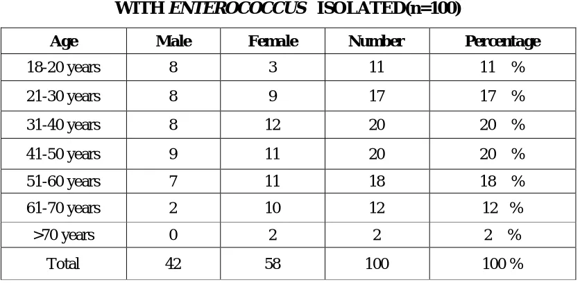
5 AGE AND GENDER DISTRIBUTION AMONG PATIENTS WITH
ENTEROCOCCUS ISOLATED 65
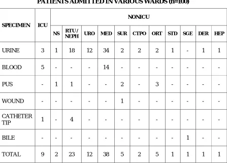
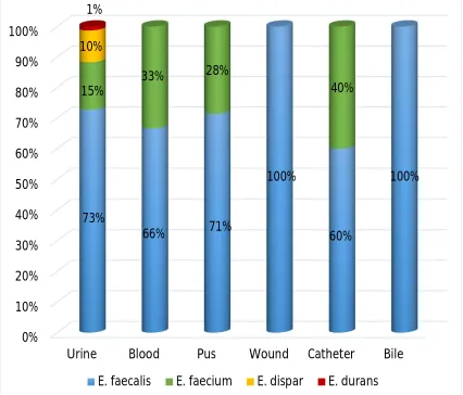
6 SAMPLE DISTRIBUTION AMONG ENTEROCOCCUS SPECIES 65 7 DISTRIBUTION OF ENTEROCOCCAL ISOLATES AMONG
PATIENTS ADMITTED IN VARIOUS WARDS 66
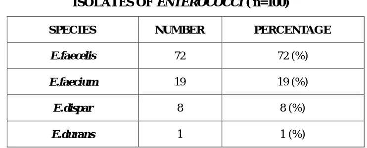
8 DISTRIBUTION OF SPECIES AMONG THE CLINICAL ISOLATES
OF ENTEROCOCCI 67
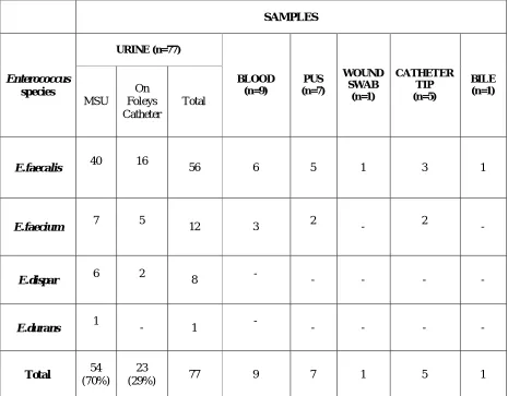
9 DISTRIBUTION OF ENTEROCOCCUS SPECIES AMONG VARIOUS
CLINICAL SPECIMENS 68
10 CLINICAL DIAGNOSIS IN CULTURE POSITIVE ENTEROCOCCAL
INFECTIONS 70
11 ASSOCIATION BETWEEN ENTEROCOCCUS INFECTION AND
RISK FACTORS 71
12 ANTIBIOTIC SUSCEPTIBILITY PATTERN: BY KIRBY BAUER
DISC DIFFUSION METHOD: FOR URINE SAMPLES 72
13
ANTIBIOTIC SUSCEPTIBILITY PATTERN: BY KIRBY BAUER DISC DIFFUSION METHOD: FOR SAMPLES BLOOD, CATHETER TIP, PUS, WOUND SWAB, BILE
74
14 HIGH LEVEL AMINOGLYCOSIDE RESISTANCE (HLAR) AMONG
ENTEROCOCCUS ISOLATES 75
15 HLG AND HLS RESISTANCE AMONG ENTEROCOCCUS SPECIES 76
16 COMPARISON OF VANCOMYCIN SCREEN AGAR AND
MICROBROTH DILUTION TEST 77
17 PROFILE OF VANCOMYCIN RESISTANCE ENTEROCOCCUS 77 18 BIOFILM FORMATION AMONG ENTEROCOCCUS ISOLATES 79
19
DISTRIBUTION OF BIOFILM PRODUCING ENTEROCOCCI IN PATIENTS WITH DEVICE ASSOCIATED INFECTIONS AND OTHER INFECTIONS
79
LIST OF CHARTS
S.NO CHARTS PAGE
NO
1 SPECIES OF ENTEROCOCCI ISOLATED FROM CLINICAL
SAMPLES 67
2 DISTRIBUTION OF ENTEROCOCCUS SPECIES AMONG
VARIOUS CLINICAL SPECIMENS 69
3 ANTIBIOTIC RESISTANCE PATTERN: BY KIRBY BAUER
DISC DIFFUSION METHOD: FOR URINE SAMPLES 73
4 PROFILE OF VANCOMYCIN RESISTANCE
ENTEROCOCCUS 78
CERTIFICATE – II
This is to certify that this dissertation work titled “DISTRIBUTION, ANTIMICROBIAL SUSCEPTIBILITY PATTERN AND DETECTION OF
BIOFILM FORMATION IN ENTEROCOCCUS SPECIES ISOLATED FROM
VARIOUS CLINICAL SPECIMENS, WITH PHENOTYPIC AND MOLECULAR
CHARACTERISATION OF VANCOMYCIN RESISTANT ENTEROCOCCUS”
of the candidate DR.R.VENNILA with registration Number 201514009 for the award of M.D. in the branch of MICROBIOLOGY. I personally verified the urkund.com website for the purpose of plagiarism Check. I found that the uploaded thesis file contains from introduction to conclusion pages and result shows 8 percentage of plagiarism in the dissertation.
1
INTRODUCTION
Enterococci are commensal microorganisms that act as potential human pathogens, causing a variety of infections in humans. Over the last decades, they have been emerged from long and being considered virtually harmless bacteria to medically important multiple–antibiotic resistant nosocomial pathogens that contribute significantly to patient morbidity and mortality, as well as health care costs.1 Enterococcus species have been recognized as a nosocomial pathogens causing diseases like bacteremia, endocarditis, complicated urinary tract infections, intraabdominal infections, pelvic infections, wound and soft tissue infections, neonatal sepsis and rarely meningitis.2 Enterococcal bacteremia is frequently associated with metastatic abcesses in multiple organs and high mortality rates.
In critically ill patients infections caused by antibiotic resistant
2
genes/ virulence factors.4 Drug of choice for aminoglycoside resistant
Enterococci is vancomycin.5
Vancomycin Resistant Enterococci (VRE) pose an emerging problem in hospitals worldwide because of widespread use of vancomycin and extended spectrum cephalosporins. Vancomycin resistant Enterococcus infections are serious because the last option for treatment of life threatening infections are glycopeptides, therefore it leads to increase in mortality rate. Enterococcus faecalis and Enterococcus faecium account for majority of vancomycin resistant Enterococcus infections. The Vancomycin resistance of Enterococcus is mediated by a group of genes like van A, van B, van C, van D and van E. These genes are transcribed in presence of vancomycin & low affinity cell wall precursors to vancomycin are synthesized.4 Horizontal gene transfer is mainly responsible for transfer of virulence genes & spread of resistance.4
3
produce biofilm are highly resistant to antibiotics than non-biofilm producers.2
4
AIM & OBJECTIVES
AIM :
To study the prevalence of vancomycin resistance among the Enterococcal
species isolated from various clinical samples such as urine, blood, pus, tissue fluids and feces obtained from the patients of a tertiary care hospital.
OBJECTIVES:
To isolate and characterize phenotypically Enterococcus species from various clinical specimens.
To study antimicrobial susceptibility pattern of Enterococci by Kirby Bauer Disk diffusion method.
To study about the detection of biofilm formation in Enterococcus species by Microtitre plate method.
To determine Minimum inhibitory concentration of vancomycin by Microbroth dilution method.
Phenotypic and genotypic characterization of vancomycin resistant
5
REVIEW OF LITERATURE
HISTORICAL PERSPECTIVES
Theircelin in 1899 used the term "enterocoque" in a french publication to describe a bacteria seen in pairs and short chains in human feces. This was later included in new genus Enterococcus proteiforms.3 Later MacCallum and Hastings in the same year had described a fatal case of endocarditis from the John hopkins hospital caused by a bacterium that was very hard and tenacious of life which they termed as "Micrococcus zymogenes" was later called as S.faecalis var zymogenes.3 They confirmed the pathogenicity of new organism by satisfying koch's postulates. Andrews and Horder first coined the name Streptococcus faecalis in 1906, for an isolate recovered from the blood of a patient with endocarditis, which is so characteristic with the organism identified in human intestine.8 Later in 1918, Orla-Jensen described Streptococcus faecium, a second organism of this group.8 In 1935, Sherman and Wing, proposed a third species
6
The members of the Genus Enterococcus are gram positive cocci that occur singly or arranged in pairs and short chains, catalase negative (except E.haemoperoxidus),few strains produce weak effervescence when grown on blood agar. They are nonmotile (except E.gallinarum and E.casseliflavus) and noncapsulated.9 Enterococci are facultative anaerobes that follows Embden Meyerhof pathway where glucose is fermented and lactic acid is produced as an end product. Enterococci are referred as typical lactic acid bacteria.10Enterococci are anaerogenic, grow at temperatures between 10 to 45°c, grow in 6.5%Nacl, grow at PH 9.6, survive at 60°c for 30 minutes and hydrolyse esculin in the presence of bile salts.5 Many of these characteristics are used to distinguish
Enterococci from nonenterococcal streptococci. In 1984 Schleifer and Kilpper-Balz provided genetic evidence used DNA-DNA and DNA-rRNA hybridization studies to prove that E.faecalis and E.faecium were different from other members of genus streptococcus and suggested to provide a separate genus8. The G+C content of DNA ranges from 32-44mol% and genomic size were 2000-3500 kb.8
Enterococci are usually alpha hemolytic or non hemolytic that show inability to lyse bovine RBCs which are commonly used in agar plates, however some E.faecalis do lyse RBCs (beta hemolysis) from human, horses and rabbits.3 On blood agar after 24 hours growth, they produce white to grey coloured colonies,1-2mm diameter and on Macconkey agar 0.5-1mm majenta pink coloured colonies are produced. Most of the species hydrolyze L-pyrollidonyl- beta-naphthylamide by producing pyrollidonyl arylamidase (PYRase)(except for
7
hydrolyze leucine β naphthylamide by producing leucine aminopeptidase
(LAPase).3 Enterococci do not synthesis porphyrin and so do not express cytochrome enzymes.11 Cytochrome oxidase activity is expressed when E.faecalis
are grown in blood containing media.
Some species produce yellowish pigment E.mundii, E.casseliflavus, E.sulfureus , E.pallens & E.gilvus. Around 37 species have been identified from clinical isolates, the predominant is E.faecalis (80-90%) & E.faecium (5-10%).Other species like E.gallinarum, E.durans, E.hirae, E.avium, E.dispar,
E.malodoratusare less frequently identified.3 The Enterococcus reacted with group D antisera, while the pyogenic streptococci reacted with group A,B,C,E,F,or G and the viridans streptococci were nongroupable. A cell wall associated glycerol teichoic acid antigen called Lancefield's group D antigen is produced by most strains.3Enterococci were classified as group D streptococci on the basis of their colony morphology and reaction with group specific antisera.
HABITAT
Enterococci are found in the faeces of most healthy adults. They are numerous in the large intestine where concentrations of 105 to 107 bacteria per gram. Due to several intrinsic characteristics they grow and survive in harsh environments. Enterococci is widespread in soil, plants, water, food and animals including mammals, birds, insects and reptiles. In humans, major habitat of
8
term "fecal Enterococci" includes four species: Enterococcus faecalis,
9
EPIDEMIOLOGY
A nosocomial infection is one for which there is no evidence that the infection was present or incubating at the time of hospital admission & it is one of the major health problem globally. Enterococci are among the leading causes of hospital acquired infections in the United States (second to third), and it is estimated that in 2004, 521,285 hospital discharges were associated with Enterococcal infections.13 The emergence of drug resistance among many bacteria has raised since hospitals serve to be the ideal ground for the development and spread of several multi-drug resistant bacteria. Over the past decade there is a shift in prevalence from gram-negative to gram-positive species as predominant cause of nosocomial infections, among which Enterococci has become one of the top three pathogens causing various infections like urinary tract infections (UTI), blood stream infections (BSI),skin and soft tissue infections (SSTI) and other miscellaneous infections.
The predominant species encountered in nosocomial infections is
E.faecalis, however in the past few decades E.faecium is on the rise. Now-a-days
E.faecium is isolated as common as E.faecalis in hospital associated infections. About 30% of Enterococcal infections are caused by VRE, among which
10
occupied by a patient colonized with VRE, invasive procedures and administration of broad-spectrum antibiotics (e.g., cephalosporins), or vancomycin.6,4 Several epidemiological studies conducted in human subjects from community showed Enterococci are resistant to various antimicrobials like ampicillin, gentamicin and vancomycin. Most of the studies have concluded that previous hospitalization and prior use of vancomycin are common factors for community dissemination of vancomycin resistant Enterococcus (VRE).
VIRULENCE FACTORS
The virulence of Enterococci is regulated by virulence coding genes present in the pathogenic islands (PAI) of the genome. The PAI of Enterococcus
was first identified in the genome of multidrug resistant strains of E.faecalis that caused nosocomial infections in 1980s.15 The ability of the organism to acquire new traits and colonize new areas enhance virulence.
HEMOLYSIN
11
GELATINASE & SERINE PROTEASE
Proteases produced by Enterococci have the capacity to hydrolyze gelatin, collagen, casein and other peptides. E.faecalis protease Gel E also modifies the critical components of immune system. The expression of gelatinase is regulated by frc locus in E.faecalis. The expression of gelatinase and serine protease genes is regulated by fsr system, which is a two component quorum-sensing regulatory system that regulates the expression of multiple genes in E. faecalis and is similar to the agr system of Staphylococcus aureus. 17,18. Mutants lacking the genes corresponding to this protein are highly attenuated in experimental endocarditis, peritonitis and endophthalmitis.
ENTEROCOCCAL SURFACE PROTEIN(ESP)
Cell wall-associated protein of E. faecalis called Enterococcal surface protein- ESP acts as an adhesin like aggregation substance. These adhesins would contribute as bacterial extracellular matrix molecules in humans. ESP variants, were reported in E. faecium clinical isolates. ESP contributes to colonization and persistence of E.faecalis in the urinary bladder wall through specific components such as mucin or uroplakin. Later studies have shown that ESP was not required, but its presence was associated with higher amounts of biofilm .8
AGGREGATION SUBSTANCE(AS)
12
substance contributes to the pathogenesis of Enterococcal infection by exhibiting resistance to killing by polymorphonuclear leukocytes and macrophages, thereby promoting intracellular survival inside macrophages. AS also contributes to adherence of Enterococcal isolates to host tissues.19
MSCRAMM Ace
The surface proteins – Ace (adhesion of collagen of E.faecalis) and Acm (homologue adhesion of E.faecium) are microbial surface components recognizing adhesive matrix molecules (MSCRAMM) structurally and functionally related to staphylococcal CNA adhesion factor.20 They are involved in attachment of bacteria to host proteins like collagen, fibrinogen and fibronectin. Similar to MSCRAMM other surface proteins playing role in virulence, are second collagen adhesion of E.faecium (Scm), Enterococcal surface protein (Espfc) of E.faecalis and of E.faecium (Espfm), surface proteins (Fms) of
E.faecium, SgrA (which binds to basal lamina components), EcbA (binds to collagen type V).3
CAPSULAR POLYSACCHARIDE
Capsular polysaccharides (on bacterial cell surface) contribute to the pathogenicity by interfering with phagocytosis by host immune cells. An operon encodes the synthesis of capsular polysaccharide in most clinical isolates of
13
LIPOTEICHOIC ACID
Lipoteichoic acid constitute the group D antigen of Enterococci, functions in modulating immune response by inducing the production of TNF and Interferons.
COCCOLYSIN
Coccolysin, an extracellular metallo-endopeptidase secreted by some
E.faealis strains mediate virulence by inactivating endothelin, the vasoactive peptide .
EXTRACELLULAR SUPEROXIDE
Extracellular superoxide, secreted in large amounts by most of E.faecalis and E.faecium strains enhances in vivo survival in mixed infection with
Bacteroids fragilis in a subcutaneous infections.21E.faecalis isolates from blood are unique to produce superoxide.
HYARULANIDASE
14
PHREMONES
They are the small peptides secreted by E.faecalis. It promotes conjugative transfer of plasmid DNA between strains. They are chromosomally encoded & elicit a specific mating response from plasmid carrying donor cells. A few phremones act as chemoattractant for neutrophils.
OTHER VIRULENCE FACTORS
AS-48 is a plasmid coded peptide produced by E.faecalis that inhibits both Gram positive and Gram negative bacteria. It causes lysis by pore generation in cytoplasmic membrane of target cells leading to depolarization and it induces lysis of some Enterococcus by activating autolysin.
Pili is present in both E.faecalis and E.faecium which mediates attachment and invasion into host tissues.
E.faecalis stress protein Gls24 responsible for Enterococcal resistance to bile salts is important in pathogenicity of endocarditis .
hyl Efm- containing plasmids found in E.faecium, increase the colonizing capacity.3
PATHOGENESIS
15
important factor that increases gastro intestinal colonization of Enterococci
(eg.VRE) is the administration of antimicrobial agents that are excreted in bile or having anti anaerobic activity without disturbing Enterococci (eg, various cephalosporins).21 Administration of broad-spectrum antibiotics favours down regulation of the intestinal expression of RegIIIγ (a bactericidal lectin produced by intestinal epithelial cells and Paneth cells), which is activity against Gram positive intestinal organisms thereby favours VRE colonization of the gastrointestinal tract . VRE colonization is also influenced by some gut anaerobic microbiota (Barnesiella species in mice) and increased stomach pH. Once
16
CLINICAL SYNDROMES
1. URINARY TRACT INFECTIONS
Enterococci are well known causative agents of nosocomial UTIs, associated with anatomic abnormalities of genitourinary tract, instrumentation, indwelling catheters, prior antibiotic use and recurrent UTIs. The centres for disease control (CDC) National Nosocomial surveillance study (NNSS) states
Enterococci being the third most common cause of catheter associated UTIs. Among them E.faecium being the predominant species (40%) followed by
E.faecalis (25%) and other species (35%). It is very difficult in the hospital setting to differentiate infection from colonization. The factors that help to differentiate are presence of leucocytes in urine along with fever, local symptoms and signs, systemic manifestations and a colony count of > 105CFU/ml.3,8 Removal of catheter itself helps to eradicate the organism. Enterococcus can cause complicated UTIs also which lead to complications like are pyelnephritis, perinephric abcess and recurrent bacteremic episodes.3,8
RISK FACTORS FOR ENTEROCOCCAL UTIs 1. Indwelling catheterization
2. Instrumentation
3. Anatomic abnormalities of genitourinary tract 4. Elderly patients
17
2. INTRA ABDOMINAL AND PELVIC INFECTIONS
Enterococci being the second most common isolate from abdominal pelvic
infections along with Gram negative and anaerobic organisms. Enterococci
produce spontaneous peritonitis and empyema in patients with cirrhosis, acute renal failure and continuous ambulatory peritonitis.9 Presence of Enterococcus in intraabdominal and pelvic infections indicates treatment failure, increases postoperative complications and mortality.22 Because of emergence of vancomycin resistant Enterococcus and increase in the multidrug resistant
E.faecium post this organism as third common agents of nosocomial surgical site infection.3
3. BLOOD STREAM INFECTIONS
18
4. ENDOCARDITIS
Enterococci are second or third most common cause of endocarditis after
Staphylococcus and Streptococcus accounting for 5-20% of endocarditis.
E.faecalis is more common , it affects both native and prosthetic valves and both community and nosocomial associated endocarditis. Sources of infection are genitourinary and gastrointestinal tract, procedures like cystoscopy, trans rectal prostatic biopsy, liver biopsy, surgeries like caesarean section & prostatectomy. Common complications are heart failure and embolisation (27-43%) and mortality ranges from 11- 35%.25
5. MENINGITIS
19
6. NEONATAL INFECTIONS
Enterococci are part of the normal adult vaginal flora and infections can be acquired by neonates during delivery. Enterococcus accounts for approximately 6% of late-onset sepsis, 5% of pneumonias, 9% of surgical site infections, 10% of blood stream infections, and 17% of UTIs in neonatal units.28 Risk factors are prolonged hospital stay, low birthweight, prematurity, prior antibiotic therapy, endovascular devices, nasogastric tubes, and several invasive procedures.3 Outbreaks of VRE causing sepsis in neonates have also been well documented in different parts of the world.
7. SKIN AND SOFT TISSUE AND OTHER INFECTIONS
Decubitus and diabetic foot ulcers are the common lesions associated with
Enterococcus and in some cases, the organisms have been associated with osteomyelitis.29 In diabetic wound infection there is transfer of the van A gene cluster (encoding proteins necessary for vancomycin resistance) from vancomycin -resistant E. faecalis to Staphylococcus aureus.30 Enterococci are rare causes of soft tissue abscesses such as liver, lung, brain abscesses.31 and breast abscess caused by E.faecium in a patient hospitalized in the critical care unit.
SPECIES OF ENTEROCOCCI
20
from sugars like arabinose, sorbitol, raffinose, sucrose, pyruvate, trehalose and tellurite reduction (0.04%), motility and pigment production.
CLASSIFICATION OF ENTEROCOCCI Group I – consists of 9 species
E.avium,E.raffinosus,E.gilvus,E.pallens,E.saccharolyticus,E.malodoratus,
E.pseudoavium, E.divriesei and E.hawaiiensis.
They produce acid from mannitol and sorbose, arginine is not hydrolysed.
Group II – consists of 8species
E.faecalis, E..faecium, E.gallinarum, E.casseliflavus, E.mundtii,
E.haemoperoxidus, E.sanguinicola, E.ythailandicus.
They produce acid from mannitol and not from sorbose and arginine is hydrolyzed. Majority of the them recovered from human sources.
Group III –consists of 6 species
E.dispar, E.canintestini, E.hirae, E.durans, E.ratti ,and E.villorum. They don’t produce acid from mannitol and sorbose, but hydrolyse arginine.
Group IV – includes 8 species
E.caccae, E.cecorum, E.aquimarinus, E.phoeniculicola, E.sulfureus, E.asini,
E.silesiacus, E.termitis.
21
Group V – consists of 6 species
E.canis, E.columbae, E.moraviensis, E.camelliae, E.hermanniensis, and E.italicus
They ferment only mannitol producing acid but not sorbose and arginine also not hydrolysed.
BIOFILM PRODUCTION BY ENTEROCOCCUS
Biofilm is a population of cells attached irreversibly on various biotic and abiotic surfaces by an extracellular matrix substances, proteins, polysaccharides and nucleic acids. It is regulated by quorum sensing systems. They are notoriously difficult to eradicate and are the source of many chronic infections. Bacteria in biofilms colonize a wide variety of medical devices like catheters, artificial pacemakers, prosthetic devices. Enterococci in biofilms are more highly resistant to antibiotics than planktonically growing Enterococci, thus the potential impact of biofilm formation could be significant.2
EPIDEMIOLOGY OF BIOFILM FORMATION
The prevalence of biofilm production varies worldwide. In most of the studies conducted E.faecalis was more common among biofilm producers than
E.faecium.
FACTORS INFLUENCING BIOFILM FORMATION
22
Other factors are, 1. Esp
2. Gelatinase
3. Multiple genes like fsr locus, gelE, epa, atn..
METHODS TO DETECT BIOFILM FORMATION 1. Tube method
2. Tissue culture plate method 3. Congo red method
TUBE METHOD
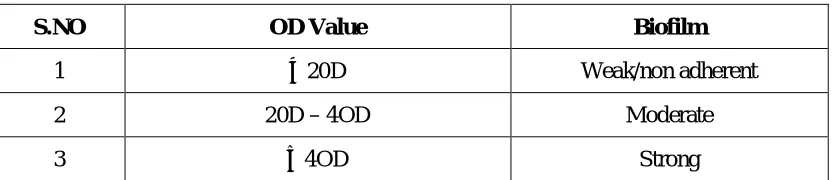
Trypticase soy broth with 2% sucrose was inoculated with loopful of microorganisms from overnight culture and incubated for 24 hours at 37oc. The tubes were then decaned and washed thrice with PBS pH 7.2 to remove any nonadherent cells. The tubes were air dried and stained with 0.1% crystal violet for 30 minutes. Excess stains were washed off with deionized water. The tubes were dried in inverted position and observed for biofilm formation. Biofilm formation was considered positive when a visible film lines the sides and bottom of each tube. Tubes were then examined and the amount of biofilm was scored as 0-absent,1-weak, 2-moderate, 3-strong. Ring formation at the liquid interface was not indicative of biofilm formation.
CONGO RED AGAR METHOD
23
(50g/land 0.8g/l) were added to Brain Heart Infusion agar. Enterococcal strains were inoculated on the congo red agar plates and incubated at 37oc for 24 hours. A positive result was indicated by black crystalline morphology. Isolates producing black colonies were considered as strong biofilm producers. Weak biofilm producers produced dark pink colonies. Non–slime producers mostly turned out as dry red colonies.
LABORATORY DIAGNOSIS
COLLECTION, TRANSPORT AND STORAGE OF SPECIMENS The standard methods of collecting blood, urine, wound swab, pus, bile, catheter tip samples, and other secretions or swab specimens are adequate.
It can be transported on any transport media or on swabs that are kept dry. The clinical samples, should be cultured as early as possible, preferably within 1 hour.
DIRECT EXAMINATION
24
As vancomycin -resistant Enterococcus (VRE) pose an important problem worldwide, hospitals should implement surveillance programs for VRE detection. In order to overcome these inherent limitations of culture-based methods of detection, conventional PCR and real-time PCR-based methods have been evaluated for direct detection in clinical and surveillance specimens. The Light Cycler vanA/vanB detection assay for screening of VRE in rectal or perianal swabs and commercially available DNA probe for the direct detection of Enterococcus in blood cultures.33
ISOLATION
Clinical specimens, can be plated onto trypticase soy agar, brain heart infusion agar or any blood agar base containing either 5% sheep, horse or rabbit blood for primary isolation of Enterococci . Samples for blood culture are inoculated into conventional blood culture systems. Most of the clinically relevant species grow well at 35o to 37ºC and do not require increased level of CO2. For specimens obtained from nonsterile sites especially when contaminated with Gram negative bacilli, selective media containing sodium azide, bile salts, antibiotics and esculin, tetrazolium can be used. Recently media with chromogenic substrates are used for isolation of Enterococci. However not all
Enterococci grow on selective media. Use of enrichment broth (Enterococcosel broth- BEA medium with 6μg vancomycin) increases the recovery rate of
25
Identification of Enterococcus species
The genus identification of Gram positive cocci, catalase negative as “Enterococcus” is based on the above said tests in genus description. Enterococal
species can be identified based on acid production from mannitol and sorbose and hydrolysis of arginine as mentioned above. Further speciation is based on acid production from sugars like arabinose, sorbitol, raffinose, sucrose, pyruvate, trehalose and reduction of 0.04% tellurite, motility and pigment production.
CULTURAL CHARACTERISTICS AND MORPHOLOGY
Enterococci are more tolerant to adverse physical and chemical conditions. Growth occurs at temperatures from 10-45oC and survive heating at 60oC for 30 minutes. Growth in 6.5% Nacl is an important characteristic feature to seperate
Enterococci from group D Streptococci which is also Bile Esculin positive.
Enterococci grow on routine nutrient agar, blood agar and MacConkey agar producing small (1-2mm), circular, translucent, convex colonies with smooth surfaces and entire edges. The ability to grow on bile salt agar medium distinguishes from most Streptococci except S.agalactiae. On MacConkey agar medium deep pink (magenta colour) colonies are formed due to lactose fermentation. On CHROM agar, they grow as blue coloured isolated pin point colonies. Enterococci may produce α or β-hemolysis on agar containing cow, rabbit, horse, or human blood but non-hemolytic on agar containing sheep blood as sheep RBCs are refractory to cytolysin mediated cell lysis. Some strains of
26
are α-hemolytic or non-hemolytic. Strains that appear to produce α-hemolytic are actually non-hemolytic strains producing peroxide. The ‘greening’ of the agar is due to peroxide action on the blood cells and not due to an α toxin.34 E. casseliflavus, E. gilvus, E. mundtii, E.pallens, and E. sulfureus produce yellow pigment on the blood-agar medium. The pigment can be detected by picking up the growth on the white cotton swab and examining the swab for a yellow colour. Chocolate agar, colistin nalidixic acid agar, 5% trypticase soy agar, 5% Brain heart infusion agar would also support growth of Enterococci .
SELECTIVE MEDIA FOR ENTEROCOCCI 1. Bile–esculin–azide agar
2. Enterococcosel agar /broth (white and surrounded by a black halo) 3. Agar containing tetrazolium salts (centre of the colony is brick-red) 4. Pfizer selective Enterococcus agar
5. Cephalexin - aztreonam- arabinose agar. 6. Oxoline esculin agar (OOA).34
Identification of Enterococcus species by commercial methods
There are several commercially available manual, semi-automated, and automated systems for the identification of Enterococcus species. These systems are reliable for the identification of E.faecalis, and to a lesser extent, E.faecium.
27
and the Gram-Positive Identification panel of the Micro Scan Walk/Away system. Approximately 80 percent of all Enterococcal isolates will be accurately identified by any one of these systems except unusual species, should be confirmed by standard reference method.
TYPING METHODS
The increasing documentation of Enterococcus as a leading nosocomial pathogen exhibiting resistance to many antibiotics together with the concept of exogenous acquisition of Enterococcus infections generated demand for strain and epidemiological studies.
CLASSIC PHENOTYPIC METHODS 1. Bio typing
2. Antibiotyping 3. Serotyping
4. Bacteriocin typing and 5. Bacteriophage typing
28
Molecular methods
Based on DNA–DNA hybridization and sequencing of the 16SrRNA genes.
They include:
1. Analysis of whole cell proteins(WCP) profiles by sodium dodecyl sulfate-polyacrylamide gel electrophoresis (SDS-PAGE)
2. Vibrational spectroscopic analysis
3. Proton magnetic resonance spectroscopic analysis 4. PCR based typing
a. Randomly amplified polymorphic DNA (RAPD) analysis b. Repetitive element sequence (REP) - PCR assay
5. Analysis of chromosomal restriction profiles by pulsed field gel electrophoresis (PFGE)
6. Field inversion gel electrophoresis (FIGE)/Counter clamped homogenous electric field electrophoresis (CHEF)
7. Multi locus enzyme electrophoresis. 8. Ribotyping
9. Sequencing analysis of the 16S rRNA gene
29
FISH (Fluorescent In Situ Hybridization) techniques (PNA FISH) been evaluated for identification of Enterococci from positive blood cultures. Analysis of chromosomal restriction profiles by pulsed field gel electrophoresis (PFGE) has been the gold standard method for strain typing and epidemiological outbreaks. It is also useful for the identification of predominant clonal complexes and resistance genes - HLAR high level amino glycosides resistance and VRE- vancomycin resistance. Among the two recent methods MLST- Multilocus sequence typing and MLVA multilocus variable number tandem repeat analysis, MLVA is less expensive and rapid compared to MLST.34
ANTIMICROBIAL SUSCEPTIBILITY AND RESISTANCE MECHANISMS (3,5,22,34)
30
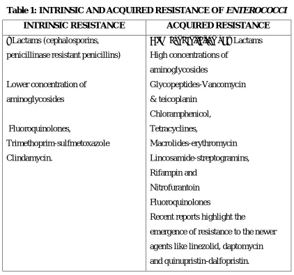
Table 1: INTRINSIC AND ACQUIRED RESISTANCE OF ENTEROCOCCI
INTRINSIC RESISTANCE ACQUIRED RESISTANCE
ΒLactams (cephalosporins, penicillinase resistant penicillins)
Lower concentration of aminoglycosides
Fluoroquinolones,
Trimethoprim-sulfmetoxazole Clindamycin.
High concentration of β Lactams High concentrations of
aminoglycosides
Glycopeptides-Vancomycin & teicoplanin
Chloramphenicol, Tetracyclines,
Macrolides-erythromycin Lincosamide-streptogramins, Rifampin and
Nitrofurantoin Fluoroquinolones
Recent reports highlight the
emergence of resistance to the newer agents like linezolid, daptomycin and quinupristin-dalfopristin.
RESISTANCE TO of β LACTAMS
β-Lactam antibiotics interfere with cell wall synthesis by inhibiting the penicillin binding proteins(PBPs) of susceptible bacteria, thus this class of antibiotics should be the first choice for the treatment of susceptible Enterococcal isolates. Many Enterococcal strains are also tolerant to β-lactams, that is, they are not killed with concentrations of antibiotics up to 16 times higher than the MIC(32ug/ml).
31
appear to be due to the expression of a resistant pbp5 allele (pbp5-R).36 which has decreased affinity to ampicillin. β-Lactam resistance in E.faecalis is mediated by the production of a β- lactamase enzyme.37
Penicillin resistance is directly proportional to the amount of PBP5 produced. Fontana et al showed that the loss of ability of a E.faecium to produce PBP5 caused this highly penicillin resistant strain to become hyper susceptible to penicillin. Unlike most Staphylococci, where β-Lactamase production is inducible, β-lactamase production in Enterococci is constitutive, low level, and inoculum dependent. Nitrocefin based test is used for reliable detection of β lactamase production in Enterococci , whereas disc diffusion and dilution methods are not reliable. Enterococci susceptible to penicillin are susceptible to ampicillin and other β lactams. But ampicillin susceptibility does not predict susceptibility to penicillin , separate testing with penicillin is needed. Ampicillin susceptibility can be used to predict imipenem susceptibility in case of E.faecalis
species.
RESISTANCE TO AMINOGLYCOSIDES INTRINSIC RESISTANCE
32
ACQUIRED RESISTANCE
Acquired resistance and high level resistance to aminoglycosides are due to conjugative plasmids or transposons. For Enterococci β-lactams are not bactericidal, but synergistic and bactericidal effect is usually achieved with the addition of an aminoglycoside. Among the aminoglycosides, gentamicin and streptomycin, are the only two compounds recommended for this synergistic effect in clinical practice. The presence of HLR to both gentamicin and streptomycin abolishes the synergistic effect in clinical practice. HLR to gentamicin is mostly due to the presence of a bifunctional aminoglycoside-modifying enzyme, 2′phosphotransferase 6′acetyltransferase, conferring resistance to gentamicin, tobramycin, netilmicin, kanamycin, amikacin but not streptomycin. HLR to streptomycin can be due to mutations in the 300S ribosomal subunit.38 and streptomycin adenyl transferase enzyme.39 Hence gentamicin and streptomycin should be tested individually to predict the resistance to aminoglycosides.
RESISTANCE TO QUINUPRISTIN-DALFOPRISTIN
-33
lactams, aminoglycosides, and glycopeptides.40 Nonsusceptibility to Q/D in
Enterococci may be due to following mechanisms,
1) The macrolide- lincosamide- streptogramin B (MLSB)-type of resistance mediated by the erm genes (encoding a 23SrRNA methyl transferase) 2) The presence of the (virginiamycin acetyl transferase) vatD and vatE
genes, which encode acetyltransferases that inactivate streptogramin A.
These genes are carried on plasmids and also confer resistance to the streptogramin and virginiamycin, (antibiotic previously used as a growth promoter in the veterinary industry).
RESISTANCE TO LINEZOLID
Linezolid resistance though rare, appears to be increasing, in patients without previous exposure to the antibiotic. Risk factors for the acquisition of nosocomial linezolid-resistant strains are peripheral vascular disease, solid-organ transplant recipients, total parenteral nutrition, and administration of piperacillin-tazobactam and/or cefepime antibiotics.
34
cfr gene may have been transferred from Enterococci to a Methicillin-resistant
Staphylococcus aureus (MRSA) isolates.32
RESISTANCE TO DAPTOMYCIN
Daptomycin resistance involves changes in genes leads to mutation of membrane protein LiaFSR involved in the cell envelope stress sensing response to antibiotics and a protein of glycerophosphoryl diester phosphodiesterase family. It abolishes the bactericidal activity of these antibiotics against these organisms.
RESISTANCE TO GLYCOPEPTIDES PHENOTYPIC DESCRIPTION
35
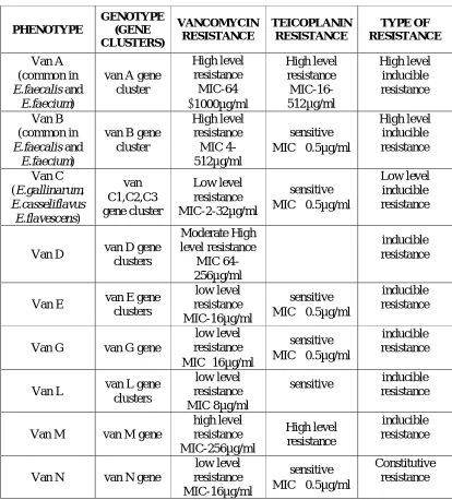
Table 2: PHENOTYPIC AND GENOTYPIC CLASSIFICATION
PHENOTYPE GENOTYPE (GENE CLUSTERS) VANCOMYCIN RESISTANCE TEICOPLANIN RESISTANCE TYPE OF RESISTANCE Van A (common in E.faecalis and
E.faecium)
van A gene cluster High level resistance MIC-64 1000µg/ml High level resistance MIC-16-512µg/ml High level inducible resistance Van B (common in E.faecalis and
E.faecium)
van B gene cluster High level resistance MIC 4-512µg/ml sensitive MIC 0.5µg/ml
High level inducible resistance Van C
(E.gallinarum, E.casseliflavus E.flavescens) van C1,C2,C3 gene cluster Low level resistance MIC-2-32µg/ml sensitive MIC 0.5µg/ml
Low level inducible resistance
Van D van D gene clusters Moderate High level resistance MIC 64-256µg/ml inducible resistance
Van E van E gene clusters
low level resistance MIC-16µg/ml
sensitive MIC 0.5µg/ml
inducible resistance
Van G van G gene
low level resistance MIC16µg/ml
sensitive MIC 0.5µg/ml
inducible resistance
Van L van L gene clusters
low level resistance MIC 8µg/ml
sensitive inducible resistance
Van M van M gene
high level resistance MIC-256µg/ml High level resistance inducible resistance
Van N van N gene
low level resistance MIC-16µg/ml
sensitive MIC 0.5µg/ml
Constitutive resistance
36
VAN A GLYCOPEPTIDE RESISTANCE
The Van A phenotype, with inducible high-level resistance to vancomycin as well as to teicoplanin, encoded by the vanA gene. Transposon 1546 is closely related genetic elements mediate this acquired inducible high-level resistance to vancomycin as well as to teicoplanin. The transfer of high level vancomycin resistance from Staphylococcus aureus via Tn 1546 was described recently. It is distributed in the following species of Enterococcus such as E.faecalis,
E.faecium, E.avium, E.casseliflavus, E.durans, E.gallinarum, E.mundtii, E.raffinosus and E.sanguinicola.
VAN B GLYCOPEPTIDE RESISTANCE
The Van B phenotype, with variable (moderate to high) levels of inducible resistance to vancomycin but not typically to teicoplanin is encoded by the van B (vanB1 and vanB2) genes. van B gene cluster consists of genes encoding polypeptides arranged to regulate glycopeptide resistance genes (van R and van S), synthesis of dipeptide D – alanyl –D-lactate (van H and van A) and hydrolysis of precursors of normal peptidoglycan (van X and van Y). The gene is located in plasmid and have been mediated by transposons Tn1547, Tn1549, Tn5382. The gene product is “D-alanine D-lactate”. It is distributed in E.faecalis, E.faecium,
E.durans and E.gallinavum.
VAN C GLYCOPEPTIDE RESISTANCE
37
chromosomal in origin corresponding to intrinsic low level glycopeptide resistance seen in E. gallinarum, E.casseliflavus and E.flavescens. Variable amounts of D–alanyl –D-alanyl relative to D-serine account for variable vancomycin resistance (Van C1, Van C2,Van C3,Van C4) among Van C phenotype and distributed in E.gallinarum (C1) and E.casseliflavus (C2-C4).
VAN D GLYCOPEPTIDE RESISTANCE
Van D is located in chromosomes and not transferable to other
Enterococci. Three clinical isolates of VRE (E.faecium )carrying van D resistance trait were first found in Boston .It is found in E.faecalis, E.faecium, E.avium and
E.gallinarum.
VAN E GLYCOPEPTIDE RESISTANCE
Recently described in E.faecalis BM 4405 with amino acid sequence identical to Van C (45%), Van B (43%) or Van D (44%).
VAN G GLYCOPEPTIDE RESISTANCE
Initially was found in E.faecalis isolates from Australia with low level resistance to vancomycin, yet susceptible to teicoplanin.
VAN L GLYCOPEPTIDE RESISTANCE
38
VAN M GLYCOPEPTIDE RESISTANCE
First described in E.faecium associated with high level resistance to and sensitive to teicoplanin. Resistance is transferable to E.faecium BM4105RF by conjugation.
VAN N GLYCOPEPTIDE RESISTANCE
Associated with low level resistance to vancomycin and sensitive to teicoplanin. Van N gene cluster determined by thermal asymmetric interlaced (TAIL) PCR, was similar to that of the Van C operons. E.faecium UCN 71 was the first strain isolated from blood culture. The peptidoglycan ends in D –serine and the resistance operon is constitutively expressed. Van N type resistance is transferable to E.faecium by conjugation. Van A and Van B are considered the most clinically relevant phenotypes and are usually associated with E. faecium
and E. faecalis isolates, while Van C resistance is an intrinsic characteristic of
E.gallinarum (vanC1 genotype) and E.casseliflavus (vanC2 and vanC3
genotypes) strains.
VANCOMYCIN DEPENDENT ENTEROCOCCI (VDE)
VDE are the Enterococci that grow only in the presence of vancomycin. First isolated in 1994 from urine of a patient receiving long term therapy with vancomycin. The phenomenon vancomycin dependence is that these
39
VANCOMYCIN RESISTANT ENTEROCOCCI (VRE) Epidemiology
Vancomycin resistant Enterococci were identified as nosocomial pathogen mainly E.faecalis and E.faecium from England in 1988 by Uttley et al.33 The presence of VRE was associated with the use of avoparcin-a glycopeptide as a growth promoter in animal feeds and was banned from European countries in 1996.3 The rates of vancomycin resistance among E. faecium clinical isolates in Europe are highest in Greece, the United Kingdom, and Portugal (10–30%), whereas rates in the Scandinavian countries and the Netherlands are <1%and latin American countries <4%. These regional differences are due to the implementation of aggressive infection control policies and higher levels of human antibiotic use in the United States. Emergence of vancomycin -resistant E. faecium in different parts are due to a unique hospital associated genetic clade that acquired the genes responsible for vancomycin resistance and other antibiotic resistance determinants.3
RISK FACTORS
Presence of immunosuppression
Presence of Co-morbid conditions like diabetes, renal failure, high APACHE (Acute physiology and Chronic Health Evaluation) score, malignancy,
Prolonged hospital stay
Residence in a long term care facility
40
Invasive procedures
Previous exposure to broad spectrum antibiotics – cephalosporins, vancomycin.
Use of enteral tube feeding/ sucralfate
Exposure to contaminated medical equipment
The most important being exposure to heath care personnel nursing to a known VRE patient.
Colonization and Infection
41
Source of infection and transmission of VRE
The source of infection could be Endogenous – patients own Gastrointestinal tract in previously colonized individuals or Exogenous as contaminated environmental surfaces and, medical devices –bed rails, linen, doorknobs, bed pans, stethoscope and blood pressure cuffs.8 Contaminated food products may be a reservoir in non hospitalized individuals.11 The most common mode of transmission is through the contaminated hands of healthcare workers in nosocomial VRE infections.11 and less commonly with contaminated equipment and contaminated surfaces.
PREVENTION AND CONTROL
The first guidelines for the control of VRE in hospitals was first published in 1994 by HICPAC
.
CDC’s Hospital Infection control practices advisorycommittee has established certain guidelines and recommendations for prevention of VRE spread.
1. Prudent use of vancomycin - inappropriate use of vancomycin is a risk factor for VRE colonization and infection and also emergence of vancomycin resistant Staphylococci. The medical staff should be educated about the appropriate or acceptable use of vancomycin (MRSA treatment, Severe antibiotic associated colitis as a second line agent, major surgical procedures involving implantation of prosthetic devices).
42
3. Implementation of surveillance procedures (feces cultures) for early detection of VRE colonization .
4. Infection control procedures aiming to limit cross contamination isolation of known VRE patients and colonizers, strict adherence to hand washing.
To minimize the nosocomial transmission of VRE, hospitals must use a multidisciplinary approach that requires participation by a variety of departments and personnel.
TREATMENT
The suggested therapeutic options for serious VRE infections are
1. Combination therapy with high dose of cell wall active agents (penicillin, ampicillin) and an aminoglycoside (if there is no acquired resistance observed for the agents).3
2. The use of ciprofloxacin and other quinolones are limited to the treatment of UTIs. Chloramphenical retains its in vitro activity against many strains of MDR E.faecium. Triple therapy (ciprofloxacin–rifampicin-gentamicin) is an excellent choice for sterilizing vegetations.
43
4. Quinupristin-Dalfopristin – parenteral semisynthetic streptogramin type A and B and is FDA approved. It is also a bacteriostatic. It is active against
E.faecium only and not active in E.faecalis
5. Daptomycin- it is an acidic lipopeptide active against both E.faecalis and
E.faecium but it is not FDA approved. It is used as an alternative in situations of therapeutic failure of the commonly used agents.
6. Oritavancin is an investigational semisynthetic glycopeptide with potent
in vitro activity against VRE.
ANTIBIOTIC THERAPY FOR MDR ENTEROCOCCI
44
MATERIALS AND METHODS
Study Design: Cross sectional study
The present study was conducted in Institute of Microbiology,
Rajiv Gandhi Government General Hospital, Madras Medical College, Chennai, India.
Study Population:
Patients attending outpatient department and inpatients of Rajiv Gandhi Government General Hospital, Madras Medical College, Chennai.
Study Period: 1 year (April 2016- March 2017)
ETHICAL CLEARANCE
Institutional Ethical committee clearance was obtained for the study under the clearance number 22042016.
INCLUSION CRITERIA Patients aged >18 years
All nonduplicate clinically significant Enterococcus isolates from samples such as urine, pus, blood, wound swab, catheter tip and other body fluids were included.
EXCLUSION CRITERIA
45
A total of 100 Enterococcus isolates were recovered from various clinical specimens such as urine, blood, pus, wound swab, sterile body fluids and central venous catheters.
METHODOLOGY
Collection and Processing of samples:
All the clinical specimens such as urine, blood, wound exudates, pus, central venous catheter and sterile body fluids from both inpatients and outpatients were collected as per standard protocol.2 Microscopic examination of the direct Gram stained smears of the specimens were done to look for cellular inflammatory response and presence of microorganisms.
CULTURE41
The urine samples were inoculated on cystine lactose electrolyte deficient agar (CLED) and 5% sheep blood agar medium by standard calibrated loop method to perform semiquantitative culture. A biphasic media using brain heart infusion broth and agar was used for isolating Enterococci from blood samples. The pus samples and other body fluids from normally sterile sites were inoculated on 5% sheep blood agar and MacConkey agar. Central venous catheter tips were processed by semiquantitative culture as per standard protocol. The inoculated media were incubated at 37°C overnight and observed for growth.
COLONY CHARACTERISTICS
46
MACCONKEY AGAR: Minute, 0.5-1mm, deep pink (magenta) colonies.
CYSTINE LACTOSE ELECTROLYTE DEFICIENT MEDIUM: Small, circular, convex, lactose fermenting colonies are seen. For urine samples, semiquantitative cultures were done using calibrated loop method and only the significant counts (105 CFU/ml in clean catch midstream sample, 102 CFU/ml in catheterised samples)42 were processed.
PRELIMINARY IDENTIFICATION OF ENTEROCOCCI Gram’s stain of smear from culture isolates
A smear was prepared from the colonies and Gram stain was done with appropriate controls. The smear was examined for the presence of Gram positive cocci arranged in pairs and short chains.
QUALITY CONTROL
Positive control-Staphylococcus aureus ATCC 25923 Negative control-Escherichia coli ATCC 25922
CATALASE TEST2
Purpose: This test is used to differentiate members of Enterococcaceae and
Streptococcaceae from members of Staphylococcaceae. Enterococci are catalase negative.
47
Procedure: A few colonies of culture to be tested were picked from the nutrient agar slope with a sterile, thin glass rod and inserted into 3% hydrogen peroxide solution held in a small, clean test tube.
Interpretation: A positive catalase reaction was indicated by rapid and sustained bubbles or effervescence (nascent oxygen). A few tiny bubbles forming after 20-30 seconds is not considered to be positive test because some bacteria possess enzymes other than catalase that can decompose hydrogen peroxide.
Quality control:
Positive control: Staphylococcus aureus
Negative control: Streptococcus species
All catalase negative Gram positive cocci in pairs & short chains were further characterized.
BILE ESCULIN TEST2
Purpose: This test is used for the presumptive identification of Enterococci and some organisms in Streptococcus bovis group. This test differentiates Enterococci
and group D Streptococci from non-group D viridans Streptococci.
48
Procedure: Two or three morphologically similar colonies were inoculated on to the slant of the bile esculin medium or streak the surface of a bile esculin plate. Incubate the tube or plate at 35°C for 24–48 hours in an ambient air.
Interpretation: Diffuse blackening of more than half of the slant within 24–48 hours indicates positive test. On plates, black haloes will be observed around isolated colonies and any blackening is considered positive.
Quality control:
Positive control: Enterococcus faecalis ATCC 29212
Negative control: Streptococcus pyogenes ATCC (19615)-no growth; no colour change or Escherichia coli ATCC (25922) - growth; no colour change
HEAT TOLERANCE TEST41
Purpose: To differentiate Enterococci from other Streptococcus bovis and
Streptococcus equinus group.
Principle: Enterococci tolerate temperature of 60oc for 30 minutes.
Procedure: An overnight broth culture of suspected colonies was streaked on blood agar plate. The same broth culture was heated for 30 minutes at 60o C. From this heated broth, another BAP is streaked. Both BAPs were incubated at 37o C for 24 -48 hours.
49
Quality control:
Positive control: Enterococcus faecalis (ATCC29212) Negative control: Streptococcus bovis (ATCC 9809)
SALT TOLERANCE TEST2
Purpose: This test is used to determine the ability of an organism to grow in high concentrations of salt. It is used to differentiate Enterococci from non
Enterococci.
Principle: This test is useful for the presumptive identification of group D
Enterococci which have the ability to grow in high salt concentration (6.5% NaCl) in the medium. This test along with bile esculin test distinguishes
Enterococcus species from the group D streptococci (S.bovis and S.equinus). Group D streptococci - bile esculin positive, salt tolerance negative; Enterococci
– bile esculin and salt tolerance positive. The brain heart infusion broth with 6.5% of NaCl is used as a test medium and bromocresol purple as an indicator for acid production.
Procedure: Two or three colonies were inoculated into 6.5% NaCl broth with or without bromothymol purple indicator and incubated at 35oc in ambient air for 48 hours.
50
Quality control:
Positive control: Enterococcus faecalis (ATCC29212) –growth; colour change to yellow.
Negative control: Streptococcus bovis (ATCC 9809) – inhibition, as demonstrated by little to no growth; no colour change.
L-pyrrolidonyl β naphthylamide test (PYR TEST)2
Principle: This test is used for the identification of Streptococcus pyogenes and
Enterococci which are PYR test positive. The presence of the enzyme L-pyrrolidonyl arylamidase hydrolyses the substrate α- pyrrolidonyl β naphthylamide and releases free naphthylamide which can be detected by the addition of N, N-methylamino-cinnemaldehyde. This detection reagent couples with the naphthylamide to form a red Schiff base.
Procedure: The colonies were inoculated in PYR broth (α- pyrrolidonyl β naphthylamide) and incubated at 37oC in ambient air for 4 hours. A drop of PYR reagent (0.01% p- dimethylamino-cinnemaldehyde) was added to the broth. Positive test was indicated by the development of cherry red colour within a minute of reagent addition.
Interpretation:
51
Quality control:
Positive control: Enterococcus faecalis (ATCC29212) or
Streptococcus pyogenes (ATCC 19615)
Negative control: Streptococcus agalactiae (ATCC 10386)
SPECIATION OF ENTEROCOCCI :
Enterococci were further identified to species level using Facklam and Collins scheme.
SUGAR FERMENTATION TEST:
For identification of species, 1% of sugars (glucose, arabinose, raffinose, lactose, mannitol, sorbitol) in peptone water with 0.002% bromothymol blue indicator was used. To each tube of sugars, 1-2 drops of 18-24 hours BHI broth culture was added and incubated at 37oC overnight.
Interpretation: Sugar fermentation was indicated by the change of colour from blue to yellow.
ARGININE DIHYDROLASE TEST2
Purpose: This test is used to differentiate decarboxylase producing organisms from other.
52
Hydrolysis of the amino acid results in an alkaline pH and colour changes to purple.
Method: Moeller’s decarboxylase basal broth with 1% arginine along with an aminoacid control were inoculated with the test strain. Both the tubes were overlaid with 4mm sterile liquid paraffin and incubated at 37oC overnight. The colour of the indicator reverting back to original purple colour indicating arginine hydrolysis was considered a positive provided the control tube remains yellow indicating fermentation.
Interpretation: Arginine hydrolysis is positive for Group 2 and Group 3
Enterococcus species. Arginine hydrolysis is negative for Group 1, Group 4 and Group 5 Enterococcus species.
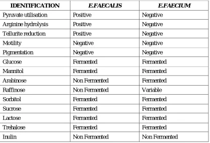
PYRUVATE UTILIZATION2
Principle: This test is used to determine the ability of an organism to utilize pyruvate. This property helps to differentiate E.faecalis (positive) and E.faecium
(negative).
53
POTASSIUM TELLURITE REDUCTION TEST2
Principle: Certain species of Enterococci are able to reduce tellurite to metallic tellurium which imparts black colour to the colonies. E.faecalis reduces tellurite in contrast to E.faecium which does not.
Procedure: 0.04% Tellurite blood agar plates were streaked with overnight nutrient broth culture and incubated at 37o c for 3 days.
Interpretation: Tellurite reduction is considered positive on appearance of black colonies within 3 days.
MOTILITY TEST2
Purpose: This test was used to identify E.gallinarum and E.casseliflavus which exhibit motility. All other Enterococcus species are nonmotile.
Hanging drop method: A drop of liquid culture was placed on the cover slip. A hollow ground slide with its concavity encircled by soft petroleum jelly was inverted over the cover slip and quickly turned around. The hanging drop was examined first with a low power objective and then with high power for the presence of motility.
Mannitol motility medium2
54
Method: The motility and fermentation of mannitol was tested by stab inoculating the isolates (including positive and negative control) into the medium and incubated at 37oc overnight detect the motility and acid production from mannitol.
Interpretation: Motility is observed by fanning around the stab area and mannitol fermentation can be detected by colour change of the medium to yellow.
Quality control:
Positive control: Escherichia coli (ATCC 25922)
Negative control: Staphylococcus aureus (ATCC 25923 )
PIGMENT PRODUCTION
Enterococcus isolates were streaked on blood agar and incubated at 37oC for 24-48 hours. A sterile white cotton swab was used to pick up colonies and observe. Pigment production is detected by yellow or orange tinge on the swab.
Enterococcus faecalis – characteristics:
Gram stain – Gram positive cocci in pairs and short chains, some exhibiting spectacle like arrangement.