Accepted Manuscript
Title: Flexible and transparent films produced from cellulose nanowhisker reinforced agarose
Authors: Reda M. Felfel, Kazi M. Zakir Hossain, Sumaya F. Kabir, Soon L. Yee, Ifty Ahmed, David M. Grant
PII: S0144-8617(18)30373-4
DOI: https://doi.org/10.1016/j.carbpol.2018.04.005
Reference: CARP 13459
To appear in:
Received date: 9-1-2018
Revised date: 14-3-2018
Accepted date: 1-4-2018
Please cite this article as: Felfel, Reda M., Hossain, Kazi M Zakir., Kabir, Sumaya F., Yee, Soon L., Ahmed, Ifty., & Grant, David M., Flexible and transparent films produced from cellulose nanowhisker reinforced agarose.Carbohydrate Polymers https://doi.org/10.1016/j.carbpol.2018.04.005
1
Flexible and transparent films produced from cellulose nanowhisker reinforced agarose Reda M. Felfel1,2●*, Kazi M. Zakir Hossain1●*, Sumaya F. Kabir1, Soon L. Yee3, Ifty Ahmed1 and
David M. Grant1
1Advanced Materials Research Group, Faculty of Engineering, University of Nottingham, UK
2Physics Department, Faculty of Science, Mansoura University, Mansoura, Egypt
3Nano and Advanced Materials Institute Limited, Hong Kong
● Shared first authorship
*Corresponding authors: Reda M. Felfel, reda.felfel@nottingham.ac.uk and Kazi M. Zakir Hossain,
ezzzh@exmail.nottingham.ac.uk
Research Highlights
Never-dried CNWs and Agarose were utilised to produce transparent and flexible nanocomposite films.
The nanocomposites films maintained more than 84% optical transparency.
The crystallinity of the nanocomposite films increased with the addition of CNWs.
A significant increase in mechanical and thermomechanical properties of the nanocomposites was obtained.
Incorporation of CNWs led to reduction in the swelling rate of the nanocomposite.
Abstract
Transparent and flexible nanocomposite films with a range of Agarose to Cellulose
Nano-Whisker (CNW) ratios were produced using dried CNWs. The incorporation of
never-dried CNWs within Agarose played an important role in the surface roughness (Ra 7 to 15 nm)
and light transparency of the films (from 84 to 90%). Surface induced crystallisation of
Agarose by CNWs was also found with increasing percentage of crystallinity (up to 79%) for
the nanocomposite films, where CNW acted as nucleating sites. The enhanced tensile
2
films compared to the control Agarose film indicated the effectiveness of the nanowhiskers
incorporation. The storage modulus of the nanocomposite films increased also to be tripled
Agarose alone as the CNWs content reached 43%. The swelling kinetics of the
nanocomposites revealed that addition of CNWs reduced the long-term swelling capacity and
swelling rate of the nanocomposite.
Keywords: Cellulose nanowhiskers, agarose, transparency, crystallinity, tensile properties, swelling kinetics.
Introduction
Polymer nanocomposites consist of at least one of the phases with a dimension of nanoscale
range (<100 nm) dispersed in a continuous polymer matrix. Polymer nanocomposites have
been heavily studied for a wide range of uses, from water treatment (Pandey, Shukla, & Singh,
2017) and energy storage (Riggs et al., 2015) to biomedical applications (Zare & Shabani,
2016), due to their unique mechanical, chemical, optical, thermal, electrical and biological
properties (Camargo, Satyanarayana, & Wypych, 2009; Müller et al., 2017). Different methods
have been developed to produce polymer nanocomposites such as melt mixing (Jordan,
Jacob, Tannenbaum, Sharaf, & Jasiuk, 2005), solution casting/evaporation (Kargarzadeh et al.,
2017), in situ polymerisation (Jordan et al., 2005) and sol-gel process (Müller et al., 2017).
Selection between those techniques is mainly dependent on the polymer matrix.
Highly transparent polymer nanocomposites have been developed utilising various
nanoparticles, e.g. quantum dot (C. Liu et al., 2017), silver nanowires, graphene oxide
(Tematio, Fosso, Krähenbühl, & Schintke, 2016), zinc sulphate (Cheng et al., 2008), gold
(Ehlert, Stegelmeier, Pirner, & Förster, 2015; Khalil, Homaeigohar, Häußler, & Elbahri, 2016),
nanoclay (Aulin, Salazar-Alvarez, & Lindstrom, 2012), silica and silica-titania (Y.-Q. Li, Fu, Yang,
& Mai, 2008), for different applications including gamma scintillation, medical (C. Liu et al.,
2017), photovoltaics, printed organic electronics, optical and optoelectronic devices (Tematio
et al., 2016). The high transparency of highly-loaded (up to 60 w%) nanocomposites was
3
al., 2008; C. Liu et al., 2017) and/or surface modification of the nanoparticles (Bin, Sugihara,
Elim, Adschiri, & Kaino, 2011; Dang et al., 2014).
Agglomeration and a lack of dispersion of the nanoparticles remain as one of the main
challenges in manufacturing polymer nanocomposites (Y. Liu et al., 2015). Absence of
chemical interaction between the phases and smaller aspect ratio nanofillers can lead to
limited mechanical properties of the nanocomposites produced (Qiang, Chunfang, JianZun, &
Yuan, 2004). It is expected that the mechanical properties of the nanocomposites will increase
by increasing the aspect ratio of the nanofiller (Cipiriano et al., 2007; Yu et al., 2017).
Therefore, carbon nanotubes (Sahoo, Rana, Cho, Li, & Chan, 2010), cellulose nanowhiskers
(CNWs) (Santamaria-Echart et al., 2017) and hydroxyapatite nanorods (Yang et al., 2017)
would be anticipated to have significantly greater reinforcement effects than nanospheres
for example (Hassanabadi & Rodrigue, 2014). Several attempts have been made to enhance
the dispersion and chemical interaction between polymer matrix and nanoparticles via
surface treatment of the nanoparticles (D. Liu, Pourrahimi, Olsson, Hedenqvist, & Gedde,
2015). The nanoparticles show good dispersion in solution such as water, however then
commonly tend to agglomerate during drying either at room temperature, in ovens or using
freeze drying processes (Rahman, Vejayakumaran, Sipaut, Ismail, & Chee, 2008).
Consequently, using an alternative process involving nanoparticle suspensions could result in
enhanced nanoparticles distribution within polymer matrices, thus leading to advanced
mechanical properties.
CNWs are usually produced via acid hydrolysis of cellulose based raw materials such as cotton,
wood and ramie (George & Sabapathi, 2015; Trache, Hussin, Haafiz, & Thakur, 2017; Xu et al.,
2013). Their aspect ratio, ratio of length to diameter or width, varies from 2 to 200 depending
on the source of the cellulose and acid hydrolysis parameters (types of acid, hydrolysis period
and temperature) (Trache et al., 2017). CNWs were selected here as a reinforcement due to
their greater theoretical tensile strength, ca. 7.6 GPa, compared to Kevlar fibres and steel wires (Trache et al., 2017). Their elastic modulus was determined using AFM and found to be
approximately 150 GPa (Iwamoto, Kai, Isogai, & Iwata, 2009; Trache et al., 2017). Due to these
mechanical properties and aspect ratio, CNWs could be regarded as ideal natural, renewable,
cytocompatible and biodegradable nanomaterials (Trache et al., 2017). Geometry and
4
employed (Khoshkava & Kamal, 2014). CNWs whilst suspended in water, showed a needle
shaped morphology with average length of ca. 170 nm and 15 nm diameter (George & Sabapathi, 2015). Small spherical shapes have been obtained by spray drying of CNWs, whilst
freeze drying yielded larger flake shaped samples (Khoshkava & Kamal, 2014) showing the
aspect ratio and morphology of CNWs can be significantly affected by the drying process.
CNWs also tend to agglomerate and form large flakes at the drying step of production due to
their high surface energy and specific surface area (Khoshkava & Kamal, 2014). Therefore, it
would be considered highly desirable to use CNWs in suspension form to maintain their
unique nano morphologies.
Agarose is a natural, biocompatible polymer obtained from seaweed (Cao, Gilbert, & He,
2009). Agarose has been heavily utilised in hydrogel form for electrophoresis purposes
(Corthell, 2014). It has also been investigated as 3D porous constructs for biomedical
applications such as regeneration of cartilage (Gadjanski, Yodmuang, Spiller, Bhumiratana, &
Vunjak-Novakovic, 2013), wound healing (Awadhiya, Tyeb, Rathore, & Verma, 2017) and
neural tissue engineering (Cao et al., 2009). Very few studies have explored the properties of
agarose in dried thin sheet form. A highly transparent film of agarose can be obtained by
drying the hydrogel at room temperature (Z. Wang et al., 2010 a; Z. Wang et al., 2010 b).
Agarose has excellent optical transparency, film-forming ability, high mechanical strength (Z.
Wang et al., 2010 b) and most interestingly it follows sol-gel transition at ca. 42oC (Medina-Esquivel, Freile-Pelegrin, Quintana-Owen, Yáñez-Limón, & Alvarado-Gil, 2008) which could
potentially prevent nanoparticle precipitation, agglomeration and thus maintaining good
dispersion.
CNWs have been effectively incorporated into numerous biodegradable polymers e.g. poly
lactic acid (PLA) (Hossain et al., 2012), poly caprolactone (PCL) (Boujemaoui et al., 2017),
polyvinyl acid (PVA) (W. J. Lee, Clancy, Kontturi, Bismarck, & Shaffer, 2016), chitosan (Celebi
& Kurt, 2015) and agar (Atef, Rezaei, & Behrooz, 2014), revealing a significant enhancement
in mechanical and physical properties of their nanocomposites. Water soluble natural
biopolymers reinforced with CNWs have been explored for wound dressing and drug coating
and delivery applications (Zakir Hossain et al., 2012).
Highly transparent agarose films containing polyoxometalates were investigated as
5
display applications (Z. Wang et al., 2010 b). Xiaodong et al. investigated the mechanical properties of agarose nanocomposite films containing varying weight fractions of nanoclay,
up to 80% (Xiaodong et al., 2005). Good agreement was observed for elastic moduli obtained
from tensile and flexural tests. The elastic modulus of the nanoclay reinforced agarose
increased gradually to 21.4 GPa, six times higher than plain agarose alone, with 60 wt% of
nanoclay concentration, whereas ductility (failure strain) for the composites decreased from
0.24 to 0.02 as the concentration of nanoclay increased to 40 wt% (Xiaodong et al., 2005).
The influence of incorporating dried CNWs on the mechanical and physical properties of agar
was explored (Atef et al., 2014; Oun & Rhim, 2015). Tensile strength and modulus of the agar
films increased by 25% and 40% by inclusion of 5wt% of CNWs, whilst the optical
transmittance decreased by ca. 18%. Both studies concluded that 5 wt% of CNWs was the
highest content to enhance or even maintain the mechanical properties of agar films.
In this study, highly transparent nanocomposite films based on agarose and never-dried (i.e.
in aqueous suspension) CNWs (used as reinforcement) were prepared using solution
casting/evaporation method at higher concentrations of CNWs (0, 16, 28 and 43 wt%). The
influence of CNWs inclusion on the morphological, surface roughness, light transparency,
crystallisation, mechanical, thermo-mechanical and swelling kinetic properties of
Agarose/CNWs blended films were investigated.
Materials and methodology Materials
DNA Grade agarose powder (Electran®, Mw 120,000 Da, gelling temperature 34-37oC and
melting point 60-90oC) was obtained from VWR international Ltd, UK. Sulphuric acid (95%
purity), amberlite MB 6113 resin and bleached cotton wool were purchased from Fisher
Scientific (UK).
Preparation of cellulose nanowhiskers (CNWs)
The CNWs were produced by acid hydrolysis of cotton using aqueous sulfuric acid (64 wt%)
for 45 min under continuous stirring (Hossain et al., 2012; Hossain et al., 2014). The residual
acid was then removed from the hydrolysed CNWs suspension via centrifugation (three cycles
at 10,000 rpm at 10oC for 20 min using de-ionised water) and dialysis under running tap water
6
followed by filtration using a grade No. 2 fritted glass filter. The CNWs suspension was treated
overnight with Amberlite resin to obtain H3O+ cation free CNWs suspension. The final
concentration of CNWs in the de-ionised water was determined to be 1.15 wt%. This kind of
as-prepared aqueous suspended CNWs was identified as never-dried CNWs (Agarwal, Ralph,
Reiner, & Baez, 2016; K.-Y. Lee & Bismarck, 2012). On the other hand, dried crystalline CNWs
were also produced via a freeze-drying process after freezing in liquid nitrogen as described
elsewhere (Labet & Thielemans, 2011).
Preparation of aqueous CNWs reinforced agarose films
The aqueous cellulose nanowhisker reinforced agarose films (Agarose-CNW) were prepared
by solution casting followed by film drying process, see Figure 1. The aqueous CNWs
suspension in de-ionised water (1.15 wt%) was sonicated for 5 min. Afterwards, different
quantities of the CNWs suspension (0, 5, 10 and 20 ml) were diluted with de-ionised water to
reach 50 ml and then a fixed amount of Agarose powder (0.3 g) was added. The mixture was
subjected to heat at 100oC with vigorous stirring using a hot plate magnetic stirrer and boiled
for 5 min until the agarose powder had fully dissolved and the solution became clear with no
obvious signs of sedimentation. The solution was then poured into a polystyrene petri-dish.
The Agarose-CNWs solution was converted into gel within 5 min as the temperature dropped
below 40oC. The nanocomposite hydrogels were allowed to dry at room temperature for 48
h to obtain flexible nanocomposite films of approximately 60 µm thickness. The
nanocomposite films produced were further dried in a vacuum oven at 50oC for 48 h to
eliminate any moisture traces present prior to testing.
7
Figure 1:A schematic diagram of the preparation process of Agarose-CNW nanocomposites films. In the solution state, CNWs and agarose chains were homogenously dispersed within the deionised water via stirring at 100oC. After casting the nanocomposite mixture and
temperature dropped below 42oC, the agarose was converted into gel and thus maintained
the uniform distribution of non-agglomerated CNWs with the agarose matrix. Water was then removed by drying at room temperature.
Final concentration of the CNWs (wt%) within various Agarose-CNW nanocomposites was
then calculated and have been tabulated (please see Table 1). The number used for the
sample codes was considered according to the calculated weight fraction of CNWs within the
nanocomposites.
Table 1: Sample codes and formulations of films prepared using never-dried CNWs and Agarose.
Sample codes
used in this study
Agarose
(g)
De-ionised
water
(ml)
Never-dried CNWs
suspension*
(ml)
CNWs
(wt%)
Agarose 0.3 50 0 0
Agarose-16 CNW 0.3 45 5 16
Agarose-28 CNW 0.3 40 10 28
CNWs/de-ionised water suspension
Agarose-CNWs solution Agarose-CNWs nanocomposite hydrogel
Agarose-CNWs nanocomposite film
8
Agarose-43 CNW 0.3 30 20 43
*Concentration of CNWs in de-ionised water 1.15%
Characterisation
Electron microscopic analysis
The surface morphology of freeze-dried CNWs was examined using scanning electron
microscopy (SEM - Philips XL30, FEI, USA) at an accelerating voltage of 12 kV and a working
distance of 10 mm. A sputtered platinum coating was employed to avoid image distortion due
to charging.
The morphology of the never-dried CNWs was characterised using transmission electron
microscopy (TEM, JEOL, 2000FXII, UK) at an accelerating voltage of 80 kV. The never-dried
CNWs sample loaded on Cu-grid (mesh size 300) was stained negatively with 2 wt% uranyl
acetate (Sigma-Aldrich, UK) to enhance the contrast.
Atomic force microscopy (AFM) analysis
AFM micrographs and roughness measurements of Agarose-CNW nanocomposite films were
performed in tapping mode utilising a Bruker Dimension Icon equipped with AFM tips (model
RTESPA-150 0.01–0.025 Ohm-cm Antinomy (n) doped Si). All measurements were carried out
over a scan area of 2 and 5 μm2. The micrographs obtained were analysed using Nanoscope
analysis software (Version 1.5) and roughness analysis (Ra) was determined from at least
three scans.
Light Transmittance
Light transparency property of the Agarose-CNW nanocomposite films was determined by
measuring the light transmittance using a UV-Vis spectrometer (PerkinElmer, Lambda-25)
over the wavelength range of 200 to 800 nm.
Polarised light microscopy and image analysis
Polarised optical microscope (Nikon Eclipse LV100ND, Nikon Instruments, Japan) equipped
with a Nikon digital sight DS-Ri1 camera and a NIS Elements software (version 4.1) was used
to examine the crystalline morphology of Agarose-CNW nanocomposites films. A small piece
9
mode between crossed polarisers at 20x optical magnification. A fixed intensity of incident
light and an exposure time of 60s were used for all specimens.
X-ray diffraction (XRD) analysis
X-ray diffraction patterns for Agarose-CNW nanocomposites were determined using a D500
diffractometer (SIEMENS) equipped with a Cu-Kα radiation source (λ= 0.154 nm) at 15 mA
and 30 kV. The samples were scanned from 5o to 45o diffraction angle (2θ) with step size of
0.05o and 2 s time interval.
The crystallinity index (CI%) of the films was calculated according to Equation 1 (Segal S., 1959;
Weimer, Hackney, & French, 1995; Xu et al., 2013):
where,I(Crysam)) denotes the most intense peak attributed to the combined crystalline and
amorphous parts of cellulose (peak intensity at 2θ=22.8o), whereas I
(am) represents the
amorphous portion of cellulose (peak intensity usually appeared at 2θ=18o).
Fourier transform infrared spectroscopy (FTIR)
The functional groups present in CNWs, pure Agarose and their nanocomposite films were
identified using a FTIR spectrometer (Tensor-27, Bruker, Germany) equipped with a standard
attenuated total reflectance (ATR) cell (Pike Technology, UK). The films were scanned in
transmittance mode over the wavenumber range from 4000 to 550 cm-1 and the spectra were
analysed using OPUS™ software (version 5.5).
Tensile properties
Tensile properties of Agarose-CNW films were determined using an Instron tensile testing
machine attached with a 100 N load cell at speed of 1 mm min-1 and 25 mm gauge length
according to standard BS EN ISO 527-1:2012. Rectangular test specimens with a dimension of
55 mm length and 5 mm width were used. Tensile strength and modulus were calculated
according to standard BS EN ISO 527-1:2012. At least 5 replicates of each film were tested.
Dynamic mechanical analysis (DMA)
) 1 ...( ... 100 I
I I
[%] CI
) am Crys (
) am ( ) am Crys
(
10
Thermo-mechanical properties of the Agarose-CNW films were measured using a DMA tester
(TA Q800 Instruments, USA) in multi-frequency strain mode to investigate the storage
modulus (E’) and tan delta of the flexible films with increasing temperature. Rectangular test specimens (25 mm length and 4 mm width) were heated from room temperature to 280oC at
a heating rate of 10oC min-1, 0.05% strain, 15 mm gap distance, 0.01N preload force, 1 Hz
frequency and 125% force track.
Swelling kinetics
The swelling capacity of the pure Agarose and Agarose-CNW nanocomposite films (10 mm
disc) were calculated gravimetrically by measuring the variation in mass of the sample after
soaking in phosphate buffered saline (PBS) media (10 ml) at 37oC for 24 h using Equation 2.
At each time point, the wet samples were taken out from the media and placed onto tissue
paper, blot dried and weighed immediately.
100.........(2)
M ) M -(M % capacity Swelling
i i
W
where, Mi is the initial mass of the dry specimen before immersion in PBS media and Mwis
the mass of wet specimen at each time point after soaking in PBS media.
Results and Discussion
Morphology and surface topography of CNWs
The morphology and surface topography of freeze-dried CNWs were characterised using SEM
and AFM micrographs. The freeze-dried CNWs exhibited flake-like morphology at
micron-range size due to the aggregation nature of CNWs (Khoshkava & Kamal, 2014) as seen in
Figure 2a. The 2D AFM images (height) of the freeze-dried CNWs presented in Figures 2b also
revealed aggregated morphology with a surface roughness value of 75 ± 7 nm from the 3D
AFM image (see Figure 2c). On the other hand, TEM image of the aqueous CNWs suspension
produced is presented in Figure 2d which revealed rod-like particles with average length and
width approximately 300 ± 100 nm and 10 ± 2 nm, reported previously (Hossain et al., 2012).
Individual rod-like morphology with the percolated network was also seen via the 2D AFM
image of aqueous CNWs (see Figure 2e) and their surface roughness was found to be 11 ± 3
nm obtained from the 3D AFM image presented in Figure 2f.
11
Figure 2: Morphology and surface topography of freeze-dried and never-dried CNWs: a) SEM image of freeze-dried CNWs (inset photograph of freeze-dried CNWs), b-c) AFM images of freeze-dried CNWs, 2D (b) and 3D (c), d) TEM image of never dried CNWs (inset photograph of never-dried CNWs suspension in deionised water, 1.15 wt%), and e-f) AFM images of never-dried CNWs, 2D (e) and 3D (f).
Pure cellulose nanowhisker films were also produced from both freeze-dried and never-dried
CNWs suspension (1.15 wt% in deionised water) via drying process at room temperature to
observe their optical transparency. The freeze-dried CNWs film appeared to be white with a
paper-like opaque appearance (see the supplementary figure, ESI 1a). This was in well
agreement with the aggregated morphology and comparatively higher surface roughness (75
± 7 nm) of the freeze-dried CNWs obtained via SEM and AFM images (as presented in Figures
2a-c). Alternatively, the aqueous CNWs prepared film presented in the supplementary figure
(ESI 1b) was observed to be translucent which was suggested to be due to their well dispersed
rod-like CNW structures as well as their comparatively lower surface roughness (11 ± 3 nm).
However, both films obtained from freeze-dried and aqueous CNWs were brittle and cracked
during the drying stage as seen in the supplementary figure (ESI 1).
Light transparency
Considering the suggested better dispersion and superior light transparency properties of the
aqueous CNWs over the freeze-dried CNWs films (Atef et al., 2014), then various contents of
aqueous CNWs were blended with Agarose to investigate the production of flexible and
a)
d)
200 µm
c) b)
e) f)
12
transparent nanocomposite films. Agarose-CNW nanocomposite films were prepared by
solution casting/evaporation process by mixing never-dried CNWs suspension with agarose
powder at high temperature (100oC) until agarose powder fully dissolved and followed by
casting and drying at room temperature, see Figure 1. Agarose is a thermo-responsive
hydrogel that could convert quickly from solution state to a gel state as the temperature
dropped below ca. 42oC (Medina-Esquivel et al., 2008). This property could potentially prevent nanoparticle precipitation and agglomeration during the drying step of the
nanocomposite films. Representative images of the flexible films (see Figures 3a-d) revealed
that the optical transparency decreased with increasing CNWs content (as expected). The
light transparency of the nanocomposite films obtained by UV/Visible spectrometry also
showed that the pure Agarose film exhibited light transparency of 91% in most of the visible
light region (at 650 nm wavelength), whereas, the Agarose-CNW nanocomposites exhibited
decreasing light transmittance with values of 90, 89 and 84% with increasing CNWs content
(ranging from 16, 28 and 43 wt%). The optical transparency of agar film was reported to
decrease more than 30% by inclusion of 10 wt% freeze-dried CNWs as a result of their
agglomeration (Atef et al., 2014). Reduction in the optical transparency by increasing the
nanoparticles content has been also reported for other highly transparent polymer
nanocomposites (Dang et al., 2014; C. Liu et al., 2017).
13
Figure 3: Representative photographs of the transparent (top) and flexible (bottom) thin films:
a) Agarose, b) Agarose-16 CNW, c) Agarose-28 CNW and d) Agarose-43 CNW. e) Light transmittance of flexible thin films obtained by UV/Visible spectrometry.
Surface roughness and polarised light micrographs
The surface topography of the Agarose-CNW films was also characterised by AFM analysis
and the 3D AFM surface topography images are presented in Figures 4a-d. The surface
roughness (Ra) of the pure Agarose film was found to be 8 ± 2 nm, whereas, the
nanocomposites exhibited surface roughness of 7 ± 2 , 7 ± 2 and 15 ± 3 nm for Agarose-16
CNW, Agarose-28 CNW and Agarose-43 CNW films, respectively. The addition of up to 28 wt%
aqueous CNWs within the nanocomposite films showed no significant change in their surface
roughness, which correlated with the optical transparency properties (89-91% transmittance
at 650 nm wavelength). However, a significant increase in surface roughness 15 ±3 nm was
found for the film containing 43 wt% aqueous CNWs suspension. This was attributed to the
a) Agarose b) Agarose-16 c) d)
CNW
Agarose-28 CNW
Agarose-43 CNW
Agarose-28 CNW
Agarose-43 CNW Agarose
e)
Agarose-16 CNW
0 20 40 60 80 100
150 250 350 450 550 650 750 850
Tr
an
smi
tt
an
ce
(%)
Wavelength (nm)
Agarose
Agarose-16 CNW Agarose-28 CNW Agarose-43 CNW
14
presence of higher amount of CNWs which tend to aggregate in the nanocomposites resulting
in a decrease in their light transparency to 84%. Nogi et al. also reported that the rough surface of the nanocellulose film significantly reduced the light transmittance property due
to light scattering effect (Nogi, Iwamoto, Nakagaito, & Yano, 2009). Further 2D AFM images
showing surface topography of the nanocomposite films produced can be seen in the
supplementary figure (ESI 2).
Figure 4: 3D AFM images showing the surface topography of the thin films: a) Agarose, b)
Agarose-16 CNW, c) Agarose-28 CNW and d) Agarose-43 CNW.
Polarised light micrographs revealed clear morphological differences between the pure
Agarose and nanocomposite films containing varying quantities of CNWs (see Figure 5). A
blank image of the crossed polarisers (completely black, seen in Figure 5a was also carried out
without any samples present. Absent of any white dots or bright field in the control Agarose
film (see Figure 5b) indicated absence of any crystalline domains within the matrix. The
nanocomposite films (see Figures 5c-e) revealed distribution of bright areas with a number of
white dots within the films which was attributed to the birefringence arising from crystalline
domains of the CNWs (Chowdhury, Peng, & Youngblood, 2017). An increase in the brightness
for the nanocomposite films indicated that the dispersion of CNWs increased with an increase
a) b)
c) d)
15
in their content within the agarose matrix. However, an increasing number of white dots
within the higher CNW-content films were observed. These were suggested to be due to the
self-association nature of CNWs and the high CNW content within the composites (Parker et
al., 2016). These limited aggregations in the nanocomposites was expected with increasing
cellulose content and correlated well with the surface roughness and optical transparency
properties of the nanocomposite films obtained. A similar aggregated morphology was also
reported for high CNWs content (up to 75 wt%) flexible films prepared using CNWs and
hydroxyethyl cellulose (Zakir Hossain et al., 2012). Aqueous CNWs films also showed a pattern
of bright field and distribution of white dots (as presented in Figure 5f) due to their
homogeneous dispersion and a percolated network of these larger domains (Xu et al., 2013).
Figure 5: Cross-polarised optical microscope images of the nanocomposite films: a) blank (no film), b) Agarose, c) Agarose-16 CNW, d) Agarose-28 CNW, e) Agarose-43 CNW and f) CNWs film.
Crystallisation properties
[image:16.595.73.506.295.575.2]XRD patterns of the CNWs, pure Agarose and Agarose-CNW flexible films are presented in
Figure 6a. The diffraction pattern of CNWs demonstrated the characteristic intense peak at
22.8o, with an additional double peak signal at 14.9o and 16.5o which was consistent with the
diffraction pattern of cellulose (reference ICDD PDF-2 database, File no. 00-050-2241)
(Abou-a) b)
d) e) f)
c)
16
Sekkina, 1986). The CNWs were calculated to contain 89.1% crystallinity, which was also
similar to the results stated in other studies (~88.6%) (Heath & Thielemans, 2010). The pure
Agarose films showed a halo◊ typical diffraction pattern of amorphous materials at 2θ = 19o
and all nanocomposite films exhibited a clear retention of the cellulose crystallites with
increasing peak intensity at diffraction angles around 14.9 o, 16.5o and 22.8o. The overall
crystallinity of the nanocomposite films was seen to increase (up to 79% for Agarose-43 CNWs
film) with the addition of CNWs into the Agarose matrix (see Figure 6b), which suggested that
the CNWs acted as nucleating agents within the Agarose matrix via the alignment of agarose
chain structure along the CNWs surfaces due to hydrogen bonding (D. Y. Liu, Yuan,
Bhattacharyya, & Easteal, 2010; Zakir Hossain et al., 2012). The increase in crystallinity of the
nanocomposites films could also be responsible for the slight reduction in their optical
transparency (S. G. Lee et al., 2009), see Figure 3e.
Figure 6: a) XRD Diffraction pattern of pure CNWs, control Agarose and nanocomposite films produced using various blends of Agarose and CNWs. ◊ = Reference cotton-based cellulose peaks identified from the ICDD patent PDF-2 database (File no. 00-050-2241), and b)
relationship between the crystallinity index (CI) and CNWs content in the flexible films. FTIR analysis
The functional groups of the CNWs, pure Agarose and the nanocomposite films were
identified using FTIR-ATR analysis as presented in Figure 7a. The pure Agarose film exhibited
a characteristic broad band around 3100-3550 cm-1 with highest peak at 3363 cm-1 which was
attributed to –OH stretching vibration. Pure CNWs also showed a similar broad spectrum peak
a) b)
0 20 40 60 80 100
0 20 40 60 80 100
C ry sta lli n ity in d e x ( %)
CNW content (wt%)
5 15 25 35 45
In te n si ty (cou n ts p er secon d )
Diffraction angle (2θ)
17
at 3336 cm-1 was attributed to free –OH stretching vibration (Trivedi, Rao, & Kumar, 2014). In
case of the Agarose-CNW nanocomposites, peaks associated with the -OH stretching vibration
were also observed at 3336 cm-1 (-OH stretching vibration peak of the CNWs), which was
suggested to be due to the formation of hydrogen bonds between -OH groups of CNWs and
agarose as indicated in Figure 7b. Similar types of hydrogen bonding were also reported for
agarose-chitosan blends (Trivedi et al., 2014). The bands at 2900 cm-1 and 1429 cm-1 indicated
the C-H stretching (for both agarose and CNWs) and bending of –CH2 (agarose only) groups,
respectively. The peak at 1162cm-1 was attributed to the C-O-C stretching bridge of glucose
ring structure of cellulose. The bands at 1057 cm-1 and 1032cm-1 represented the C-O
stretching at position C-6 and C-3 in the saccharide structure, respectively (Q. Li, Zhou, &
Zhang, 2009). Agarose-CNW nanocomposites also showed the characteristics absorbance
bands of agarose at 931 cm-1 (due to 3,6-anhydrogalactose bending), 1151 and 1040 cm-1
(attributed to C―O stretching vibration) (Chaudhary, Vadodariya, Nataraj, & Meena, 2015)
and 1635 cm-1 (due to the C=O stretching vibration) (Trivedi et al., 2014).
18
Figure 7: a) FTIR spectra of pure CNWs, control Agarose and nanocomposite films produced using various blends of Agarose and CNWs, and b) schematic illustration for the expected chemical interaction between agarose and CNWs within the nanocomposite films (green dot lines represent the hydrogen bonding).
Mechanical properties
The tensile properties of the nanocomposite films are shown in Figure 8a, where all the
Agarose-CNW nanocomposite films displayed an increase in tensile strength and modulus
properties when compared to the pure Agarose film. For example, ca. 30% and 90% increase in tensile strength and modulus was observed for Agarose-16 CNW compared to pure Agarose
film (tensile strength 46 ± 2 MPa and modulus 1.7 ± 0.1 GPa). An increase in tensile properties
was expected due to the reinforcing effect of the CNW nanofillers (longitudinal tensile
modulus of cotton based CNWs 57-105 GPa (Rusli & Eichhorn, 2008)). However, no significant
Agarose Transmi ttan ce (%) 500 1000 1500 2000 2500 3000 3500 4000
Wave number (cm-1)
O O O O OH O H O OH O O O O OH O H
O HO
OH O O H OH O H O O H O O H OH O O OH OH OH O
H O O
19
change was seen in tensile strength (ca. 90 MPa) of the nanocomposites films as the CNWs increased from 16 to 42 wt%. Moreover, the difference in the tensile modulus between
Agarose-43 CNW film (4.2 ± 0.1 GPa) compared to Agarose-28 CNW film (4.1 ± 0.3 GPa) was
insignificant. This was suggested to be due to the tendency of CNWs to agglomerate (Xu et
al., 2013) within the matrix (at higher CNWs loading content) which was in a good agreement
with observations from the polarised light microscopy images.
From the stress-strain curves (see Figure 8b), a decrease in strain at break was seen with the
addition of CNWs for all Agarose-CNW nanocomposites in comparison with the pure Agarose
film, suggesting the addition of CNWs made the Agarose-CNW nanocomposite films relatively
rigid (Shumigin, Tarasova, Krumme, & Meier, 2011), which was expected for nanocomposite
materials (Moon, Martini, Nairn, Simonsen, & Youngblood, 2011).
Figure 8: a) Tensile strength and modulus curves and b) representative stress-strain curves of control Agarose and various Agarose-CNW nanocomposite films. Five replicates (n=5) of each specimen were tested and the mean (±SD) is presented.
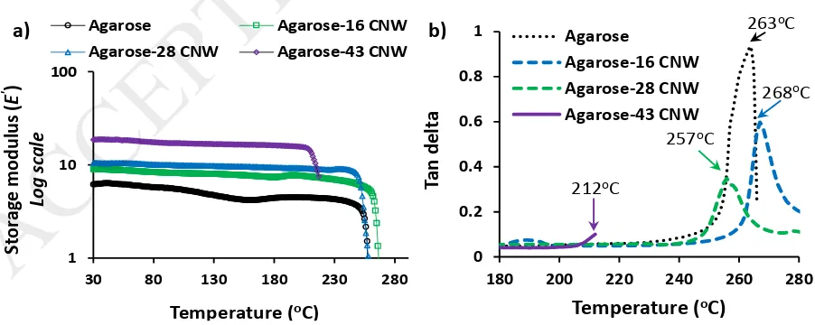
Thermo-mechanical properties
DMA was used to obtain the storage modulus of the nanocomposite films as a function of
temperature (Figure 9a). The DMA test showed that the Agarose-CNW nanocomposite films
demonstrated good stability against temperature up to and beyond 200oC as shown in Figure
9. The addition of CNWs within the Agarose matrix showed a significant increase in storage
modulus properties for all compositions with increasing CNWs content over the whole
temperature range investigated (25 to 280oC). The storage modulus of the nanocomposite
a) b)
0 1 2 3 4 5 0 20 40 60 80 100 120 Te ns ile mo du lu s (G Pa ) Te ns ile s tr en gth (MPa ) Tensile strength Tensile modulus 0 20 40 60 80 100 120
0 0.1 0.2 0.3
Te n si le s tr es s (M Pa ) Strain
Agarose Agarose-16 CNW
Agarose-28 CNW Agarose-43 CNW
20
films increased to reach ca. 300% of Agarose alone as the CNWs content increased to 43 wt%. Nanofillers blended with polymer was also reported to improve the storage modulus of the
matrix at higher temperatures due to their higher interfacial surface area, and surface
induced crystallisation within the nanocomposites (Petersson, Kvien, & Oksman, 2007; Zakir
Hossain et al., 2012).
Tan delta curves of the Agarose-CNW flexible films are presented in Figure 9b. Agarose-16
CNW film revealed that the tan delta peaks had shifted slightly to a higher temperature region
(268oC) as compared to pure Agarose film (263oC), which indicated that the incorporated
CNWs governed the reduction in the segmental motions of the Agarose matrix in the blends
through CNW-Agarose interactions (Petersson et al., 2007). However, 28 and 43 wt% addition
of CNWs within the nanocomposite films resulted in a shift of tan delta peaks to lower
temperature region (257oC and 212oC, respectively). This was suggested to be due to the
thermal degradation of cotton based acid hydrolysed CNWs which was reported to be
initiated at around 200oC with major decomposition at 261oC (Zakir Hossain et al., 2012). At
higher nanofiller loading it was anticipated that the presence of a considerable amount of
aggregated CNWs (as confirmed earlier by the white dots obtained via polarised light
microscopy images) was believed to be loosely bound within the matrix. These poorly bound
aggregated CNWs were expected to degrade after 200oC as they are not firmly protected by
the matrix thus leading to heat induced failure of the nanocomposite films, which was also
obvious from the tan delta curve (film broke at ~212oC) of the Agarose-43 CNW film.
Figure 9: a) storage modulus curves and b) tan delta curves of thin films produced using various composites of Agarose and CNWs. Five replicates (n=5) of each specimen were tested and the mean (±SD) is presented.
a) b)
0 0.2 0.4 0.6 0.8 1
180 200 220 240 260 280
Ta
n
d
el
ta
Temperature (oC) Agarose
Agarose-16 CNW Agarose-28 CNW Agarose-43 CNW
212oC
257oC
268oC
263oC
1 10 100
30 80 130 180 230 280
St or ag e m odu lus ( E ') Log s cal e
Temperature (oC)
Agarose Agarose-16 CNW Agarose-28 CNW Agarose-43 CNW
[image:21.595.79.530.518.698.2]21 Swelling kinetics
The swelling capacity, i.e. percentage of weight change due to water absorption, of control
Agarose and Agarose-CNW nanocomposite films in PBS media at 37oC with swelling time is
presented in Figure 10a. The incorporation of CNWs within the matrix led to a significant
decrease in the swelling capacity of the nanocomposites suggesting the presence of a greater
degree of CNW-CNW interaction which was believed to decrease the hydrophilicity by
reducing the water holding capacity (W. Wang, Wang, Kang, & Wang, 2011; Zakir Hossain et
al., 2012).
A swelling kinetics model (Schott’s second order) was also introduced (Equation 3) (Schott,
1992; Zakir Hossain et al., 2012) to evaluate the theoretical swelling capacity and initial
swelling rate of the Agarose-CNW nanocomposite films (W. Wang et al., 2011).
) 3 ...( ... 1
1
t Q is k t Q
t
Here, Qt (g/g) denotes the swelling capacity of the films at time t (s), Q∞ (g/g) is the theoretical
equilibrium swelling capacity and Kis (g/g) is the initial swelling rate constant.
A plot of t/Qt against t was drawn using the experimental swelling data of Agarose-CNW films
(see Figure 10b) and the linear trend line obtained for these experimental values showed high
regression coefficients (>0.99), which justified the use of Schott’s second-order swelling
kinetics model for these nanocomposite films in PBS media (W. Wang et al., 2011). The
theoretical equilibrium swelling capacity (Q∞) and the initial swelling rate constant (C. Liu et
al.) were calculated using the linear regression equation: Y= a + bX, (where, Y=t/Qt, a=1/Kis,
b=1/Q∞ and X=t) obtained from the linear lines and plotted against CNWs content in the
nanocomposite films in Figure 10c. The calculated theoretical swelling kinetic parameters (Q∞
and Kis) of the nanocomposite films were found to decrease with addition of CNWs content
which suggested that the incorporation of CNWs into the matrix played a significant influence
on decreasing swelling rate of the Agarose-CNW nanocomposite films. The values of Q∞ and
Kis decreased by ca. 50% of their values (280 g.g-1 and 0.6 g.g-1.s-1) as the amount of CNWs
reached 43 wt%.
22
Figure 10: a) Swelling kinetic curves and b) Corresponding plots of t/Qt against t of the
nanocomposite films produced using various blends of Agarose and CNWs. c) Relationship between the swelling rate constant and equilibrium swelling capacity with CNWs content in PBS media at 37oC. Five replicates (n=5) of each specimen were used in the swelling
experiment and the mean (±SD) is presented.
Most of the highly transparent polymer nanocomposites reported in the literature were
obtained by in situ polymerisation technique and /or surface modification of the
nanoparticles (Althues et al., 2007; Bin et al., 2011; Dang et al., 2014; C. Liu et al., 2017). In
this study, a highly transparent Agarose-CNW nanocomposite was achieved, using
conventional solution casting method and non-functionalised nanoparticles, benefiting from
two characteristics of the nanocomposite constituents; sol-gel property of Agarose and
suspension of the non-agglomerated CNWs. It was also found that incorporation of CNWs into
the Agarose matrix provided significant increases in mechanical, thermomechanical and
a) b)
c) 0 100 200 300 400 500 0 0.1 0.2 0.3 0.4 0.5 0.6 0.7
0 10 20 30 40 50
Q∞ (g g -1) K is (g g -1 s -1)
CNW content (wt%)
Swelling rate constant
Equilibrium swelling capacity 0 50 100 150 200 250 300 350
0 25000 50000 75000 100000
Q
t (
g g
-1)
Time (s)
Agarose Agarose-16 CNW Agarose-28 CNW Agarose-43 CNW
y = 0.0035x + 1.4898
y = 0.0055x + 2.3073 y = 0.0065x + 2.7992
y = 0.0083x + 3.6551
0 100 200 300 400 500 600 700 800
0 25000 50000 75000 100000
23
crystallinity and decrease in the swelling capacity of the nanocomposites when compared to
control Agarose films. These modified capabilities offer many potential applications for
cellulose based composite films in tissue engineering, drug coating and encapsulation.
Conclusions
This study presents a morphological, surface topographical and film forming properties of
freeze-dried and aqueous suspended CNWs. The freeze-dried CNWs were seen to aggregate
into flake-like geometries with higher surface roughness (Ra ~75 nm) compared to the well
dispersed needle-like aqueous CNWs (Ra ~11 nm). Due to higher surface roughness the
freeze-dried CNWs were found to form a white, opaque and brittle film, while the aqueous CNWs
film was brittle and translucent. However, transparent and flexible films based on a range of
Agarose-CNWs nanocomposite films were prepared in this study. Incorporation of
never-dried CNWs within the matrix was seen to be well dispersed despite some limited aggregation
within higher CNWs loaded film (43 wt%), which played an influential role on the surface
roughness (Ra below 15 nm for all nanocomposite films) and optical transparency properties
of the nanocomposite films (transparency more than 84%). An increase in crystallinity of
nanocomposite films with CNWs content suggested that the CNWs acted as nucleating sites
to induce crystallisation within the Agarose matrix. The enhanced tensile strength, modulus
and storage modulus properties of the nanocomposite films compared to the control Agarose
film confirmed the reinforcing effect of the nanowhiskers The incorporation of CNWs also had
a significant influence on controlling the swelling properties of Agarose-CNW blend films in
aqueous media.
References
Abou-Sekkina, M. M., Sakran, M.A. and Saafan, A.A. (1986). Industrial & Engineering Chemistry Product Research and Development 25, 676.
Agarwal, U. P., Ralph, S. A., Reiner, R. S., & Baez, C. (2016). Probing crystallinity of never-dried wood cellulose with Raman spectroscopy. Cellulose, 23(1), 125-144.
Althues, H., Simon, P., & Kaskel, S. (2007). Transparent and luminescent YVO4 : Eu/polymer nanocomposites prepared by in situ polymerization. Journal of Materials Chemistry, 17(8), 758-765.
Atef, M., Rezaei, M., & Behrooz, R. (2014). Preparation and characterization agar-based nanocomposite film reinforced by nanocrystalline cellulose. International Journal of Biological Macromolecules, 70(Supplement C), 537-544.
24
Aulin, C., Salazar-Alvarez, G., & Lindstrom, T. (2012). High strength, flexible and transparent nanofibrillated cellulose-nanoclay biohybrid films with tunable oxygen and water vapor permeability. Nanoscale, 4(20), 6622-6628.
Awadhiya, A., Tyeb, S., Rathore, K., & Verma, V. (2017). Agarose bioplastic-based drug delivery system for surgical and wound dressings. Engineering in Life Sciences, 17(2), 204-214. Bin, C., Sugihara, O., Elim, H. I., Adschiri, T., & Kaino, T. (2011). An approach of high refractive index
and highly transparent polymer nanocomposite fabrication. 17th Microopics Conference (MOC) (pp. 1-2).
Boujemaoui, A., Cobo Sanchez, C., Engström, J., Bruce, C., Fogelström, L., Carlmark, A., & Malmström, E. (2017). Polycaprolactone Nanocomposites Reinforced with Cellulose
Nanocrystals Surface-Modified via Covalent Grafting or Physisorption: A Comparative Study. ACS Applied Materials & Interfaces, 9(40), 35305-35318.
Camargo, P. H. C., Satyanarayana, K. G., & Wypych, F. (2009). Nanocomposites: synthesis, structure, properties and new application opportunities. Materials Research, 12, 1-39.
Cao, Z., Gilbert, R. J., & He, W. (2009). Simple Agarose−Chitosan Gel Composite System for Enhanced Neuronal Growth in Three Dimensions. Biomacromolecules, 10(10), 2954-2959.
Celebi, H., & Kurt, A. (2015). Effects of processing on the properties of chitosan/cellulose nanocrystal films. Carbohydrate Polymers, 133(Supplement C), 284-293.
Chaudhary, J. P., Vadodariya, N., Nataraj, S. K., & Meena, R. (2015). Chitosan-Based Aerogel
Membrane for Robust Oil-in-Water Emulsion Separation. ACS Applied Materials & Interfaces, 7(44), 24957-24962.
Cheng, Y., Lu, C., Lin, Z., Liu, Y., Guan, C., Lu, H., & Yang, B. (2008). Preparation and properties of transparent bulk polymer nanocomposites with high nanophase contents. Journal of Materials Chemistry, 18(34), 4062-4068.
Chowdhury, R. A., Peng, S. X., & Youngblood, J. (2017). Improved order parameter (alignment) determination in cellulose nanocrystal (CNC) films by a simple optical birefringence method. Cellulose, 24(5), 1957-1970.
Cipiriano, B. H., Kashiwagi, T., Raghavan, S. R., Yang, Y., Grulke, E. A., Yamamoto, K., . . . Douglas, J. F. (2007). Effects of aspect ratio of MWNT on the flammability properties of polymer
nanocomposites. Polymer, 48(20), 6086-6096.
Corthell, J. T. (2014). Chapter 3 - Agarose Gel Electrophoresis. In Basic Molecular Protocols in Neuroscience: Tips, Tricks, and Pitfalls (pp. 21-25). San Diego: Academic Press
Dang, A., Ojha, S., Hui, C. M., Mahoney, C., Matyjaszewski, K., & Bockstaller, M. R. (2014). High-Transparency Polymer Nanocomposites Enabled by Polymer-Graft Modification of Particle Fillers. Langmuir, 30(48), 14434-14442.
Ehlert, S., Stegelmeier, C., Pirner, D., & Förster, S. (2015). A General Route to Optically Transparent Highly Filled Polymer Nanocomposites. Macromolecules, 48(15), 5323-5327.
Gadjanski, I., Yodmuang, S., Spiller, K., Bhumiratana, S., & Vunjak-Novakovic, G. (2013). Supplementation of Exogenous Adenosine 5′-Triphosphate Enhances Mechanical
Properties of 3D Cell–Agarose Constructs for Cartilage Tissue Engineering. Tissue Engineering Part A, 19(19-20), 2188-2200.
George, J., & Sabapathi, S. N. (2015). Cellulose nanocrystals: synthesis, functional properties, and applications. Nanotechnology, Science and Applications, 8, 45-54.
Hassanabadi, H. M., & Rodrigue, D. (2014). Effect of Particle Size and Shape on the Reinforcing Efficiency of Nanoparticles in Polymer Nanocomposites. Macromolecular Materials and Engineering, 299(10), 1220-1231.
Heath, L., & Thielemans, W. (2010). Cellulose nanowhisker aerogels. Green Chemistry, 12(8), 1448-1453.
25
Hossain, K. M. Z., Hasan, M. S., Boyd, D., Rudd, C. D., Ahmed, I., & Thielemans, W. (2014). Effect of Cellulose Nanowhiskers on Surface Morphology, Mechanical Properties, and Cell Adhesion of Melt-Drawn Polylactic Acid Fibers. Biomacromolecules, 15(4), 1498-1506.
Iwamoto, S., Kai, W., Isogai, A., & Iwata, T. (2009). Elastic Modulus of Single Cellulose Microfibrils from Tunicate Measured by Atomic Force Microscopy. Biomacromolecules, 10(9), 2571-2576.
Jordan, J., Jacob, K. I., Tannenbaum, R., Sharaf, M. A., & Jasiuk, I. (2005). Experimental trends in polymer nanocomposites—a review. Materials Science and Engineering: A, 393(1), 1-11. Kargarzadeh, H., Mariano, M., Huang, J., Lin, N., Ahmad, I., Dufresne, A., & Thomas, S. (2017). Recent
developments on nanocellulose reinforced polymer nanocomposites: A review. Polymer, 132, 368-393.
Khalil, R., Homaeigohar, S., Häußler, D., & Elbahri, M. (2016). A shape tailored gold-conductive polymer nanocomposite as a transparent electrode with extraordinary insensitivity to volatile organic compounds (VOCs). Scientific Reports, 6, 33895.
Khoshkava, V., & Kamal, M. R. (2014). Effect of drying conditions on cellulose nanocrystal (CNC) agglomerate porosity and dispersibility in polymer nanocomposites. Powder Technology, 261(Supplement C), 288-298.
Labet, M., & Thielemans, W. (2011). Improving the reproducibility of chemical reactions on the surface of cellulose nanocrystals: ROP of ε-caprolactone as a case study. Cellulose, 18(3), 607-617.
Lee, K.-Y., & Bismarck, A. (2012). Susceptibility of never-dried and freeze-dried bacterial cellulose towards esterification with organic acid. Cellulose, 19(3), 891-900.
Lee, S. G., Lee, J. S., Ha, J.-W., Park, I. J., Lee, S.-B., & Lee, J. D. (2009). Effect of crystallinity on surface morphology and light transmittance of poly(vinylidene fluoride-co-hexafluoropropylene) thin film. Journal of Applied Polymer Science, 114(5), 3331-3337.
Lee, W. J., Clancy, A. J., Kontturi, E., Bismarck, A., & Shaffer, M. S. P. (2016). Strong and Stiff: High-Performance Cellulose Nanocrystal/Poly(vinyl alcohol) Composite Fibers. ACS Applied Materials & Interfaces, 8(46), 31500-31504.
Li, Q., Zhou, J., & Zhang, L. (2009). Structure and properties of the nanocomposite films of chitosan reinforced with cellulose whiskers. Journal of Polymer Science Part B: Polymer Physics, 47(11), 1069-1077.
Li, Y.-Q., Fu, S.-Y., Yang, Y., & Mai, Y.-W. (2008). Facile Synthesis of Highly Transparent Polymer Nanocomposites by Introduction of Core–Shell Structured Nanoparticles. Chemistry of Materials, 20(8), 2637-2643.
Liu, C., Li, Z., Hajagos, T. J., Kishpaugh, D., Chen, D. Y., & Pei, Q. (2017). Transparent Ultra-High-Loading Quantum Dot/Polymer Nanocomposite Monolith for Gamma Scintillation. ACS Nano, 11(6), 6422-6430.
Liu, D., Pourrahimi, A. M., Olsson, R. T., Hedenqvist, M. S., & Gedde, U. W. (2015). Influence of nanoparticle surface treatment on particle dispersion and interfacial adhesion in low-density polyethylene/aluminium oxide nanocomposites. European Polymer Journal, 66(Supplement C), 67-77.
Liu, D. Y., Yuan, X. W., Bhattacharyya, D., & Easteal, A. J. (2010). Characterisation of solution cast cellulose nanofibre - Reinforced poly(lactic acid). Express Polymer Letters 4(1), 26-31. Liu, Y., Sun, D., Askari, S., Patel, J., Macias-Montero, M., Mitra, S., . . . Maguire, P. (2015). Enhanced
Dispersion of TiO2 Nanoparticles in a TiO2/PEDOT:PSS Hybrid Nanocomposite via Plasma-Liquid Interactions. Scientific Reports, 5, 15765.
Medina-Esquivel, R., Freile-Pelegrin, Y., Quintana-Owen, P., Yáñez-Limón, J. M., & Alvarado-Gil, J. J. (2008). Measurement of the Sol–Gel Transition Temperature in Agar. International Journal of Thermophysics, 29(6), 2036.
Moon, R. J., Martini, A., Nairn, J., Simonsen, J., & Youngblood, J. (2011). Cellulose nanomaterials review: structure, properties and nanocomposites. Chem Soc Rev, 40(7), 3941-3994.
26
Müller, K., Bugnicourt, E., Latorre, M., Jorda, M., Echegoyen Sanz, Y., Lagaron, J. M., . . . Schmid, M. (2017). Review on the Processing and Properties of Polymer Nanocomposites and
Nanocoatings and Their Applications in the Packaging, Automotive and Solar Energy Fields. Nanomaterials, 7(4), 74.
Nogi, M., Iwamoto, S., Nakagaito, A. N., & Yano, H. (2009). Optically Transparent Nanofiber Paper. Advanced Materials, 21(16), 1595-1598.
Oun, A. A., & Rhim, J.-W. (2015). Effect of post-treatments and concentration of cotton linter cellulose nanocrystals on the properties of agar-based nanocomposite films. Carbohydrate Polymers, 134(Supplement C), 20-29.
Pandey, N., Shukla, S. K., & Singh, N. B. (2017). Water purification by polymer nanocomposites: an overview. Nanocomposites, 3(2), 47-66.
Parker, R. M., Frka-Petesic, B., Guidetti, G., Kamita, G., Consani, G., Abell, C., & Vignolini, S. (2016). Hierarchical Self-Assembly of Cellulose Nanocrystals in a Confined Geometry. ACS Nano, 10(9), 8443-8449.
Petersson, L., Kvien, I., & Oksman, K. (2007). Structure and thermal properties of poly(lactic acid)/cellulose whiskers nanocomposite materials. Composites Science and Technology, 67(11-12), 2535-2544.
Qiang, X., Chunfang, Z., JianZun, Y., & Yuan, C. S. (2004). The effects of polymer-nanofiller
interactions on the dynamical mechanical properties of PMMA/CaCO3 composites prepared by microemulsion template. Journal of Applied Polymer Science, 91(5), 2739-2749.
Rahman, I. A., Vejayakumaran, P., Sipaut, C. S., Ismail, J., & Chee, C. K. (2008). Effect of the drying techniques on the morphology of silica nanoparticles synthesized via sol–gel process. Ceramics International, 34(8), 2059-2066.
Riggs, B. C., Adireddy, S., Rehm, C. H., Puli, V. S., Elupula, R., & Chrisey, D. B. (2015). Polymer Nanocomposites for Energy Storage Applications. Materials Today: Proceedings, 2(6), 3853-3863.
Rusli, R., & Eichhorn, S. J. (2008). Determination of the stiffness of cellulose nanowhiskers and the fiber-matrix interface in a nanocomposite using Raman spectroscopy. Applied Physics Letters, 93(3), 033111-033113.
Sahoo, N. G., Rana, S., Cho, J. W., Li, L., & Chan, S. H. (2010). Polymer nanocomposites based on functionalized carbon nanotubes. Progress in Polymer Science, 35(7), 837-867.
Santamaria-Echart, A., Ugarte, L., Gonzalez, K., Martin, L., Irusta, L., Gonzalez, A., . . . Eceiza, A. (2017). The role of cellulose nanocrystals incorporation route in waterborne polyurethane for preparation of electrospun nanocomposites mats. Carbohydrate Polymers,
166(Supplement C), 146-155.
Schott, H. (1992). Swelling kinetics of polymers. Journal of Macromolecular Science, Part B, 31(1), 1-9.
Segal S., C. J. J., Martin A.E., Conrad C.M. . (1959). An empirical method of estimating the degree of crystallinity of native cellulose using the X-ray diffractometer. Textile Research Journal, 29, 786-794.
Shumigin, D., Tarasova, E., Krumme, A., & Meier, P. (2011). Rheological and Mechanical Properties of Poly(lactic) Acid/Cellulose and LDPE/Cellulose Composites. MATERIALS SCIENCE
(MEDŽIAGOTYRA), 17(1), 32-37.
Tematio, C., Fosso, N., Krähenbühl, J., & Schintke, S. (2016). Conductive polymer nanocomposites for transparent circuits and thin films. 2016 18th International Conference on Transparent Optical Networks (ICTON) (pp. 1-5).
Trache, D., Hussin, M. H., Haafiz, M. K. M., & Thakur, V. K. (2017). Recent progress in cellulose nanocrystals: sources and production. Nanoscale, 9(5), 1763-1786.
Trivedi, T. J., Rao, K. S., & Kumar, A. (2014). Facile preparation of agarose-chitosan hybrid materials and nanocomposite ionogels using an ionic liquid via dissolution, regeneration and sol-gel transition. Green Chemistry, 16(1), 320-330.
27
Wang, W., Wang, J., Kang, Y., & Wang, A. (2011). Synthesis, swelling and responsive properties of a new composite hydrogel based on hydroxyethyl cellulose and medicinal stone. Composites Part B: Engineering, 42(4), 809-818.
Wang, Z., Zhang, R., Ma, Y., Peng, A., Fu, H., & Yao, J. (2010 a). Chemically responsive luminescent switching in transparent flexible self-supporting [EuW10O36]9--agarose nanocomposite thin films. Journal of Materials Chemistry, 20(2), 271-277.
Wang, Z., Zhang, R., Ma, Y., Zheng, L., Peng, A., Fu, H., & Yao, J. (2010 b). Transparent and flexible phosphomolybdate-agarose composite thin films with visible-light photochromism. Journal of Materials Chemistry, 20(6), 1107-1111.
Weimer, P. J., Hackney, J. M., & French, A. D. (1995). Effects of chemical treatments and heating on the crystallinity of celluloses and their implications for evaluating the effect of crystallinity on cellulose biodegradation. Biotechnology and Bioengineering, 48(2), 169-178.
Xiaodong, L., Hongsheng, G., Wally, A. S., Dongling, F., Vivek, T., Michael, A. S., . . . Michael, L. M. (2005). Structural and mechanical characterization of nanoclay-reinforced agarose nanocomposites. Nanotechnology, 16(10), 2020.
Xu, X., Liu, F., Jiang, L., Zhu, J. Y., Haagenson, D., & Wiesenborn, D. P. (2013). Cellulose Nanocrystals vs. Cellulose Nanofibrils: A Comparative Study on Their Microstructures and Effects as Polymer Reinforcing Agents. ACS Applied Materials & Interfaces, 5(8), 2999-3009.
Yang, H., Liang, T., Qi, X., Jiang, H., Deng, Y., Wang, P., & Gao, H. (2017). Solvothermal synthesis of hydroxyapatite nanorods with assistance of green polymer. Materials Science and
Engineering: C, 79(Supplement C), 9-14.
Yu, H.-Y., Zhang, H., Song, M.-L., Zhou, Y., Yao, J., & Ni, Q.-Q. (2017). From Cellulose Nanospheres, Nanorods to Nanofibers: Various Aspect Ratio Induced Nucleation/Reinforcing Effects on Polylactic Acid for Robust-Barrier Food Packaging. ACS Applied Materials & Interfaces, 9(50), 43920-43938.
Zakir Hossain, K. M., Jasmani, L., Ahmed, I., Parsons, A. J., Scotchford, C. A., Thielemans, W., & Rudd, C. D. (2012). High cellulose nanowhisker content composites through cellosize bonding. Soft Matter, 8(48), 12099-12110.
Zare, Y., & Shabani, I. (2016). Polymer/metal nanocomposites for biomedical applications. Materials Science and Engineering: C, 60(Supplement C), 195-203.