Expression of a
glycosylphosphatidylinositol-anchored ligand, growth hormone, blocks
receptor signalling
Franc¸ois GUESDON*1, Yahia KAABI†1, Aiden H. RILEY†, Ian R. WILKINSON†, Colin GRAY†, David C. JAMES‡,
Peter J. ARTYMIUK§, Jon R. SAYERS* and Richard J. ROSS†2
*Department of Infection and Immunity, University of Sheffield, Medical School, Beech Hill Road, Sheffield S10 2RX, U.K., †Department of Human Metabolism, University of Sheffield, Medical School, Beech Hill Road, Sheffield S10 2RX, U.K., ‡Department of Chemical and Biological Engineering Chemistry, University of Sheffield, Mappin Street, Sheffield S1 3JD, U.K., and§Department of Molecular Biology and Biotechnology, University of Sheffield, Firth Court, Western Bank, Sheffield S10 2TN, U.K.
'
&
$
%
Synopsis
We have investigated the interaction between GH (growth hormone) and GHR (GH receptor). We previously demon-strated that a truncated GHR that possesses a transmembrane domain but no cytoplasmic domain blocks receptor signalling. Based on this observation we investigated the impact of tethering the receptor’s extracellular domain to the cell surface using a native lipid GPI (glycosylphosphatidylinositol) anchor. We also investigated the effect of tethering GH, the ligand itself, to the cell surface and demonstrated that tethering either the ecGHR (extracellular domain of GHR) or the ligand itself to the cell membrane via a GPI anchor greatly attenuates signalling. To elucidate the mechanism for this antagonist activity, we used confocal microscopy to examine the fluorescently modified ligand and receptor. GH–GPI was expressed on the cell surface and formed inactive receptor complexes that failed to internalize and blocked receptor activation. In conclusion, contrary to expectation, tethering an agonist to the cell surface can generate an inactive hormone receptor complex that fails to internalize.
Key words: cytokine antagonist, growth hormone, growth hormone receptor, glycosylphosphatidylinositol anchor, signal transduction, receptor trafficking
Cite this article as: Guesdon, F., Kaabi, Y., Riley, A.H., Wilkinson, I.R., Gray, C., James, D.C., Artymiuk, P.J., Sayers, J.R. and Ross, R.J. (2012) Expression of a glycosylphosphatidylinositol-anchored ligand, growth hormone, blocks receptor signalling. Biosci. Rep. 32, 653–660
INTRODUCTION
We have been investigating the interaction of GH (growth hor-mone) with its receptor and investigating approaches to manipu-late receptor signalling. We observed that a patient heterozygous for a mutation in the GHR (GH receptor) had a short stature and GH insensitivity suggesting that the mutation created a dom-inant negative receptor [1]. The mutation encoded a truncated receptor that had a normal extracellular and transmembrane do-main but lacked the essential cytoplasmic signalling dodo-main. We demonstrated that the dominant negative action occurred as the truncated receptor was highly expressed on the cell surface,
. . . .
Abbreviations used:CMV, cytomegalovirus; GFP, green fluorescent protein; GH, growth hormone; GHR, growth hormone receptor; ecGHR, extracellular domain of GHR; GPI, glycosylphosphatidylinositol; HEK, human embryonic kidney; LHRE, lactogenic hormone response element; LL, long linker; PE, phycoerythrin; RFP, red fluorescent protein; RL,Renillaluciferase; wtGH, wild-type GH.
1 These authors made an equal contribution to this study.
2 To whom correspondence should be addressed (email r.j.ross@sheffield.ac.uk).
complexed with the full-length receptor, but the complex failed to signal or internalize [2]. Based on these observations, we pro-posed that anchoring a truncated receptor to the cell surface would generate an antagonist and by using a synthetic lipid anchor we demonstrated this was the case [3]. Following on from this work we have been examining the impact of anchoring proteins us-ing naturally occurrus-ing GPI (glycosylphosphatidylinositol) lipid anchors.
GPI anchors are common components of the eukaryotic cell membrane and examples include ALP (alkaline phosphatase) and DAF (decay-accelerating factor) [4]. GPI-anchored proteins are tethered to the cell membrane through a glycolipid moiety and have no transmembrane or cytoplasmic domains. Recombinant
ing a fusion of the ecGHR (extracellular domain of GHR) with a GPI anchor and also asked the question; what would happen if we anchored the ligand itself to the cell surface? As expec-ted, anchored receptor blocked signalling but to our surprise we found that the anchored ligand also blocked receptor signalling and internalization.
EXPERIMENTAL
Plasmids
The reporter construct pUC18–LHRE (lactogenic hormone re-sponse element)–Luc, containing STAT5 (signal transducer and activator of transcription 5) binding element, LHRE fused to the minimal tk (thymidine kinase) promoter and the firefly lu-ciferase cDNA (GHR) has been described previously [2]. The
phRL-CMV expression vector, encoding RL (Renillaluciferase)
under control of the CMV (cytomegalovirus) promoter, was from Promega. The expression vector pCR3.1gpi, encoding the mam-malian Thy-1 GPI signal sequence under control of the CMV promoter was a gift from C. Beghadi (University of Lausanne, Lausanne, Switzerland). The cDNAs encoding human GH and the ecGHR were cloned without stop codons upstream of the Thy-1 GPI signal sequence in pCR3.Thy-1gpi to generate the ecGHR–GPI and GH-GPI constructs. The human GH cDNA was cloned in the same vector to generate the wtGH (wild-type GH) construct, en-coding non-anchored wtGH. GH–LL (long linker)–GPI was gen-erated by insertion in GH–GPI of a sequence encoding a flexible
LL consisting of five repeats of a Gly4Ser (tetraglycine-serine)
motif between the coding sequences of GH and the Thy-1 signal sequence. The GHR–GFP (green fluorescent protein) construct, encoding the full-length GHR fused at its C-terminal end to the GFP, was generated by inserting the open GHR coding sequence into the pTagGFP vector (Evrogen). The GH–RFP (red fluor-escent protein) construct, encoding GH fused at its C-terminal end to the RFP, was made by inserting the open GH coding se-quence into pTagRFP. Adding the Thy-1-GPI signal to GH–RFP then generated the GH–RFP–GPI construct. All the constructs were subjected to DNA sequencing carried out within our Core Genetics Facility (Faculty of Medicine, Dentistry and Health, University of Sheffield).
remove the residual unbound stain. PE was excited at 488 nm and
the emitted light was detected through a 585+−21 nm band pass
filter. Data were acquired on a FACSort flow cytometer (Bec-ton Dickinson) using the CellQuest data acquisition and analysis software housed in our Core Flow Cytometry Facility (Faculty of Medicine, Dentistry and Health, University of Sheffield).
Triton X-114 phase partitioning
Transfected HEK-293 cells (1×10⁶) producing ecGHR–GPI,
GH–GPI or GH–LL–GPI were washed in PBS and lysed in
250μl of lysis buffer (20 mM Tris/HCl, 150 mM NaCl, 1 mM
EDTA, 2 % Triton X-114 and 0.0 005 % Bromophenol Blue). The cells were then left on ice with frequent stirring for 20 min.
The resulting lysate was centrifuged at 13 000gfor 5 min at 0◦C
to remove cellular debris. The supernatant was incubated at 30◦C
for 5 min and then centrifuged at 4000gfor 3 min at room
tem-perature to separate the clear aqueous phase from the detergent phase containing Bromophenol Blue.
Western blotting
Samples were separated by SDS/PAGE (12 % gels) and blot-ted on to the PVDF membrane. The primary antibodies were mouse anti-GHR mAb263 (Biogenesis used at a 1:2500 dilution ratio) and anti-human GH (rabbit) polyclonal antibody [NIH (Na-tional Institutes of Health), used at 1:10000 dilution]. The second-ary antibodies were anti-mouse or anti-rabbit HRP (horseradish peroxidase)-linked IgG (Amersham Pharmacia Biotech) used at 1:10000 dilution. They were detected using an enhanced chemi-luminescence substrate (Roche Diagnostics) and light sensitive X-ray films.
GH signalling bioassay
Figure 1 GPI-anchored proteins associate with plasma membrane
(A–C) Detection of GPI-anchored proteins at the surface of transfected cells. HEK-293 cells were transfected with ecGHR–GPI (A), GH–GPI (B) or GH–LL–GPI (C), labelled with antibodies to GHR or GH and subjected to flow cytometry. Histograms from transfected cells (dark grey curves) are shown overlaid onto control histograms from untransfected cells (light grey). (D) Phase partitioning and molecular masses of GPI-anchored proteins. Cells transfected with ecGHR–GPI, GH–GPI or GH–LL–GPI were subjected to phase partitioning and the resulting detergent (Deter.) and aqueous (Aqu.) phases were probed by Western blotting with antibodies to GHR or GH, as appropriate. The positions and molecular masses (kDa) of standard proteins are shown next to the first immunoblot.
18 h after transfection, then lysed after a further 6 h. The fire-fly and RL activities were measured using the Promega dual luciferase reporter assay kit. The firefly luciferase values were normalized to those of the phRL-CMV-encoded RL to correct for variations in transfection efficiency and cell number. The res-ults were expressed as fold-induction relative to the unstimulated control of each plate. For experiments requiring autocrine GH stimulation, HEK-293–GHR cells were transfected twice. The first transfection used 100 ng/well (12-well plate) of wtGH con-struct. The other cDNA expression constructs and the reporter constructs were co-transfected in the second transfection 18 h later.
Laser scanning confocal microscopy
HEK-293 cells were grown on poly-L-lysine-coated
glass-bottomed microwell culture dishes (MatTek Corporation) for 16 h before being transfected using Lipofectamine2000 (Invitrogen). Confocal microscopy was carried out 2 days after transfection using a Zeiss 510 NLO laser scanning confocal microscope fitted
with×20 NA 0.8 and×40 NA 1.2W (water) objective lenses.
Fluorescence images of GFP were obtained using 488 nm laser excitation, NFT490 dichroic and LP505 emission filter. Images of RFP were obtained using 543 nm laser excitation, NFT545 dichroic and LP560 emission filters. Images were acquired at
1024×1024 (pixel dwell time 0.8μs) pixels and 1.3μm optical
slices. Background fluorescence was subtracted from the im-ages using ImageJ 1.37c software (NIH) [20]. Co-localization of fluorophores was identified by Intensity Correlation Analysis, determining the positive product of the difference of the means with the ImageJ plug-in developed at the Wright Cell Imaging Facility, Toronto, Canada.
RESULTS
GPI-anchored GH proteins demonstrate cell membrane expression
We wished to examine the impact of anchoring ecGHR and GH to the cell surface and we also raised the question whether the length of linker between GH and its anchor would alter bio-logical activity. The following constructs were tested: wtGH with no anchor (wt GH), GH linked to GPI either through a short linker (GH–GPI) or LL (GH–LL–GPI), and ecGHR linked to GPI (ecGHR–GPI). Flow cytometry of HEK-293 cells transfected with the GPI-anchored proteins: GH–GPI, GH–LL– GPI and ecGHR–GPI showed that transfection efficiencies were
>90 % and that for all the GPI-anchored proteins there was a
Figure 2 Inhibition of signalling by GPI-anchored proteins
(A) HEK-293–GHR cells were co-transfected with the pUC18–LHRE–Luc and phRL–CMV reporter constructs, with or without the wtGH, ecGHR–GPI, GH–GPI or GH–LL–GPI cDNA construct, as indicated, and stimulated 18 h later with various doses of exogenous GH. Normalized reporter levels are expressed relative to values in cells transfected with reporter constructs only and not exposed to GH. Means+−S.E.M. ofn=4 (ecGHR–GPI, GH–LL–GPI) orn=6 are shown. (B) HEK-293–GHR cells were transfected with the reporter constructs but no cDNA and were then challenged with conditioned media from untransfected cells or cDNA-transfected cells expressing ecGHR-GPI or GH–GPI, as indicated. Stimulation with exogenous GH, reporter assays and data analysis were carried out as in (A). Means+−S.E.M. ofn=3 are shown. (C) To test the inhibition of the autocrine GH response, HEK-293–GHR cells were pre-transfected with 100 ng of wtGH construct 18 h before being transfected a second time with the reporter constructs and various amounts of plasmid encoding ecGHR-GPI, as indicated. Data analysis was carried out as in (A) and (B) and one-way ANOVA with Bonferroni’spost-hoccomparison was performed. Means+−S.E.M. ofn=6 are shown. One, two or three asterisks correspond toP<0.05,P<0.01 or
P<0.001, respectively. (D) As in (C), except that the GH–GPI expression construct was used instead of ecGHR–GPI.
assessed by phase partitioning (separation of protein into either a detergent or aqueous phase) to demonstrate the presence or absence of a GPI anchor (Figure 1D). The recombinant pro-teins ecGHR–GPI, GH–GPI, GH–LL–GPI migrated at their pre-dicted molecular masses and all partitioned into the detergent-enriched phase with no detectable protein in the aqueous phase, as is expected for GPI-anchored proteins. These res-ults confirmed that all the three GPI-anchored proteins were expressed at the cell surface and retained their GPI lipid moiety.
GPI-membrane-anchored GH and ecGHR block GH signalling
A dual luciferase promoter–reporter bioassay for functional ligand–receptor-mediated signalling was employed to examine the action of GPI-anchored proteins. HEK-293 cells stably ex-pressing human GHR (HEK-293–GHR) and transiently trans-fected with the bioassay reporter constructs showed a dose-dependent response to exogenously administered GH (Fig-ure 2A). When the wtGH expression plasmid was co-transfected with the reporter constructs, high reporter expression was seen independent of exogenous GH, indicating that the expression of wtGH led to autocrine/paracrine stimulation. In contrast, HEK-293–GHR cells transfected with the anchored ecGHR, ecGHR– GPI, showed no increase in basal reporter expression and no response to the exogenously administered GH (Figure 2A). This demonstrated that, as expected, ecGHR–GPI acted as an ant-agonist. To our surprise, cells transfected with GH–GPI did not behave like the wt-GH-transfected cells but instead showed
no increase in basal reporter expression and, like the ecGHR– GPI-transfected cells, no response to the exogenously added GH (Figure 2A). We questioned whether an LL between GH and GPI could overcome any steric hindrance and restore sig-nalling; however, the GH–LL–GPI construct inhibited the bioas-say as effectively as GH–GPI (Figure 2A). Thus, both the GH– GPI and GH–LL–GPI constructs behaved as antagonists of GH signalling.
We considered the possibility that the inhibitory effects of the GPI-linked proteins might be caused by the partial release of these proteins into the culture medium. To examine this, we challenged untransfected cells with GH in the presence of con-ditioned media from cells transfected with either ecGHR–GPI or GH–GPI. The conditioned media did not inhibit the response to GH (Figure 2B), indicating that the ecGHR–GPI- and GH– GPI-expressing cells were not secreting any detectable soluble inhibitor of GH signalling.
Figure 3 Biological activities of the fluorescent proteins
(A) HEK-293 cells were subjected to the promoter–reporter bioassay in the absence of co-transfected cDNA or in the presence of cDNA constructs encoding wt GHR or GHR–GFP, as indicated. (B) The activities of fluorescent labelled GH-RFP and GH–RFP–GPI proteins were tested with the bioassay in the HEK-293–GHR-expressing cell line as had been done for the non-fluorescent wtGH and GH–GPI in Figure 2(A). All the data were analysed as in Figure 2 and are means+−S.E.M. of
n=4 (A) orn=6 (B).
Signalling properties of fluorescent fusion proteins
As a preparation to investigating the mechanisms of inhibition of GH action by the GPI fusion proteins, we generated a red-fluorescent GPI-anchored GH (GH–RFP–GPI), a red-red-fluorescent non-anchored GH (GH–RFP) and a green fluorescent receptor (GHR–GFP) and assessed the biological activities of the fluor-escent proteins. The activity of fluorfluor-escent labelled GHR–GFP was assessed with the dual-luciferase promoter–reporter bioas-say in the HEK-293 cells, which endogenously only express low levels of GHR. In the absence of a co-transfected GHR cDNA, the cells only show weak activation of reporter expression in response to exogenously administered GH (maximum fold
ac-tivation 1.65+−0.26), but the response to the GH is dramatically
increased when they are transfected with a wt GHR cDNA
con-struct (maximum fold activation 16.9+−1.5, Figure 3A). A
sim-ilar but quantitatively smaller increase was observed when the fluorescent receptor construct, GHR–GFP, was substituted for
the wtGHR (maximum fold induction 6.5+−0.9, Figure 3A).
The activities of fluorescent labelled GH–RFP and GH–RFP– GPI proteins were tested in the HEK-293–GHR cell line (Fig-ure 3B). Cells expressing GH–RFP showed constitutively high
levels of reporter expression (8.2+−1.5-fold activation of reporter
expression relative to cells expressing no cDNA) and this was not significantly increased when the cells were exposed to increas-ing amounts of exogenously administered GH (Figure 3B). Ex-pression of GH–RFP–GPI activated reporter exEx-pression, albeit to a lesser extent than that observed in cells expressing GH–
RFP (5.7+−0.7-fold activation). This intermediate level of
re-porter activity suggested that the expression of GH–RFP–GPI partially activated the GH signalling machinery; however, no significant increase in reporter activation was observed when the cells expressing GH–RFP–GPI were exposed to increas-ing doses of exogenous GH (Figure 3B). This suggested that while GH–RFP–GPI differed from GH–GPI and GH–LL–GPI by being a partial agonist, it shared with the other GPI
con-structs the ability to block the action of exogenously administered GH.
GPI-anchored GH forms cell surface complexes with wt receptor
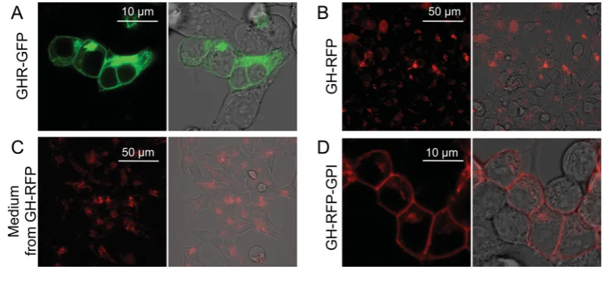
Figure 4 Intracellular localization of fluorescent fusion proteins
(A,B) HEK-293 cells imaged after transfection with either the green fluorescent GHR fusion construct, GHR–GFP (A) or the red fluorescent cytokine construct, GH–RFP (B). (C) HEK-293 cells that have not been transfected were imaged after a 3 h incubation in conditioned media from the GH–RFP-expressing cells. The intracellular red fluorescence indicates that the cells had internalized GH–RFP present in the conditioned media from the GH–RFP-expressing cells. (D) HEK-293 cells imaged after transfection with the GPI-anchored fluorescent GH construct, GH–RFP–GPI. Each confocal fluorescence image is shown both alone (left) and overlaid onto a phase contrast image of the same field (right). In contrast, anchored GH–RFP–GPI is predominantly found on the cell surface (C).
To analyse the interactions between the GH constructs and the GH receptor, we then co-transfected the GH–RFP expres-sion plasmid with the GH-based constructs (Figure 5). In cells co-transfected with GHR–GFP and GH–RFP, the recombinant receptor showed both membrane and cytoplasmic localizations (Figure 5A). However, in contrast to what had been observed in the single transfection experiments (Figure 4A), the intracel-lular pool of GHR–GFP molecules was not evenly distributed throughout the cytoplasm but appeared instead to accumulate in discrete intracellular locations (Figure 5A) similar to that seen for internalized GH–RFP from media (Figure 4C). Overlaying the red and green fluorescent images showed that a fraction of the GHR–GFP pool co-localizes with GH–RFP (Figure 5C), and this was confirmed by the co-localization analysis (Fig-ure 5D). Similar results were observed when cult(Fig-ure media containing non-anchored GH–RFP were added to GHR–GFP-transfected cells (Figures 5G–5K). These results are entirely consistent with the distributions of GH–RFP observed previ-ously in cells that were not expressing GHR–GFP (Figures 4B and 4C). They indicate that co-localization of GH–RFP and the receptor reflects internalization of the receptor–hormone complex.
We then observed the distributions of co-transfected GHR– GFP and GH–RFP–GPI (Figures 5I–5L). The results show that the two pools of fluorescent proteins are entirely co-localized, with the majority of each construct being located on the cell mem-brane and only a minor fraction of each pool adopting an intra-cellular distribution (Figure 5I–5L). Thus, GH–RFP-GPI forms a complex with GHR–GFP on the cell surface, which does not internalize.
DISCUSSION
We have examined the interaction between GH and its receptor and investigated the impact of anchoring either the extracellular domain of the receptor or the GH ligand itself to the cell surface via attachment of an endogenous GPI anchor. Both the receptor’s extracellular domain and ligand blocked receptor signalling when tethered to the cell membrane through a GPI anchor.
GH binding to its receptor results in a conformational change that includes rotation in the transmembrane domain and trig-gers signalling and internalization [9]. We questioned whether increasing the length of linker to the cell membrane could restore agonistic activity to our GPI-anchored hormone, as too short a linker could restrict the movement of the molecule. However, the GH–LL–GPI construct inhibited GH signalling as efficiently
as GH–GPI despite the presence of a 70 ˚A (1 ˚A=0.1 nm) long
glycine/serine-rich linker that was intended to relieve any con-formational or spatial restraints between the GH moiety and the GPI domain.
Figure 5 GPI-anchoring of GH blocks GHR internalization
This Figure examines the co-localization of GHR and GH with and without GPI-anchored GH–GPI. In cells co-expressing the non-anchored GHR–GFP (A,C,D) and GH–RFP (B–D) proteins, a fraction of the GHR–GFP pool co-localizes with the GH–RFP inside cells (A–D). Co-localization is indicated by yellow colouring in the overlay image (C) and is confirmed in the intensity correlation image (D). The same distributions are observed when cells expressing GHR–GFP only are incubated with exogenously provided GH–RFP (E–H), indicating that intracellular co-localization reflects the internalization of the receptor–ligand complexes. By contrast, when GHR–GFP is co-expressed with the anchored GH–RFP–GPI (I–L), neither protein is internalized.
The cytoplasmic domain of the GHR is essential not only for activating the GHR but also for triggering internalization of the hormone receptor complex [10]. However, signalling is not re-quired for receptor internalization as a receptor with a mutated cytoplasmic domain that fails to signal still internalizes [11], and the GH antagonist Pegvisomant binds the receptor dimer, pre-vents signalling but is internalized [12]. Truncated receptors that lack a cytoplasmic domain act as dominant negative inhibitors of signalling not only because they heterodimerize with the full-length receptor but also because they prevent internalization and therefore accumulate on the cell surface [2]. It has been repor-ted that both GH and its receptor may translate to the nucleus and correlate with proliferative activity [13]; thus a failure of the hormone receptor complex to internalize could again prevent the biological activity of GH. Our results suggest that GH–GPI not only occupies the receptor to prevent signalling but also holds the receptor complex at the cell surface. GHR is present in ca-veolae and lipid rafts which are lipid-rich microdomains of the plasma membrane [14,15]. Various membrane proteins are con-centrated in these lipid microdomains including GPI-anchored proteins such as the Thy-1 cell surface antigen [16]. Thus, GPI
anchoring of GH or ecGHR increases the probability that the GPI-anchored proteins will co-segregate with the native GHR on the cell membrane and constrain the GHR in a conformation that cannot be readily internalized.
In some cell types, autocrine actions of GH are more potent than exogenously administered GH [17–19]. Moreover, autocrine GH producing cells become unresponsive to externally admin-istered GH [20,21]. Both GH–GPI and ecGHR–GPI inhibited the action of intracellularly generated GH, and by increasing the ratio of GH–GPI to wtGH, the inhibition was increased although never complete. However, it should be recognized that trans-fection experiments result in relatively high expression levels of endogenous GH compared with physiological conditions. We attempted to purify ecGHR–GPI and GH–GPI and investigate whether the GPI fusions could be re-inserted into the cell mem-branes, however despite repeated attempts we were unable to purify the proteins (results not shown). This may relate to either the presence of a lipid moiety or the low level of expression of cell surface proteins.
FUNDING
This work was supported by the University of Sheffield [grant
num-ber RF102780] and the Saudi Arabian Cultural Bureau (to Y.K.).
REFERENCES
1 Ayling, R. M., Ross, R., Towner, P., Von Laue, S., Finidori, J., Moutoussamy, S., Buchanan, C. R., Clayton, P. E. and Norman, M. R. (1997) A dominant-negative mutation of the growth hormone receptor causes familial short stature. Nat. Genet.16, 13–14 2 Ross, R. J., Esposito, N., Shen, X. Y., Von Laue, S., Chew, S. L.,
Dobson, P. R., Postel-Vinay, M. C. and Finidori, J. (1997) A short isoform of the human growth hormone receptor functions as a dominant negative inhibitor of the full-length receptor and generates large amounts of binding protein. Mol. Endocrinol.11, 265–273
3 Bowles, C. E., Wilkinson, I., Smith, R. A., Moir, A. J., Montgomery, H. and Ross, R. J. (2007) Membrane reinsertion of a
myristoyl-peptidyl anchored extracellular domain growth hormone receptor. Endocrinology148, 824–830
4 Cross, G. A. (1990) Glycolipid anchoring of plasma membrane proteins. Annu. Rev. Cell Biol.6, 1–39
5 Medof, M. E., Nagarajan, S. and Tykocinski, M. L. (1996) Cell-surface engineering with GPI-anchored proteins. FASEB J.10, 574–586
6 Cunningham, B. C., Ultsch, M., de Vos, A. M., Mulkerrin, M. G., Clauser, K. R. and Wells, J. A. (1991) Dimerization of the extracellular domain of the human growth hormone receptor by a single hormone molecule. Science254, 821–825
7 Woodhouse, L. J., Mukherjee, A., Shalet, S. M. and Ezzat, S. (2006) The influence of growth hormone status on physical impairments, functional limitations and health-related quality of life in adults. Endocr. Rev.27, 287–317
receptor as being critical for ligand-mediated internalization and down-regulation. J. Biol. Chem.270, 17210–17214
11 Alves dos Santos, C. M., ten Broeke, T. and Strous, G. J. (2001) Growth hormone receptor ubiquitination, endocytosis, and degradation are independent of signal transduction via Janus kinase 2. J. Biol. Chem.276, 32635–32641
12 Maamra, M., Kopchick, J. J., Strasburger, C. J. and Ross, R. J. (2004) Pegvisomant, a growth hormone-specific antagonist, undergoes cellular internalization. J. Clin. Endocrinol. Metab.89, 4532–4537
13 Conway-Campbell, B. L., Wooh, J. W., Brooks, A. J., Gordon, D., Brown, R. J., Lichanska, A. M., Chin, H. S., Barton, C. L. and Boyle, G. M. Parsons et al. (2007) Nuclear targeting of the growth hormone receptor results in dysregulation of cell proliferation and tumorigenesis. Proc. Natl. Acad. Sci. U.S.A.104, 13331–13336 14 Yang, N., Huang, Y., Jiang, J. and Frank, S. J. (2004) Caveolar and
lipid raft localization of the growth hormone receptor and its signaling elements: impact on growth hormone signaling. J. Biol. Chem.279, 20898–20905
15 Galbiati, F., Razani, B. and Lisanti, M. P. (2001) Emerging themes in lipid rafts and caveolae. Cell106, 403–411
16 Brown, D. A. and London, E. (1998) Functions of lipid rafts in biological membranes. Annu. Rev. Cell Dev. Biol.14, 111–136 17 Goh, E. L., Pircher, T. J. and Lobie, P. E. (1998) Growth hormone
promotion of tubulin polymerization stabilizes the microtubule network and protects against colchicine-induced apoptosis. Endocrinology139, 4364–4372
18 Kaulsay, K. K., Mertani, H. C., Tornell, J., Morel, G., Lee, K. O. and Lobie, P. E. (1999) Autocrine stimulation of human mammary carcinoma cell proliferation by human growth hormone. Exp. Cell Res.250, 35–50
19 Mylonas, P. G., Matsouka, P. T., Papandoniou, E. V., Vagianos, C., Kalfarentzos, F. and Alexandrides, T. K. (2000) Growth hormone and insulin-like growth factor I protect intestinal cells from radiation induced apoptosis. Mol. Cell. Endocrinol.160, 115–122 20 Liu, N., Mertani, H. C., Norstedt, G., Tornell, J. and Lobie, P. E.
(1997) Mode of the autocrine/paracrine mechanism of growth hormone action. Exp. Cell Res.237, 196–206
21 van den Eijnden, M. J. and Strous, G. J. (2007) Autocrine growth hormone: effects on growth hormone receptor trafficking and signaling. Mol. Endocrinol.21, 2832–2846
Received 8 August 2012; accepted 13 August 2012