INTRODUCTION
Quantifying the nuclear DNA (nDNA) methylation of cytosine bases at specific autosomal genetic loci has been widely used to estimate an individual’s age for forensic and medical purposes [1-5]. The existence of methylated cytosines within mtDNA has been controversial. Originally proposed four decades ago, the presence of mitochondrial methylation has been difficult to confirm [6, 7]. Maekawa et al. showed signals for mitochondrial methylation in 2004, [8] and Infantino and co-workers provided the first evidence of methylated bases (5-methyl-2′-deoxycytidine) present in human mtDNA using mass spectrometry[9]. MtDNA methylation has now been reported in the form of methylcytosine (mC) and hydro-xymethylcytosine (hmC) modification[10], and a mitochondrially targeted DNA methyltransferase 1 enzyme (mtDNMT1) is suggested to be responsible[11, 12, 13, 14-18].
Research Paper
MtDNA methylation has been proposed as a cause of ageing and disease [13, 19, 20]. A comprehensive profile of methylation levels across the mitochondrial genome and across ages would be an invaluable starting-point to address the role of epigenetics in these biological phenomena. Here, we describe the next generation sequencing analysis of blood-derived, bisulphite-treated mtDNA taken from a cohort of subjects with ages ranging from 18 to 91, using the Illumina MiSeq sequencing platform.
We selected functional regions of interest (ROI) within the mitochondrial genome and designed primers for methylation analysis – for example, the displacement loop (D-loop)/hypervariable regions that contain the promoters for the ‘heavy’ and ‘light’ strand polycistronic mRNAs, specific mitochondrial genes, and regions where published evidence already exists for methylation. [12] In addition, two replicated sites of mtDNA mutation were chosen because of their potential
www.impactaging.com
AGING,
Febr
uary 20
16
, Vol
8
No
2
Quantification
of
global
mitochondrial
DNA
methylation
levels
and
inverse
correlation
with
age
at
two
CpG
sites
Shakhawan
K.
Mawlood
1,
Lynn
Dennany
2,
Nigel
Watson
2,
John
Dempster
1,
and
Benjamin
S.
Pickard
1
1 Strathclyde Institute of Pharmacy and Biomedical Sciences, University of Strathclyde, Glasgow, Scotland, UK 2 Centre for Forensic Science, Department of Pure and Applied Chemistry, University of Strathclyde, Glasgow, Scotland, UK
Key words: mitochondrial DNA, epigenetics, ageing, age prediction, Illumina NGS Received: 10/26/15; Accepted: 01/20/15; Published: 02/17/16
Correspondence to: Shakhawan K Mawlood, PhD; E‐mail: Shakhawan.mawlood@strath.ac.uk
Copyright: Mawlood et al. This is an open‐access article distributed under the terms of the Creative Commons Attribution License,
which permits unrestricted use, distribution, and reproduction in any medium, provided the original author and source are credited
to create or destroy CpG dinucleotides: an A11778G associated with Leber`s Heriditary Optic Neuropathy, [21] and a T8993G mutation, which is associated with a neurological phenotype [22, 23].
RESULTS
The mitochondrial genome sequence (GRCH38 or Anderson/Cambridge reference sequence) [24] consists of 16,565 bp in which 435 predicted CpG sites are present (Supplementary 1). We used bisulphite treatment and high-throughput sequencing (Illumina) to assess methylation levels within regions of interest chosen because of their functional importance within the mito-chondrial genome. In total the study queried methylation levels at 133/435 CpG sites in the first sample set of DNA from 41 individuals.
We identified unambiguous evidence for non-zero mtDNA methylation at 54 of the 133 CpG sites sequenced with average read depth of 2000 (Fig. 1). Methylation levels were, on average, very low (often between 2-6%) but showed regional differences across the mitochondrial genome and, more importantly, a great variance between individuals, as the methylation maxima and averages indicate (Fig.1).
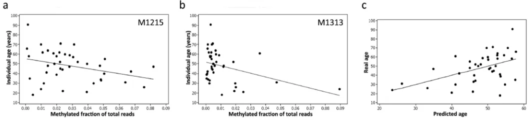
The correlation between methylation and age was determined in the data set sample using Pearson’s correlation coefficient for the 54 sites. Sites MT1215 and MT1313, both in the 12S RNA gene (MT-RNR1) showed significant methylation changes (hypomethylation) with increasing age (MT1215, 0.322 p=0.043. mt1313 R=-0.383, p=0.015). These findings were consistent with age-related changes reported in the general 12S RNA gene region by others [13, 26] .
Figure 2. Methylation levels (expressed as a fraction) at two CpG sites within the 12S RNA gene, M1215 (a) and M1313
(b), correlate with age and can be used to construct an accurate predictive model (c) using Pearson’s correlation.
Figure 1. Mean methylation levels at 54 CpG sites across the mitochondrial genome (category labels denote
reference base position). While methylation levels are typically 2‐6%, considerable inter‐individual variation was
[image:2.612.44.550.296.438.2] [image:2.612.42.573.538.661.2]In the next step, multivariate linear regression was applied to the methylation data from both M1215 and M1313 (Fig.2a, b) and a linear regression prediction model was developed relating age to methylation.
The prediction accuracy of the model and contribution of particular predictors were assessed using the adjusted R2 statistic, which measures the proportion of age variation explained by the developed model. The model yielded an R2=0.509 indicating that 50% of the variation in methylation could be explained by age. The mean absolute deviation (MAD) between predicted and chronological age was 9.3 years (Fig.2c).
The second sequenced library dataset (n=41 individuals) was used to evaluate the model. From this set, 31 samples (64.5%) had ages correctly predicted to within a ± MAD. The analysed samples were also divided into three age categories: I: 18–39 years, II: 40–59 years, III: 60 years and older. Prediction results were significantly better for samples from category II (83.3% correct predictions) than for categories I (53.8%) and III (20% correct predictions), suggesting that innate or environmental methylation-determining factors, in addition to age, are at play in early and late life.
DISCUSSION
Although previous reports have shown correlation of mtDNA damage and mutation with aging, [27] it was only very recently that studies suggested that mitochondrial methylation could be used as an ageing biomarker [13, 15, 28]. Dzitoyeva and co-workers have reported the existence of mitochondrial DNA (hydroxy)methylation and its increase with ageing in subregions the mouse brain.[10] Our study provides the most comprehensive map of methylation levels at 133 of the available 435 CpG sites in the mitochondrial genome from 82 individuals. Quantified changes in the methylation state (hypomethylation) of two 12S RNA gene CpG sites (M1215 and M1313) correlate with age. This novel discovery opens new avenues to investigate the mtDNA changes associated with health status and in the processes of criminal investigation. The CpG pair at M1215 and M1313 could only have been detected using high throughput sequencing, such as by Illumina MiSeq300 v2. Therefore, such sensitive and advanced methodologies can solve the issue of failure to detect low-level methylation in the mtDNA genome that may explain reported absence of mitoepigenetics [29, 30], its failure as a health tool marker [8] or its description as having an unusual CpG pattern.[14] Also contributing to this confusion might have been the use of quantification techniques such as ELISA and MeDIP that may struggle with low-level methylation.
The correlation between M1215/M1313 methylation and age was especially accurate for younger and middle-aged individuals in contrast to the poorer predictive power after the age of 60. This might reflect differences in individual lifetime metabolic health or exposure to environmental influences, thus giving rise to discrepancies between biological and chronological age. Furthermore, we speculate that these life-accumulated environmental influences cause the substantial general mtDNA methylation variance observed between individuals. Alternatively, methylation differences might also reflect varying representation of cell-types in blood samples – again, a potential surrogate for cytological investigation that might provide useful insights into immunological status. Our study did not differentiate between 5mC and 5hmC as the chemical conversion by sodium bisulphite (the gold standard detection technique for methylation studies) distinguishes only converted cytosines from non-methylated cytosines. The study has some limitations that have been taken into account in data interpretation. Further studies are needed to isolate mtDNA and analyse its methylation in specific subtypes of blood cells, such as platelet progenitors and lymphocytes. Further, mitochondrial DNA analysis from other populations and from male samples needs to be carried out to assess the generality of our findings. Lastly, the impact on mtDNA methylation of determinants of ill health, including smoking, pollution, obesity and chronic life stress should be studied to quantify external influences.
To conclude, Illumina sequencing of 82 human blood samples indicated clear methylation patterns in mtDNA at 54 CpG sites in mitochondrial genome and that the level of the methylation was variable among different ages (18-91 years). Two of these CpG sites (M1215 and M1313) showed stronger correlation between predicted and chronological age with only about 9 years mean absolute difference (MAD). This finding implies that mtDNA methylation will be an available biological marker for forensic age-prediction and health status measurement.
PATIENTS, MATERIALS AND METHODS
lifestyle. 100 µL aliquots were immediately placed in Eppendorf tubes and DNA extracted. The extracted DNA was quantified by Stratagene 3005X qPCR instrument (Agilent Technologies, CA, USA), using the Quantifiler® Human DNA Quantification Kit cat. # 4343895 (Life Technologies, CA, USA).
Region of interest selection. The mitochondrial sequence (GRCH38 or Anderson/Cambridge reference sequence) [24] consists of 16,565 bp in which 435 predicted CpG sites are present (Supplementary 1). Regions of interest (ROI) were chosen on the basis of biological function: the promoters for both ‘heavy’ and ‘light’ strand of the D-loop regions, polycistronic mRNAs and most of the functional genes of mitochondria. In addition, two replicated sites site of
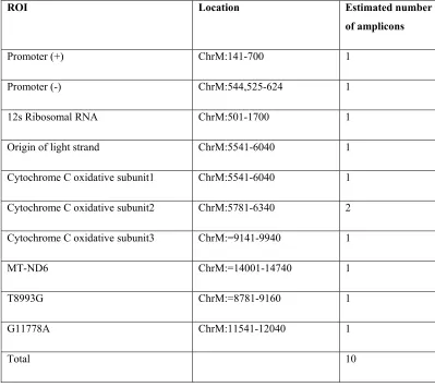
[image:4.612.105.504.326.681.2]mtDNA mutation were chosen because of their potential to create or destroy CpG dinucleotide: A11778G associated with Leber`s Heriditary Optic Neuropathy, [21] and a T8993G mutation, which is associated with a neurological phenotypes [22, 23]. Primer sequences used to amplify the RsOI (each amplicon 100-300bp in length) and the amplicon sequence (before and after bisulphite conversion) are in Supplementary 2. Primers targeting 10 ROI amplicons (Table 1) were designed using Methyl Primer Express Software v1.0 (Applied Biosystem, Foster City, California), synthesised (Applied Biosystems, Foster City, California, and Life Technologies, CA, USA), and successfully passed amplification quality control criteria. Some locations within each ROI were impossible to amplify due to repeat sequences or high CpG content.
Table 1. 10 Amplicons which passed Illumina quality control criteria. The location of each ROI in
mitochondria genome (ChrM) and estimated amplicon number in the mDNA are shown as well.
ROI Location Estimated number
of amplicons
Promoter (+) ChrM:141-700 1
Promoter (-) ChrM:544,525-624 1
12s Ribosomal RNA ChrM:501-1700 1
Origin of light strand ChrM:5541-6040 1
Cytochrome C oxidative subunit1 ChrM:5541-6040 1
Cytochrome C oxidative subunit2 ChrM:5781-6340 2
Cytochrome C oxidative subunit3 ChrM:=9141-9940 1
MT-ND6 ChrM:=14001-14740 1
T8993G ChrM:=8781-9160 1
G11778A ChrM:11541-12040 1
Bisulphite analysis and library construction. The mtDNA was modified (non-methylated C residues converted to U) via a bisulphite conversion step, EZ DNA Methylation-DirectTM Kit (Cat. 5020 and 5021, Zymo Research).[25] Multiplex PCR amplification of the RsOI (Fluidigm Access ArrayTM System, BioMark, USA) generated pools of amplicons that also employed a universal forward tag common sequence 1 (CS1), and universal reverse tag common sequence 2 (CS2) (CS1= ACACTGACGACATGGTTCTACA-3', CS2 = 5'-TACGGTAGCAGAGACTTGGTCT-3'). Each indivi-dual amplicon pool was subsequently barcoded. Two age-matched libraries, each consisting of 41 individuals, were created and purified (ZR-96 DNA Clean & Concentrator™ - ZR, Cat.# D4023) and then prepared for massively parallel sequence by Illumina next generation sequencing (NGS) using a MiSeq V2 300bp Reagent Kit (cat. # MS-102-2001), (paired-end sequencing protocol) according to the manufacturer’s guidelines.
ACKNOWLEDGEMENTS
The authors thank all the volunteers who donate sample and Keith Booher [kbooher@zymoresearch.com] from Zymo Research Corporation for the Illumina Next Generation Sequencing. Special thanks go to Olga Krasnova, Production Editor of Aging journal [krasnova@impactaging.com] for her guide and un-conditional help.
Funding
This work was sponsored by Ministry of Higher Education/ Kurdistan Regional Government (KRG) and supported by Criminal Evidence Laboratory/ Ministry of Interior/ KRG.
Conflict of interest statement
The authors have no conflict of interests to declare.
REFERENCES
1. Yi SH, Xu LC, Mei K, Yang RZ, Huang DX. Isolation and
identification of age‐related DNA methylation markers for
forensic age‐prediction. Forensic Science International: Genetics.
2014; 11:117‐125.
2. Burgess DJ. Human epigenetics: Showing your age. Nature
Reviews Genetics. 2012; 14:6.
3. Bocklandt S, Lin W, Sehl M, Sánchez F, Sinsheimer J.
Epigenetic Predictor of Age. PLoS ONE. 2011; 6:e14821.
4. Koch CM, Wagner W. Epigenetic‐aging‐signature to determine
age in different tissues. Aging (Albany NY). 2011; 3:1018‐10127.
5. Weidner CI, Lin Q, Koch CM, Eisele L, Beier F, Ziegler P, et al.
Aging of blood can be tracked by DNA methylation changes at
just three CpG sites. Genome Biology. 2014; 15:R24.
6. Manev H, Dzitoyeva S, Chen H. Mitochondrial DNA: a blind
spot in neuroepigenetics. Biomolecular concepts. 2012; 3:107‐
115.
7. Hong EE, Okitsu CY, Smith AD, Hsieh C‐L. Regionally specific
and genome‐wide analyses conclusively demonstrate the
absence of CpG methylation in human mitochondrial DNA.
Molecular and cellular biology. 2013; 33:2683‐90.
8. Maekawa M, Taniguchi T, Higashi H, Sugimura H, Sugano K,
Kanno T. Methylation of mitochondrial DNA is not a useful marker
for cancer detection. Clinical chemistry. 2004; 50:1480‐1481.
9. Stewart SA, Weinberg RA. Senescence: does it all happen at
the ends? Oncogene. 2002; 21:627‐630.
10. Stewart SA, Weinberg RA. Telomeres: cancer to human
aging. Annu Rev Cell Dev Biol. 2006; 22:531‐557.
11. Shock LS, Thakkar PV, Peterson EJ, Moran RG, Taylor SM.
DNA methyltransferase 1, cytosine methylation, and cytosine
hydroxymethylation in mammalian mitochondria. Proceedings
of the National Academy of Sciences. 2011; 108:3630‐3635.
12. Chinnery PF, Elliott HR, Hudson G, Samuels DC, Relton CL.
Epigenetics, epidemiology and mitochondrial DNA diseases.
International journal of epidemiology. 2012; 41:177‐187.
13. Iacobazzi V, Castegna A, Infantino V, Andria G. Mitochondrial
DNA methylation as a next‐generation biomarker and diagnostic
tool. Molecular Genetics and Metabolism. 2013; 110:25‐34.
14. Bellizzi D, D'Aquila P, Scafone T, Giordano M, Riso V, Riccio A,
et al. The Control Region of Mitochondrial DNA Shows an
Unusual CpG and Non‐CpG Methylation Pattern. DNA Research.
2013; dst029.
15. Byun H‐M, Panni T, Motta V, Hou L, Nordio F, Apostoli P, et
al. Effects of airborne pollutants on mitochondrial DNA
methylation. Part Fibre Toxicol. 2013; 10:18.
16. O'Sullivan M, Rutland P, Lucas D, Ashton E, Hendricks S,
Rahman S, et al. Mitochondrial m. 1584A 12S m62A rRNA
methylation in families with m. 1555A> G associated hearing
loss. Human molecular genetics. 2014; ddu518.
17. Metodiev MD, Lesko N, Park CB, Cámara Y, Shi Y, Wibom R,
et al. Methylation of 12S rRNA is necessary for in vivo stability of
the small subunit of the mammalian mitochondrial ribosome.
Cell metabolism. 2009; 9:386‐397.
18. Rebelo AP, Williams SL, Moraes CT. In vivo methylation of
mtDNA reveals the dynamics of protein–mtDNA interactions.
Nucleic acids research. 2009; gkp727.
19. Feng S, Xiong L, Ji Z, Cheng W, Yang H. Correlation between
increased ND2 expression and demethylated displacement loop
of mtDNA in colorectal cancer. Molecular medicine reports.
2012; 6:125‐130.
20. Pirola CJ, Gianotti TF, Burgueño AL, Rey‐Funes M, Loidl CF,
Mallardi P, et al. Epigenetic modification of liver mitochondrial
DNA is associated with histological severity of nonalcoholic fatty
liver disease. Gut. 2012; gutjnl‐2012‐302962.
21. Komaki H, Akanuma J, Iwata H, Takahashi T, Mashima Y,
Nonaka I, et al. A novel mtDNA C11777A mutation in Leigh
syndrome. Mitochondrion. 2003; 2:293‐304.
22. Harding A, Holt I, Sweeney M, Brockington M, Davis M.
Prenatal diagnosis of mitochondrial DNA8993 T‐‐‐‐G disease.
American journal of human genetics. 1992; 50:629.
23. Holt I, Harding A, Petty R, Morgan‐Hughes J. A new
mitochondrial disease associated with mitochondrial DNA
heteroplasmy. American journal of human genetics. 1990;
24. Butler JM. Forensic DNA typing biology, technology, and
genetics of STR markers. Amsterdam; Boston: Elsevier Academic
Press. 2005.
25. RESEARCH Z. EZ DNA Methylation‐Direct™ Kit. Zymo
Research. 2014.
26. Ghosh S, Sengupta S, Scaria V. Comparative analysis of
human mitochondrial methylomes shows distinct patterns of
epigenetic regulation in mitochondria. Mitochondrion. 2014;
18:58‐62.
27. Xu C, Qu H, Wang G, Xie B, Shi Y, Yang Y, et al. A novel
strategy for forensic age prediction by DNA methylation and
support vector regression model. Scientific reports. 2014;
5:17788.
28. Giordano M, Cristiani C, Crocco P, D'Aquila P, De Rango F,
Pisani F, et al. Methylation of the human mitochondrial 12S
rRNA gene is correlated with aging. 12th International FISV
Congress, Rome, Italy. 2012; 9.5.
29. Park J‐L, Kwon O‐H, Kim JH, Yoo H‐S, Lee H‐C, Woo K‐M, et al.
Identification of body fluid‐specific DNA methylation markers for
use in forensic science. Forensic Science International: Genetics.
2014; 13:147‐53.
30. Antunes JP, Madi T, Balamurugan K, Bombardi R, Duncan G,
McCord B. DNA methylation markers as a powerful technique to
discriminate body fluids present in crime scenes. Proceedings of
the 24th International Symposium on Human Identification.
2014.
SUPPLEMENTARY DATA
Please browse the Full text version of this manuscript to see the Supplementary material.
Supplementary 1. The regions of interest (ROI) with their sequences and primer sequences (forward and reverse). The single strand sequences of the ROI after bisulphite sodium conversion with the primers sequence (forward and reverse).
Supplementary 2. Reference Sequence: The Mito-chondrial sequence (hg38:2013) shows all the 345 CG (underlined and bold CG) and 54 assessed sites indicated by red text with underlined and bold CG.