THE USE OF INHIBITORS IN TEE A HA. LYSIS OF THE GROWTH CYCLE OF VACCINIA VIRUS
b y
K .B . EÄSTERBROOK
A T h e s is
s u b m i t t e d f o r t h e D e g re e o f D o c to r o f P h i lo s o p h y i n th e A u s t r a l i a n N a t i o n a l U n i v e r s i t y , C a n b e r r a ,
TABLE OF CONTENTS
STATEMENT* 1
INTRODUCTION• 2- 5
PAPERS PRESENTED:
1* The Multiplication of Vaccinia Virus 6-29
in Suspended KB Cells*
2* Analysis of the Early Stages of
Vaccinia Virus Infection of KB Cells 30-52
Using- Sodium Azide.
3* Interference with the Maturation of
Vaccinia Virus by Isatin 5J>-6Q
4 - thios e mi carbaz one.
4* The Effect of 5-B^om °(leo:xyuridine 011 69-85
the Multiplication of Vaccinia Virus. GENERAL DISCUSSION:
1. Inhibitors and Growth Cycle Studies* 87-116
2. The Growth Cycle of Vaccinia Virus* 117-145
STATEMENT
Chapter 4 embodies the results of an investigation
carried out along lines suggested by Dr. C.I. Davern. The
remainder of the thesis is the work of the candidate.
INTRODUCTION
Substances interfering with viral multiplication may
be studied with one of two aims in view; either the development
V
of an efficient chemotherapeutic agent or the analysis of the
mechanism by which the interference is produced. Numerous
compounds have been investigated and different methods of attack used in pursuit of the former aim without important practical results, and the relevant literature has become large (see Hurst
and Hull, 1956). By contrast, attempts to analyse the mechanism
of action of inhibitors and their use as tools in the dissection
of the growth cycle have been few. It is with this latter aim
that the work presented in this thesis has been designed and carried out.
The growth cycle of a virus is complex and can be
divided for ease of discussion into a number of more or less well
defined stages. Since viruses lack the essential enzymes
required for the synthesis and utilization of precursor material they are compelled to take-over or modify existing enzymes, concerned normally in the growth and replication of more highly
organised life-forms. This process of take-over constitutes the
3
host for viral multiplication»
In response to information supplied by the virus particle, the enzyme systems of the cell commence to synthesise abnormal proteins and nucleic acids which possess configurations
characteristic of the infecting virus. Viral nucleic acid is
then removed from the replicating pool by a process of assembly in which quanta of nucleic acid become enclosed in protein
envelopes to give units bearing superficial resemblance at least
to the parent virus. Finally some process of maturation occurs
and the units acquire the biological property of infectivity. This last stage may coincide with the release of the particles from the cell e.g, in the case of influenza virus, or may be essentially an intracellular process.
When viral multiplication is followed in a population of cells, these stages tend to become obscured as a result of asynchrony of initiation and differences in the rate of synthesis
in different cells. In studies involving populations of cells,
it is the first cell to enter a stage that determines the time
of commencement of the stage. Since, however, viral multiplication
It is possible, at least in theory, to inhibit viral multiplication at any stage with a resultant reduction or abolition of the normal yield of infectious virus particles* So far, however, the demonstration of such specific inhibition has not often been made because of inadequate criteria of
definition of any given stage* Alteration in the infectious virus yield as a criterion for inhibition is capable only of indicating interference somewhere in the sequence and not of indicating the stage at which it occurs.
The aim of the present study was to investigate the growth cycle of vaccinia virus by analysing the mode and time of action of certain inhibitors.
A
consideration of the5
a c id and p r o t e i n and th e p ro d u c tio n o f i n f e c t i o u s v i r u s .
The i n h i b i t o r s s tu d ie d in c lu d e d th o se i n t e r f e r i n g
w ith a ) th e su p p ly o f energy f o r p r e c u r s o r s y n t h e s i s , b ) th e
s y n th e s is o f v i r a l n u c le ic a c i d , c ) th e s y n th e s is o f v i r a l
p r o t e i n and d ) th e m a tu ra tio n o f v i r a l com ponents. P re lim in a r y
e x p erim en ts w ith i n h i b i t o r s o f p r o t e i n s y n th e s is proved un
s a t i s f a c t o r y and a t t e n t i o n was th e r e f o r e c o n c e n tr a te d on th e
e f f e c t s o f sodium a z id e , 5 -b rom odeoxyuridine and i s a t i n ß
-th io se m ic a rb a z o n e . The r e s u l t s o b ta in e d w ith th e s e i n h i b i t o r s
have b een p re s e n te d i n the form o f p a p ers su b m itte d f o r
p u b l i c a t i o n . The g e n e r a l d is c u s s io n w hich fo llo w s r e l a t e s the
e x p e rim e n ta l f in d i n g s s e t o u t i n th e s e p a p e rs w ith o th e r p u b lis h e d
work, and a n a tte m p t i s th e n made to d e s c r ib e th e grow th c y c le
o f v a c c in ia v i r u s , u s in g a l l a v a i l a b l e in f o r m a tio n .
Of th e f o u r p ap ers p re s e n te d , th e f i r s t two a r e under
c o n s id e r a tio n f o r p u b lic a tio n by the e d i t o r s o f V iro lo g y , and
th e t h i r d i s to be su b m itte d s h o r t l y to th e same j o u r n a l . The
r e s u l t s r e p o r te d i n the l a s t p a p er may be su b m itte d a t a l a t e r
THE MULTIPLICATION OF VACCINIA VIRUS
7
INTRODUCTION
The infection and maintenance of cells in suspension provides a system in which it is possible to analyse virus-cell interaction in cells exposed to uniform and controlled conditions.
The growth cycle of vaccinia virus in tissue culture has been followed under a variety of conditions including cells
in suspension (Furness and Youngner, 1959} Smith and Sharp,
i960). However, no attempt has been made to determine the number
of cells contributing, at a given instant, to the total virus
yield. This paper describes the infection of suspended KB cells
and their maintenance under conditions which permit a determination of the proportion of infected cells present and an estimate of the multiplicity of infection, and allow repeated sampling of the cell population and its constituent single cells.
The succeeding paper describes the use of this system to analyse the effect of sodium azide on the multiplication of vaccinia virus.
MATERIAIS AND METHODS
V irus. The chorioallantoic membrane (CAM) of 11 day-
old chick embryos was infected with a suspension prepared from a single pock of the Mill Hill (V-MH, Fenner, 1958) strain of
e x t r a c t e d w ith f lu r o c a r b o n (G e s s ie r e t a l . , 1956), and a s to c k
p r e p a r a t io n was d i s t r i b u t e d i n t o ampules and s to r e d a t -60° .
B efo re use thawed p r e p a r a tio n s were d is p e r s e d w ith a n u l t r a s o n i c
8.7
9.5
d r i l l . Such p r e p a r a t io n s c o n ta in e d 10 EFU p e r ml and 10
w e ll d is p e r s e d s i n g l e p a r t i c l e s p e r ml when examined w ith th e
e l e c t r o n m ic ro sc o p e .
M edia. Many non-immune s e r a c o n ta in s u b s ta n c e s
i n h i b i t o r y t o v a c c in ia v i r u s . Some o f t h i s a c t i v i t y i s d e s tr o y e d
by h e a ti n g a t 56° f o r 30 m in u tes b u t h e a t - s t a b l e i n h i b i t o r s
re d u c in g th e i n f e c t i v i t y by a p p ro x im a te ly 50^ a re p r e s e n t i n
many s e r a . A few specim ens o f serum fo u n d to be a lm o s t f r e e
from t h i s i n h i b i t o r y a c t i v i t y were r e s e r v e d f o r th e p r e p a r a t io n
o f a d s o r p tio n medium and n o n - in h i b ito r y grow th medium. A l l s e r a
were h e a te d a t 56° f o r 30 m in u tes b e fo re u s e .
F o u r ty p e s o f medium were used f o r d i f f e r e n t p h ases o f
th e e x p e rim e n ts .
1 . Growth medium f o r grow th o f c e l l m onolayers and f o r
grow th c y cle e x p e rim e n ts i n which e x t r a c e l l u l a r v ir u s was n o t
a s s a y e d . T h is c o n s is te d o f Vjffo human serum and 0 .5 ^ la c ta lb u m in
h y d ro ly s a te i n Hanks* B alan ced S a l t S o lu t io n (BSS).
2 . N o n - in h ib ito r y growth medium. The human serum
9
which interfered with the assay of extracellular virus. For
experiments involving such assays a medium containing 20$ selected non-inhibitory calf serum in E a g l e ’s solution (Eagle 1955&) was used.
3* Adsorption medium. Adsorption of virus and
washing of cells was carried out with more dilute non-inhibitory
calf serum. Adsorption medium consisted of 0*59$ calf serum
(non-inhibitory) in BSS.
4. Maintenance medium. The yeast-extract maintenance medium of Robertson et al. (1955) was used for certain experiments
on adsorption and growth in serum-free medium. This consisted
of 0.1$ Yeastolate (Difco) and 0.25$ glucose in Hanks’ BSS.
Cells. KB cells (Eagle, 1955^) were grown as mono-
layers in flat screw-capped bottles. For the production of cell
suspensions a 24 hour culture was usually chosen, half confluent
and with few cells in the medium. The monolayer was washed
briefly with trypsin-versene solution (0.02596 trypsin, 0.01$ EDTA) and incubated for 3-5 minutes at 37° with 5 ml per bottle of the
same solution. Cells still adhering to the glass were removed
by shaking. An equal volume of adsorption medium at room
temperature was added and the cells dispersed by expulsion through
centrifugation (135g* for 2 minutes), the cells were counted
in a Neubauer chamber. Cell v i a b i l i t y was assayed by ability
to exclude trypan blue (0#5$ in phosphate buffered saline), and on this basis most cell suspensions contained about 95fo
viable cells. Centrifuge tubes used in cell manipulations
were treated with a silicone#
Infectivity Titrations# Virus was titrated on the
CAM of 11 day-old embryonated eggs under the conditions described
by Westwood et al. (1957)» Results were read on the second day
after incubation at 36°. Virus dilutions were made in isotonic
saline containing 0#5^ gelatine.
Cell-associated virus (CAV) was assayed after cell disruption and dispersal of the virus particles by one cycle of
freezing and thawing followed by 30 seconds treatment with an
ultrasonic drill.
5
Staining of Antigen, Cell samples containing about 10
cells vrere washed in isotonic saline to remove traces of serum
protein. Smears prepared from these washed cells were dried
at room temperature for 1 hr. After fixation in acetone for
11
o b s e r v a tio n s w ith a n OSRAM HBO 200 m ercury vapour lamp as
l i g h t s o u rc e . A t o t a l o f 500 c e l l s were counted i n e ac h sm ear.
C o m p lem en t-F ix atio n . S u sp e n sio n s o f i n f e c t e d c e l l s
5
(5*10 c e l l s p er m l) were d is r u p te d by tre a tm e n t f o r 50 seco n d s
w ith an u l t r a s o n i c d r i l l . A f te r c e n t r i f u g a t i o n to remove c e l l
d e b r i s , th e amount o f a n tig e n was a ssa y e d by complement f i x a t i o n
t e s t s w ith a hyperimmune r a b b i t a n t i - v a c c i n i a serum . The immune
serum , a t a d i l u t i o n o f 1 :8 0 , and 3 MHD o f complement were u sed
i n a dropw ise t e s t . F i x a t i o n was a llo w ed to o ccu r o v e rn ig h t a t
4 ° . The d i l u t i o n of a n t i g e n f i x i n g 50^ o f th e complement was
ta k e n as th e t i t e r .
E x p e rim en ta l P ro ced u re f o r S in g le Cycle Growth C u rv e.
One to two m i l l i o n c e l l s i n 1 ml a d s o r p tio n medium were t r a n s
f e r r e d to a 3” x ■J** tu b e , which was c lo s e d w ith a s i l i c o n e - r u b b e r
s to p p e r . The a p p r o p r ia te v ir u s d i l u t i o n was added i n a volume
o f 0.05 m l. A d so rp tio n was a llo w ed to p ro ceed f o r 30 m in u tes
a t room te m p e ra tu re , th e c e l l s b e in g s t i r r e d by a sm all m ag n etic
s t i r r e r r o t a t i n g i n c o n ta c t w ith th e bottom o f th e tu b e .
F o llo w in g a d s o r p tio n , th e c e l l s were washed th r e e tim e s w ith 5 ml
volumes of a d s o r p tio n medium ( c e n tr if u g e d a t 135 g f o r 2 m in u te s ),
re d u c in g th e f r e e v i r u s to a th o u sa n d th of t h a t o r i g i n a l l y ad d ed .
Evidence for the Retention of Cellular Integrity by Infected Cells and for the Prolonged Association of
Virus with Such Cells.
Medium used for Incu-bationa
Cell Count (log No. per ml)
Virus Infectivity (log PFU per ml)
CAV
? r e e Virus
0 hours 24 hours 24 hours after infection Free Virus Cell-associated
Virus
4(y/oHuman
Serum
5.0 4.9 5.52 6.85 5.55
lj/o Human Serum
5.0 5.0 4.48 6.96 2.48
7
%
Human Serum5.0 5.0 4.40 6.82 2.42
2CP/o Calf Serum
5.5 5.5 4.40 6.40 2.00
Yeast Extract Maintenance
5.5 5.5 4.48
t
6.42 1.94
a
[image:14.525.67.515.51.670.2]12
per ml in 10 to 2C ml of warmed growth medium contained in
glass stoppered 6” x 1 ” tubes (growth tube). These were
flushed with 5$ CC^-air mixture and incubated over magnetic
stirrers in a water bath maintained at 37° + 0.25°.
The concentration of 10^ cells per ml in the growth
tube was chosen for several reasons. (l) At cell concentrations
less than 10^ per ml the suspended cells did not survive satis
factorily and virus yields fell. (2) Single cells could be
isolated from such suspensions without concentrating the sample* (3) Smears for fluorescent staining could be made from a
convenient volume of the cell suspension.
RESULTS
The Prolonged Association of Vaccinia Virus with Infected Cells.
Previous workers (e.g. Overman and Tamm, 1957» Joklik
and Rodrick, 1959» Furness and Youngner, 1959) have noted that vaccinia virus produced in infected cells remains associated with
them for prolonged periods. A large number of growth cycle
experiments showed that this was true in suspended KB cells also. Table 1.1 shows typical results.
This property allowed a) estimation of the proportion
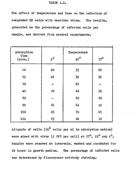
The e f f e c t o f te m p e ra tu re and tim e on th e i n f e c t i o n o f
suspended KB c e l l s w ith v a c c in ia v i r u s . The r e s u l t s ,
p re s e n te d a s th e p e rc e n ta g e of i n f e c t e d c e l l s p e r
sam ple, a r e d e riv e d from s e v e r a l e x p e rim e n ts.
A d so rp tio n Time
(m ins. ) 4 °
Tem perature
22° 37°
10 40 35 60
15 42 52 52
30 - 63
-40 70 66 56
60 - 60 60
80 62 64 45
150 65 70 25
210 65 62 12
c
A liq u o ts of c e l l s (1CD c e l l s p e r ml i n a d s o r p tio n medium)
were mixed w ith v ir u s (2 EFU p e r c e l l ) a t 37°» 22° and 4°*
Samples were removed a t i n t e r v a l s , washed and in c u b a te d f o r
24 ho u rs i n grow th medium. The p e rc e n ta g e o f in f e c t e d c e l l s
[image:16.525.53.518.58.676.2]13
after infection and b) calculation of total virus yield of individual cells and populations of cells by determination of the infectivity released into non-inhibitory medium by the disruption of washed cells*
Adsorption of Virus
It was found in preliminary experiments that the most suitable medium for the washing of cells and subsequent
adsorption of virus was Hanks* BSS containing a small amount of
serum. To determine the efficiency of adsorption of virus to
KB cells suspended in this medium, aliquots containing 10^ cells in 1 ml adsorption medium (which contained 0*5^ non-inhibitory calf serum) were equilibrated for 30 minutes at 57°, 22° and 4° and then infected with a virus inoculum of 2 PPU per cell.
Samples were removed at intervals, washed, resuspended in growth medium at a concentration of 10^ cells per ml and incubated at
37° for a total period of 24 hours. A n estimate of the
efficiency of virus uptake by cells was obtained from counts of
fluorescent cells at the end of this time. Maximum uptake was
reached by 30-40 minutes at 4° and 22° (Table 1*2), At 37° no
TIME OF ADDITION OF ANTISERUM (MINUTES OF INCUBATION AT 37 °)
FIG . 1 .1 The A c q u is itio n of Serum R e s is ta n c e by I n f e c te d G e l l s .
C e lls were in f e c te d and in c u b a te d a t 37°» At th e tim es
in d i c a t e d 1 ml sam ples were removed, t r a n s f e r r e d to s e p a r a te
grow th tu b es and in c u b a te d i n th e p re se n c e of I s 30 h y p e r
immune serum . A f te r 24 h o u rs in c u b a tio n , sam ples were
[image:18.525.46.515.63.554.2]14
Im m ediately a f t e r a n a d s o r p tio n p e rio d o f 30 m in u tes
a t 22 c r 37 only 75?a o f th e v ir u s in p u t co u ld be re c o v e re d
a s i n f e c t i o u s v i r u s , w hereas i n c o n tr o l tu b e s la c k in g o n ly c e l l s
th e whole o f th e in p u t was r e c o v e re d . About o n e - t h i r d o f th e
r e s i d u a l i n f e c t i v i t y was i n the s u p e r n a ta n t f l u i d and th e r e s t
f ir m ly bound to the c e l l s . S u b seq u en t e x p erim en ts s u g g e ste d
t n a t p a r t a t l e a s t o f th e m is sin g 25^ o f th e t o t a l inoculum had become n o n - in f e c tio u s (gone i n t o " e c l i p s e " ) i n th e c e l l s . I t
c an be i n f e r r e d t h a t th e p ro c e ss r e s p o n s ib le f o r t h i s lo s s i n
i n f e c t i v i t y s t a r t s when th e v ir u s i s added to th e c e l l s and i n
a l l growth curve e x p erim en ts z e ro tim e has been ta k e n as th e tim e
o f a d d i t i o n o f v ir u s to th e c e l l s . A p e rio d of a b o u t 1 h o u r
e la p s e d betw een z ero tim e and th e commencement o f in c u b a tio n a t
3 7 °.
I n a l l e x p e rim e n ts, 9&/o o f th e unadsorbed v ir u s was
re c o v e re d i n th e f i r s t w ash.
P e n e t r a t i o n o f V iru s
The p e n e tr a ti o n o f v i r u s was i n v e s t i g a t e d by d e te rm in in g
th e r a t e o f a c q u i s i t i o n o f serum r e s i s t a n c e . A p o p u la tio n o f
c e l l s was exposed to v ir u s f o r 3 0 m inutes a t 2 2 °, washed, and d i l u t e d i n grow th medium. A f t e r i n c r e a s in g tim e s o f in c u b a tio n
Acquisition of Serum Resistance and Loss of Infectivity of Cell-Associated Virus.
Period of Incubation (minutes)
Acquisition of Serum
Resistance0- log V^/V'
Eclipse*3 of CAV log V0/ V
0 0.056 0.0
30 0.268 0.076
60 0.377 0.201
90 0.745 0.377
120 0.745 0.569
180 1.523 0.638
Cells were infected and incubated in growth medium. At the
times stated, the titer of CAV was measured and 1 ml aliquots removed and incubated separately in the presence of hyperimmune
serum* After a total of 24 hours incubation, samples of cells
were removed from all tubes and stained with fluorescent antibody,
a. V* » titer of CAV still accessible to antibody calculated
from the proportion of cells shown by fluorescent
staining to be uninfected following antibody treatment
(proportion negative « e~m ).
b. Eclipse used here to describe the process by which
15
small tubes in the presence of a 1:30 dilution of specific
hyperimmune serum* At the end of 24 hours incubation, samples
of cells were removed from all the tubes and stained with
fluorescent antibody* Virus rapidly became resistant to
neutralization (Fig, 1*1), the 5O/o end-point being reached after
only 40 minutes incubation of the virus-cell complex (Table 1*3).
In the same experiment, the titer of cell-associated
virus at various times after infection was also measured* It is
apparent from Table 1*3> in which the rate of acquisition of
serum resistance is compared with the loss of infectivity of
cell-associated virus, that the former process occurred at a
faster rate than the latter. Thus, on the average, a virus
particle associated with a cell first became resistant to antibody
and then became non-infectious*
Virus Growth
The development of new viral material was followed by
three methods; the specific staining of antigen in cells by
fluorescent antibody, complement-fixation, and the titration of
infectious virus*
Development of Antigen. In a population of cells
receiving a virus inoculum of 2 PFU per cell and stained with
TIME AFTER INFECTION (HOURS)
FIG. 1.2# Developm ent o f a n tig e n i n c e l l s in f e c t e d w ith
in p u t m u l t i p l i c a t i o n o f 20
(o),
8 (a ) and 2 ( x ) . C e llsremoved a t th e i n d ic a te d tim es were s t a i n e d w ith
[image:22.525.68.516.35.608.2]16
visible on the surface of the cells at the end of adsorption. This persists until, after about 4-,5 hours, new virus antigen
appears as small fooi in a few cells. These foci increase in
size until the cytoplasm of the cell is full of fluorescent
material. Concurrently with this, the proportion of cells
containing antigen increases (Fig. 1.2). As Cairns (i960)
found with cells in monolayers, the curve of antigen production
rises much more steeply with high multiplicities. With a viral
inoculum of 20 PFU per cell, antigen was first seen after three hours, and by eleven hours all cells were brilliantly fluorescent. The maximum proportion of fluorescing cells was reached by 24 hours and counts were routinely made at this time.
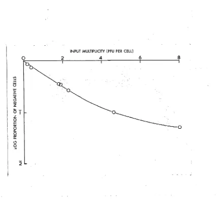
With milltiplicities insufficient to infect all cells,
the proportion of cells containing antigen at 24 hours is related
to the virus input as shown in Fig. 1.3. The relation between
the function plotted is linear when few cells in the population
are multiply infected (i.e. with viral inocula 4 2 PFU per cell)
but at higher multiplicities deviates somewhat from linearity. This may be partly due to variation in cell size but the major
factor is probably variation in susceptibility. At high
FIG. 1.3. Relation between the input multiplicity and the proportion of cells containing antigen at 24 hours#
Cells were mixed with varying concentrations of virus,
adsorption allowed to proceed in adsorption medium for 30
minutes at room temperature, the cells washed and then
incubated in growth medium for 24 hours at 37°• Cell
[image:24.525.77.510.48.455.2]17
The presence of sufficient antigen to fix complement was observed in the first sample tested, at 6 hours after
infection* Thereafter the titer rose rapidly to reach a
maximum value of 32 at about 20 hours (Table 1*4).
'.Development of Infectious Virus. Alterations in the titer of
infectious virus were followed by frequent titrations of the virus content of suspended cell cultures up to 48 hours after
the end of the adsorption period* Two virus inocula were used,
2 PFU per cell and 20 FFU per cell. The results will be
discussed in terms of changes in the infectious virus content
of suspended cell populations, and of individual cells; and the
relation between antigen stained by fluorescent antibody and infectious virus.
1. Infectious Virus in Cell Populations. Fig. 1*4
illustrates typical growth curves obtained from populations of cells suspended in growth medium after infection with viral
inocula of 20 and 2 PFU per cell. Titration of unadsorbed virus
indicated that the adsorbed multiplicities were 15 and 1*5
respectively but immediately following the start of the
incubation only two-thirds of this could be detected by assay on
the CAM. At two-hourly intervals volumes of 1 ml were removed
The P r o d u c tio n of C om plem ent-Fixing A n tig e n i n C e lls
I n f e c te d w ith V a c c in ia V iru s .
Time a f t e r I n f e c t i o n A n tig e n T i t e r
1 0
6 4
9 8
12 16
18 32
21 32
30 32
C e lls were i n f e c t e d and in c u b a te d i n grow th medium.
At th e tim e s g iv e n 5*10^ c e l l s were removed, washed
and re su sp e n d e d i n 1 ml g e l a t i n e s a l i n e . A f te r
d i s r u p t i n g the c e l l s and rem oving any p a r t i c u l a t e
m a t e r i a l by c e n t r i f u g a t i o n , th e r e s u l t i n g a n tig e n
p r e p a r a t i o n was d i l u t e d s e r i a l l y and t i t r a t e d f o r
c o m p le m e n t-fix in g a c t i v i t y w ith a f i n a l d i l u t i o n o f
18
of smears for fluorescent staining. The other half was spun
down, the supernatant discarded and the cells resuspended in
gelatine saline and stored at -6o°. At the completion of the
experiment all samples were thawed, treated with the ultrasonic
drill and titrated on the CAM* Cell counts were made on
alternate samples and remained constant.
The curves for viral infectivity are similar and can
be divided into three stages: (l) fall in titer, from 0 to 5
hours, (2) rapid increase , from 5 to 9 or 10 hours, and (3)
slower logarithmic increase from 10 to 30 hours. On incubation,
the titer of cell-associated virus fell until at 4 hours only
10-15$ of the virus cell-associated at 0 hours could be detected.
Prom the fifth to the ninth hour, with a n inoculum of 20 PPU per cell, and the fifth to the tenth hour with 2 PFTJ per cell, the infectivity titer rose rapidly to reach a level of about 16 PFU
per infected cell* Thereafter there was a slower increase to a
maximum of about 150 PPU per infected cell by the thirtieth hour* The titer fell slightly during the subsequent 18 hours*
2. Infectious Virus in Single Cells. At intervals
single cells were isolated from each of the growth tubes used for
the experiment described above. Approximately 20 cells were
19
transferred individually with a micropipette to tubes
containing 0*5 ml gelatine saline. The contents of the tubes
were frozen, thawed and sonicated and the entire sample was
titrated on five eggs. The results are shown in Table 1*5*
With an inoculum of 20 PFU per cell all nine cells assayed at the end of the adsorption period yielded virus, the
average number of PFU per cell being 9* Six hours later
only-five out of twenty cells were positive, and each of these yielded
only one PFU per cell. By the eleventh hour 90/o of the cells
sampled yielded virus, and this situation persisted throughout
the rest of the experiment. There was a gradual increase in
the mean virus yield per cell«
With an inoculum of 2 PFU per cell no yielders were found at 7 hours, but at 11 hours 'JOfo of cells yielded a mean of
19 PFU per cell, and the proportion remained about the same for the remainder of the experiment, although the yield per cell rose
steadily. Virus yields of yielders in samples of single cells
were fairly evenly distributed around the mean value.
3* Relation between Development of Antigen and Infectious
Virus. In the early stages of the growth cycle there was a
divergence between infectivity and the presence of antigen in
A n a ly s is of th e Y ie ld s o f 2 0 S in g le C e ll s I s o la te d a t I n te r v a ls du ri n g - th e G ro w th C y c le « ra rH H 0 O
9
<d 0 0 PH 3 CO Ö •H § • H O o > Ch O ■n rH 0 •HT i - e a p i ON CM KN KN UN UN C— CD C—u n k n c — t — u ncmctncm •
o
O ON rH -v)" CO NN CM rH CM CM UN rH
rH
3 Ä .
•55 >
u a x p a i x 00 O KN '«j* ^ UN t— CO C—UN O ON O VO UN rH O V£> VO CM CO UN CO H H CM K N M - "cf
s T I e D q . u 9 0 S 8 j o n x d °/o
LTN O UN UN UN UN UN UN
O CO ON ON ON ON ON ON ON CCM UN O '- C— CO 00 COO VO o t o o o i
o o o o
^ UN
«--1
8 8
KN 'vj- o
i—( rH 0 o v 1 o o n
UN CT\
CM CM CM
fH
V—✓ 20
0 -2 4 9 1--i rH w ' d iH 0 1 O CT\
u no n
rH rH rH LfN rH rH rH
•H 1 0 0 -1 4 9
rH KN C— CM KN KN O CM
H •H
> UN CTN !
C— ON rH rH CM UN KN ’v}- rH rH •*3' O
X! .
•H £
CQ 1
o
UN f — KN CO I A K N H ^ VO H O H rH UN H
rH
O «
u no n
CM -s}- 0 0 CM VO UN KN KN CM KN CM <vt- UN ^ ^
CM
J VO UN VO ’ t ' t W H KN tN- ON VO CM KN KN
0
i
UN
1
o K N I A O O H C M H O H KN CM CM KN KN KN
o O H C v l ^ O O H C M OUN OCM VO VO C— ^ CM CM
( s r n o q )
U 0 T X 0 8 J X I X
ainij,
rH UN ON f~ - H UN ON H t s - H H H W K N K N ' s r
t r H UN ON C~— rH ON r H r H r H CM KN
X n d u i
O
20
3 h o u rs and was p r e s e n t i n a la r g e p r o p o r tio n of c e l l s a f t e r
5 h o u r s . I n c o n t r a s t , th e f i r s t in c r e a s e i n t i t e r o f c e l l -
a s s o c i a t e d v i r u s co u ld n o t be d e te c te d u n t i l a f t e r 5 h o u r s .
S i m i l a r l y , w ith th e low er m u l t i p l i c i t y , 15$ o f c e l l s c o n ta in e d
a n t i g e n a t 5 h o u rs w hereas th e i n f e c t i v i t y was a t i t s lo w e st
l e v e l a t t h i s tim e .
Under norm al c o n d itio n s , how ever, th e tim e o f f i r s t
ap p ea ra n ce o f new v ir u s i s obscured by th e p e r s i s t e n c e o f
r e s i d u a l u n e c lip s e d v i r u s . The p ro d u c tio n o f new v ir u s i s
p ro b a b ly b e t t e r i n d i c a t e d by a change i n s lo p e o f th e e c l i p s e
cu rv e th a n by a n a b s o lu t e in c r e a s e i n t i t e r . T his h as been
shown u n e q u iv o ca b iy by th e use o f sodium a z id e ( E a s te rb ro o k , 196 1 ).
K in e tic e x p e rim e n ts w ith a z id e allo w ed a n a c c u r a te a sse ssm e n t t o
be made o f th e p r o d u c tio n o f new a n tig e n and new i n f e c t i o u s v i r u s ,
and showed t h a t th e s e e v e n ts co u ld n o t be s e p a r a te d i n tim e .
Fate o f the I n f e c t in g Virus P a r t ic le
The f i r s t s ta g e o f the grow th c y c le i s c h a r a c t e r i s e d
by a f a l l i n t i t e r o f c e l l - a s s o c i a t e d v i r u s . To o b ta in more
ev id en ce on th e f a t e o f th e i n f e c t i n g v ir u s p a r t i c l e s , a su sp e n sio n
o f c e l l s was i n f e c t e d w ith an inoculum o f 9 FFU p e r c e l l
(A dsorbed m - 4*5) and t r a n s f e r r e d to a grow th tu b e c o n ta i n in g
21
were measured in the usual way, attention being concentrated
on the first seven hours of the growth cycle* The results are
shown in Pig* 1*5. There was a profound fall in cell-
associated virus from 4*5
FFU
per cell to 0.04 FFU per cell inthe first 3 hours of incubation* The initial fall was very
rapid, from 4.5 to 1.2 PFU per cell during the hour occupied by manipulations between addition of virus to cells in the adsorption tube and resuspension of cells in the growth tube. There was a rapid rise in infectivity between the fifth and seventh hour.
The infectivity of single cells during this early stage of the growth cycle was measured by two different methods,
titration of single cells for cell-associated virus, and for
their ability to initiate a plaque (Table 1.6)* The latter
property was measured by mixing washed cells with chick embryo fibroblasts and allowing the latter to form a monolayer (Abel,
to be published). The percentage of infected cells at 1 and
5 hours was 90, the same as that determined by single cell titration and fluorescent antibody staining at the end of the
growth period. In contrast, the data from Pig. 1.5 shows that
Titration of cells taken at intervals
during a growth cycle in non-inhibitory
medium.
Time after addition of Virus to cells (hours)
Percentage of Cells
Initiating Plaques
Yielding Virus
Containing Antigen
1 88 -
-5 90 0
-7 - 25
22
v i r u s . The v ir u s w hich h as i n i t i a t e d i n f e c t i o n i n th e c e l l s
has p a sse d thro u g h a s ta g e when i t s i n f e c t i v i t y co u ld n o t be
d e m o n stra te d .
V irus R elea se
I t has b een shown p re v io u s ly (T ab le l , l ) t h a t v ir u s
produced i n c e l l s rem ain s a s s o c i a t e d w ith them f o r lo n g p e r io d s .
The fo llo w in g e x p erim en ts were c a r r i e d o u t t o d eterm in e th e
l o c a t i o n o f i n f e c t i o u s c e l l - a s s o c i a t e d v i r u s .
A f te r 26 h o u rs in c u b a tio n an in f e c te d c e l l s u s p e n s io n 5
was washed and c o n c e n tr a te d to 5x10 c e l l s p e r m l. An a l i q u o t
of 1 ml was exposed to hyperimmune serum ( f i n a l d i l u t i o n 1 :1 0 0 )
f o r 10 m in u tes a t 37°» th e n d i l u t e d I s 100 i n g e l a t i n e s a l i n e .
A f te r d i s r u p t i o n o f th e c e l l s , r e s i d u a l v ir u s was a ssa y e d and
compared w ith a second a l i q u o t t r e a t e d i n th e same way b u t w ith
th e o m issio n o f a n tis e ru m , A t h i r d a l i q u o t was s o n ic a te d b e fo re
exposure to a n tis e r u m . The same p ro ced u re was perform ed on
c e l l s in c u b a te d f o r 48 h o u rs a f t e r i n f e c t i o n . The r e s u l t s
(T able 1 ,7 ) show t h a t a t b o th tim e s th e m ajor p a r t of th e t o t a l
i n f e c t i o u s v ir u s was n o t n e u t r a l i z e d by a n tis e ru m .
An a tte m p t was a l s o made to remove i n f e c t i o u s v i r u s
from th e c e l l s u r f a c e by th e a c t i o n o f t r y p s i n o r v e rs e n e . A f te r 5
28 ho u rs in c u b a tio n , 3x10 c e l l s were washed and d iv id e d i n t o
The Inaccessibility of Cell-Associated
Virus to Antiserum.
Treatment prior to Assay Virus Titer (PFU per inl x 104 )
26 hours 48 hours
Antib ody-re sis tant 1. Treated with antiserum 64 96 2. Diluted 1:100
virus in suspension 3. Disrupted
Antib ody-resis tant 1. Disrupted
2. Treated with antiserum 3 4 virus in disrupted 3. Diluted 1:100
suspension
Total virus content 1. Disrupted 77 128 2. Diluted 1:100
of suspension
Cells were infected and incubated in growth medium. After
[image:36.525.83.517.40.495.2]23
b ) 0.02^6 v e rsen e c ) rem ained u n tr e a te d , f o r 30 m inutes a t 3 7 °.
The c e l l s were th e n washed tw ice w ith g e l a t i n e s a l i n e and f r o z e n .
On t i t r a t i o n no d if f e r e n c e c o u ld he d e te c te d betw een c e l l s
exposed to t r y p s i n o r v e rse n e and th o s e n o t t r e a t e d .
Both e x p e rim e n ts i n d i c a t e t h a t m ost of th e v ir u s
produced rem ains i n t r a c e l l u l a r .
Absence o f Secondary I n f e c t i o n i n th e Growth Tube
I t was im p o rta n t t o d e cid e w h eth er seco n d ary i n f e c t i o n
o c c u rre d under th e c o n d itio n s d e s c r ib e d . C e lls were in f e c te d
w ith s e v e r a l m u l t i p l i c i t i e s o f v ir u s and a f t e r a d s o r p tio n each
p r e p a r a t io n was d iv id e d i n t o two p a r t s . One o f each o f th e s e
was d i l u t e d w ith an e q u a l volume of u n in f e c te d c e l l s so t h a t th e
f i n a l c o n c e n tr a tio n o f a l l p r e p a r a t io n s was 10^ c e l l s p e r m l.
A ll th e sam ples were th e n in c u b a te d f o r 24 h o u rs . E x am in atio n
o f s ta i n e d sm ears made a t th e end o f t h i s p e rio d showed t h a t
only h a l f as many c e l l s i n th e p r e p a r a t io n s mixed w ith f r e s h
c e l l s were i n f e c t e d a s i n th e c o n t r o l s . W ith m u l t i p l i c i t i e s
o f a b o u t 1, 60-7O/o o f c e l l s showed f lu o r e s c e n c e by th e 24th h o u r
and no more f lu o r e s c e d on p ro lo n g ed in c u b a tio n . I t was
p o s s ib le t h a t c e l l s n o t i n f e c t e d i n i t i a l l y became in s u s c e p t i b l e
due to t h e i r m aintenance i n s u s p e n s io n . However, i f c e l l s
were m a in ta in e d i n s u s p e n s io n i n a grow th tu b e , a t a
FIG, 1,6. Effect of prior incubation of KB cells in
suspension for twenty-four hours on the time of production
of virus.
o Cells incubated for 24 hours before infection.
24
to 10° per ml in an adsorption tube and infected in parallel with a freshly prepared cell suspension they were equally susceptible to infection (Fig. 1.6).
Both types of experiment indicate that secondary infection in the growth tube does not occur to a significant extent.
DISCUSSION
The aim in the experiments described in this paper was to obtain an overall picture of the growth cycle of vaccinia in suspended cells by a method which could be used for
quantitative studies on the development of poxviruses under
conditions of metabolic and specific inhibition. For such
studies it is desirable a) that the multiplicity of the
infecting virus should be known, b) that the growth of virus
should be restricted to a single cycle, c) that the virus
yields of individual cells and populations of cells should be easily determined at intervals throughout the growth cycle, d) that the production of antigen by individual cells could be compared with their content of infectious virus and e) that the number of cells responsible for the observed yield of
infectious virus should be known. The method described
The rem oval o f u n adsorbed v ir u s from c e l l s i n
su sp e n sio n i s e a s i l y and r a p i d l y a c h ie v e d and a t th e s t a r t o f
in c u b a tio n more th a n 99/o o f the t o t a l v ir u s p r e s e n t i s c e l l -
a s s o c i a t e d . V a c c in ia v ir u s i s v e ry slo w ly r e l e a s e d from
in f e c t e d c e l l s and t h i s s i t u a t i o n i s m a in ta in e d th ro u g h o u t th e
grow th c y c le .
Counts o f th e p r o p o r tio n o f c e l l s s t a i n i n g w ith
f l u o r e s c e n t a n tib o d y a t th e end o f th e grow th c y c le p ro v id e s a
read y e s tim a te o f th e p r o p o r tio n o f c e l l s in f e c t e d by a g iv e n
inoculum . I t g iv e s in fo r m a tio n on la r g e numbers o f c e l l s
o th e rw ise o b ta in a b le o n ly , a f t e r d i l u t i o n , by in f e c t e d c e l l
co u n ts o r s i n g le c e l l a s s a y . The e s tim a te i s , o f c o u rs e ,
a c c u ra te only i f v i r u s p r o d u c tio n i s l i m i t e d to th o s e c e l l s
i n i t i a l l y i n f e c t e d . D ir e c t e x p e rim e n ts showed t h a t sec o n d a ry
i n f e c t i o n d id n o t o ccu r t o a s i g n i f i c a n t e x te n t and th e f a i l u r e
of c o lc h ic in e (50 mg p e r ml) t o a f f e c t th e p r o p o r tio n o f c e l l s
s ta i n e d (u n p u b lish e d r e s u l t ) e lim in a te d the p o s s i b i l i t y t h a t
d i v i s i o n o f i n f e c t e d and u n in f e c te d c e l l s o c c u rre d a t d i f f e r e n t
r a t e s d u r in g th e grow th c y c le .
Noyes and W atson
(1955)
and C a irn s(i960)
have shownt h a t i n m onolayers s t a i n e d w ith f l u o r e s c e n t a n tib o d y th e number
of c e l l s s t a i n e d was p r o p o r tio n a l to th e amount o f v i r u s added*
26
suspended KB c e l l system ( F ig ,
3 ) .
Vfith sm all v i r a l i n p u t s ,th e d e v i a t i o n from a 45^ slo p e was s l i g h t and th e v i r u s
m u l t i p l i c i t y (m) can be d e riv e d from th e e x p r e s s io n ,
P r o p o r tio n p o s i t i v e = l - e ” m. W ith l a r g e r in p u ts th e e s tim a te
o b ta in e d i n t h i s way becomes i n c r e a s i n g l y in a c c u r a te and su ch
m u l t i p l i c i t i e s have to be c a l c u la te d a p p ro x im a te ly from th e
s t a i n e d c e l l c o u n ts o b ta in e d w ith a 5 - f o ld o r 1 0 - f o ld d i l u t i o n
o f th e v i r u s , p u t up i n p a r a l l e l w ith th e more c o n c e n tra te d
p r e p a r a t i o n . T h is d e v i a t i o n from l i n e a r i t y has been o b serv ed
i n o th e r v i r u s - c e l l sy stem s (M arcus, 1959) and i t has been
p o s s ib le to i s o l a t e c lo n e s of c e l l s h a v in g g r e a t e r and more
uniform s u s c e p t i b i l i t y t o th e v ir u s e s s tu d ie d (NDV - M arcus, 1959»
p o l i o v i r u s - D a rn e ll and Sawyer, 1959)* D o u b tle ss s i m i l a r c lo n e s
w ith h ig h u n ifo rm s u s c e p t i b i l i t y to v a c c in ia v i r u s co u ld be
i s o l a t e d and i n th e s e , th e h ig h m u l t i p l i c i t i e s used i n r e
co m b in atio n e x p erim en ts m ight be d e term in e d w ith re a s o n a b le
a c c u ra c y .
Repeated sam pling from a s in g le growth tube o b v ia tes
the need f o r time r e p l i c a t e s . In d iv id u a l c e l l s can be
is o la t e d w ith out d i f f i c u l t y and t h e ir v ir u s assayed a t any time
in the growth c y c le . S in ce v a c c in ia v ir u s remains c e l l
-a s s o c i-a te d f o r so lo n g , the v ir u s r e le -a s e d on d is r u p tio n
to s a m p lin g .
D u rin g th e s e e x p e rim e n ts, s e v e r a l f a c t s emerged
w hich a r e o f g e n e r a l i n t e r e s t i n th e c o n s id e r a tio n o f th e
grow th c y c le o f v a c c in ia v iru s* A dsorbed v ir u s r a p id ly
becomes r e s i s t a n t to th e b lo c k in g a c t i o n of a n tib o d y and th e
r a t e o f t h i s r e a c t i o n exceeds th e r a t e o f lo s s o f v ir u s
i n f e c t i v i t y . Thus a n i n f e c t i n g v i r u s p a r t i c l e becomes
r e s i s t a n t t o a n tib o d y n e u t r a l i z a t i o n b e fo re i t becomes non-
in f e c tio u s * Such a f in d i n g p ro v id e s ev id en ce t h a t v a c c in ia
v i r u s l o s e s i t s i n f e c t i v i t y a s th e r e s u l t o f a n i n t r a c e l l u l a r
r e a c t i o n r a t h e r th a n a s th e r e s u l t o f i r r e v e r s i b l e b in d in g to
th e c e l l s u r f a c e . U n til r e c e n t l y a group o f w orkers (M a itla n d
and P o s t l e t h w a i t e , 1959) i n s i s t e d t h a t i t had n o t been s a t i s
f a c t o r i l y d e m o n strated t h a t v a c c in ia v ir u s underw ent e c l i p s e
o f th e ty p e now re g a rd e d a s c h a r a c t e r i s t i c of v ir u s grow th.
S in ce th e n th e s e w orkers have o b ta in e d e v id en c e o f a p ro fo u n d
f a l l i n i n f e c t i v i t y on in c u b a tio n o f a c e l l - v i r u s complex
( P o s tle th w a ite and M a itla n d , i9 6 0 ) a s have F u rn ess and Youngner
(1959) u s in g monkey k id n e y c e l l s i n m onolayers and s u s p e n s io n .
I n th e p r e s e n t e x p e rim e n ts, e v id en c e was o b ta in e d o f th e ty p e
w id ely a c c e p te d as i n d i c a t i v e o f an e c l i p s e p h a se , v i z . a f a l l
28
and a failure to recover virus from single cells at a time
when the infected cell count was 90/o, The fall in titer of
infectious virus observed during this phase was variable under
conditions conducive to subsequent virus growth. If, however,
new virus growth was inhibited by sodium azide, infectivity fell by 9 9 and sometimes as much as 99*0^ (Easterbrook, 1961). This demonstrates that the apparent extent of eclipse is
determined by two processes, the loss of infectivity of input
virus and the production of new virus. Conclusive evidence of
such an eclipse is provided by electronmicroscopic (Higashi, 1959) and autoradiographic (Cairns, i960) observations of the
development of DN/L pools lacking mature virus particles 6 hours after infection.
Cairns (i960) has shown that with the same strain of vaccinia (V-MH) added to KB cell monolayers to give a multip
licity of 6, new antigen and new DNA were first detectable 4
hours after infection. With suspended cells infected with an
inoculum of 20 PFU per cell and subsequently stained with
fluorescent antibody, newly-developed antigen was visible 3
hours after infection and very obvious at 5 hours; sufficient
antigen to fix complement was present before 6 hours. New
r a p i d l y u n t i l a b o u t th e t e n t h h o u r . By t h i s tim e , w ith h ig h
m u l t i p l i c i t i e s , a l l c e l l s c a p a b le o f p ro d u c in g v ir u s were
d o in g s o (a s judged by f l u o r e s c e n t a n tib o d y s t a i n i n g ) . From
th e t e n t h u n t i l a b o u t th e t h i r t i e t h h o u r th e i n f e c t i v i t y t i t e r
ro s e l o g a r i t h m i c a l l y , d o u b lin g e v e ry 6 J - h o u r s . W ith low er
m u l t i p l i c i t i e s (inoculum of 2 PFU p e r c e l l ) asynchrony caused
a p ro lo n g e d and more g ra d u a l i n i t i a l r i s e , b u t once a g a in when
a l l i n f e c t e d c e l l s had s t a r t e d t o produce v ir u s th e i n f e c t i v i t y
t i t e r r o s e l o g a r ith m i c a lly a t a s i m i l a r r a t e . With th e t e s t e d
l i m i t s , ( in p u ts from 2 to 20 PFU p e r c e l l ) th e f i n a l a v erag e
y i e l d p e r i n f e c t e d c e l l was th e same.
The f a i l u r e of a n tib o d y t o n e u t r a l i z e more th a n a sm all
p a r t o f th e i n f e c t i o u s v i r u s p r e s e n t even a t 48 h o u rs a f t e r
i n f e c t i o n , an d th e f a i l u r e of t r y p s i n o r v e rsen e to remove such
v i r u s from c e l l s , show t h a t m ature v a c c in ia v in o s rem ain s i n an
i n t r a c e l l u l a r p o s i t i o n f o r a c o n s id e ra b le tim e a f t e r i t s
p ro d u c tio n , Ihe m a tu r a tio n of v a c c in ia i s th u s d is tin g u is h e d
from t h a t o f th e m y x o v iru ses. I n th e l a t t e r case assem bly
and m a tu r a tio n occur a t , o r n e a r , th e c e l l s u r f a c e and more th a n
95rfo o f th e i n f e c t i o u s v i r u s p r e s e n t a t any tim e d u rin g th e
grow th c y c le c a n be n e u t r a l i z e d by s p e c i f i c a n tib o d y ( F r a n k lin
29a
SUMMARY
The multiplication of vaccinia virus was investigated in suspensions of KB cells, assays being made by infectivity titrations and by staining' cells with fluorescent antibody. Under the conditions described, 50-60^6 of the input virus was adsorbed in 30 minutes, free virus was reduced to less than l°/0
by a single wash and virus multiplication was restricted to a single cycle.
Adsorbed virus became serum resistant and soon after wards there was a fall of 80-90^ in the infectivity titer of
cell-associated virus. Assay of single cells in the early
stages of the growth cycle indicated the existence of a true eclipse phase.
Infectivity began to rise between the 5th and 7th hour
and when all infected cells were producing virus the infectivity
titer rose logarithmically between the 10th and 50th hour. The
maximum titer reached was approximately 150 PFU per infected cell and was independent of the multiplicity of infection between m » 1.5 and m = 15 at least. Most of the newly-formed infectious virus was not accessible to antibody, showing that maturation was an intracellular process and mature virus remained in an intracellular location.
REFERENCES
CAIRNS, J. (i960). The initiation of vaccinia infection,
Virology 603-62%
EAGLE, H. (1955a), The specific amino acid requirements of a
human carcinoma cell (strain HeLa) in tissue culture. J. Exp, Med, 102, 37-48»
EAGLE, H. (l955fr)» Propagation in fluid medium of a human
epidermoid carcinoma Strain KB. Proc. Soc. Exp. Biol.
Med. 89, 362-364.
DARNELL, J.E. Jr. and SAWYER, T.K. (1959)* Variation in plaque forming ability among parental and clonal strains of Heia cells. Virology _8, 223-229»
EASTERBROOK, K.B. (196I). Analysis of the early stages of
vaccinia virus infection of KB cells using sodium azide. Virology (in press).
FENNER, F. (1958 ). The biological characters of several strains
of vaccinia, cowpox and rabbitpox viruses. Virology 502-529.
FRANKLIN, R.M. and HENRY, C.H. (i960). The multiplication of fowl plague virus in tissue cultures of chick embryo cells Virology 10, 406-418»
FURNESS, G. and YOUNGNER, J.S. (1959)» One-step growth curves
for vaccinia virus in cultures of monkey-kidney cells.
and rapid method for isolating viruses by selective
fluorocarbon deproteinization. Trans. N.Y. Acad. Sei. 18, 7 01-70%
HIGASHI, N. (1959)* Electron microscopy of viruses in thin sections of cells grown in culture. Progr. med. Virol. 43-72.
JOKLIK, W.K. and RODRICK, J.Mc.N. (1959). Biochemical studies on vaccinia virus in cultured cells. I. Incorporation of adenine-8-C^ into normal and infected cells. Virology 1
596
-416
#MAITLAND, H.R. and POSTLETHWAITE, R. (1959). Vaccinia virus in He La cells. Symp. Soc. Gen. Microbiol. (A. Isaacs and B.W.
Lacey, Eds.) Cambridge University Press.
MARCUS, P.I. (1959). Host-cell interaction of animal viruses, II. Cell-killing particle enumeration : survival curves at low multiplicities. Virology j?, 5 4 6-5 6 5.
NOYES, W.F. and WATSON, B.K. (1955). Studies on the increase of vaccine virus in cultured human cells by means of the fluorescent antibody technique. J. Exp. Med. 102, 257-242. OVERMAN, J.R. and TAMM, I. (1957). Multiplication of vaccinia
virus in the chorioallantoic membrane in vitro. Virology
POSTLETHWAITE, R. and MAITLAND, H.B. (i960). The eclipse phase of vaccinia virus growing in chick embryo monolayers and some technical procedures which affect its demonstration.
\
J.
Hyg. 58, 133-145*ROBERTSON, H.E., BRÜNIER, K.T. and SY VERTON, J.T. (1955).
Propagation in vitro of poliomyelitis viruses. VII. pH
change of HeLa cell cultures for assay. Proc. Soc. Exp. Biol. Med. 88, 119-122.
RUBIN, H., FRANKLIN, R.M. and BALUDA, M. (1957). Infection and
growth of Newcastle Disease virus (NDV) in cultures of
chick embryo lung epithelium. Virology 587-600.
SMITH, K.O. and SHARP, D.G. (i960). Interaction of virus with
cells in tissue culture. I. Adsorption on and growth of
vaccinia virus in L cells. Virology 11, 519-532*
WESTWOOD, J.C.N., PHIPPS, P.H. and BOULTER, E.A. (1957). The titration of vaccinia virus on the chorioallantoic
membrane of the developing chick embryo. J. Hyg. 55*
ANALYSIS OF THE EARLY STAGES OF VACCINIA VIRUS
31
.
INTRODUCTION
Thompson (1947) examined a large number of chemicals for their possible inhibitory activity on the growth of vaccinia
virus in tissue culture* Amongst these, sodium azide was found
to reduce or prevent the multiplication of vaccinia.
Preliminary experiments indicated that azide mi^it specifically inhibit an early stage of viral multiplication and the
development of more refined techniques for the analysis of
vaccinia virus multiplication (Cairns, i960; Easterbrook, 1961)
prompted a re-investigation of its effect, which is reported here.
MATERIALS AND METHODS
All materials used in this study have been fully
described in the preceding paper (Easterbrook, 1961). Heat-
inactivated virus (designated by the prefix H-) was prepared as described by J o k lik et al. (i960).
The general experimental method was as follows. KB
cells which had been grown as a monolayer were dispersed with trypsin-versene and 10^ cells were suspended in 1 ml of BSS, containing 0*5^ non-inhibitory calf serum in small siliconed tubes ("infection tubes"). They were infected by adding 0.1 ml of a virus suspension to the cells and stirring them for 30
w ashing i n th e c e n tr if u g e and th e c e l l s were d i l u t e d to a
c o n c e n tr a tio n o f 10^ c e l l s p er ml i n growth medium i n l a r g e r
s i l i c o n e d tu b e s ( “grow th tu b e s “ ) . The c e l l s w ere a g i t a t e d
w ith m ag n etic s t i r r e r s and in c u b a te d a t 37° i n a w a te r b a th .
A liq u o ts were removed a t i n t e r v a l s f o r a s s a y p u rp o se s.
I n f e c t i o u s v ir u s was t i t r a t e d on th e c h o r i o a l l a n t o i c membrane
(CAM) and th e p r o p o r tio n of c e l l s c o n ta in in g a n tig e n was
d e term in e d by f l u o r e s c e n t a n tib o d y s t a i n i n g . At any s ta g e
sodium a z id e co u ld be added o r removed by w ash in g th e c e l l s and
re s u s p e n d in g them i n grow th medium.
RESULTS
The M inimal E f f e c t i v e C o n c e n tra tio n o f Sodium A zid e.
The m inim al e f f e c t i v e c o n c e n tr a tio n of a z id e r e q u ir e d
to i n h i b i t v i r a l s y n th e s is was d eterm in ed by in c u b a tin g i n f e c t e d
KB c e l l s i n in c r e a s i n g c o n c e n tr a tio n s o f a z i d e . Samples were
ta k e n a f t e r 30 h o u rs in c u b a tio n f o r i n f e c t i v i t y t i t r a t i o n and
a n tig e n s t a i n i n g .
The r e s u l t s (T ab le 2 .1 ) i n d i c a t e t h a t sodium a z id e
c o m p le tely i n h i b i t e d v i r a l m u l t i p l i c a t i o n a t c o n c e n tr a tio n s o f
3 • 10~^M o r g r e a t e r . There was some e f f e c t a t 10“ ^M b u t none
a t lO ^M a z id e . I n a l l s u b se q u e n t e x p erim en ts a z id e was used a t
TAB IE 2.2.
The Effect of Azide on Virus Infectivity
Incubation Time Titer (PPU per ml x 105) at 37
(in hours) Cells incubated in
Growth Medium Growth Medium + Azide (3.10”5m)
0 8.7 8.4
6 6.1 6.8
Sodium a z id e co u ld e x e r t i t s i n h i b i t o r y e f f e c t i n
one of s e v e r a l ways, o r perhaps by a co m b in atio n o f th e s e .
I t co u ld i n a c t i v a t e f r e e v ir u s , i t c o u ld so a l t e r c e l l s t h a t
th e y were s u b se q u e n tly unable to ta k e up o r produce v i r u s , o r
i t c o u ld a f f e c t one o r more s p e c i f i c s ta g e s i n th e growth
c y c le o f th e v i r u s .
The E f f e c t o f Azide on V a cc in ia V iru s I n f e c t i v i t y
To t e s t w h eth er a z id e had any d i r e c t e f f e c t on th e
i n f e c t i v i t y o f f r e e v a c c in ia v i r u s , s to c k v i r u s was d i l u t e d i n
grow th medium w ith o r w ith o u t th e a d d i t i o n of a z id e and
in c u b a te d a t 37°* At i n t e r v a l s sam ples were d i l u t e d i n
g e l a t i n e s a l i n e and t i t r a t e d on th e CAM.
The r a t e o f i n a c t i v a t i o n i n grow th medium was no t
in c r e a s e d by th e p re se n c e o f a z id e , th e i n a c t i v a t i o n d u rin g 22
h o u rs being- a b o u t 6
(
yfa i n b o th media (T able 2.
2).
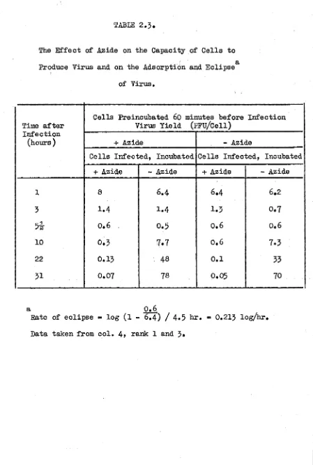
The E f f e c t of A zide on th e C a p a c ity o f C e l ls to Produce V irus
and on th e A d so rp tio n and E c lip s e o f V iru s
The p o s s i b i l i t y t h a t c e l l s exposed t o a z id e a r e re n d e re d
in c a p a b le of s u p p o r tin g v i r a l m u l t i p l i c a t i o n b ecau se of
perm anent p h y s io lo g ic a l im pairm ent was i n v e s t i g a t e d . I n th e
same ex p erim en t th e e f f e c t o f a z id e on th e a d s o r p tio n and
TABLE 2,3
The Effect of Azide on the Capacity of Cells to
a Produce Virus and on the Adsorption and Eclipse
of Virus.
Time after
Cells Preincubated 60 minutes before Virus Yield (pFü/Cell)
Infection
Infection
(hours) + Azide - Azide
Cells Infected, Incubated Cells Infected, Incubated
+ Azide - Azide + Azide - Azide
1 8
6.4
6.4
6.23 1.4 1.4 1.3 0.7
5
&
0.6 0.5 0.6 0.610 0.3 7.7 0.6 7.3
22
0.13 48 0.1 3331 0.07 78 0.05 70
a 0.6
Rate of eclipse « log (l -