PHARMACOGENETICS
IN WARFARIN
THERAPY
Thesis submitted in accordance with the
requirements of the University of Liverpool for the
degree of Doctor in Philosophy
by
Azizah Ab Ghani
ii has not been presented, nor is currently being presented, either wholly or in part for
any other degree of qualification.
Azizah Ab Ghani
This research was carried out in the Department of Molecular and Clinical
Pharmacology, in the Institute of Translational Medicine, at The University of
iii CONTENTS
ABSTRACT iv
ACKNOWLEGEMENTS v
ABBREVIATIONS vi
CHAPTER 1: General introduction 1
CHAPTER 2: Development and validation of a warfarin dosing algorithm 47
CHAPTER 3: Incorporating genetic factor into HAS-BLED,
a bleeding risk score 75
CHAPTER 4:Pharmacogenetics of warfarin in a paediatric population 102
CHAPTER 5: Warfarin pharmacogenetic in children:
A genome-wide association study 135
CHAPTER 6: Validation of a novel point of care Hybeacon® genotyping
method on prototype PCR instrument Genie 1 for CYP2C9
and VKORC1 alleles 155
CHAPTER 7: Final discussion 178
iv phase because of its narrow therapeutic range and large inter-individual variability. Therefore, the aim of this thesis was to investigate the use of pharmacogenetics and clinical data to improve warfarin therapy.
Genetic variants in cytochrome P450 2C9 (CYP2C9) and vitamin K epoxide reductase (VKORC1) are known to influence warfarin dose. Therefore we developed a pharmacogenetic dosing algorithm to predict warfarin stable dose prospectively in a British population based on 456 patients who started warfarin in a hospital setting and validated it in 262 retrospectively recruited patients from a primary care setting. The pharmacogenetic algorithm which included CYP2C9*2, CYP2C9*3 and VKORC1-1693 together with body surface area, age and concomitant amiodarone use, explained 43% of warfarin dose variability. The mean absolute error of the dose predicted by the algorithm was 1.08 mg/day (95% CI 0.95-1.20). 49.6% of patients were predicted accurately (predicted dose fell within 20% of the actual dose).
The HAS-BLED score, a bleeding risk score has recently been suggested for use in the management of patients with atrial fibrillation. We validated HAS-BLED performance in predicting major bleeding using a prospective cohort with 6 months follow-up (n=482) (c-statistic 0.80 95% CI (0.71-0.90). Factors significantly associated with major bleeding in our cohort (p≤0.1) were concurrent amiodarone use, labile INR, concurrent clopidogrel use, bleeding predisposition, concurrent aspirin use and CYP2C9*3. Adding a genetic covariate (CYP2C9*3) to the HAS-BLED score did not significantly improve its performance in predicting major bleeding. Considering CYP2C9*3 is a rare allele, our study was underpowered and requires further investigation in a larger cohort.
A retrospective study of 97 Caucasian children was conducted to gain greater understanding of the factors that affect warfarin anticoagulant control and response in children. Results from multiple regression analysis of genetic and non-genetic factors showed that indication for treatment (Fontan or non-Fontan group), VKORC1 -1693, and INR group explained 20.8% of variability in proportion time in which INR measurements fell within the target range (PTTR); CYP2C9*2 explained 6.8% of the variability in INR exceeding target range within the first week of treatment; CYP2C9*2, VKORC1 -1693, age and INR group explained 41.4% of warfarin dose variability and VKORC1 -1693 explained 8.7% of haemorrhagic events. The contributions of CYP2C9 and VKORC1 polymorphism were small in the above outcomes. We therefore went onto explore other genetic markers using genome-wide scanning. Two SNPs on chromosome 5, rs13167496 and rs6882472 were found to be significantly associated at a genome-wide significance level with PTIR. However, none of SNPs were significantly associated with warfarin stable dose, INR values exceeding the target range within the first week of treatment and bleeding complications. Because of our small sample size, these findings will need to be validated in a replication cohort.
Finally, we have validated and evaluated the performance of Genie HyBeacon®, a point of care therapy (POCT) instrument to genotype 135 samples for CYP2C9*2, CYP2C9*3 and VKORC1 -1693. We showed that the instrument accuracy was >98% (agreement with ABI Taqman® genotyping), it was relatively simple to use and had a
good turn-around time (1.6 hours) making it suitable for clinical use.
v ACKNOWLEDGEMENT
First and foremost, I would like to express my sincere gratitude to my supervisors
Professor Munir Pirmohamed, Dr Andrea Jorgensen and Dr Ana Alfirevic for all
their invaluable support, assistance and guidance throughout my PhD studies. Their
expertise and directions enabled me to complete the work presented in this thesis. I
would also like to acknowledge the Malaysia Ministry of Health for giving me the
funding.
My special thanks to Dr Eunice Zhang for her invaluable assistance,
encouragement, advice and helpful suggestion throughout my degree. A big thank
you to all the clinicians and nurses and the patients for taking part in the study,
without whom, the study would never have never been possible. Many thanks to Dr
Laura Sutton who provided superb statistical expertise with all analysis undertaken in
children study.
I am also grateful for support, friendship and assistance in the lab offered by
the other members of The Wolfson Centre for Personalised Medicine. You have all
made my stay in Liverpool unforgettable.
Finally, words of thanks will never enough for my husband for his patience,
sacrifice and tremendous support throughout the project until completion of this
thesis. My love and thanks also goes to my children who give me strength and
courage to go on. Lastly but not the least, I owe deep gratitude towards my family
especially my beloved mum and dad for their endless support, patience and
vi
bp base pairs
CALU calumenin
CEU Caucasians in Utah, USA
CHB Han Chinese in Beijing, China
CI confidence interval
CIVC cysteine132-isoleucine-valine-cysteine135
CPIC Clinical Pharmacogenetics Implementation Consortium
CYP1A1 cytochrome P450, family 1, subfamily A, polypeptide 1
CYP1A2 cytochrome P450, family 1, subfamily A, polypeptide 2
CYP2C18 cytochrome P450, family 2, subfamily C, polypeptide
18
CYP2C19 cytochrome P450, family 2, subfamily C, polypeptide
19
CYP2C8 cytochrome P450, family 2, subfamily C, polypeptide 8
CYP2C9 cytochrome P450, family 2, subfamily C, polypeptide 9
CYP3A4 cytochrome P450, family 3, subfamily A, polypeptide 4
CYP3A5 cytochrome P450, family 3, subfamily A, polypeptide 5
CYP4F2 cytochrome P450, family 4, subfamily F, polypeptide
12
DNA deoxyribonucleic acid
EDTA ethylenediaminetetraacetic acid
EPHX1 epoxide hydrolase 1
Factor II/FII coagulation factor 2
Factor IX/FIX coagulation factor 9
Factor VII/FVII coagulation factor 7
Factor X/FX coagulation factor 10
FDA Food and Drug Administration
GGCX -glutamyl carboxylase
GGCX gamma-glutamyl carboxylase
gla gamma-carboxyglutamic acid
vii
GWAS genome-wide association study
HWE hardy–weinberg equilibrium
IBD identity by descent
INR international normalised ratio - ratio
IWPC International Warfarin Pharmacogenetics Consortium
JPT Japanese in Tokyo, Japan
K1 vitamin K1 (phyloquinone)
KH2 reduced vitamin K, or hydroquinone
KH2 reduced vitamin K or hydroquinone
KO vitamin K 2,3 epoxide (oxidised vitamin K)
LD linkage disequilibrium
MAF minor allele frequency
MK-4 vitamin K2 (Menoquinone or MK-n) homologous.
MK-n has a variables side chain length of isoprene units
an ‘n’ stands for the number of isoprenoid residues in
the chain
M-PVA polyvinyl alcohol particles
NCBI National Center for Biotechnology Information
NSAIDs nonsteriodal anti-inflammatory drugs
OAC oral anticoagulant
PBX3 Pre-B-Cell Leukemia Homeobox 3
PCR polymerase chain reaction
POCT point of care therapy
PTTR percentage or proportion time that a patient was within
targeted therapeutic range
RCT randomised control trial
RCT randomised controlled trial
ROC receiver operating curves
SNPs single polymorphisms
ST18 Suppression of tumorigenicity 18
TF tissue factor
TIFAB TRAF-Interacting Protein With Forkhead-Associated
Domain, Family Member B
1
Chapter 1
2
C
ONTENTS1.1THE GENETIC BASIS OF INTER-INDIVIDUAL DIFFERENCES IN DRUG RESPONSE ... 3
1.1.1 Genetic variants ... 3
1.1.2 Pharmacogenetics and pharmacogenomics ... 4
1.2WARFARIN ... 6
1.2.1 Warfarin chemistry ... 7
1.2.2 Pharmacokinetics of warfarin and the genes involved ... 8
1.2.2.1 Absorptions and distribution ... 8
1.2.2.2 Metabolism and excretion ... 8
1.2.3 Pharmacodynamics of warfarin and the genes involved ... 10
1.2.3.1 Effect of warfarin on the blood clotting cascade ... 10
1.2.3.2 The Vitamin K Cycle and the mechanism of warfarin action ... 13
1.3INDICATION AND MONITORING WARFARIN RESPONSES ... 15
1.4CURRENT WARFARIN DOSING ALGORITHM ... 17
1.5ANTICOAGULATION CONTROL IN PATIENTS RECEIVING WARFARIN... 19
1.6OVER-ANTICOAGULATION AND BLEEDING RISK IN PATIENTS RECEIVING WARFARIN ... 21
1.7UNDER-ANTICOAGULATION AND THROMBOEMBOLISM RISK IN PATIENTS RECEIVING WARFARIN ... 22
1.8FACTORS INFLUENCING THE WARFARIN RESPONSE ... 23
1.9GENETICS AND WARFARIN DOSE REQUIREMENT ... 26
1.9.1 CYP2C9 gene and SNPs that affect variability in warfarin dose ... 26
1.9.2 VKORC1 gene and SNPs that affect variability of warfarin dose ... 30
1.9.3 Other genes and variability in warfarin dose ... 35
1.10GENETICS AND OTHER WARFARIN-RELATED OUTCOMES ... 36
1.10.1 Genetics and over-anticoagulation during warfarin treatment ... 36
1.10.2 Genetics and major bleeding risk during warfarin treatment ... 37
1.10.3 Genetics and time to stable INR in patients treated with warfarin ... 38
1.11ONTOGENY WHICH MAY CONTRIBUTE TO CHANGES IN WARFARIN PHARMACOKINETICS ... 38
1.12ONTOGENY WHICH MAY CONTRIBUTE TO CHANGES IN WARFARIN PHARMACODYNAMICS ... 42
CHAPTER 1
3
1.1
The genetic basis of inter-individual differences in drug response
One of the major challenges in prescribing medicines is to give the right
medicine to the right patients at the right dose. There are often large differences among
individuals in the way they respond to medications, in term of toxicity, treatment
efficacy or both. Potential causes for variability in drug response include age, weight,
organ function and drug interaction. In addition, inheritance differences in the
metabolism and drug disposition, and genetic polymorphisms in the target of drug
therapy (e.g. receptor, target protein) also have an influence on the efficacy and
toxicity of medications.
An adverse drug reaction is one example of a toxic response, contributing
significantly to morbidity and mortality, which imposes a considerable financial
burden on the healthcare system. For example, a meta-analysis of 39 prospective
studies in the US revealed that adverse drug reactions (toxicity effects) accounted for
more than 2.2 million serious cases and over 100,000 deaths, and are one of the leading
causes of hospitalisation and death (Lazarou et al. 1998).
1.1.1 Genetic variants
The human genome consists of approximately 3 billion base pairs (bp) which
reside in the 23 pairs of chromosomes within the nucleus of all our cells (Venter 2001).
Variation within the human genome occurs approximately once every 300-3,000 bp if
the genome of two unrelated individuals are compared, which is less than 1% of the
entire human genome (Sachidanandam et al. 2001, Belmont et al. 2005). Variants that
are present in at least 5% of the population are called common variants, the variant
which frequency lower than these (1-4%) are called low frequency variant and variants
4 Genetic variations include insertions or deletions, copy number variations,
variable numbers of tandem repeats and single nucleotide polymorphisms. Of these,
the most common variations are single nucleotide polymorphism (SNPs). The SNP
can occur in coding or non-coding regions of the genome. Nonsynonymous SNPs that
occur in a coding region may alter the amino acid sequence and therefore, change
protein structure and/or function. Some DNA sequences do not encode proteins but
may have a regulatory role in influencing the gene expression level, timing, or tissue
specificity. The function of the remainder, and the vast majority, of the DNA sequence
is not yet known and is the subject of many investigations.
1.1.2 Pharmacogenetics and pharmacogenomics
The first example of a pharmacogenetic trait was described by Pythagoras and
dates back to 510 BC, when he noted that certain individuals who ate fava beans (broad
beans) developed red blood cell haemolysis (Rowan 1859). This condition, now
known as favism, and it it is known to be affected in people of Mediterranean origins.
Susceptibility to favism is inherited as a sex-linked trait and appears to be closely
related to deficiency of the enzyme glucose-6-phosphate dehydrogenase (G6PD).
(Meletis and Konstantopoulos 2004). Genetic variation in human was recognised as
important predictors of drugs response variability in 1950’s when researchers found
that variability of drug concentration in plasma or urine correspond to a specific
inherited phenotype of drug response (Kalow and Genest 1957, Kalow and Gunn 1957,
Kalow and Staron 1957).
The term ‘pharmacogenetics’ was first coined in 1959 (Vogel 1959) and can
be defined as the study of the variability in drug response due to heredity.
Pharmacogenetics focusing on genome, particularly the variation in nucleotide
sequence of candidate genes with respect to drug action. In 1977, the newer term of
CHAPTER 1
5 interchangeably; however, pharmacogenomic signifies the availability of the
knowledge and technology advances in high-throughput DNA and mRNA analysis to
elucidate genetic determinants of drug effect and toxicity, and to study the effect of
therapeutic agents on the pattern of gene expression in the tissue. Therefore
pharmacogenomics focuses on gene, RNA transcripts, and their encoded protein
(example: the genome, transcriptome and proteome) and seek to define the effect of
drugs on gene expression patterns and protein synthesis in cells, tissue and organ
systems (Winkelmann et al. 2003).
Advances in pharmacogenetics truly began in the last decade following the
completion of the Human Genome Project (Craig Venter et al. 2001) and the
International HapMap Project (Thorisson et al. 2005), along with the rapid
development of genotyping and sequencing technologies which have facilitated the
assessment of the whole genome and have greatly affected pharmacogenetic
discoveries (Pirmohamed 2011). Recently, the genomes of 1,092 individuals
representing 14 populations across Europe, Africa, Asia and Americas were published
and 38 million validated SNPs, 1.4 million short insertions and deletions, and more
than 14,000 larger deletions have been identified in the human genome (Abecasis et
al. 2012). This has enabled a deep characterisation of human genome sequence
variations and, therefore, serves as a foundation for investigating the relationship
between genotype and phenotype.
Results from a systematic review suggest that adverse drug reactions could be
reduced through the use of pharmacogenomics knowledge (Phillips et al. 2001). To
date, few tests (genotype of phenotype) have made it to clinical trials (Roden and
Tyndale 2011). However, it is possible that a patient’s genetic make-up can be used
to predict patient’s response to a specific drug, enabling the best possible treatment to
6 with environmental factors. Hence, a holistic approach that takes into account both
environmental and genetic factors is needed to ensure that patients receive the right
drug, at the right dosage, at the right time to maximise efficacy and minimise toxicity.
1.2
Warfarin
The discovery of warfarin began 90 years ago when a new cattle disease
characterised by fatal bleeding following the consumption of spoiled sweet clover was
reported in North America. Later, Link identified dicoumarin as a haemorrhagic agent
that is produced by spoiled sweet clover. In 1948 warfarin was patented and promoted
as a rodenticide (Link 1959). Three years later, it was reported that a patient who took
a high dose of warfarin in a suicide attempt was treated without any complication by
blood transfusion and an injection of vitamin K. This incident acted as a catalyst for
studying the effect of this drug in humans.
Today, warfarin is the most commonly used oral anticoagulant (OAC)
world-wide for the prevention of thromboembolic events in high-risk patients (Fuster et al.
2006, NICE 2006, Singer et al. 2008, Wallentin et al. 2010). The drug is now
prescribed annually to 0.5-1.5% of the world-wide population (Johnson et al. 2011).
The effectiveness of this drug has been demonstrated in a recent pooled analysis of
studies, showing that warfarin reduces the risk of stroke by 64% compared with a
placebo (Hart et al. 2007). Although it is highly efficacious, warfarin’s narrow
therapeutic index and wide inter-individual variability makes its dosing notoriously
challenging (Jacobs 2006, Kimmel 2008). Inappropriate warfarin dosing has been
reported to the US Food and Drug Administration (FDA) as one of most frequent
reasons for emergency room visits (Shehab et al. 2010). Perhaps the most serious
complication is a major haemorrhage and the rate of major haemorrhages in patients
CHAPTER 1
7 Executive Steering Committee for the SPORTIF V Investigators 2005, Wallentin et
al. 2010).
1.2.1 Warfarin chemistry
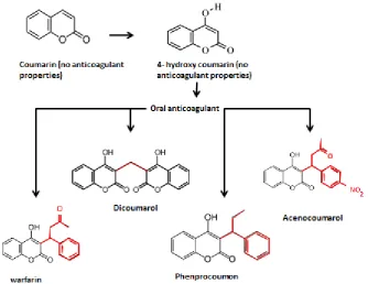
Warfarin is a coumarin derivative. The structures of coumarin and its
derivatives are as shown in Figure 1-1. The enolic benzopyrene structure is essential
[image:15.595.129.464.265.523.2]to their common pharmacological action as vitamin K antagonists (VKA).
Figure 1-1. The structure of coumarin and derivatives adapted from Au and Rettie (2008).
The enolic benzopyrene structure (shown in red) which is important for the molecule’s property as a vitamin K antagonist anticoagulant.
The empirical formula for warfarin is C19H15NaO4. Coumarin is insoluble in
water but a 4-hydroxy substitution confers weak acidic properties on the molecule
making it slightly soluble under weak alkaline conditions. Warfarin contains a single
chiral centre at the C9 carbon which gives two enantiomers, ‘R’ and ‘S’ warfarin (West
et al. 1961). In stable anticoagulated patients, free concentrations of S-warfarin range
8 (Chan et al. 1994). Despite its free concentration being almost the same as that of
R-warfarin, S-warfarin is three to five times more potent than its R-enantiomer in
inhibiting vitamin K epoxide reductase complex (VKORC) activity. Thus the
anticoagulation effects of warfarin are mainly attributed to S-warfarin.
1.2.2 Pharmacokinetics of warfarin and the genes involved
1.2.2.1Absorptions and distribution
Warfarin is completely absorbed after oral administration and reaches peak
concentration in the blood within four hours (Pyörälä et al. 1971). In the blood, it
binds extensively to plasma proteins, primarily albumin (O'Reilly 1969). The genes
involved are presented in Table 1-1 with their known function.
Table 1-1: Genes involved in the transport of warfarin.
Protein Name Gene Function
Alpha-1-acid glycoprotein 1, Orosomucoid 1
ORM1 A plasma glycoprotein that functions as a carrier in the blood (Otagiri et al. 1987, Nakagawa et al. 2003)
Alpha-1-acid glycoprotein 2, Orosomucoid 2
ORM2 A plasma glycoprotein that functions as a carrier in the blood (Otagiri et al. 1987, Nakagawa et al. 2003)
P-glycoprotein, Multidrug resistance protein 1
ABCB1 (MDR1)
A cellular efflux for xenobiotics (Kroetz et al.
2003). Warfarin is a weak inhibitor and may be a substrate (Sussman et al. 2002)
Table adapted from Wadelius and Pirmohamed, 2007.
1.2.2.2Metabolism and excretion
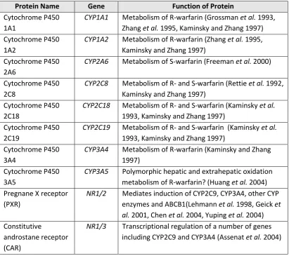
Warfarin is predominantly oxidised by cytochrome P450 in the liver to
inactivated hydroxylated metabolites. S-warfarin is oxidised by CYP2C9 (primarily,
~90%), CYP2C8, CYP2C19 and CYP2C18, whereas R-warfarin is oxidised by
CYP1A2 (primarily, ~60%), CYP3A4, CYP2C8, CYP2C18, CYP2C19 and CYP3A5
CHAPTER 1
9
Table 1-2: Genes associated with the warfarin metabolising enzymes, cytochrome P450. Protein Name Gene Function of Protein
Cytochrome P450 1A1
CYP1A1 Metabolism of R-warfarin (Grossman et al. 1993, Zhang et al. 1995, Kaminsky and Zhang 1997) Cytochrome P450
1A2
CYP1A2 Metabolism of R-warfarin (Zhang et al. 1995, Kaminsky and Zhang 1997)
Cytochrome P450 2A6
CYP2A6 Metabolism of S-warfarin (Freeman et al. 2000)
Cytochrome P450 2C8
CYP2C8 Metabolism of R- and S-warfarin (Rettie et al. 1992, Kaminsky and Zhang 1997)
Cytochrome P450 2C18
CYP2C18 Metabolism of R- and S-warfarin (Kaminsky et al.
1993, Kaminsky and Zhang 1997) Cytochrome P450
2C19
CYP2C19 Metabolism of R- and S-warfarin (Kaminsky et al.
1993, Kaminsky and Zhang 1997) Cytochrome P450
3A4
CYP3A4 Metabolism of R-warfarin (Kaminsky and Zhang 1997)
Cytochrome P450 3A5
CYP3A5 Polymorphic hepatic and extrahepatic oxidation metabolism of R-warfarin? (Huang et al. 2004) Pregnane X receptor
(PXR)
NR1/2 Mediates induction of CYP2C9, CYP3A4, other CYP enzymes and ABCB1(Lehmann et al. 1998, Geick et al. 2001, Chen et al. 2004, Yuping et al. 2004) Constitutive
androstane receptor (CAR)
[image:17.595.114.526.91.456.2]NR1/3 Transcriptional regulation of a number of genes including CYP2C9 and CYP3A4 (Assenat et al. 2004)
10
1.2.3 Pharmacodynamics of warfarin and the genes involved
Warfarin exerts its anticoagulant effect by inhibiting the recycling of vitamin K which
is important in the blood clotting cascade. The recycling of vitamin K process is
particularly important because the amount of vitamin K in the diet and its levels in the
body are limited (Stafford 2005).
1.2.3.1Effect of warfarin on the blood clotting cascade
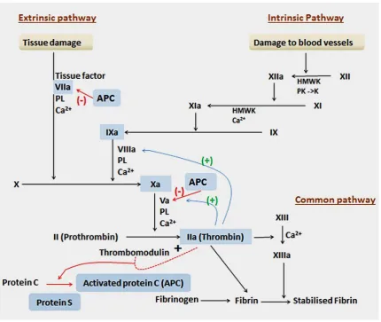
The classic theory of blood coagulation was described using the Cascade and
Waterfall model in the 1960s (Davie and Ratnoff 1964, Macfarlane 1964), as portrayed
CHAPTER 1
[image:19.595.114.545.68.431.2]11 Figure 1-2. . Blood clotting cascade (adapted from Rang and Dale (2012)).). Extrinsic pathway: Any trauma to the tissue-activated endothelial cell leads to exposure of the Tissue Factor. This is a cellular receptor for factor VII, which, in the presence of Ca2+, undergoes an active site transition. This results in rapid autocatalytic activation of factor VII to VIIa. The tissue factor, VIIa and Ca2+ formed an extrinsic tenase complex. This compex convert Factor X (X) to Factor Xa (Xa). In the process acidic phospholipids (PL) function as surface catalyst.
Intrinsic pathway: This pathway is initiated when Factor XII (XII) (from the blood) makes contact with a negatively charged surface. Once a small amount of Factor XIIa accumulates, it will convert prekallikrein (PK) to kallikrein (K) which in turn, accelerates the production of Factor XIIa. Factor XIIa cleaves Factor XI (XI) to form Factor XIa (XIa). Next, Factor X1a cleaves Factor IX (IX) to Factor IXa (IXa). Finally, Factor IXa and Factor VIIa, together with Ca2+ and
12 This model proposed that the process of coagulation can be divided into three
distinct parts: extrinsic (so called because some components come from outside
circulating blood), intrinsic (so called because all the components were present in
circulating blood) and common pathway (process that initiate factor Xa by either
pathway and eventually leads to generation of a fibrin clot). When a blood vessel is
injured a cascade of reactions aimed at forming fibrin is initiated. The components
(called factors) are present in the blood as inactive precursors (zymogen) of proteolytic
enzymes and co-factors. They are activated by proteolysis, the active forms being
designated by the suffix ‘a’. This including vitamin K-dependent coagulation factors:-
Factor II, VII, IX, X, protein C and protein S. However, they require vitamin K in
reduced form for their biological activity. Factor II, VII, IX, X plays a role to activate
blood clotting process whole protein C and S plays roles as regulators in the blood
clotting process. The anticoagulant effect of warfarin is due to the sequential
depression of Factor VII (t½ = 4-6 hours), Protein C (t½ = 8 hours), Factor IX (t½ =24
hours), Protein S (t½ =30 hours), Factor X (t½ =48-72 hours) and Factor II (t½ = 60
hours). The genes that are associated with vitamin K-dependent clotting factors are
described in Table 1-3.
Table 1-3. Genes associated with vitamin K-dependent clotting factors.
Protein Name Gene Protein Function
Coagulation Factor II, prothrombin
F7 Converts fibrinogen to fibrin; activates FV, FVIII, FXI, FXIII, protein C (Berkner 2000, Dahlback 2005)
Coagulation Factor VII
F7 Is converted to FIX and then to FXa (Berkner 2000, Dahlback 2005)
Coagulation Factor IX F9 Makes a complex with FVIIIa and then converts FX to its active form (Berkner 2000, Dahlback 2005)
Coagulation Factor X F10 Converts FII to FIIa in the presence of factor Va (Berkner 2000, Dahlback 2005)
Protein C PROC Activated protein C counteracts coagulation together with protein S by inactivating FVa and VIIIa (Berkner 2000, Dahlback 2005)
[image:20.595.116.528.552.752.2]Protein S PROS1 Participates in many processes, for example, potentiation of agonist-induced platelet aggregation (Berkner 2000, Dahlback 2005)
CHAPTER 1
13
1.2.3.2The Vitamin K Cycle and the mechanism of warfarin action
Reduced vitamin K, or hydroquinone (KH2), plays an important role as a
cofactor for -glutamyl carboxylase (GGCX), which catalyses the post-translational
carboxylation of a specific glutamic acid residue to -carboxyglutamic acid (gla) in a
variety of vitamin K-dependent proteins (Presnell and Stafford 2002). The
carboxylation process is essential for the biologic functions of vitamin K-dependent
proteins involved in blood coagulation.
During the carboxylation process, KH2 is oxidised to vitamin K 2,3 epoxide
(KO). KO then undergoes electron reduction to give the reduced forms K1 and KH2.
The reduction and subsequent re-oxidation of vitamin K, coupled with carboxylation,
is known as the vitamin K cycle Figure 1-3.
[image:21.595.114.493.399.640.2]14 Warfarin binds to the vitamin K epoxide reductase enzyme (VKOR) and
prevents VKOR from recycling vitamins KO and K1 to vitamin KH2 (Silverman
1981). The binding is tight and seems irreversible because of the structural similarity
between warfarin and vitamin K (Fasco and Principe 1982). Due to depletion of KH2,
vitamin K dependent clotting factor is not activated. As a consequence, the
coagulation is reduced due to a decrease in thrombin generation. The genes that are
involved in the vitamin K cycle are presented in Table 1-4.
Table 1-4. Genes involved in the vitamin K cycle.
Protein Name Gene Protein Function
Vitamin K epoxide reductase
VKORC1 A hepatic epoxide hydrolase that catalyses the reduction of vitamin K. The target of warfarin (Bell
et al. 1972, Li et al. 2004, Rost et al. 2004a) Apolipoprotein E APOE Serves as a ligand for a receptor that mediates the
uptake of vitamin K (Saupe et al. 1993, Kohlmeier et al. 1996)
Epoxide hydrolase, microsomal
EPHX1 A hepatic hydrolase in the endoplasmic reticulum that may be complexed with VKOR (Cain et al. 1997, Loebstein et al. 2005, Morisseau and Hammock 2005)
NAD(P)H dehydrogenase, quinine 1
NQO1 A detoxifying enzyme that has the potential to reduce the quinine form of vitamin K (Wallin and Hutson 1982, Berkner and Runge 2004, Ross and Siegel 2004)
Calumenin CALU Binds to vitamin K epoxide reductase complex and inhibits the effect of warfarin (Wallin et al. 2001, Wajih et al. 2004)
Gamma-glutamyl carboxylase
GGCX Carboxylates vitamin K-dependent coagulation factors and protein in the vitamin K cycle (Wu et al.
1997, Rost et al. 2004b)
[image:22.595.113.527.309.662.2]CHAPTER 1
15
1.3
Indication and monitoring warfarin responses
Warfarin is widely used for the treatment and prevention of thrombosis and
thromboembolism, the formation of blood clots in blood vessels and their migration
elsewhere in the body. The aim of warfarin treatment is to maintain a patient’s
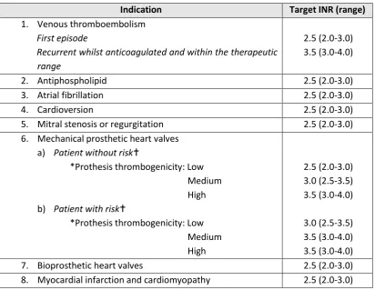
international normalised ratio (INR) within the therapeutic range, as recommended by
the British Committee for Standards in Haematology fourth edition and shown in Table
[image:23.595.114.527.286.605.2]1-5 (Keeling et al. 2011).
Table 1-5. Indications and recommended target INR (adults).
Indication Target INR (range)
1. Venous thromboembolism
First episode
Recurrent whilst anticoagulated and within the therapeutic range
2.5 (2.0-3.0) 3.5 (3.0-4.0)
2. Antiphospholipid 2.5 (2.0-3.0)
3. Atrial fibrillation 2.5 (2.0-3.0)
4. Cardioversion 2.5 (2.0-3.0)
5. Mitral stenosis or regurgitation 2.5 (2.0-3.0) 6. Mechanical prosthetic heart valves
a) Patient without risk
*Prothesis thrombogenicity: Low Medium High b) Patient with risk
*Prothesis thrombogenicity: Low Medium High 2.5 (2.0-3.0) 3.0 (2.5-3.5) 3.5 (3.0-4.0) 3.0 (2.5-3.5) 3.5 (3.0-4.0) 3.5 (3.0-4.0)
7. Bioprosthetic heart valves 2.5 (2.0-3.0)
8. Myocardial infarction and cardiomyopathy 2.5 (2.0-3.0) *Prosthesis thrombogenicity: Low: Carbomedics (aortic position), Medtronic Hall, St Jude Medical (without silzone); Medium: Bjork-Shiley, other bileaflet valves; High: Starr-Edwards, Omniscience, Lillehei-Kaster.
16 The INR is the ratio of patient’s prothrombin time to a normal (control) sample,
raised to the power of International Sensitivity Index (ISI) value. It measures the time
it takes for the blood to clot compared to an international standard. It has been
established by the World Health Organization (World Health Organization) and the
International Committee on Thrombosis and Hemostasis for monitoring the effect of
VKA (International Committee on Thrombosis and Haemostasis 1985). Values above
the ‘therapeutic range’ will place the patient at an increased risk of haemorrhagic
complication, while low values may lead to thrombosis; both scenarios have
potentially dangerous consequences, including serious morbidity and death (Baglin et
al. 2007).
Although the majority of warfarin usage occurs in adults, it is also the mainstay
of oral anticoagulation therapy in children and adolescents. Warfarin is recommended
as thrombo-prophylaxis for heart valve replacement, cardiac catheterization,
post-surgical correction of congenital cardiac defects (e.g. shunt insertion), and
haemodialysis (Monagle et al. 2012). The recommended INR for children with
[image:24.595.118.522.542.786.2]different condition are shown in Table 1-6 (Keeling et al. 2011).
Table 1-6. Indications and recommended target INR (children).
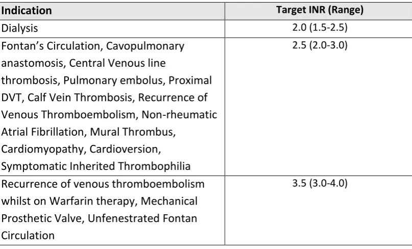
Indication Target INR (Range)
Dialysis 2.0 (1.5-2.5)
Fontan’s Circulation, Cavopulmonary anastomosis, Central Venous line
thrombosis, Pulmonary embolus, Proximal DVT, Calf Vein Thrombosis, Recurrence of Venous Thromboembolism, Non-rheumatic Atrial Fibrillation, Mural Thrombus,
Cardiomyopathy, Cardioversion, Symptomatic Inherited Thrombophilia
2.5 (2.0-3.0)
Recurrence of venous thromboembolism whilst on Warfarin therapy, Mechanical Prosthetic Valve, Unfenestrated Fontan Circulation
CHAPTER 1
17 After treatment is started, the INR response is monitored frequently until a
stable dose-response relationship is obtained; thereafter, the frequency of INR testing
is reduced. It has been recommended that patients have their INR measured at least
every 12 weeks (Baglin et al. 2006). Studies have shown that more frequent testing
leads to tighter anticoagulant control and reduces bleeding and antithrombotic
complications in patients on OAC (Cannegieter et al. 1995, Palareti et al. 1996).
However, it would be inconvenient and expensive to monitor patients this frequently
in primary or secondary care.
Today, many point of care (POC) devices such as CoaguChek® XS (Roche
Diagnostics), INRatio® (Hemosense) and ProTime®/ProTime 3 (International
Technidyne Corporation) are available commercially which enables patients to test
their own INR at home using a finger prick sample of blood. These devices are easy
to use and can generate immediate results. Because patients can test their INR at home,
this leads to greater convenience, especially for those living in remote rural areas or
depend on carers to get them to and from the hospital. Research has shown that
home-testing of the INR leads to better anticoagulation control and improved quality-of-life
(Newall et al. 2006, Smith et al. 2012, Ansell 2013, Bereznicki et al. 2013, Gaw et al.
2013).
1.4
Current warfarin dosing algorithm
In current clinical practice, there is no standardised warfarin dosing algorithm
(Ansell et al. 2008, Keeling et al. 2011). Adult patients are typically initiated with
5mg for 1-2 days but the dose can range from 3-10mg of warfarin depending on age
and disease (Harrison et al. 1997, Crowther et al. 1999, Ansell et al. 2008). Then, the
subsequent dosing is based on the INR response (Table 1-5 and Table 1-6). For
18 valves it is 2.5-3.5 (depending on thrombosis prosthesis thrombogenicity). Prosthesis
thrombogenicity is related to the biomaterial and valve design features (Edmunds Jr
1996). If the INR result is higher than the targeted value the dose will be decreased,
whereas if it is lower the dose will be increased. In patients with a stable dose the INR
will be monitored every two to six weeks, although practice varies widely.
There is no generally accepted method designed for increasing, decreasing, or
maintaining the weekly warfarin dose based on the current INR. In the Randomized
Evaluation of Long-term Anticoagulation Therapy (RE-LY) trials, the algorithm dose
recommendations for atrial fibrillation were as follows (Van Spall et al. 2012): no
change for INR 2.00 to 3.00; increase 15% change for INR ≤1.50, increase 10% for
INR 1.51 to 1.99, decrease 10% for INR 3.01 to 4.00. For INR 4.00 to 4.99, the
recommendation was to hold the dose for 1 day and then to reduce it by 10%. For INR
5.00 to 8.99, the dose was to be held until the INR was therapeutic and then decreased
by 15% per week. It was suggested that the dose be calculated on a weekly, rather
than daily basis, because the recommended dose changes were small and difficult to
achieve with a daily dosing regimen. Subsequently, weekly INR monitoring was also
recommended for out-of-range INR values.
There is lack of specific evidence regarding the safety and efficacy of OAC
drugs in children, especially new-borns, as they are physiologically different to adults
(Andrew et al. 1988, Andrew et al. 1992, Andrew 1995). Current guidelines for
anti-thrombotic therapy in children have recommended an initial warfarin dose of 0.2
mg/kg, with more frequent monitoring of INR than with adults (Monagle et al. 2008).
This dosing recommendation was proposed by Michelson et al. (1998), based on six
publications (Carpentieri et al. 1976, Hathaway 1984, Bradley et al. 1985, Woods et
al. 1986, Doyle et al. 1988, Andrew et al. 1994) and has been evaluated in a
CHAPTER 1
19 found that infants required an average of 0.33 (±0.20) mg/kg and teenagers 0.09
(±0.05) mg/kg of warfarin to maintain an INR of 2.0 –3.0 (Streif et al. 1999).
1.5
Anticoagulation control in patients receiving warfarin
The safety and efficacy of warfarin therapy are dependent on maintaining the
INR within the target range. To attain INR values within the target range, patients are
routinely monitored and their therapeutic dosage adjusted when necessary. Regardless
of the INR within the therapeutic range (at a level at which the incidence of both
thromboembolic and bleeding complications is lowest), adverse events are still
reported, but in smaller percentages than for those with time out of range (Cannegieter
et al. 1995).
Percentage or proportion of time that the patient was within the targeted
therapeutic range (PTTR) is used to summarise INR control over time. It has been
suggested that this is also an evaluation of the effectiveness of anticoagulant therapies,
including warfarin (Rosendaal et al. 1993). A high amount of time spent in the
therapeutic range is associated with a lower risk of thromboembolic events and
bleeding events (Jones et al. 2005, Rose et al. 2009b).
The relationship between PTTR and the benefit of OAC was examined by
Connolly et al. (2008), using the PTTRs of patients from 526 centres, within 15
countries involved in the ACTIVE W trial (Atrial Fibrillation Clopidogrel Trial With
Irbesartan for Prevention of Vascular Events) and who were randomised to OAC. In
patients with a PTTR above 65%, OAC had a marked benefit, reducing vascular events
by >2-fold (relative risk, 2.14; 95% confidence interval, 1.61 to 2.85; P<0.0001). A
population-average model predicted that a PTTR of 58% would be needed to
confidently predict that patients would benefit from being on OAC therapy. Similar
20 ximelagatran, among individuals with atrial fibrillation assigned to warfarin (White et
al. 2007). Those individuals with poor control (defined as PTTR less than 60%), had
higher rates of mortality (4.20% vs. 1.69%) and major bleeding (3.85% vs. 1.58%)
compared with the good control group (defined as PTTR greater than 75 %), (P<0.01).
The poor control group also had higher rates of myocardial infarction (1.38 vs. .62 %,
P = 0.04) and of stroke or systemic embolic event (2.10 vs. 1.07 %, P = 0.02).
In practice, it is recognised that long-term INR stability is difficult to achieve
because of unexpected INR fluctuations in patients. For example, a study conducted
with a British population (n=2223) reported that patients who were treated with
warfarin were inside in the target range only for 67.9% of the time. (Jones et al. 2005).
This finding is supported by a further meta-regression analysis which reported a mean
PTTR in all studies of 64% (van Walraven et al. 2006), although PTTR has been
reported as low as 29% in some studies (Samsa et al. 2000, Sarawate et al. 2006).
The PTTR also increases as the duration of INR monitoring increases. In the
above British population, during the first three months of warfarin therapy, the PTTR
was 48%; after two years, the PTTR increased to 70% (Jones et al. 2005). Similarly,
in a systematic review and meta-analysis of forty studies reporting the PTTR in
patients with VKA for treatment of venous thromboembolism, the mean PTTR was
54.0% in the first three months of treatment and increased to 72% in months four to
twelve and over (Erkens et al. 2012).
Rose et al. (2010) demonstrated that younger age, female sex, lower income,
black race, frequent hospitalisations, poly-pharmacy, active cancer, substance abuse,
psychiatric disorders, dementia, and chronic liver disease were all associated with
lower PTTR. Processes related to warfarin management have also been shown to
CHAPTER 1
21 out of range INR, selection of INR target and loss to follow-up (Rose et al. 2009a,
Rose et al. 2011, Rose et al. 2012, Rose et al. 2013).
1.6
Over-anticoagulation and bleeding risk in patients receiving
warfarin
Over-anticoagulation is a common problem in warfarin therapy and can lead
to a major or life-threatening bleed. It is a measurable parameter for the analysis of
bleeding risk in a modestly-sized population. There is strong evidence that higher INR
values are associated with bleeding risk (Hull et al. 1982, Saour et al. 1990, Altman et
al. 1991, Kearon et al. 2003, Finazzi et al. 2005) (Table 1-7). An INR higher than 4
places a patient at a greater risk of bleeding (Cannegieter et al. 1995, Palareti et al.
1996, Hylek et al. 2000, Hirsh et al. 2003, Pagano and Chandler 2012) and the risk of
intracranial hemorrhage increases approximately 2-fold for every 1 unit rise in INR
(Hylek et al. 1994).
Table 1-7. Relationship between INR and bleeding risk. Authors, studied population,
anticoagulant used
Target INR Range (n)
Duration of Therapy
Incidence of bleeding (%):
Hull et al. (1982), deep vein thrombosis, warfarin
3.0–4.5 (96) vs. 2.0–2.5 (96)
3 months 22.4 vs 4.3 P=0.015
Saour et al. (1990), mechanical prosthetic heart valves, warfarin
2.3–2.7 (125) vs. 1.3-2.7 (122)
3.4 years 42.4 vs 21.3 P<0.002
Altman et al. (1991), mechanical prosthetic heart valves,
acenocoumarol
3.0–4.5 (99) vs. 2.0–2.9 (99)
11.2 months 24.0 vs 6.0 P<0.02
Kearon et al. (2003), recurrent venous thrombosis, warfarin
1.5-1.9 (369) vs. 2.0-3.0 (36)
2.4 years 8.4 vs 10.6 P=0.26
Finnazi et al. (2005), antiphospholipid syndrome, warfarin
3.0–4.0 (54) vs. 2.0–3.0 (55)
22
1.7
Under-anticoagulation and thromboembolism risk in patients
receiving warfarin
In contrast to the bleeding risk associated with VKA, there has been much less
research on the thromboembolism risk with under-anticoagulation in patients receiving
warfarin. It is known that that under-anticoagulation (INR ≤ 1.5) has been associated
with a 16-fold increase in the rate of thromboembolism (Rose et al. 2009b).
Hylek et al. (1996) demonstrated that atrial fibrillation patients with INRs of
1.7 had nearly twice the risk of stroke compared those with INRs of 2. Those with
INRs of 1.5 had nearly three times the risk and those with INRs of 1.3 had a seven-
fold greater risk. Similarly, Palareti et al. (2005), have shown that the relative risk of
recurrence thromboembolism (VTE) was significantly higher in those who spent
more time at an INR<1.5 especially in first 90 days of oral anticoagulant therapy.
Dentali et al. (2012) conducted a study evaluating the risk of thromboembolic
events in patients with isolated subtherapeutic levels of anticoagulant therapy, who
were receiving warfarin because of high-risk conditions such as the presence of
prosthetic mechanical heart valves or moderate-to-high risk AF (Stroke score
CHADS2=3). The sub therapeutic INR value (defined as 0.5–1.0 INR units below the
lower limit of the patient-specific target INR range) was associated with a risk of
thromboembolic events occurring within 2 weeks of the targeted INR being reached.
Based on their observations, patients were exposed to an increased risk of
thromboembolism due to a prolong period of inadequate anticoagulation, rather than a
single low INR. In contrast, other studies did not replicate this association (Clark et
al. 2008, Dentali et al. 2009), possibly due to the patient’s risk profile being unknown
(Clark et al. 2008) or the inclusion of patients with a single sub-therapeutic INR
CHAPTER 1
23
1.8
Factors influencing the warfarin response
Many clinical factors influence the warfarin dose requirement and response.
These include age, weight, height, ethnicity, disease, medications, diet, alcohol,
smoking and adherence.
Warfarin dose requirements fall with increasing age (Loebstein et al. 2001,
Kamali et al. 2004, Sconce et al. 2005, Gage et al. 2008); it has been postulated that
this is due to a reduction in liver size with increasing age (Wynne et al. 1995). Hillman
et al. (2004) showed that age, body surface area and the male gender account for
14.6%, 7.5% and 4.7% of variability of warfarin, respectively. Inter-ethnic differences
in warfarin dose requirements have been reported between African American,
European American and Asian populations. As compared with Caucasians,
African-Americans require a higher dose (Gage et al. 2004) and Asians require lower doses on
average (Dang et al. 2005, Lee et al. 2006).
The presence of other diseases and medications can affect the
pharmacokinetics of warfarin, for example renal failure. Although warfarin is
completely metabolised through the liver, chronic renal impairment can alter the
degree of protein binding and bioavailability, thereby increasing the risk of adverse
events (Grand'Maison et al. 2005). Many drugs can inhibit the activity of CYP2C9
enzymes. These include amiodarone, cimetidine, isoniazid and trimetoprim (Smith et
al. 2004, Greenblatt and Von Moltke 2005, Holbrook et al. 2005, Plakogiannis and
Ginzburg 2007). Lower warfarin dosing is required to maintain the targeted INR when
these drugs are administered together. Conversely, higher warfarin doses are needed
when an inducer of CYP2C9 such as rifampicin, phenobarbital or carbamazepine is
co-administered with warfarin. Common drugs interactions with warfarin are
24
Table 1-8. Common drug interactions with warfarin and proposed mechanism. INCREASED Effect of warfarin DECREASED Effect of warfarin PHARMACOKINETICS
Inhibition of S-warfarin clearance
By inhibition CYP2C9 enzyme: trimethoprim/sulfamethoxazole, amiodarone, fluvastatin, fluvoxamine, isoniazide, phenylbutazone, sertraline, gemfibrozil, amiodarone, metronidazole, fluconazole
Inhibition of R-warfarin Clearance:
Inhibition of CYP3A4 enzyme:
cimetidine, omeprazole), Clarithromycin, Erythromycin, variconazole, metronidazole, fluconazole
By inhibition CYP1A2 enzyme: Ethanol
By inhibition CYP1A2/CYP3A4 enzymes: Ciprofloxacin
Increase warfarin absorption: acarbose
PHARMACOKINETICS
Induction of S-warfarin Clearance
By induction CYP2C9 enzyme: Rifampicin, secobarbital, carbamazepine, phenytoin, phenobarbiturate, primidone, St john’s wart
Induction of R-warfarin Clearance
By induction CYP1A2 enzyme: Cigarette smoking
Alteration of warfarin absorption:
cholestyramine
PHARMACODYNAMIC
Inhibition of synthesis of vitamin K:
Second and third generation cephalosporins
Increase catabolism of clotting factors:
Levothyroxine
Inteference with other pathways of
hemostasis: Acetylsalicylic acid (aspirin) and Nonsteriodal anti-inflammatory drugs (NSAIDs)
PHARMACODYNAMIC
Alteration in Dietary Vitamin K Content: Vitamin K, Vitamin K containing foods
Patient lifestyle also contributes to the warfarin response. As warfarin is a
vitamin K antagonist, patients taking warfarin are likely deficient in regenerated
vitamin K. Therefore, it is a common practice to administer a supra-physiological
doses of vitamin K to reverse the over-anticoagulation effect of warfarin (Hanley et
al. 2004). Several studies have confirmed an inverse relationship between warfarin
maintenance dose requirement and dietary vitamin K intake (Franco et al. 2004, Khan
CHAPTER 1
25 Chronic alcohol intake (long-term) may activate warfarin metabolising
enzymes, and as a result, decrease the anticoagulation effect by increasing warfarin
metabolism (Weathermon and Crabb 1999). For this reason, chronic alcohol drinkers
will need a higher dose of warfarin. Active smokers also need a higher dose of
warfarin due to components in tobacco smoke which can induce activation of CYP1A2
enzymes (Ansell et al. 2008).
Many studies have validated the contribution of genetic polymorphisms to the
warfarin response. CYP2C9*2, CYP2C9*3 and VKORC1-1639 polymorphisms have
been validated in many studies to contribute to warfarin stable doses. These SNPs
explain up to 40% of dose variability (Sconce et al. 2005, Gage et al. 2008, Jonas and
McLeod 2009). In 2007, the US FDA updated the warfarin label to include that the
CYP2C9 and VKORC1 genotypes may be useful in determining the optimal initial
dose of warfarin based on findings regarding pharmacogenetic effects (Thompson
26
1.9
Genetics and warfarin dose requirement
As genetic associations with warfarin responses vary between ethnicities, this
section will compare the response to warfarin doses in different populations/
ethnicities.
1.9.1 CYP2C9 gene and SNPs that affect variability in warfarin dose
The CYP2C9 gene maps to chromosome 10q24.2, contains nine exons, and
codes for a 60kDa microsomal protein. Patients expressing the wild-type gene
encoding CYP2C9 have the CYP2C9*1 genotype (Arg144/Ile359). The most
commonly studied SNPs in this gene are CYP2C9*2 and CYP2C9*3. In patients
expressing the CYP2C9*2 variant, arginine is replace by cysteine (Arg144Cys).
Arg144 is encoded by exon 3 and is located in helix C, which forms part of the putative
P450 reductase binding site of the protein (Rettie and Jones 2005). Any changes are
expected to contribute to a change in enzymatic function. In contrast, CYP2C9*3 is a
genotype produced when isoleucine is replace by leucine (Ile359Leu). Ile359 is
located in exon 7 and maps close to the active site. Replacement of this amino acid
leads to a change in the Vmax and Km of the CYP2C9 substrate (Rettie and Jones
2005). CYP2C9*2 and CYP2C9*3 produce enzyme variants with catalytic activities
involving S-warfarin that are reduced by 30% and 95% respectively compared with
the wild type (Rettie et al. 1999).
The minor allele frequency for CYP2C9*2 (rs1799853) is 0.12 in Americans
and Europeans and much lower in Africans (0.02) (Abecasis et al. 2012). CYP2C9*2
has not been reported in Asian populations except in Malays and Indians in Malaysia
(South East Asians) (Gan et al. 2004, Ngow et al. 2009). The minor allele frequency
of CYP2C9*3 (rs1057910) is 0.06 in both Americans and Europeans, 0,04 in Asians
CHAPTER 1
27 Figure 1-4 shows the effect of CYP2C9 polymorphisms on warfarin dose
variation in different populations. One allele, *2, gives a 6-45% dose reduction
compared with wild type. This contributes to a 13-25% dose reduction in Caucasian
Americans (Higashi et al. 2002, Kealey et al. 2007, Limdi et al. 2007), 5-14% in
Caucasian Europeans (Scordo et al. 2002, Topic et al. 2004, Sconce et al. 2005, Mark
et al. 2006, Markatos et al. 2008) and 26-45% in Asian populations (Gan et al. 2004,
Tanira et al. 2007). Another allele, *3, gives a 15-48% dose reduction. This
contributes to a 28-41% dose reduction in the Caucasian American population
(Higashi et al. 2002, Kealey et al. 2007, Limdi et al. 2007), 25-48% in the Caucasian
European population (Scordo et al. 2002, Topic et al. 2004, Sconce et al. 2005, Mark
et al. 2006, Markatos et al. 2008) and 12-38% in the Asian population (Gan et al. 2004,
Tanira et al. 2007, Yildirim et al. 2008, Huang et al. 2009, Ohno et al. 2009).
Supposedly, the *3 allele has a greater effect on variation in warfarin dosing; in
contrast to this, studies by Tanira et al. (2007) and Gan et al. (2004) revealed that the
*2 allele had a greater influence on warfarin dose in the Omani population and the
Indian group of the Malaysian population. However, these studies have a small sample
sizes and so these findings need to be treated with caution. In addition, there are many
other possible factors which can influence warfarin dose such as diet, drugs and
lifestyle.
If having one *3 allele gives a great influence on warfarin dosing,
homozygosity for the *3 allele gives the greatest influence on warfarin dose (Figure
1-5). Homozygosity for the *3 allele gives a 20-80% dose reduction compared with
wild type (Higashi et al. 2002, Scordo et al. 2002, Gan et al. 2004, Topic et al. 2004,
Huang et al. 2009, Ohno et al. 2009).
There is no clear association between an individual’s genotype and the warfarin
28 that CYP2C9 explained the 6% warfarin dose variation in the Caucasian population
but had no effect on dose variation in the African American population. Similar
findings were observed by Limdi et al. (2007). The inconsistent findings might be the
result of low prevalence of the variant CYP2C9 genotype in these populations, rather
than the lack of effect.
A recent meta-analysis study (Jorgensen et al. 2012), has shown that no
significant associations were found between CYP2C9*2 and warfarin stable dose
requirements for either Asian or white patients butsignificant associations were
observed between CYP2C9*3 and stable dose for the White, Chinese, Japanese and a
mixed Indian, Chinese and Malaysian populations. Dose reduction observed by
comparing the heterozygotes CYP2C9*3 to wild-type showed that white population
required the largest dose reduction (approximately 1.80mg/day less), followed by
mixed Indian, Chinese and Malaysian populations (1.50mg/day less) and Chinese
29
Figure 1-4. Percentage dose reduction by CYP2C9 polymorphisms (one allele compared with wild-type).
Abreviation:
USA: United state AA: African American W: White M’sia: Malaysia I: India
M: Malay C: Chinese
30
1.9.2 VKORC1 gene and SNPs that affect variability of warfarin dose
In 2004, two independent study groups discovered the VKORC1 gene (Li et al.
2004, Rost et al. 2004a). The gene for human VKORC1, which maps to the short arm
of chromosome 16, contains three exons and two introns. The membrane topology of
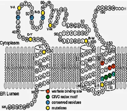
VKORC1 has been investigated by Tie et al. (2005), as illustrated in Figure 1-6. The
protein is 163 amino acids in length and is an integral membrane protein with three
trans-membrane domains.
Alignment of amino acid sequences has identified several conserved amino
acids and functional motifs. Five conserved polar amino acids have been proposed to
form the active centre of VKORC1 (Goodstadt and Ponting 2004). These are two
conserved cysteine residues located within the cytoplasmic loop (Cys43 and Cys51 in
human VKORC1), one conserved serine or threonine (Ser57/Thr57) located within the
cytoplasmic region and two conserved cysteine residues (Cys 132 and Cys 135)
predicted to be partially buried in the ER membrane form a possible CIVC redox motif.
In photo-affinity labelling and site-directed mutagenesis experiments, a
hydrophobic sequence motif, Thr-Tyr139-Ala (TYA) was identified as essential for the
dicoumarol binding site (Ma et al. 1992). This was supported by evidence that
mutation at residue Y139 in the rat produces warfarin-resistant phenotypes. Although
tyrosine and phenylalanine differ by only one hydroxyl group, substitution led to
CHAPTER 1
[image:39.595.114.528.69.433.2]31
32 The first identified SNPs in the VKORC1 gene were reported by D’Andrea et
al. (2005). Rieder et al. (2005) grouped 10 common SNPs to two main haplotypes, A
and B, where A required a low dose and B required high-dose warfarin. Similar
findings were observed by Giesen et al. (2005), who further extended the number of
main haplotypes to VKORC1*1, VKORC1*2, VKORC1*3, VKORC1*4. VKORC1*1
is identified as ancestral, VKORC1 *2 corresponds to the low-dose group A, as defined
by Rieder et al., (2005), while VKORC1*3 and *4 correspond to the high-dose group
B.
The effect of VKORC1*2 (the lower dose phenotype), VKORC1 1173
(rs9934438) and VKORC1 -1693 (rs9923231) has been extensively studied in Asian
populations. Both SNPs are in near perfect linkage disequilibrium (LD). The minor
allele frequency is 0.44 in American, 0.40 in European, 0.07 in African and 0.92 in
Asian population (Abecasis et al. 2012). The effects of polymorphisms are described
in Figure 1 7(A-C). In Japanese populations, heterozygosity for these allele gave a
~49% dose reduction whilst homozygosity gave a ~67% dose reduction compared with
the wild-type carrier (Kosaki et al. 2006, Kimura et al. 2007, Ohno et al. 2009). In the
Chinese population, the heterozygous variant gave ~8-51% dose reduction and the
homozygous variant gave ~43-71% dose reduction (Wang et al. 2008, Huang et al.
2009). The Korean population was shown to be more sensitive to the variant allele;
the heterozygous variant led to a 60% dose reduction and the homozygous variant also
gave a very similar dose reduction (Cho et al. 2007).
In white American Caucasians, both SNPs contributed to a 24-27% dose
reduction in heterozygotes and 40-49% in the homozygotes (Li et al. 2006, Schelleman
et al. 2007). However, there were contradictory findings about the association of these
SNPs in the African American population. For example, Shellemann et al. (2007)
CHAPTER 1
33 warfarin dose in African American people. In contrast, a study by Limdi et al. (2008)
found similar SNP genotypes were associated with a lower warfarin dose (p<0.005)
among African Americans.
Perini et al. (2008) examined the distribution of VKORC1 -1693 in the
Brazilian population. The heterozygous variant for VKORC1 -1693 contributed to a
20-36% dose reduction while the homozygous variant contributed to 40-46%.
In European populations, studies from Italy on VKORC1 1173 and Greece on
VKORC1 -1693 demonstrated a 20-35% dose reduction for the heterozygous variant
and 60-70% for the homozygous variant (Borgiani et al. 2007, Markatos et al. 2008).
Only VKORC1*3 (rs 7294), the higher dose phenotype has been studied for its
association with warfarin dose. The minor allele frequency is 0.35 in both Americans
and Europeans, 0.48 in Africans and 0.07 in Asians (Abecasis et al. 2012). In Japanese
populations, the heterozygous condition contributed to a 30% dose increment (Kimura
et al. 2007). In American Caucasians, heterozygosity contributed up to a 19% dosage
increment while homozygosity contributed up to 39% (Kimura et al. 2007). In
European populations, Borgiani, et al. (2007) found that the heterozygous variant
contributed up to 60% and the homozygous variant contributed up to 90% of the dose
increment.
In the same meta-analysis as mentioned earliar (Jorgensen et al. 2012), a
significant association was observed between the VKORC1 rs9923231 SNP and stable
dose in both the white and Chinese ethnic groups, with heterozygotes requiring 1.45
mg/day less than wild-types in both ethnic groups. A significant difference was also
observed between VKORC1 rs7294 carriers (heterozygotes) and wild-types the white
population. Carriers of VKORC1 rs7294 required 1.80 mg/day more than the
34
(A) rs9934438 (VKORC1*2 1173C>T)
(B) rs9923231 (VKORC1*2-1693G>A)
(C) rs7294 (VKORC1*3)
Legends:
Heterozygous Homozygous
CHAPTER1
35 In summary, variants of both CYP2C9 and VKORC1 have a significant
influence on warfarin dose requirement. Many researchers have evaluated the
relationship between genotype and dose to build a statistical model that incorporates a
variety of factors (i.e., clinical, demographic and genetic) to estimate the influence of
each factor on dose requirement. Such analyses have demonstrated that
polymorphisms in VKORC1 account for approximately 40% of the variance in
warfarin dose while CYP2C9 explains less than 10% (Schalekamp and De Boer 2010).
Together with clinical data, they can explain up to 60% of warfarin variation (Wadelius
et al. 2009, Lenzini et al. 2010). Still, another 40% remains unrecognised and this
could result in over or under dosing. Considering the difference of minor allele
frequencies in different populations which may contribute to the different effect of the
polymorphism to warfarin dose, pharmacogenetics algorithm for a specific population
is needed.
1.9.3 Other genes and variability in warfarin dose
A recent study identified a non-synonymous variant (rs2108622) in exon 2 of
a cytochrome P450 family 4, subfamily F, polypeptide 2 gene (CYP4F2) which led to
an increase in warfarin requirement in a European–American cohort of patients
(Caldwell et al. 2008). CYP4F2 is a vitamin K1 oxidase and that carriers of the
CYP4F2 V433M allele (rs2108622) have a reduced capacity to metabolize vitamin K1
(McDonald et al. 2009). Therefore, patients with the rs2108622 polymorphism are
likely to have elevated hepatic levels of vitamin K1, necessitating a higher warfarin
dose.
Caldwell et al., (2008) then repeated the study to validate the result in three
different sites at Marshfield University, the University of Florida and Washington
University in St Louis. The variant predicted an additional 4% (Washington
36 variability of warfarin dose per T allele (minor allele). The effect of this SNP has been
confirmed in a Swedish population, where it accounted for 1% of dose variability
(Takeuchi et al. 2009), Italians where it explained 7.5% of dose variability (Borgiani
et al. 2009) and North Americans where each copy of the minor allele elevated the
dose by ~0.5mg/day (Cooper et al. 2008). The effect was not present in
African-American populations since the minor allele frequency is low in this population
(Cavallari et al. 2010).
Contradictory findings were shown in an association study of warfarin dose
and polymorphisms of GGCX, Factor II, Factor VII, Factor X, EPHX1, CALU,
ABCB1, and CYP3A4 (D'Ambrosio et al. 2004, Wadelius et al. 2004, Aquilante et al.
2006, Vecsler et al. 2006, Rieder et al. 2007, Saraeva et al. 2007). It was also
discovered that the CYP3A5*3, CYP1A1*1F, CYP2C19 and PROC gene variants did
not correlate with warfarin dosage variation (Wadelius et al. 2004, Saraeva et al.
2007).
1.10
Genetics and other warfarin-related outcomes
Warfarin-related bleeding is the most problematic side effect of this drug. As
such, the relationship between genetic variation and bleeding risk has become an area
of research interest.
1.10.1Genetics and over-anticoagulation during warfarin treatment
Carriers of CYP2C9*2 and CYP2C9*3 are more frequently
over-anticoagulated (INR ≥4) than wild-type carriers (Higashi et al. 2002, Wadelius et al.
2009, Molden et al. 2010, Ma et al. 2012). A systematic review and recent
meta-analysis have demonstrated that both CYP2C9*2 and CYP2C9*3 carriers were
susceptible to over anticoagulation (INR ≥4) when compared to the CYP2C9
