1
Culture Conditions Govern Mouse Embryonic Stem Cell Behaviour:
Dependence on Heparan Sulfate and Optimisation of Synthetic Polymer
Substrates
Thesis submitted in accordance with the requirements of the University of
Liverpool for the degree of Doctor in Philosophy.
By
Chloe Jayne Williams
3
Acknowledgements
First I would like to thank my supervisors Prof. Jerry Turnbull, Dr. Patricia Murray and Prof. David Edgar for their support, motivation and patient guidance throughout. I feel extremely
lucky to have had Jerry as my primary supervisor; at times when I felt like my only option was to give up, his kind words, compassion and encouragement kept me going – a brilliant
role model.
Special thanks go to fellow students and staff at The University of Liverpool and specifically to all members of Lab B, past and present, for their technical know-how, especially Dr.
Sophie Thompson and Dr. Scott Guimond. I accredit Sophie with keeping me sane as we regularly discussed the latest Eastenders storyline, and re-lived our teenage youth through Radio One’s golden years in the hope of forgetting the frustrating lab problems!
Many thanks also go to the CASE partners SpheriTech Ltd, specifically Don Wellings and Andy Gallagher, not only for providing us with novel polymers but for their input regarding
materials and methods for chapter 5. Dr. Victoria Kearns and Prof. Rachel Williams of the Clinical Engineering department at the University of Liverpool provided invaluable advice
regarding materials analysis.
On a personal level, I would like to thank my mum, an inspiration to whom I owe my hardworking ethos. Selflessly, mum would listen to me ramble about my latest experiments
during coffee breaks (sometimes cake if we were in non-diet mode!). Thanks go to my younger brother Matt (equally as inspirational as he completes his GCSEs at this time) for repeatedly asking those basic questions that could so easily have been lost; ‘what is a stem
4
both proud. I look back fondly on their constant Dr. jokes and light-hearted, unique sense of
humour that so importantly kept me smiling.
Special thanks too, go to Alex. She constantly provided me with personal support, encouragement and laughs; a hug when I needed one and a kick-up the a** when I lost sight of the finish. Black coffee seemed in endless supply, and I hope one day I can repay the
favour.
Finally, I would like to thank the BBSRC for the financial support and likewise, my CASE
5
Contents
1. Introduction ... 14
1.1 Embryonic Stem Cells ... 14
1.1.1 Origins of ESCs ... 14
1.1.2. Early embryonic development ... 16
1.1.2 Human ESCs (hESCs) and mouse ESCs (mESCs) ... 17
1.1.3 ESC pluripotency ... 18
1.1.4 In vitro expansion of mESCs (maintenance of pluripotency) ... 23
1.1.5 Embryoid Body (EB) ... 24
1.1.7 Lineage commitment of mESCs ... 25
1.2 Heparan Sulfate ... 37
1.2.1 Glycosaminoglycans (GAGs) ... 37
1.2.2 HS structure ... 38
1.2.3 HS Biosynthesis ... 39
1.2.4 Core proteins ... 42
1.2.5 Heparin: a close relative of HS ... 44
1.3 Heparan sulfate in development ... 45
1.3.1 Early embryonic development ... 45
1.3.2 HS-protein interactions ... 46
1.4 Biomaterials ... 54
1.4.1 Overview of biomaterials ... 54
1.4.2. Optimising polymer surfaces to enhance polymer-cell interaction ... 60
1.5 Work leading to current studies ... 61
2. Materials and Methods ... 62
2.1 Materials ... 62
2.1.1 Solutions ... 62
2.1.2 Buffers ... 63
2.1.3 Cell lines ... 63
2.1.4 Biomaterials ... 64
2.2 Methods... 65
2.2.1 Cell Culture ... 65
2.2.2 Immunochemistry and histology ... 71
2.2.3 Real Time Polymerase Chain Reaction... 75
2.2.4 Compositional disaccharide analysis of HS ... 77
6
2.2.6 Statistical Analysis ... 83
3. Variations in mESC culture condition influences behavior ... 84
3.1 General Introduction ... 84
3.2 mESCs maintained in the absence of feeders and/or serum, remain undifferentiated and display typical behaviour during 2D culture ... 86
3.3 mESC development in the EB model is dependent upon 2D culture conditions prior to EB formation ... 91
3.3.1 EB characterisitcs including morphology, size and cavitation all differ depending on 2D mESC culture conditions ... 91
3.3.2 mESC differentiation capacity is impaired in EBs generated from serum-free feeder-free conditions ... 97
3.3.3 Differentiation of extraembryonic endoderm and BM formation within EBs is influenced by mESC pre-culture conditions ... 101
3.4 Differentiation of mesendoderm and ectoderm within EBs is influenced by mESC pre-culture conditions ... 110
3.5 Discussion ... 113
4. Heparan Sulfate is crucial for normal mESC behaviour ... 125
4.1 General Introduction ... 125
4.2. Defective EB development attributed to a lack of serum, can be rescued with exogenous porcine mucosal heparin (PMH) ... 130
4.3 HS-deficient EXT1-/- mESCs require serum and feeders for normal behaviour ... 136
4.3.1 HS-deficient mESCs cannot be maintained in the absence of serum during 2D expansion ... 136
4.3.2 HS-deficient EBs do not display characteristic morphological development ... 141
4.3.3 HS-deficient EBs show restricted EEE differentiation ... 146
4.4 HS structure and abundance differs depending on mESC culture conditions ... 154
4.4.1 mESC culture condition influences the structure of cell-surface HS ... 155
4.4.2 Culture conditions influences the structure of HS synthesised by mESCs ... 159
4.4.3 Levels of HS biosynthetic enzyme expression in E14 mESCs differs depending on culture condition ... 167
4.5 Discussion ... 171
5. Optimisation of polymers for the expansion of mESCs and KSCs in vitro ... 179
5.1 General Introduction ... 179
5.2 Hydrogels for cell attachment or viability ... 188
5.3 Poly-Ɛ-lysine based polymers support mESC viability and self renewal ... 198
5.4. Growth of kidney stem cells on poly-Ɛ-lysine ... 203
5.4.1. Poly-Ɛ-lysine-based polymers +/- RGD support KSCs-GFP ... 203
7
5.6 Discussion ... 216
6. General Discussion and Future Directions ... 224
References ... 234
8 Table of Figures
1.1. Early embryonic development ... 16
1.2. mESC pluripotency ... 19
1.3. EB development ... 26
1.4. Laminin ... 33
1.5. GAG repeating unit ... 38
1.6. HS biosynthesis. ... 40
Table 1.1 HS biosynthetic enzymes and their respective roles ... 41
1.7 HS master regulator ... Error! Bookmark not defined. Table 1.2 Classifications of biomaterials ... 55
Figure 1.8. Polystyrene application in cell culture. ... 58
Table 1.3 Uses of common polymers in medical applications and corresponding monomers ... 59
Table 2.1 Synthetic SpheriTech hydrogels and corresponding compositional ratios. ... 64
Figure 2.1 Schematic outlining the three distinct 2D culture conditions employed during E14 mESC expansion ... 66
Figure 2.2 Schematic diagram depicting the trans-well set-up ... 70
Table 2.4 HS compounds adsorbed to surface of poly-Ɛ-lysine macroporous. ... 71
Figure 2.3 Diagram outlining the quantification of a Nanog immunostain. ... 75
Figure 2.4 Diagram outlining the quantification of differentiating EBs with regard to laminin expression. 75 Table 2.5 Heparitinase enzymes for HS digestion to aid disaccharide analysis ... 79
Table 2.6 Order of disaccharide standards elution during HPLC-SAX analysis with time ... 81
Figure 2.6 Schematic of CPD set-up. ... 82
Figure 3.1. E14 mESC display typical morphology in the absence of feeders and/or FBS. ... 88
Figure 3.2 E14 mESCs express Oct4 and Nanog in the absence of feeders and/or FBS. ... 89
Figure 3.3 The proportion of Oct4 and Nanog positive E14 mESCs was comparable in all three culture conditions. ... 89
Figure 3.4 E14 mESC proliferation rate is dependent on culture condition. ... 90
Figure 3.5 Serum-free feeder-free EBs are significantly smaller than EBs derived from mESCs cultured with serum +/- feeders.. ... 93
Figure 3.6 Serum-free EBs are smaller than EBs conditioned with serum. ... 93
Figure 3.7 E14 mESCs EB development and morphology is different and dependent on 2D growth conditions prior to EB formation. ... 94
Figure 3.8 EB development is influenced by 2D growth conditions prior to EB formation. ... 96
Figure 3.9 Percentage of Oct4+ and Nanog+ cells within 7d EBs depends on prior 2D culture conditions.. 98
Figure 3.10 Localisation of Oct4 positive mESCs EBs changes with different mESC culture conditions. .. 99
Figure 3.11 Oct4 mRNA levels are significantly higher for serum-free feeder-free conditioned day 7 EBs.. ... 99
Figure 3.12 Localisation of Nanog differs depending on culture condition.. ... 100
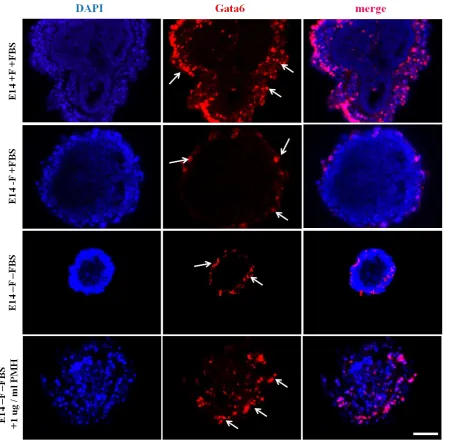
Figure 3.14 Gata6 mRNA levels differ depending on culture condition. ... 104
Figure 3.13 Gata6 positive cell numbers vary depending on culture condition. ... 104
Figure 3.15 Primitive endoderm marker, AFP levels vary depending on culture condition.. ... 105
Figure 3.16 AFP mRNA levels do not change between conditions. ... 105
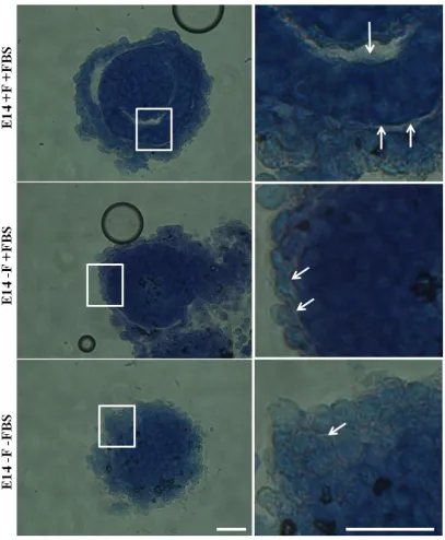
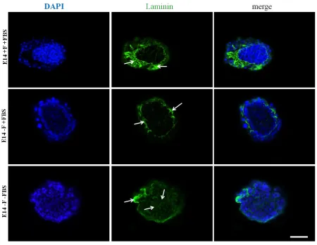
Figure 3.17 2D mESC culture conditions affect laminin distribution in EBs. ... 106
Figure 3.18 2D ESC culture conditions affect laminin distribution in EBs. ... 107
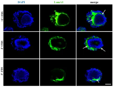
Figure 3.20 LamA1 expression levels differ depending on culture conditions.. ... 109
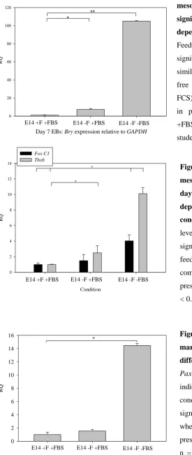
Figure 3.21 Levels of nascent mesoderm marker, Bry are significantly different for day 7 EBs depending on 2D culture conditions. ... 112
Figure 3.22 Levels of paraxial mesoderm makers Foxc1 and Tbx6 in day 7 EBs significantly differ depending on 2D culture pre-condition. ... 112
9
Table 3 Summary of 2D culture conditions prior to EB formation and the effects they have on subsequent
EB development. ... 115
Figure 4.1 Exogenous PMH affects the distribution of Oct4 positive cells within EBs. ... 131
Figure 4.2 Exogenous PMH induces Gata6 expression in peripheral EB cells.. ... 132
Figure 4.3 Effect of exogenous PMH on laminin expression. ... 134
Figure 4.4 Exogenous PMH was shown to rescue a typical EB differentiation marker pattern expression in 7 day serum-free conditioned EBs. [ ... 135
Figure 4.5 HS-deficient EXT1-/- mESC expansion was not supported in serum-free and feeder-free 2D culture conditions.. ... 138
Figure 4.6 HS-deficient EXT1-/- mESC maintained in serum-free 2D conditions are not viable by 48 h. 139 Figure 4.7 Oct4 and Nanog expression in HS-deficient EXT1-/- mESCs cultured in the presence or absence of feeder cells.. ... 139
Figure 4.8 The percentage of Nanog positive mESCs is higher in HS-deficient EXT1-/- mESCs compared to E14 mESCs but only in the absence of feeders. ... 140
Figure 4.9 HS-deficient EBs are significantly smaller compared to E14 EBs. ... 142
Figure 4.10 HS-deficient EXT1-/- EBs display some signs of normal development but lack cavitation, despite culture condition. ... 143
Figure 4.11 HS-deficient EBS lack cavitation and a complete BM. ... 145
Figure 4.12 The distribution of Oct4 positive cells is more extensive throughout HS-deficient EBs compared to E14 EBs and a lack of feeders enhances this effect. ... 148
Figure 4.13 The percentage of Oct4 positive cells within HS-deficient EBs is higher than in E14 EBs. .... 149
Figure 4.14 Oct4 expression in day 7 EBs is significantly higher in HS-deficient EXT1-/- EBs compared to E14 EBs. ... 149
Figure 4.15 HS-deficient EBs display abnormal localisation of Gata6 positive cells. ... 150
Figure 4.16 HS-deficient EBs display significantly lower Gata6+ cells in day 7 EBs compared to E14 EBs.. ... 150
Figure 4.17 Laminin is abnormally localised in HS-deficient EBs. ... 151
Figure 4.18 Abnormal intracellular LamA1 expression in HS-deficient EBs. ... 152
Figure 4.19 HS-deficient EBs display significantly lower LamA1 expression compared to day 7 E14 EBs derived from the same culture conditions... 153
Figure 4.20 Expression of 10E4 HS epitope was detected in all three conditions but localisation differed and primary expression was by feeder cells. ... 156
Figure 4.21 Levels of HSPGs on the surface of mESCs, identified by an antibody specific to the 3G10 HS-epitope, differed in three different culture conditions.. ... 157
Figure 4.22 10E4 and 3G10 epitope expression levels and localisation differs depending on the mESC culture condition.. ... 158
Figure 4.23 Quantitation of HS detected from equal volumes of starting material demonstrates differences in HS levels depending on source or culture condition. ... 161
Figure 4.25 HPLC-SAX analysis of soluble HS purified from media components, Advanced DMEM and serum (FBS), demonstrates the abundance of HS and disaccharide compositional make-up. ... 163
Table 4.1 HPLC-SAX analysis demonstrated differences in soluble HS purified from conditioned media, from different culture conditions. ... 165
Figure 4.27 EBs generated from mESCs cultured in the absence of serum express higher levels of EXT2 than EBs generated from mESCs cultured in the presence of serum. ... 168
Figure 4.28 Levels of the HS biosynthetic enzyme NDST1 are significantly different depending on mESC 2D culture condition. ... 169
Figure 4.29 Levels of the HS modification sulfatase enzyme Sulf1 are different depending on mESC culture condition but Sulf2 levels are unchanged. ... 170
Table 5.1 Outlining some common hydrogels successfully employed as biomaterials ... 183
Figure 5.1 Poly-E-lysine polymer, showing the monomer subunit, n = 25 - 30. ... 184
10
Figure 5.3 Methacrylic-based hydrogels 6, 15 and 16 do not support fibroblast attachment or proliferation. ... 193 Figure 5.4 Acrylic-based hydrogels 7, 9 and 11 display little evidence of supporting fibroblast attachment
and spread.. ... 194 Figure 5.5 Acrylic-based hydrogels 12, 13 and 14 display little evidence of supporting fibroblast attachment and spread.. ... 195 Figure 5.6 Fibroblast morphology varies according to ability to spread. STO fibroblasts were seeded onto
two different control conditions, one to encourage cell attachment and spread (fibronectin-coated cover slip) and another to discourage cell attachment (uncoated glass cover slip). Cell morphology was assessed accordingly, 48 h post seeding. ... 196 Figure 5.7 Fibroblast viability was completely lost in all but three hydrogels. ... 197 Figure 5.8 mESCs seeded onto poly-Ɛ-lysine polymers, attach, proliferate and retain alkaline phosphatase
expression up to 10 days.. ... 200 Figure 5.9 E14 mESCs maintain Nanog expression when seeded onto poly-Ɛ-lysine-based macroporous
polymers and topography of polymer directs location of mESCs. ... 201 Figure 5.10 Poly-Ɛ-lysine topography and architecture. ... 202 Figure 5.11 Poly-Ɛ-lysine macroporous polymers (+/- RGD) support KSCs-GFP attachment and
proliferation up to 10 days.. ... 205 Figure 5.12 Synthetic poly-Ɛ-lysine coupled with RGD, supports KSCs-GFP proliferation at a higher rate
than poly-Ɛ-lysine not coupled with RGD. ... 206 Figure 5.13 KSCs-GFP populate the pores of the poly-Ɛ-lysine or on the surface of the spheres, and
fibroblast-like processes appear to be lost compared to standard tissue culture plastic. ... 207 Figure 5.14 Tissue culture plastic coated with per-sulfated heparin, outperforms other heparin analogue
coatings, in supporting KSC-GFP attachment and spreading... 209 Figure 5.15 PMH and per-sulfated heparin structures absorbed to the surface of tissue culture plastic best
support KSC-GFP proliferation compared to tissue culture plastic coated with 6-O and 2-O desulfated heparin or NAc heparin structures. ... 212 Figure 5.16 Surface topography of poly-Ɛ-lysine macroporous polymers is affected by coating with
different HS-mimetic structures. ... 213 Figure 5.17 KSC-GFP proliferation on poly-Ɛ-lysine macroporous polymers altered depending on heparin
structure.. ... 214 Figure 5.18 KSC-GFP-polymer interaction alters depending on the level/pattern of sulfation exhibited by
11
Abstract
Human embryonic stem cell (hESC)-based therapies will only become viable once we eliminate the use of animal-derived material during ESC scale-up. Some groups have demonstrated the expansion of hESCs in xeno-free systems but the effect on downstream self-renewal and differentiation is poorly understood. Heparan sulfate (HS) is a master regulator of cellular behavior but the role of HS during ESC expansion is unclear, as is the exogenous source of HS in cultures. It has been shown that mESCs synthesise low levels of low-sulfated HS, but it is unclear if culture condition has any impact. In the studies here, three discrete culture conditions were employed for E14 mESC expansion along with immunostaining and RT-qPCR to study marker expression for differentiation to the three lineages and corresponding BM synthesis. SAX-HPLC was used to characterise soluble HS from cells/medium/serum. A varierty of polymers were tested as synthetic alternatives for ESC expansion. It was found that HS-deficient embryoid bodies (EBs) (derived from EXT1-/- mESCs in normal culture conditions) remained in a pluripotent state and lacked a typical differentiation pattern. Furthermore, HS-deficient mESCs could not be maintained in the absence of serum, highlighting a link between serum and HS. EBs derived from E14 mESCs cultured in the absence of serum displayed unusual differentiation patterns, which were rescued by exogenous porcine mucosal heparin (PMH). Feeder cells displayed cell-surface HS but feeder-cell conditioned medium (CM) was predominantly an unsulfated structure. An array of low and highly sulfated HS structures were identified in serum-alone. 10-fold more HS was purified from serum-free feeder-free (-F –FBS) CM compared to the other mESC CM (with/without feeders but in the presence of serum; +/-F +FBS). Furthermore, unlike +/-F +FBS conditions, highly sulfated HS disaccharide UA2S–GlcNS6S was the major constituent in –F-FBS and Sulf2 levels were significantly reduced. Poly-Ɛ-lysine macroporous substrates supported mESC and kidney-derived stem cells (KSCs-GFP) adherence and proliferation, further enhanced by adsorbing RGD or per-sulfated HS structures to the surface of the poly-Ɛ-lysine. The key conclusions from these studies were that serum is a source of HS, without which, mESCs behave uncharacteristically; that synthetic HS-mimetic structures could represent an alternative to serum; and poly-Ɛ-lysine shows great promise to replace current animal-derived coating materials for ESC expansion.
12
Abbreviations
Adv DMEM Advance Dulbecco's Modified Eagle Medium
BM Basement membrane
BMP Bone morphogenic protein BODIPY Boron-dipyrromethene
Bry Brachyury
cDNA Complimentary deoxyribonucleic acid CEE Columnar epiblast epithelium
CM Conditioned media
CNS Central nervous system DAPI 4',6-diamidino-2-phenylindole DMEM Dulbecco's Modified Eagle Medium DNA Deoxyribonucleic acid
DNAse Deoxyribonucleic acid enzyme
EB Embryoid body
EC Embryonic carcinoma
ECM Extracellular matrix
EDTA Ethylenediaminetetraacetic acid
EEE Extraembryonic endoderm
EG Embryonic germ
EMT Epithelia-mesechymal transition
ESC Embryonic stem cell
EXT1 Exostosin-1
EXT2 Exostosin-2
F Feeder layer
FBS Fetal bovine serum
FGF Fibroblast growth factor
GAPDH Glyceraldehyde-3-phosphate dehydrogenase
GAG Glycosaminoglycan
HPLC High performance liquid chromatography
HS Heparan sulfate
13 Klf Kruppel-like factor
LIF Leukaemia inhibitory factor
LIFR Leukaemia inhibitory factor receptor
Min Minutes
NA N-Acetyl
NDST N-Decetyl Sulftransferase
NS N-Sulfatase
PBS Phosphate buffered saline PCR Polymerase chain reaction
PE Primitive endoderm
PƐL Poly-Ɛ-lysine
PFA Paraformaldehyde
PGC Primordial germ cell PMH Porcine muscosal heparin PMHS Porcine mucosal heparan sulfate Rpm Revolutions per minute
RT Room temperature
RT-qPCR Real-time quantitative polymerase chain reaction RNA Ribonucleic acid
SAX Strong anion exchange
SEM Secondary electron microscopy SSEA Stage-specific embryonic antigen
STAT Signal transducer and activator of transcription Su1f1 Sulfatase enzyme 1
Sulf2 Sulfatase enzyme 2
14
1. Introduction
1.1Embryonic Stem Cells
Embryonic stem cells are pluripotent cells that can differentiate to become any cell type
whilst maintaining the ability to replicate indefinitely. As a result, embryonic stem cells have massive potential in medicine, both therapeutically in repairing damaged/diseased tissue, for example, in Parkinson’s disease or diabetes, and for implementation in drug screening.
However, before stem cell-based therapies can be developed or employed clinically, it is necessary to understand how embryonic stem cells are regulated, both in maintenance of their
pluripotency and direction of their differentiation.
1.1.1 Origins of ESCs
Since 1970, four different classes of pluripotent cells have been isolated; embryonic stem cells (ESCs), embryonic germ (EG) and epiblast stem cells (EpiSCs) from the embryo, and embryonic carcinoma (EC) cells isolated from adults. All three have the capacity for self-renewal and differentiation but discrete differences underpin their respective therapeutic potential.
Embryonic stem cells (ESCs)
Embryonic stem cells (ESCs) are derived from the inner cell mass (ICM) of a pre-implanted blastocyst (Evans and Kaufman 1981; Martin 1981) and represent pluripotent cells with the ability to replicate indefinitely in an undifferentiated state, whilst maintaining the ability to
give rise to all cell lineages once stimulated correctly, such is the ability of ESCs to generate high cell numbers.
Embryonic germ (EG) cells
Embryonic germ (EG) cells are derived from primordial germ cells (PGC) of the post-implanted embryo, destined to form eggs or sperm. In the human, EG cells can be isolated
15
from PGCs in the mouse embryo at 8.5 day post-coitum (Labosky, Barlow et al. 1994;
Donovan and de Miguel 2003).
Epiblast Stem Cells (EpiSCs)
Epiblast-derived stem cells (EpiSCs) are derived from the very early (pre-implanted
blastocyst stage) embryo but pre-gastrulation.
Embryonal carcinoma (EC) cells
A teratoma represents an encapsulated tumour composed of various tissues foreign to their site of origin, of which there are two classifications; malignant and benign. Benign teratomas
have limited growth ability and are represented by well-differentiated somatic tissues. Malignant teratomas (teratocarcinomas) in contrast, contain undifferentiated stem cells with
an unlimited proliferative ability and tendency to metastasize. Teratocarcinomas, first formed experimentally in adult mice by grafts of early mouse embryonic tissues, represent the origin of EC cells. Most EC cell lines, despite retaining some capacity to differentiate, display poor
differentiation in vivo andtypically form tumours (Stevens 1970; Damjanov, Damjanov et al. 1987).
Consequently, EC cells do not present much potential in any clinical capacity, largely owing to their tumourigenic nature. Furthermore, EC cells are aneuploid and cannot proceed through meiosis to produce mature gametes, unlike ESCs which stably retain euploid chromosome
constitution, crucial for meiosis and underpins genetic manipulation technology. Although EG cells form chimeras following injection into the blastocyst and give rise to the three germ
layers (Stewart, Gadi et al. 1994), their genomic imprint can be erased, therefore negating the ability to employ them as therapeutic agents (Smith 2001). Subsequently, ESCs represent the
16 1.1.2. Early embryonic development
ESCs, owing to their defining pluripotent characteristic, theoretically can mimic in vivo
development in an in vitro model. Embryonic development is a complex, highly sequential process whereby the zygote diversifies into every cell type, as a consequence of
differentiation, proliferation and growth, which begins with cleavage (mitotic divisions after fertilisation) and gastrulation (polarisation of the embryo), outlined in Figure. 1.1.
1.1. Early embryonic development Schematic outlining the early embryonic development cascade which gives rise to extraembryonic and embryonic tissues (from stemcells.nih.gov)
Given that ESCs are isolated at the ICM stage of embryonic development, they possess the
ability to give rise to all three germ layers; endoderm, ectoderm and mesoderm, underpinning the massive clinical potential, since every cell type in the adult human originates from one of
17
Ectoderm (outer layer):
Outer surface - epidermal (skin)
CNS - neuron of brain
Neural crest - pigment cell
Mesoderm (middle layer):
Dorsal - notochord
Paraxial - bone tissue
Intermediate - tubule cell of kidney
Lateral - red blood cells
Head - facial muscle
Endoderm (inner layer):
Digestive tube - stomach cell
Pharynx - thyroid cells
Respiratory tube - lung cell
Consequently, any population of ESCs theoretically can be directed to differentiate and become a specific cell type to suit specific clinical applications; for example, ESCs stimulated to become neurons for a Parkinson’s disease-based application (Marchetto,
Brennand et al.; Zhang, Duan et al.) or ESCs stimulated to become insulin-producing cells for diabetes treatment (Soria, Roche et al. 2000).
However, any clinical application will initially require the expansion of ESCs to suitably high cell numbers, therefore, understanding and foremost exploiting pluripotency is crucial.
1.1.2 Human ESCs (hESCs) and mouse ESCs (mESCs)
18
stem cell (hESC) research, mouse embryonic stem cells (mESCs) are often used as a model
(de Wert and Mummery 2003).
hESCs were isolated as recently as 1998 from the ICM of a pre-implanted blastocyst at the 4-5 day stage (Thomson, Itskovitz-Eldor et al. 1998), almost 20 years after the derivation of mESCs from 3.5-4 day embryo (Stevens 1970). Consequently, much of the research,
speculation and prospects of hESCs is founded on the well defined and deeply investigated, murine model. Furthermore, the difference in timing of ESC isolation in the embryo (4-5 day
for hESCs compared to 7.5day for mESCs) is likely to underpin some of the major differences in ESC behaviour. Nonetheless, many similarities are apparent, justifying the
usage of mESCs throughout this project.
In culture, like mESCs, hESCs express the pluripotency marker and transcription factor, Oct4 (Smith 2001) and similar antigens, SSEA-1 for example (Ginis, Luo et al. 2004; Zeng, Miura
et al. 2004), although hESCs display a relatively slow proliferation rate compared to that of mESCs. hESCs do not respond to the leukaemia inhibitory factor (LIF), the major factor maintaining mESC pluripotency in vitro, and furthermore, it is unclear if the STAT pathway,
so crucial in mESCs pluripotency, governs any part of hESC pluripotency. In the developing embryo, trophectoderm cells, which give rise to the placenta, do not differentiate in the
mouse model, as identified in the human model.
1.1.3 ESC pluripotency
As mentioned, ESCs are characterised by their ability to self-renew, a property governed by extracellular signals coupled to a complex, timely activated intracellular signalling cascade,
to activate transcription programs (summarised in Fig. 1.2.).
1.1.4.1 Transcriptional regulators of ESC pluripotency
19
Oct-4, a 352 amino acid protein belonging to class V of POU transcriptional factors, is initially expressed in all blastomeres of the developing embryo, constantly expressed in ICM
cells and maintained throughout the epiblast (Pesce and Scholer 2001; Sterneckert, Hoing et al. 2012). Targeted disruption of Oct4 was shown to result in an ICM lacking pluripotent
properties (Nichols, Zevnik et al. 1998) therefore Oct4 has long been identified as a marker of ESC pluirpotency. Steady-state expression of Oct4 is shown to be crucial; high-expressing Oct4 ESCs are driven towards mesoderm and endoderm differentiation, whilst low-level Oct4
expressing ESCs become trophectoderm (Niwa, Miyazaki et al. 2000; Niwa 2001).
20
Nanog
Nanog, a homeobox-containing protein has more recently been identified as a marker of
pluripotency (Chambers, Colby et al. 2003; Mitsui, Tokuzawa et al. 2003) expressed in mESCs and hESCs and EC and EG cells (Yamaguchi, Kimura et al. 2005). Nanog-null embryos fail to survive beyond implantation due to failure to specify the pluripotent epiblast
and Nanog deletion in mESCs was shown to result in ESC differentiation (Mitsui, Tokuzawa et al. 2003; Ivanova, Dobrin et al. 2006), highlighting the importance of Nanog in mESC
pluripotency. Interestingly, unlike Oct4, Nanog functions independently of the LIF-STAT3 pathway since over-expression of mESCs renders ESCs independent from STAT3
stimulation, but cannot abrogate the requirement for Oct4 (Orkin, Wang et al. 2008; Silva, Nichols et al. 2009).
Sox2, Klfs and other regulators of ESC pluripotency
Genome-wide studies have highlighted co-localisation of Sox2 with Oct4 and Nanog in ESC
chromatin (Chambers and Tomlinson 2009) in an organised transcriptional network to maintain pluripotency. Sox2 (SRY (sex determining region-Y)-box 2) is another transcription
factor thought to regulate pluripotency, largely attributed to its interaction with Oct4 (Masui, Nakatake et al. 2007; Kashyap, Rezende et al. 2009).
Krüppel-like factors (Klfs) are evolutionarily conserved zinc finger-containing transcription
factors, shown to participate in the maintenance of mESC pluripotency (Bourillot and Savatier). Klf2, Klf4 and Klf5 have more recently been particularly highlighted as influential,
since triple knockdown of Klf2/Klf4/Klf5 was shown to induce ESC differentiation (Jiang, Chan et al. 2008; Parisi, Passaro et al. 2008; Hall, Guo et al. 2009). Klf2 and Klf4 are more efficient at reprogramming cells into iPS cells than Klf5 though, suggesting a hierarchical
21
expression of Oct4, Nanog and Sox2, however disparity occurs in activation of Klfs; Klf2 is activated by Oct4 whilst Klf4 and Klf5 are activated by Nanog (Bourillot, Aksoy et al. 2009)
and furthermore Klf4 and Klf5 (not Klf2) are regulated by STAT3, suggesting these link extrinsic regulators to the core pluripotency nectwork (Schulz, Kolde et al. 2009).
Neural repressor REST and Sall4 are two more transcription factors shown to be of great importance in maintenance of ESC pluripotency. Deletion of REST results in loss of ESC pluripotency (Ballas, Grunseich et al. 2005; Singh, Kagalwala et al. 2008) and mutation in
Sall4 leads ESCs to re-specify to trophoblast cells, since Sall4 is known to activate Oct4 (Elling, Klasen et al. 2006; Sakaki-Yumoto, Kobayashi et al. 2006).
1.1.4.2. Mechanisms and signalling pathways maintaining mESC pluripotency
LIF/STAT3 pathway
The leukaemia inhibitory factor (LIF) was the first purified factor shown to govern the
undifferentiated state of mESCs in vitro (Moreau, Donaldson et al. 1988; Smith, Heath et al. 1988; Williams, Hilton et al. 1988), originally identified as a macrophage
maturation-inducing factor during the inhibition of leukaemia (Patterson 1994), hence the name.
LIF is a glycoprotein, which belongs to the IL – 6 cytokine family shown to function via the binding to LIF receptors, gp190 (LIFR) and gp130. LIF binds LIFR and forms a
heterodimeric complex with gp130, recruiting tyrokinase JAK on its cytoplasmic domain, in turn, upon phosphorylation, creating sites for signal transducer and activator of transcription
(STAT) proteins (Boeuf, Hauss et al. 1997; Niwa, Burdon et al. 1998; Turkson, Ryan et al. 2001). The LIF-STAT3 pathway has specifically been identified as critical in mESC self-renewal (Niwa, Burdon et al. 1998; Niwa, Ogawa et al. 2009). Activation of STAT3 is shown
22
into primitive endoderm, although visceral and parietal endoderm is inhibited (Shen and Leder 1992; Murray and Edgar 2001). LIF could therefore facilitate mESC pluripotency via
inhibition of visceral endoderm differentiation, supported by Mountford et al., who demonstrated that a lack of primitive endoderm, attributed to transfection with Oct4 promoter
gene, results in maintained pluripotency, despite the lack of LIF (Mountford, Nichols et al. 1998).
Activin/Nodal pathway
The transforming growth factor beta (TGF-β) is a superfamily, including 30 proteins, with a broad array of biological functions, of which Activin and Nodal are members. They signal upon formation of heteromeric complexes of type I and II receptors; active type II receptor
kinase, phosphorylates type I receptor, thus activating intracellular signalling cascades, SMAD being an important example (Miyazawa, Shinozaki et al. 2002).
It has been demonstrated that these signalling pathways are important in maintaining an mESC niche (Ramalho-Santos, Yoon et al. 2002; Chng, Vallier et al. 2011). Nodal-null mice
result in limited Oct4 expression (Conlon, Lyons et al. 1994) and inhibition of Activin/Nodal signalling by Smad7 expression, resulted in decreased mESC propagation (Ogawa, Saito et
al. 2007), emphasising the importance of Activin/Nodal signalling in ESC pluripotency.
Bone Morphogenic Protein (BMP)
The bone morphogenic proteins (BMPs) were originally identified as facilitators of ESC
23
the LIF pathway to maintain the ESCs self-renewal (Ying, Nichols et al. 2003). In the
absence of LIF however, BMPs function to facilitate ESC differentiation.
Wnt
Unlike LIF/STAT3 and BMP, Wnt signalling, thus far, is the only signalling pathway thought
to be active in maintaining pluripotency in both human and mouse ESCs (Sato, Meijer et al. 2004; Sokol 2011). Wnt proteins were originally identified during wing development in
Drosphilia studies, owing to the name. Wnts represent a group of secreted, lipid-modified
proteins shown to function to maintain mESC pluripotency via inactivation of a serine kinase GSK-β (a consequence of activation of cytoplasmic signal protein, Disheveled) to stabilise β-catenin (cell-cell adhesion gene regulator) (Sato, Meijer et al. 2004; Miki, Yasuda et al.
2011).
1.1.4 In vitro expansion of mESCs (maintenance of pluripotency)
Since the 1960s, in vitro cell propagation has been achieved via expansion of certain cell populations on tissue culture plastic (polystyrene) dishes (Curtis, Forrester et al. 1983), corresponding to tailored culture conditions. Expansion of pluripotent mESCs and hESCs,
has long been achieved using serum supplementation and a fibroblast feeder cell layer as substrate (Heath and Smith 1988; Smith, Heath et al. 1988; Brook and Gardner 1997) and
more recently, leukaemia inhibitory factor (LIF) supplementation for the maintenance of mESCs pluripotency (Niwa, Burdon et al. 1998; Sun and Shi 1998).
mESCs typically form compact colonies, and display high nuclear:cytoplasmic ratio.
24
density is low enough, furthermore highlighting the importance of a proper experimental
niche for the maintenance and proliferation of ESCs.
1.1.5 Embryoid Body (EB)
An interesting feature of mESCs is that, once grown in suspension culture, they clump together to form aggregates known as embryoid bodies (EBs). EBs represent a
well-established model system of early development (Robertson 1987), since the three-dimensional structure mimics a developing embryo at the egg cylinder stage in vivo (Denker
2004), demonstrating differentiation to all three primary germ layers; endoderm, ectoderm and mesoderm, as discussed previous.
EB development persists once ESCs aggregate to form clusters by day 2. This is followed by
differentiation of extraembryonic primitive endoderm (PE) at the periphery of the EB by day 3 (Murray and Edgar 2000). At day 4, visceral and parietal endoderm cells are present, a
result of differentiation from their PE precursors. PE cells deposit a BM at their basal surface, resembling that of the BM which is present between the primitive endoderm and epiblast in the early embryo. Parietal endoderm cells deposit a thick BM similar to Reichert’s
membrane, which is found between parietal endoderm and trophectoderm in the early embryo, although the BM in the blastocyst between the ICM and PE cells is much thinner
(Inoue, Leblond et al. 1983) since the parietal endoderm cells cannot migrate away from core of the EB as they would normally do from the ICM. BM deposition initiates polarisation of
inner cells forming primitive ectoderm epithelium, separating pluripotent inner cells from differentiated outer cells. Some inner ESCs differentiate to epiblast to form columnar epiblast epithelium (CEE), whilst other inner ESCs undergo programmed cell death (Coucouvanis and
Martin 1995), representing the start of cavitation, a phenomenon typically seen by day 7. CEE cells then further differentiate to definitive endoderm, ectoderm and mesoderm, such as
25 1.1.7 Lineage commitment of mESCs
1.1.7.1 Transcriptional regulators of ESC differentiation during lineage commitment
ESC differentiation is dependent on silencing the self-renewing, highly complex signaling cascade described previously and the response of ESCs to specific environmental cues to make fate decisions. In vitro, ESCs can be directed to differentiate, instead of undergoing self
renewal, via a combination of aggregation culture and the removal of LIF (Keller 1995; Smith 2001), the method employed throughout this study, although two other methods also
exist both consistent with the removal of LIF. One involves expansion of ESCs on stromal cells (Nakano, Kodama et al. 1994), while the other entails expansion of ESCs on ECM
proteins (Nishikawa, Nishikawa et al. 1998). Upon differentiation in vitro, analogous to ESC differentiation in vivo, a specific patterning exists correlating to a specific order of transcriptional networks. Throughout this project, key markers of these transcriptional
26
1.3. EB development Schematic diagram outlining the important stages of EB development once ESCs are grown in suspension. Aggregation of ESCs occurs at day 2, followed by differentiation of cells on the periphery of EB to extraembryonic primitive endoderm by day 3. Visceral and parietal endoderm develop from primitive endoderm precursors by day 4. Parietal endoderm cells contribute to thick BM deposition, specifically a Reichert’s-like BM. Some inner ESCs differentiate to epiblast and upon contact with BM become polarised and become columnar primitive ectoderm (CEE), whilst epiblast cells that do not deposit BM undergo programmed cell death, resulting in cavitation of EB. Primitive ectoderm undergoes further differentiation to mesoderm, definitive endoderm and definitive ectoderm, although this process is chaotic.
Mesoderm differentiation
Mesoderm, as outlined briefly, forms all tissues of the adult human with the exception of the nervous system, skin epidermis and epithelia and can be categorized into paraxial,
intermediate and lateral plate. Accordingly, a vast array of transcription factors and activated signaling cascades occur according to patterning stage, some of which are used in this
project.
Mesoderm differentiation in the early developing embryo is initially identified by the epithelial to mesenchymal transition (EMT), a pivotal point during gastrulation and formation
E-27
cadherin expression (Frame and Inman 2008).Brachyury, a T-box transcription factor (Inman and Downs 2006) marks the specification of mesoderm at the time of gastrulation and is
transiently expressed from E7 to 8.5 in the mouse intermediate and axial mesoderm (Wilkinson, Bhatt et al. 1990; Kispert and Herrmann 1994), and is therefore a useful marker
of mesoderm differentiation in vitro. Mutations in the Bry gene result in eventual embryonic lethality, owing to insufficient mesoderm and absence of the notochord, although development is normal up to the primitive streak (Herrmann 1992; Herrmann and Kispert
1994). Paraxial, intermediate and lateral plate mesoderm differentiation occurs as a consequence of successful patterning due to somite divisions into sclerotom, dermatome and
myotome. In the mouse, the first genes known to be asymmetrically identified at this stage are members of the TGFβ family member Nodal, around the node (Jones, Kuehn et al. 1995; Beddington and Robertson 1999; Gritsman, Talbot et al. 2000), followed by Lefty2,
and Pitx2 in the left LPM and Lefty1 in the left floor plate of the neural tube (Lowe, Yamada et al. 2001; Brennan, Norris et al. 2002; Saijoh, Oki et al. 2003). The paraxial mesoderm
develops via the formation of two cylinders of tissue either side of the notochord and after weeks 4 and 5, blocks of this tissue called somites bud off to the cranial end, to begin governing segmentation. The t-box transcription factor Tbx-6 is known to work downstream
of Nodal and has important roles in the presomitic mesoderm and formation of the somite borders (Chapman, Agulnik et al. 1996; Hadjantonakis, Pisano et al. 2008). Scleratome,
which gives rise to vertebral body and arch surrounding neural tube is thought to be initiated and maintained by sonic hedgehog (Shh) and Noggin, molecules produced by the neural tube (Dockter and Ordahl 1998; Dockter 2000), functioning to antagonize BMP4 (Hirsinger,
Duprez et al. 1997). Epithelial-mesenchymal conversion of the dermatome, which contributes to skin on the dorsal side of the body and the myotome, which develops to form skeletal
28
tube (Brill, Kahane et al. 1995). The intermediate mesoderm gives rise to the gonads, kidneys and adrenal cortex (Evseenko, Zhu et al.); Pax-2 and Osr1 are important transcription factors
first seen after gastrulation in the mediolateral mesoderm (James and Schultheiss 2005) and the forkhead transcription factors Foxc1 and Foxc2 are crucial (Wilm, James et al. 2004),
demonstrated by Foxc1-deficient mice lacking mesoderm differentiation (Aitola, Carlsson et al. 2000).
Despite the complexity, mesoderm differentiation is the most well studied and well
categorized of the three germ lineages, perhaps underpinned by some default mechanism, given the ease of differentiating mESCs to hematopoetic, vascular and cardiac lineages
(Doetschman, Eistetter et al. 1985). Differentiation of ESCs to mesoderm in the EB model is often achieved using combinations of exogenous proteins (Liu, Wang et al.; Torres, Prieto et al.) and provides little information on positional information for cell types (Era); therefore
much work is still needed to optimize for mesoderm-derived cells using this model.
Endoderm-differentiation
Endoderm is classically defined as the inner layer of the embryo, whose main derivative is the epithelia of the digestive tract, from which organs such as the liver and pancreas develop. Analogous to endoderm development in the EB model, in vivo endoderm develops in close
association with mesoderm in vertebrates, and most endoderm cells are derived from the primitive streak (Lawson, Meneses et al. 1991; Wells and Melton 1999). Extraembryonic
endoderm structures, referred to in this project, are defined as primitive endoderm, parietal endoderm and visceral endoderm and differ from definitive endoderm, although they do share
many transcriptional markers.
29
and segregate to form an outer layer detected by E4.0 shortly before implantation (Gardner 1982). Likewise during EB development, endoderm differentiation persists via the
differentiation of cells on the EB periphery to extraembryonic primitive endoderm (PE), as outlined previously.
GATA factors have been identified as key regulators of both extraembryonic and definitive
endoderm differentiation, via regulation of primitive endoderm differentiation (Murakami, Okumura et al. 2005; Okumura, Matsumoto et al. 2005). GATA factors are evolutionarily
conserved transcriptional regulators, comprised of six members (Gata1 - 6). Gata1-3 are generally expressed in hematopoietic lineages; for instance Gata2 and Gata3 have been
suggested to play a role in early embryonic patterning in Xenopus and Zebrafish (Zon, Mather et al. 1991), specifically detected at the pre-primitive streak stage in the chick (Sheng and Stern 1999). Gata4-6 in contrast are mainly found in mesoderm and endoderm lineages
(Molkentin 2000; Ralston and Rossant 2005) evidently crucial in early development (Simon 1995) and Gata6specifically, is employed in this project.
Gata6 is a key regulator of endoderm differentiation, demonstrated neatly since Gata6-null
embryos lack primitive endoderm differentiation, and visceral and parietal endoderm are consequently lacking also (Morrisey, Tang et al. 1998; Cai, Capo-Chichi et al. 2008).
Moreover, ectopic Gata6 expression can bypass the requirement of Grb2, crucial in Nanog repression and primitive endoderm differentiation (Hamazaki, Kehoe et al. 2006; Wang,
Smedberg et al. 2011), further highlighting the importance of Gata6 in primitive endoderm differentiation.
Alpha-fetoprotein (AFP), Sall4, Sox7, Sox17 and HNF-4 (Duncan, Manova et al. 1994)
30
protein of the mammalian embryo identified in the embryonic yolk sac and fetal liver (Tomasi 1977; Spear and Tilghman 1990), although expression is shown to substantially
decrease after birth. AFP is thought to be secreted by the visceral endoderm (Dziadek 1978), although it is proposed that synthesis and expression may depend on interactions of visceral
endoderm with underlying ectoderm tissue. Hepatocyte nuclear factor 4 (HNF-4) is a transcription factor identified in liver extracts as a DNA binding protein (Sladek 1994), of which primitive endoderm differentiation is thought to depend, attributed to its interaction
with Gata6(Duncan, Nagy et al. 1997; Morrisey, Tang et al. 1998). Sall4, a component of the spalt-like zinc-finger family of transcription factors, is also demonstrated to be crucial in
differentiation to primitive endoderm (Elling, Klasen et al. 2006), and proposed to be specifically important for the Nanog repression step of primitive endoderm differentiation (Frankenberg, Gerbe et al. 2011). Sox7 and Sox17 are thought to govern parietal endoderm
differentiation through interactions with GATA factors (Futaki, Hayashi et al. 2004), and one proposed model is that Sox7 competes with Gata4 for FGF3 binding (Murakami, Shen et al.
2004). Interestingly Sox7 and Sox17 are thought to depend and affect BM formation, specifically attributed to Laminin alpha 1 (LamA1) interactions (Niimi, Hayashi et al. 2004).
Ectoderm
In vertebrates, ectoderm is dissected into the external (or surface) ectoderm, the neural crest, and the neural tube. Cell lineages derived from the ectoderm differentiate to form the
epidermis; skin, hair, and nails, the brain, and the nervous systems. Formation of these neural tissues commence when the notochord, derived from the mesoderm, induce over-lapping areas of ectoderm to form the neural plate. The neural plate subsequently folds to form the
31
form the adult nervous system. Specific ectodermal lineages include oligodendrocytes, type-1 and -2 astrocytes, and neuron progenitors.
The relationship and interactions between BMP4, Noggin and Chordin is thought to largely underpin ectoderm differentiation, specifically inhibition of BMP4-Noggin-Chordin
interactions is thought to be required for folding of the neural plate (Piccolo, Sasai et al. 1996; Zimmerman, De Jesus-Escobar et al. 1996). Pax2 and Pax6 are transcription factors that contain paired box DNA binding domains, and are evolutionarily conserved in ectoderm
development. Eye development is a classic demonstration of the importance of Pax6 for example; Pax6 mutants lose functionality of the eye, consistent with Drosophila, mice and
humans (Hogan, Horsburgh et al. 1986; Quiring, Walldorf et al. 1994). Pax6 expression is detected in a number of regions of the developing mouse central nervous system, including the presumptive retina from the headfold stage onwards (Walther and
Gruss 1991; Walther, Guenet et al. 1991; Stoykova and Gruss 1994; Grindley, Davidson et al. 1995) and therefore represents a useful marker of differentiation to ectoderm in the EB
model.
1.1.7.2 Basement membrane
Basement membranes (BM) represent the earliest extracellular matrices produced during
embryogenesis. In the early embryo, primitive endoderm cells deposit BM at their basal surface, present between the PE and epiblast. Parietal endoderm cells deposit a thick BM
similar to Reichert’s membrane, which is found between parietal endoderm and trophectoderm in the early embryo (Austria and Couchman 1991).
BMs are synthesised to provide structural support onto which epithelial tissues grow and for
compartmentalisation, as a specialised type of extracellular matrix (ECM) localised between epithelial and mesenchymal tissues (Amenta, Clark et al. 1983; Martinez-Hernandez and
32
instances, BMs function as semi-permeable membrane, controlling the passage of macromolecules by size and charge and more recently, it has been shown that BMs influence
cell behaviour; certain BM components are shown to promote cell adhesion and proliferation in the early stages of blood vessel development (Navaratnam 1991) and direct definitive
endoderm differentiation in mESCs (Higuchi, Shiraki et al. 2010).
Components of BM largely underpin their structure and function; laminin and collagen IV are major constituents (Kleinman, Cannon et al. 1985; Liotta, Rao et al. 1986; Martin and Timpl
1987) along with proteoglycans and nidogen (Carlin, Jaffe et al. 1981; Timpl, Fujiwara et al. 1984; Leivo and Engvall 1988). BMs are connected to local cells via a network of integrins,
who preferentially bind laminin and collagen IV in mass molecular self-assembly (Yurchenco, Tsilibary et al. 1986; Timpl and Brown 1996). Throughout this project, identification and analysis of laminin expression, is employed as a method of assessing EB
development and mESC differentiation.
Laminin
Laminin, a family of extracellular matrix heterotrimeric glycoproteins, are the major non-collagenous constituent of BMs. In mammals they have been shown to be involved in diverse
developmental processes including cell adhesion, differentiation, migration and metastasis, to name a few.
Laminins are composed of three non-identical chains (alpha, beta, gamma) as a laminin heterodimer in a cross-shaped structure. Three short arms are formed by a different chain and one long arm is composed of the three assembled coiled chains (Figure 1.4; Yurchenco and
Mathus, 2000.). Of the three chains which make up the laminin heterodimer, beta 1 and gamma 1 are detected at the 2-cell stage, whilst alpha 1 is detected at the 8-16 cell stage
33
1.4. Laminin-1 Schematic diagram outlining the structure of laminin-1 as a model for other laminins, outlining the relationship of the 3 chains; α, β and γ joined together in a coiled-coil to form a crucifix shape. The heparin-binding domain (G) extends from the coil. Roman numberals indicate regions that vary between structures (Yurchenco and Mathus, 2000).
Mammals possess 15 different heterotrimers (compared to 2 or 3 in invertebrates) formed via combinations of 5 alpha, 4 beta and 3 gamma (Aumailley, Bruckner-Tuderman et al. 2005). Synthesis of the laminin heterodimer is thought to occur initially with dimerisation between
beta and gamma chains intracellularly, followed by incorporation of the alpha subunits, to facilitate secretion. Ultimately, this model predicts that the alpha subunit dictates extracellular secretion (Morita, Sugimoto et al. 1985; Kumagai, Kadowaki et al. 1997; Goto,
Aoki et al. 2001). Interestingly, research has shown that while the alpha subunit can be secreted alone as a monomer, secretion of beta and gamma chains requires simultaneous
expression of all three chains and their heterotrimeric assembly (Yurchenco, Quan et al. 1997).
34
requires the long arm to be tethered to receptors on cell surface (Urbano, Torgler et al. 2009). Laminins interact with many different receptors, including integrins, α dystroglycan and
sulphated carbohydrates (sulphatides, heparin, heparan sulphates and HNK-1) (Miner and Yurchenco 2004). The importance of such interactions are not fully defined, but it appears
that laminins employ integrins as a predominant method of mediating cellular response (Martin-Bermudo and Brown 1999; Urbano, Torgler et al. 2009).
The importance of laminin in development is well-documented and different laminin
isoforms have proven crucial during different development stages. Laminin-111 (alpha 1, beta 1 and gamma 1) deficient mice lack BM formation and embryogenesis fails to persist
beyond pre-implantation (Smyth, Vatansever et al. 1999; Scheele, Falk et al. 2005); moreover conditional laminin-111 knockout (to bypass embryonic lethality) displayed defects in cerebellum development in mice (Heng, Lefebvre et al. 2011). Laminin-511 (alpha 5, beta 1,
gamma 1) deficient mice lack a normal developing intestine (Mahoney, Stappenbeck et al. 2008).
1.1.7.3 Signalling pathways important in lineage commitment
Bone Morphogenic Protein (BMP)
The bone morphogenic proteins (BMPs) as mentioned, were discovered over 40 years ago as
orchestrators of bone formation (Urist 1965) and play a crucial role in ESC differentiation (as well as self renewal described previous). Since then, twenty BMPs have been identified, of
which many have been discovered to be essential in early embryonic development, often linked to activation of Nodal signaling (Mishina 2003). BMP4 is highly expressed in extraembryonic ectoderm and primitive streak before gastrulation (Winnier, Blessing et al.
35
necessary for visceral endoderm differentiation (Sirard, de la Pompa et al. 1998; Yang, Li et al. 1998). BMP2 is also expressed in extraembryonic tissues and although BMP2-null mice
can gastrulate, there is evidence of abnormal embryonic development (Zhang and Bradley 1996; Kishigami and Mishina 2005). Likewise, BMP6 and BMP7 null mice do not display
restricted patterning during embryonic development (Dudley, Lyons et al. 1995; Luo, Hofmann et al. 1995).
Fibroblast growth factor (FGF) signaling pathway
There are 22 members of the fibroblast growth factors (FGFs) family and 4 identified FGF receptors (Ornitz and Itoh 2001; Reuss and von Bohlen und Halbach 2003), representing extracellular signalling proteins that act through receptor tyrosine kinase activity. Tyrosine
phosphorylation occurs upon ligand stimulation and has been shown to activate MAPK pathways (Chen, Li et al. 2000; Li, Wang et al. 2007; Suwinska and Ciemerych 2011). The
importance of FGF during development has long been highlighted, since mutations in different FGF species are shown to be embryo lethal after pre-implantation (Feldman, Poueymirou et al. 1995; Arman, Haffner-Krausz et al. 1998). Furthermore, an FGF docking
adaptor protein, FRS2a, is shown to control intracellular response of mESCs to FGF (Hadari,
Gotoh et al. 2001; Gotoh, Manova et al. 2005).
The role of FGFs in ESC behaviour however is inconsistent. Some studies suggest that FGFs have a role in ESC self-renewal (De Felici, Farini et al. 2009; Lanner and Rossant 2010;
Hsieh, Intawicha et al. 2011). For instance, FGF4, specifically has been shown to be regulated by Oct4 and Sox2, markers of ESC pluripotency (Yuan, Corbi et al. 1995) and, basic FGF (FGF2) is shown to support hESC self-renewal via stabilisation of the signalling
36
contrast, a more long-standing idea is that FGF influences differentiation. This is supported by recent data suggesting that the role of LIF in maintenance of mESC pluripotency is to
block FGF signaling downstream of ERK (Silva and Smith 2008; Ying, Wray et al. 2008). Ornitz et al., identified that many FGFs are crucial in early development (Ornitz 2000; Wang,
Ai et al. 2004); FGF2 has been shown to be crucial in brain development (Qiao, Meyer et al. 2003) and blood vessel formation (Sperinde and Nugent 2000), furthermore, FGF4-null mESCs were shown to fail to respond to differentiation inducers (Ids), suggesting a specific
role for FGF4 in mESC differentiation (Wilder, Kelly et al. 1997; Kunath, Saba-El-Leil et al. 2007).
Overall, much is already known of the intrinsic factors that regulate ESC self-renewal and differentiation, but much less is known about the role of extracellular factors, such as growth
factors and the extracellular matrix (ECM), in regulating ESC behaviour.
One master extrinsic regulator however, is heparan sulfate (HS), a polysaccharide synthesised by all mammalian cells shown to mediate many signalling pathways responsible for cell
37 1.2 Heparan Sulfate
Heparan Sulfate (HS) is a linear polysaccharide and a class of glycosaminoglycan (GAG),
made by virtually all mammalian cells. In biological terms, it is often referred to as heparan sulfate proteoglycan (HSPG), since this GAG is commonly found attached to a protein.
Interest in HSPGs has grown massively of late with the realisation that this complex macromolecule plays fundamental roles in growth factor signalling and morphogenesis.
1.2.1 Glycosaminoglycans (GAGs)
Glycosaminoglycans (GAGs) are large linear polysaccharides, of which there are five classifications:
Hyaluronan
Chondroitin Sulfate (CS)
Dermatan Sulfate (DS)
Heparin/heparin sulphate (HS)
Keratan Sulfate (KS)
Hyaluronan, a major carbohydrate component of the ECM and found in abundance in load-bearing joints, is unique since it is the only GAG which is unsulfated and not found attached to a protein (Laurent 1989; Chen and Abatangelo 1999). KS, fibrous filaments found in the
cornea, does not contain a uronic acid disaccharide component, unlike the other GAGs. DS is the predominant GAG found in skin (Trowbridge and Gallo 2002; Trowbridge, Rudisill et al.
2002), and more recently has been linked to cardiovascular disease and tumourgenesis (Tollefsen). Chondroitin and heparin/HS are the most abundant GAGs. CS, commonly found as the proteoglycan aggregan, is a major component of cartilage, loss of which results in
38
The biological activity and significance of HS, like any GAG, is attributed to its structure and biochemical make-up; one classic example is that all GAGs function in part to maintain ECM
hydration, attributed to their hydrophilicity and net negative charge (Prydz and Dalen 2000).
1.2.2 HS structure
HS is not a single molecule but instead a diverse family of related molecules, consisting of
repeating uronic acid-glucosamine disaccharide subunits, arranged in sulfated (NS) and unsulfated (NA) regions with spacers between; Figure 1.5. Variability in substitution of the
disaccharide subunits with N-sulfate, N-acetyl and O-sulfate groups means that theoretically 48 disaccharides could exist, although to date, only 24 have been identified. Generally, the
same set of disaccharides exist in most tissues but their relative content varies quantitatively (Esko and Lindahl 2001); HS and heparin being an exemplar. HS and heparin display identical building blocks but the disaccharide subunits are present in different proportions;
SO4 content is lower in HS than heparin, therefore resulting in a fine structure that can encode for higher diversity of information (Rabenstein 2002).
39 1.2.3 HS Biosynthesis
Biosynthesis of HS occurs sequentially in the Golgi apparatus, where a Xyl-Gal-Gal-GlcA
linker oligosaccharide is initially built up, attached to the serine residues of proteins (which become the core proteins). The HS chain is then produced via the activation of
membrane-bound enzymes. These enzymes are present in multiple isoforms, where expression is spatially and temporarily regulated giving rise to different GAGs during different stages of development (Raman, Sasisekharan et al. 2005; Nairn, Kinoshita-Toyoda et al. 2007).
Elongation of the HS chain occurs via alternating addition of N-acetyl-glucosamine (GlcNAc) residues followed by D-glucuronic acid (GlcA), mediated by two copolymerase
enzymes (GlcNAc transferase II and GlcA transferase II) products of EXT1 and EXT2 genes. The HS chain is then transported across the Golgi apparatus and the nascent chain is modified by several enzymes (summarised in Table 1.1); firstly replace NDSTs which can replace
N-acetyl with N-sulfate, then O-sulfates can be added at C2 by 2-O-glucuronic/iduronic sulfotransferase (2OST), and finally further O-sulfation of HS can occur at C6 and C3
positions of glucosamine residues if 6-O-sulfotransferase and 3-O- sulfotransferase are present, respectively (Whitelock and Iozzo 2005). Modifications are not template-driven though, and therefore these reactions do not modify the entire chain to completion, resulting
40
41
Table 1.1 HS biosynthetic enzymes and their respective roles
Enzyme Name Function
EXT1 Exostosin1 First step of chain initiation; adds alternating units of glucuronic acid (GlcA) and GlcNAc to the non-reducing end of the chain (Busse, Feta et al. 2007; Presto, Thuveson et al. 2008)
EXT2 Exostosin 2 First step of chain initiation; adds alternating units of glucuronic acid (GlcA) and GlcNAc to the non-reducing end of the chain (Busse, Feta et al. 2007) HS20ST Heparan sulfate 2-O
sulfotransferase
Transfers the sulfo group from 3'-phosphoadenosine 5'-phosphosulfate (PAPS) to the 2-OH position of the uronic acid adjacent to N-sulfated glucosamine (Xu, Song et al. 2007) HS30ST1-6 Heparan sulfate 3-O
sulfotransferases (6 isoforms)
Transfers the sulfo group from 3'-phosphoadenosine 5'-phosphosulfate (PAPS) to the 3-OH position of the glucosamine which are N-sulfated
HS60ST1-3 Heparan sulfate 6-O sulfotransferases (1-3)
Transfers the sulfo group from 3'-phosphoadenosine 5'-phosphosulfate (PAPS) to the 6-OH position of the glucosamine which is sulfated or acetylated (if adjacent to an N-sulfated glucosamine)
NDST1-3 N-deactylase/N sulfotransferases 1-3
Replaces acetyl groups with sulfate groups at the N-position of the glucosamine
Sulf1 6-O-endosulfatase Removes 6-O sulfate groups (Dhoot, Gustafsson et al. 2001; Morimoto-Tomita, Uchimura et al. 2002) Sulf2 6-O-endosulfatase Removes 6-O sulfate groups (Morimoto-Tomita,
Uchimura et al. 2002; Holst, Bou-Reslan et al. 2007)
C-5 epimerase
42
Ultimately, HS biosynthesis dictates the resultant HS molecule, which largely governs HS-protein interactions, according to sulfation pattern. A simple demonstration of this is how
increased HS sulfation of HS is typically correlated with increased signalling activity (Baldwin, ten Dam et al. 2008). However, the core protein represents a method of classifying
overall HSPGs as an alternative to degree of sulfation, and often underpins their functionality in development, since the core proteins carry the chains for display at specific locations.
1.2.4 Core proteins
Commonly, HS GAGs are attached to a core protein, of which there are three basic types, each representing a different mode of cell-interaction and therefore determines if that specific
HSPG is expressed on the cell surface or in the ECM(Gandhi and Mancera 2008);
Syndecans
Glypicans
Basement membrane proteins
Syndecans are classified into four subfamilies; syndecan1, 2, 3 and 4, based on distinct gene
products which vary on the size of the extended extracellular domain onto which associated GAG is attached. They contain a conserved transmembrane domain and a characteristically small cytoplasmic domain (Lopes, Dietrich et al. 2006). Syndecans are detected on
blastomere surfaces at the morula stage but restricted in the blasteol cavity (Jokimaa, Inki et al. 1998; San Martin, Soto-Suazo et al. 2004).
Glypicans are categorised into three subfamilies; Gpc1 and 2, Gpc3 and 4 and Gpc5 and 6; all distinct gene products with 14 highly conserved cysteine residues which form an extended region (2 or 3 Ser-Gly sequences) near the plasma membrane; the glycosylphosphatidyl
43
ECM proteins, as the name suggests, represent ECM constituents, given that these HSPGs, unlike syndecans and glypicans, are secreted from the cell into the ECM. Theyare thought to
have a multi-domain structure (Lindblom, Carlstedt et al. 1989; Kallunki and Tryggvason 1992) and there are three predominant sub-types (Iozzo, Cohen et al. 1994; Murdoch, Liu et
al. 1994);
Agrin
Perlecan
Collagen XVIII
Agrin is responsible for the formation, maintenance and regeneration of the neuromuscular junction, widely expressed in the central nervous system (CNS) (Kroger and Schroder 2002)
and accumulating evidence suggests it has a major role in Alzheimers disease (Cole and Halfter 1996; Bezakova and Ruegg 2003). Perlecan is more broadly expressed throughout
early embryonic development, identified in underlying uterine epithelia and trophoblast. In the pre-implanted mouse embryo, expression is transient before the morula stage and by the blastocyst stage, perlecan expression is enhanced (Rohde, Janatpore et al. 1998; Abraham,
Riggs et al. 2010). It is also expressed in interstitial matrix tissue of cartilage and bone (Schofield, Gallagher et al. 1999; Farach-Carson, Hecht et al. 2005). The C-terminal domain
of Collagen XVIII, endostatin, is reported to have angiogenesis properties and promotes tumour growth via inducing apoptosis of endothelial tissues (Seppinen and Pihlajaniemi;
Marneros and Olsen 2005). Proteoglycans are therefore diverse molecules, depending on the number of GAG chains, type of core protein and class of GAG side chain (Turnbull and
Gallagher 1991; Lyon, Deakin et al. 1994; Maccarana, Sakura et al. 1996).