Dissertation on
COMPARISON OF HISTOPATHOLOGY IN
EMERGENCY AND DELAYED
APPENDICECTOMY SPECIMENS
Submitted in partial fulfilment of the requirement for the award of the degree of
M.S. BRANCH – I (GENERAL SURGERY)
DEPARTMENT OF GENERAL SURGERY
GOVT. STANLEY MEDICAL COLLEGE & HOSPITALS THE TAMILNADU Dr. M.G.R. MEDICAL UNIVERSITY,
CERTIFICATE
This is to certify that this dissertation on COMPARISON OF
HISTOPATHOLOGY IN EMERGENCY AND DELAYED APPENDICECTOMY SPECIMENS presented herein byDr.JESSIMA SUBAHANI.K., is the original work done in the Department of General Surgery, Government Stanley Medical College and Hospitals, Chennai in
partial fulfilment of requirements of M.S. Branch-I (General Surgery)
examination of The Tamilnadu DR.M.G.R.Medical University to be held
in April 2013 under guidance and supervision, during the academic period
of 2010-2013.
Prof.Dr.C.BALAMURUGAN Additional Professor Department of General Surgery
Govt.Stanley Medical College &Hospitals
Chennai
Prof.Dr.P.DARWIN Prof. &Head of the Department
Department of General Surgery Govt.Stanley Medical
College &Hospitals Chennai
Dr.GEETHALAKSHMI
Dean
Govt. Stanley Medical College& Hospitals Chennai
DECLARATION
I, DR.JESSIMA SUBAHANI.K, solemnly declare that this
dissertation, titled “COMPARISON OF HISTOPATHOLOGY IN
EMERGENCY AND DELAYED APPENDICECTOMY
SPECIMENS” is a bonafide record of work done by me in the Department of General Surgery, Government Stanley Medical College and
Hospitals, Chennai under the guidance of my unit chief
PROF.DR. BALAMURUGAN M.S.,Addl. Prof. of surgery.
This dissertation is submitted to The Tamilnadu DR.M.G.R.
Medical University, Chennai in partial fulfilment of regulations for the
award of M.S. (General Surgery) examination, to be held in APRIL 2013.
Place: Chennai
ACKOWLEDGEMENT
I thankDr.GEETHALAKSHMI,M.D., Dean , for permitting me to
conduct this study in Government Stanley Medical College and Hospitals,
Chennai-1.
I owe my sincere thanks to Prof. DR.DARWIN M.S., Prof. & Head of the Department of General Surgery, Government Stanley Medical
College and Hospitals, for his valuable guidance, constant encouragement
and suggestion during my study.
I have great pleasure in expressing my gratitude and humbleness
towards my Unit Chief and guide Prof. DR.BALAMURUGAN, M.S.,
who guided and encouraged me through this study.
I owe my humble thanks to Prof.DR.DEEPAK KABIR,M.S., our
former Chief who helped me understand the basic concepts.
I express my profound gratitude to Prof.DR.MARY LILY M.D.,
Head of the Department of Pathology for her valuable help and guidance
during my study.
I am thankful to our Surgical Registrar DR.SIVA KUMAR.M.S.,
I am extremely thankful to the Assistant professors of my unit
Dr.Jim Jebakumar.M.S., Dr. Kumaresan,M.S., Dr. Venkatesh.M.S.,for their valuable suggestion and help.
I also thank my Colleagues, CRRIs, and hospital workers for helping
me Last, but not the least, I thank all my patients with gratitude for their
CONTENTS
Chapter No. Title Page No.
1. INTRODUCTION 1
2. AIM OF STUDY 2
3. REVIEW OF LITERATURE 3
4. MATERIALS AND METHODS 56
5. OBSERVATIONS & RESULTS 59
6. DISCUSSION 74
7. SUMMARY AND CONCLUSION 79
8. BIBLIOGRAPHY 80
9. ANNEXURE
PROFORMA & PATIENT INFORMATION MODULE ETHICAL COMMITTEE
TURN IT IN SCREEN SHOT
INTRODUCTION
Appendicectomy is the most common emergency abdominal
surgery performed worldwide. The common age groups involved are the
adolescents and the young adults .Operative management is the key
procedure in acute appendicitis, with acceptable negative
appendicectomies.
Presently the trend is towards a conservative management in an
acute phase with or without definitive surgery at a later date. The
present study evaluates the histopathology of the appendicectomy
specimens retrieved during emergency and delayed appendicectomies
and compares the progress of the disease event, evaluating the
AIM OF THE STUDY
1. To analyze the role of histopathology in appendicectomy
specimens
REVIEW OF LITRATURE
Leonardo da
Vinci
1492 Showed appendix in drawings ; called it
"orecchio" (little ear); published in the 18th
century
Berengario da
Carpi
1521 First person to describe the appendix
Andreas
Vesalius
1543 Showed the appendix in a drawing but did
not describe it in the text
Jean Fernel 1544 Early description of appendicitis
Lorenz Heister 1711 description of perforated appendix with
abscess formation
Giovanni
Battista
Morgagni
1719 First detailed anatomic description of
appendix
Claudius Amyand
1736 Performed the first appendectomy.
Mestivier 1759 Described perforation of the appendix by a
pin; considered perforation the cause of the
abscess; the second unequivocal case
John Hunter 1767 Described gangrenous appendix at autopsy
John Parkinson 1812 Described autopsy findings, with perforated
appendix containing a fecalith in a 5 year
old.
Louyer-Villemay 1824 Described fatal gangrenous appendix in two
young men; first clinical history of acute
suppurative appendicitis
Francois Melier 1827 Presented six autopsy descriptions of
appendicitis and suggested that perhaps
surgical removal of the appendix was in
order
Goldbeck 1830 Described acute suppurative appendicitis
but said cause was irritation of caecum; first
use of term "perityphlitis"
Guillaume
Dupuytren
1835 Ascribed RLQ abscesses to pericaecal
origin without mention of appendix
Stokes 1838 Used large doses of opium to treat intra
Thomas Addison
and
1839 Described symptoms of appendicitis; stated
that appendix was the cause of many or
Richard Bright most of the inflammatory processes of the
right iliac fossa
A. Grisolle 1839 Advocated drainage of abdominal abscesses
following watchful waiting until fluctuation
Volz 1846 Identified the appendix as the origin of
RLQ inflammatory process
Henry Hancock 1848 Recommended earlier operation for
drainage of abscesses
Willard Parker 1867 Recognized obstructive origin of
appendicitis; reported four cases of abscess
secondary to perforated appendix; advised
surgical drainage after the 5th day of the
disease, but did not advise operation before
perforation
Lawson Tait 1880 Removed a gangrenous appendix; in 1890
Abraham Groves 1883 Removed an inflamed appendix; not
published until 1934
Mikulicz 1884 Removed the appendix but patient did not
survive
Krönlein 1884 Perhaps, rather than Amyand in 1736, was
first to perform appendectomy
Charter-Symonds
1885 Extra peritoneal removal of fecalith
Reginald Heber
Fitz
1886 Advocated early surgical removal of acute
appendix; first used term "appendicitis"
R.J. Hall 1886 Successfully removed perforated appendix
within an irreducible inguinal hernia with
pelvic abscess
John Homans 1886 Operated on an 11-year-old boy, draining
Thomas G.
Morton
1887 Successful operative removal of perforated
appendix with draining of abscess
Edward R. Cutler 1887 Performed one of the first "clean"
unruptured appendectomies; reported in
1889
Henry Sands 1888 Removed two fecaliths and closed the
perforation of the appendix
Charles
McBurney
1889 Described abdominal point tenderness
(McBurney's point)
June,
1894
Presented "gridiron incision" (McBurney's
incision) to Chicago Medical Society
(CMS)
Lewis L.
McArthur
July,
1894
Published his vertical midline incision
technique, which was postponed from
presentation at June meeting of CMS
G.R. Fowler 1894 Advocated "cuffing" of appendiceal stump
R.H.M. Dawbarn 1895 Advocated invagination of appendiceal
William Henry
Battle
1897 Advocated a vertical incision through the
lateral edge of the right rectus sheath;
others also advocated it, and incision
sometimes is referred to as
Battle-Jalaguier-Kammerer incision
A.C. Bernays 1898 Reported 71 consecutive appendectomies
without mortality
Harrington, Weir,
and Fowler
1899 Described medial extension of gridiron
incision by dividing lateral portion of
rectus sheath (Fowler-Weir extension)
A.J. Ochsner 1902 Advocated nonoperative treatment to
localize spreading peritonitis
John B. Murphy 1904 Reported 2000 appendectomies without
death
H.A. Kelly 1905 Advocated against "ligating, amputating,
and burying the little stump"
A. E. Rockey, 1905 Each advocated transverse skin incision
(later called Rockey-Davis incision)
Arthur Rendle
Short
1925 Investigated appendicitis as "a disease of
LeGrand Guerry 1926 Cited 2,959 personal cases of
appendectomy
A.J.E. Cave 1936 Described appendiceal duplications and
abnormalities
D.C. Collins 1951 Described agenesis of the appendix
Skandalakis et al. 1962 Collective review of cases of smooth
muscle tumors of the colon and appendix
as reported in the world literature
O'Neill 1966 Described use of appendix as fallopian
tube
E. Higa et al. 1973 Described proliferative epithelial tumors of
appendiceal mucosa
de Kok 1977 Laparoscope-aided appendectomy with
mini-laparotomy
A.P. Dhillon, L.
Papadaki, J. Rode
1982 to
1983
Studied subepithelial neuroendocrine cells;
immunoreactivity for serotonin
EMBRYOLOGY
The caecal bud arises as a diverticulum from the post-arterial
segment of the mid gut loop from which develop the caecum and the
appendix due to a differential growth pattern.
The appendix is visible at about the eighth week of gestation. At
first, it projects from the apex of the caecum. As the caecum grows, the
origin of the appendix shifts medially toward the ileocaecal valve. The
taenia coli also show a medial displacement towards the base of the
appendix where they converge.
This shift does not occur in 5-15% of individuals. In these cases,
the appendix is funnel-shaped. If the appendix is of normal shape, it is
still located symmetrically on the caecal apex.
The appendix is circular in cross-section up to 12 weeks of
gestation and later becomes lobed. Villi are found in the 4th and 5th
months, disappearing before birth. In the wall of the appendix a few
lymph nodules appear by the 7th month. They increase up to puberty and
later gradually decrease. Obliteration of the lumen is common in elderly
Congenital Anomalies
Because of its seemingly vestigial nature, one would expect to
find great variability of the appendix, but this is not the case.
Appendiceal variations are few, and are all rare. In few patients, there
might be absent appendix with or without caecum. In the presence of
caecum, the most frequent cause is the lack of the differential growth,
seen as more than four haustrations in the caecum. Also before
committing on absent appendix, one must look for it in the retrocaecal
and ileocaecal regions thoroughly. Also we should look for appendiceal
autoamputation, intussusception or volvulus.
Ectopic appendix
Historically appendix has been found in the thorax, associated
with malrotation and diaphragmatic defect, in lumbar region and within
the posterior caecal wall.
Duplication of appendix
It may be the “double barrel type”, “bird-type” or “taenia
There may be anomalies like diverticula and presence of
heterotrophic mucosa.
Normally the caecum migrates to the right iliac fossa, the site of
classical pain of appendicitis. If this is arrested during development it
results in a subhepatic appendix.
Left sided appendix -occurs with
1)Situs invursus totalis-In situs inversus totalis the appendix is
present in the left iliac fossa , causing difficulty in diagnosis on
inflammation.
2) A wandering caecum with a long mesentery or a very long
appendix crossing the midline
ANATOMY
The appendix is a narrow tube like structure arising from the
posteromedial caecal wall, 2 cm below the ileocaecal junction. The
average length is 9cm –can vary between 2-20 cm. The average
diameter at the base is 0.6 cm. The base of the lumen is guarded by a
The position of the appendix is marked by the convergence of the
three taenia coli over the caecum. The position of the appendix is not
attributed to the percentage of its involvement in inflammation.
The base of the appendix is a fixed one where as the tip can be in
any of the following positions:
Retrocaecal -- Commonest --- 74%
Pelvic -- 21%
Paracaecal -- 2.5%
Subcaecal -- 1.5%
Pre-ileal -- 1%
Post ileal -- 0.5%
As appendix is a part of caecum, there is no true mesentery. The
mesoappendix is a fold of peritoneum arising from the lower surface of
the mesentery of the terminal ileum and it encloses the appendicular
artery and veins. The mesentery frequently appears to be too short for
The appendicular artery originates from the ileocolic artery, less
often an ileal branch, or from a caecal artery. The vein and artery to the
appendix lie in the edge of the mesentery.
Occasionally there may be an accessory appendicular artery
known as “Accessory appendicular artery of Sheshachalam” traversing
the mesoappendix, which is a frequent cause of bleeding after main
vessel ligation in appendicectomy.
The appendicular vein ends in caecal veins to become the
ileocolic vein, and drains in to the right colic vein. Lymphatic drainage
is through a chain of nodes lying along the appendicular, ileocolic, and
superior mesenteric arteries .From these lymph reaches the celiac lymph
nodes and the cisterna chyli.
The celiac and superior mesenteric ganglia give the sympathetic
nerve supply and the vagus nerve provides parasympathetic innervation
Pain sensation is through the eighth thoracic spinal nerve, or by
HISTOLOGY
The appendiceal wall is similar to the wall of the colon. It is
formed by
1.The serosa
2. A muscular layer composed of the longitudinal and circular
layers. At the appendiceal base, the longitudinal muscle produces a
thickening that is related to all caecal taeniae.
3. The submucosa, which contains many lymphoid islands.
4. The mucosa.
The columnar epithelial cells and attenuated
antigen-transporting membrane or M cells cover the mucosa. Even though the
association between columnar epithelial cells and lymphocytes within
the epithelial layer of the gut and other organs is well known, much
work remains to establish the real role of interactions between
Function
Although in humans the appendix appears to be vestigial as a
digestive organ, it emerges as a fully developed and functional lymphoid
organ. The intramural lymphoid nodules do not have communicating
channels with the serosal lymphatics and they play a role in the Gut
Associated Lymphoid Tissue chain,
secreting IgA.These maintain the mucosal immunity from the
invading bacteria. Also appendix is of late used as a sphincter in
reconstructed bladder from ileum and as a replacement of short segment
of ureter, with its blood supply.
Surgical Anatomy of Appendicectomy:
The incision for appendicectomy is made over McBurney's point.,
which is at right angles to a line between the anterior superior iliac spine
and the umbilicus at two-thirds the distance from the umbilicus.
One-third of the incision should be above the line; two-One-thirds should be
INCIDENCE
Acute appendicitis is relatively rare in infants. Its incidence
increase in childhood and early adult life and peaks in the teens and
early 20s . Later the risk declines.
Before puberty, the incidence of appendicitis is equal in both
sexes.
In teenagers and young adults the male-female ratio increases to
3:2. The incidence of male preponderance declines in the elderly.
There is8.6%lifetime risk for males and 6.7% life time risk for
females for appendicitis and 12.0% and 23.1% life time risk for
ETIOPATHOGENESIS
Luminal obstruction is the prime etiological event in appendicitis.
This may be due to hyperplasia of the submucosal follicles, which
occurs in around 60% of patients, especially children and forms the
‘catarrhal appendicitis’.
Other important cause is a fecolith or fecal stasis, usually seen in
adults making around 35 % of the patients. Remaining patients present
due to rare cause like foreign body obstruction (4%), parasitic
infestation and tumors (1%).
Obstruction is followed by an increase in mucus production in to
the lumen causing increased intra luminal pressure. The stasis of mucus
causes a bacterial overgrowth in the region converting the mucus into
pus which further rises the intraluminal pressure, causing distension of
the appendix, felt as the visceral pain, commonly in the epigastric and
periumbilical regions due to common dermatomal supply by the nerves
to these regions.
With increasing intra luminal pressure, there is obstruction of
lymphatics causing appendicular edema, known as acute appendicitis.
pain to the right lower quadrant. This is perceived as a migrating
abdominal pain in patients with appendicitis.
As the obstruction progresses, venous obstruction ensues leading
to a swollen appendix and translocation of the gut bacteria occurs,
commonly known as acute suppurative appendicitis. Gangrene and
perforation occur at last with venous thrombosis and arterial
compromise.
Following perforation if the exudates is walled off by the
omentum it forms an appendicular mass, where in further suppuration
may occur to progress to an appendicular abscess or resolve later
without much symptoms. In young children and elderly people due to
smaller omentum and atrophied omentum respectively, this localisation
does not occur and hence they are prone for diffuse peritonitis.
Bacteriology of appendicitis –the common organisms
areEscherichia coli, Streptococcus viridans, Bacteroides and
CLINICAL PRESENTATION
SYMPTOMS
Pain abdomen is the most common symptom. The specific
characteristics of the pain, its migratory nature and duration are all
reliable indicators of acute appendicitis. The patients’ pulmonary,
gynaecological and recent genitourinary symptoms also give clue to
exclude other disorders.
The migratory nature of pain is present in only 50% of
individuals. Some will present with vague pain, pain in the suprapubic
region and dysuria. Other than abdominal pain, nausea and anorexia are
other two reliable symptoms with an onset within 24-36 hours.
Uncommon symptoms include increased frequency of
micturition, due to bladder irritation, presence of diarrhoea in patients
with a pelvic appendix.
SIGNS
Patients can have a low-grade fever ( 38°C). Abdominal
examination shows decreased bowel sounds, tenderness and local
McBurney's point. The normal appendix is mobile hence the site of
maximal pain and tenderness can vary.
Peritoneal irritation can be demonstrated by the voluntary and
involuntary guarding on percussion, or rebound tenderness (Blumberg’s
sign)
Rovsing Sign- presence of tenderness in the right iliac fossa when
giving a gentle thrust in the left iliac fossa.
Pointing sign-on asking the patient to point the site of maximum
tenderness, the patient points to the right iliac fossa.
Dunphy’ s sign- pain in the right iliac fossa on coughing.
Aaron’s sign-pain referred to the epigastric region in appendicitis
Cope’s Psoas test-Patient feels pain on extension of right hip-seen
in retrocaecal appendicitis.
Obturator sign-seen as pain on internal rotation of hip, as with
pelvic appendicitis
Diffuse peritonitis is indicative of free abdominal perforation. An
lower quadrant. Rectal and pelvic examinations are most likely to be
negative. However, if the appendix is located within the pelvis,
tenderness on abdominal examination may be minimal, whereas anterior
tenderness may be elicited during rectal examination as the pelvic
peritoneum is manipulated. Pelvic examination with cervical motion
may also produce tenderness in this setting.
If the appendix perforates, abdominal pain becomes intense and
more diffuse, and abdominal muscular spasm increases, producing
rigidity. The heart rate rises, with an elevation of temperature above
39°C. The patient may appear ill and require a brief period of fluid
resuscitation and antibiotics before the induction of anaesthesia.
Occasionally, pain may improve somewhat after rupture of the
appendix, although a true pain-free interval is uncommon.
Risk factors for perforation of the appendix:
infancy and elderly people.
Immunocompromised
Diabetics
Pelvic appendix
Previous abdominal surgery
The varied positions of the appendix cause variable clinical
picture:
– Retrocaecal appendix: Right lower quadrant / right flank pain
with ureteric irritation
– Pelvic appendix: urinary symptoms and pelvic pain; pelvic
inflammatory disease must be ruled out.
– Subhepatic appendix: Due to caecal malrotation; presents
gallbladder symptoms
– Upper or lower midline appendix: Epigastric or hypogastric
pain
LABAROTORY DIAGNOSIS
The WBC is slightly elevated in nonperforated appendicitis, and
markedly elevated in the presence of perforation. The WBC can be
normal in patients with acute appendicitis, particularly in early cases. A
rising value on serial studies of WBC helps for an accurate diagnosis.
Also the differential count shows neutrophilia.
Patients with non specific abdominal pain should undergo a
routine urine analysis to rule out urinary tract infection and ureteral
calculus which is marked by significant hematuria with colicky
abdominal pain and testing directed at this diagnosis is indicated.
Presence of a urinary tract infection, does not exclude the
diagnosis of acute appendicitis but it should be identified and treated. In
a patient with appendicitis a few white blood cells may be present in
urine due to inflammation of the ureter by the adjacent appendix.
Other laboratory tests that may be useful are serum liver enzymes
and amylase to help in diagnosing liver, gallbladder, or pancreatic
disease, in patients complaining of mid-abdominal or right upper
All women of childbearing age group should have a beta-HCG
done to rule out ectopic or concurrent pregnancy as ectopic pregnancy
requires prompt diagnosis and treatment and ionising radiation exposure
for diagnostic purposes can be avoided with concurrent pregnancy.
C - reactive protein
Levels of C-reactive proteins in appendicitis can been measured
and correlated with the severity of the disease process. As it’s an acute
phase protein, it holds value in early identification of perforation.
Bilirubin levels, D-dimer studies and pro calcitonin levels though
suggested are only supplementary to CRP levels in denoting the
severity.
CLINICAL SCORING
Using Bayesian analysis, these historic, physical, and laboratory
data have been incorporated into a variety of clinical scoring systems,
several of which have been evaluated prospectively. The Alvarado or
MANTRELS score is perhaps the most commonly used, and is actually
a quantification of the most common signs, symptoms and laboratory
ALVARADO SCORING:
VARIANT SCORE
MIGRATORY PAIN 1
ANOREXIA 1
NAUSEA 1
TENDERNESS IN RIF 2
REBOUND TENDERNESS 1
ELEVATED TEMPERATURE 1
LEUCOCYTOSIS 2
SHIFT TO LEFT(NEUTROPHILIA) 1
ALVARADO-INTERPRETATION
Total of 5 to 6 possible appendicitis
Total of 7 to 8 probable appendicitis
In setups where differential count cannot be done a Modified
Score is calculated for a total of nine points.
The scoring system is adjunct to clinical examination and is no
replacement for clinical suspicion, as stated by a study by Saidi and
Chavda. They are more useful in interpreting situations where the
diagnosis appears confused.
Special Situations
INFANTS:
-Relatively rare in infants below 3 years of age.
- Diagnosis is often delayed & incidence of perforation and
postoperative morbidity is high.
CHILDREN
- Usually associated with vomiting and have anorexia and
insomnia during an acute attack, with absent bowel sounds in early
ELDERLY
- At very high risk for gangrene and perforation
- As the abdominal wall is lax or obese the signs and symptoms
are masked or very minimal.
- Coexisting medical condition, late presentation cause more
morbidity & mortality.
Pregnancy
It is the most common extrauterine acute abdominal condition
during pregnancy, occurring in 1:1500–2000 pregnancies.
There is a diagnostic delay as the early symptoms mimic the non
specific symptoms of pregnancy.
Obstetric teaching has been that as pregnancy progresses the
caecum and appendix are gradually pushed to the right upper quadrant
of the abdomen.
However, pain in the right lower quadrant of the abdomen
remains the cardinal feature of appendicitis in pregnancy. Abortion rate
Immunocompromise
Appendicitis can also affect patients who have undergone organ
transplantation, on chemotherapy, have blood borne malignancy, or are
HIV infected.
Hepatitis, pancreatitis, acalculous cholecystitis, intra-abdominal
opportunistic infections, secondary malignancies (like Kaposi’s
sarcoma), graft-versus-host disease, and typhlitis should also be
considered here. Hence there can be a diagnostic delay and late
presentation to surgical evaluation, ending with perforation.
As abdominal pain is a common symptom in patients with
HIV/AIDS, its difficult to differentiate surgical from non surgical
condition Fever and leucocyte count also do not help in these subset of
patients and hence some advocate CT scan.
Once diagnosed with appendicitis, these patients should also
undergo appendicectomy as their immunocompromised state is not a
DIFFERENTIAL DIAGNOSIS: Gastrointestinal Meckel's diverticulitis Intestinal obstruction Diverticulitis Duodenal ulcer Gastroenteritis Intestinal obstruction Intussusception Mesenteric lymphadenitis Neoplasm (carcinoid, carcinoma, lymphoma) Gynaecologic Ectopic pregnancy Ovarian torsion Endometriosis Pelvic inflammatory disease
Ruptured ovarian cyst
(follicular, corpus luteum) Tubo-ovarian abscess Systemic Porphyria Diabetic ketoacidosis
Sickle cell disease
Henoch-Schönlein
purpura
Genitourinary Other
nephrolithiasis Parasitic infection
Pyelonephritis Psoas abscess
Torsion testis Rectus sheath hematoma
UTI
Wilms' tumor
Prostatitis
With atypical clinical presentations, radiographic studies take on
increased importance in the diagnosis of acute appendicitis.
Role of plain radiograph
The plain x ray findings that can be found are
1. Fluid level in terminal ileum or caecum
2. Gas and ileus of caecum and ascending colon
3. Blurred right psoas shadow
5. Right lower quadrant showing increased soft tissue density
6. Deformed caecal gas shadow
7. Presence of fecolith
8. Intraperitoneal gas
Barium enema is used by certain centres and the findings in it are:
Persistently appendix being not visualised
Partially visualised appendix
Caecum showing pressure defect
Irritable caecum or terminal ileum on screening
These procedures are now outdated by ultrasound.
In general, these tests are useful only if they clearly demonstrate
appendicitis. Negative or equivocal studies do not rule out appendicitis.
Ultrasonography
The criteria for diagnosing appendicitis are:
1. The inflamed appendix is seen as a tubular structure from
2. It is aperistaltic and noncompressible.
3. A diameter of 6 mm is essential for diagnosing acute
appendicitis.
4. Active inflammation is marked by circumferential colour
in colour Doppler.
5. For retrocecal appendices, probe is placed in an oblique
plane, adjacent to the caecum.
6. Pelvic appendix is best seen by trans vaginal ultrasound in
women.
7. Fecoliths are seen as bright, echogenic foci with clean
distal acoustic shadowing.
8. There may be decreased or no perfusion shown by color
Doppler US in gangrene.
9. Once perforation occurs, the distended appendix is no
longer visualised.
Though the criteria for the diagnosis of appendicitis focus on the
Classi cation of Acute Appendicitis According to
Ultrasonographic Findings
Pathological
diagnosis
Layer of the
wall of
appendix
Submucosal layer
TypeI Catarrhal Clear absent hypertrophy
TypeII phlegmonous indistinct Hypertrophied
Type III Gangrenous Disrupted Indistinct and partly
CT CRITERIA FOR THE DIAGNOSIS OF ACUTE APPENDICITIS:
The type and quality of the CT scan done influence the
visualisation of the appendix. Thin slice Helical CT and dynamic cine
view are of help.
The important influencing factors are:
i. Size of the appendix
ii. Amount of periappendiceal fat
iii. Amount of ileocaecal bowel opacification.
About 67-100% of symptomatic adult patients can be identified,
response being higher with rectal contrast.
The following are the features of a normal appendix:
a. Appears like a ring like or tubular pericaecal structure
which is collapsed or partially filled with air, contrast material or fluid.
b. Wall thickness of 1-2 mm
c. Mesoappendix may be identified
In patients with non-perforating appendicitis the organ is
minimally distended, fluid filled with 5-6 mm diameter and has a
homogenous fat attenuation of the mesoappendix.Such a condition is
seen only in 5 % of the patients. Majority of them have higher degrees
of luminal distension and transmural inflammation of size 7-15 mm
diameter, which are further enhanced by intravenous contrast. Also
mural stratification is seen as “target sign”.
Radionuclide Imaging:
- Radio labelled autologous leukocytes have been developed that
have a high sensitivity and specificity in the diagnosis of appendicitis.
-99m Tc-labelled intact polyvalent human immune globulin and -99m Tc
labelled anti-granulocyte antibody Fab fragments also have high
TREATMENT
Indications for surgery:
Any patient with suspected appendicitis who has
(1) Continuous pain and fever showing an increasing trend
(2) A rising leucocyte count or
(3) Worsening clinical examination findings should undergo
appendicectomy or at least a diagnostic laparoscopy. Serial physical
examination holds an important role in patients presenting with atypical
symptoms. Alvarado scoring can guide for decision. Imaging studies
TREATMENT
Role of Conservative management in Appendicitis
Coldrey in 1959 reported that appendicitis could be treated with
antibiotics alone and following him many authors had their trials on the
same.Cochrane database metaanalysis in 2011 has shown that
Appendicectomy is still the gold standard in appendicitis and that
antibiotics are useful in preop,post-operative situations and in certain
special conditions alone where surgery is contraindicated.
Surgical management
The treatment of acute appendicitis is appendicectomy.
Appendicectomy can be done as either an Emergency Procedure
or as an Interval procedure. Studies by Ingraham et al showed that a
delay in appendectomy under monitored situations do not alter the
overall length of hospital stay, operative time or rate of complications.
In conditions where a preoperative diagnosis of an appendicular
mass is made, conservative management followed by Interval
Appendicectomy is the procedure of choice.Gahukamble and
appendicectomy benefits patients with thickened, fibrotic, lumen
obliterated and chronically inflamed appendix after resolution of
appendicular mass.
Preoperative preparation
All patients especially those with a presumed diagnosis of
peritonitis should be adequately prepared before being taken to the
operating room. Intravenous fluid replacement and resuscitation as
rapidly as possible as should be made especially when peritonitis is
suspected. Nasogastric suction if peritonitis and profound ileus are
present and temperature > 39 degree celsius.
Broad spectrum antibiotic to cover gram-negative, anaerobic
organism preoperatively to control and reduce incidence of wound
sepsis. Antibiotics should be continued in case of gangrenous and
ruptured appendix, while single dose is sufficient for early appendicitis.
Examination under Anaesthesia
All patients abdomen should be examined after induction of
appropriate anaesthesia, such examination may reveal other diagnosis
Uncomplicated appendicitis without palpable mass
In these circumstances appendicectomy should be performed.
Earlier the diagnosis made sooner the appendicectomy is performed,
better the prognosis.
The recommended incisions are
- Grid-iron incision
- Transverse skin crease Lanz incision
- Rutherford Morison’s if appendix is Para / retrocecal and fixed
Methods to be adopted in special circumstances:
- If the base of the appendix is inflamed, instead of crushing it is
transfixed close to caecal wall gently after which appendix is amputated
and stump invigilated. Invagination of the stump avoids the theoretical
risk of bacterial spillage in to peritoneum and also adhesion.
- When the base is gangrenous, two sutures are placed through the
wall of the caecum close to the base of gangrenous appendix, the
- Retrograde appendicectomy- with a retrocaecal appendix and
adhesion, the base is divided first and then proceeded distally. This
sometimes requires mobilisation of the caecum from its lateral
peritoneal attachments.
- Drainage of peritoneal cavity: Usually unnecessary, may be
needed when large amount of pus is present in the retrocaecal space or
pelvis.
Laparoscopic appendicectomy
Most valuable aspect of laparoscopy is as diagnostic tool and if
required to be used as therapeutic tool.
As females have a higher negative appendicectomy rate and
elderly people a higher perforation rate a diagnostic laparoscopy is
suggested in these age group as suggested by Marudhanayagam et al.
Pneumoperitoneum is established with an open method with an
empty bladder in moderate Trendelenburg tilt of operating table. The
appendix is found and appendicectomy done as in conventional method.
Patients who undergo laparoscopic appendicectomy have less
higher in patients operated on for gangrenous or perforated appendicitis
when compared to open method.
Problems encountered during appendicectomy
If the appendix appears normal, other possible diagnosis
according to age and sex should be excluded – terminal ileitis, Meckel’s
diverticulitis, tubal or ovarian causes. The appendix is usually removed
to avoid future diagnosis difficulties.
When the appendix is not found-Caecum is mobilised and should
trace along the taeniae coli up to their confluence.
An appendicular tumor is found- an appendicectomy is sufficient
for tumors smaller than 2 cms, where as a right hemicolectomy is
mandatory for larger ones.
An appendiceal abscess is present and not able to remove the
appendix- local peritoneal toileting is done, any abscess present is
drained and intravenous antibiotics are given.
In rare situations a caecectomy or a partial right hemicolectomy
Appendicitis complicating Crohn’s disease
Appendicectomy can be done if the caecal part at the base of the
appendix is healthy.
- Appendix is rarely involved in crohn’s disease –if involved, a
conservative approach may be warranted.
Appendix abscess
- CT or USG guided percutaneous drain, if it fails, a midline
laparotomy is needed.
Pelvic abscess
A complication that occurs irrespective of position of the
appendix- it causes spiking pyrexia, pelvic pressure, discomfort, and
tenesmus.
- Rectal examination reveals a boggy mass in pelvis.
-Pelvic USG or CT confirms the diagnosis
Management of an appendix mass
For an appendiceal mass with satisfactory patient condition,
conservative OCHSNER- SHERREN regimen is adapted by careful
record of the patient’s general condition, extent of mass, its periodic
examination, intravenous fluids and antibiotics.
Clinical detoriation or evidence of peritonitis is indication for
early laparotomy. If an abscess is present, it should be drained under
radiological control or open method.
Postoperative complications
Comparatively uncommon and relies on the amount of peritonitis
present at the time of operation and other coexisting morbidities that
may predispose to complications.
Wound infection: Commonest complication -occur in 5 to
10% of all patients.
Intrabdominal abscess: Relatively rare after use of
preoperative antibiotics. Fever, malaise, poor appetite after 5-7 days of
surgery suggest an intraperitoneal collection, the common sites are those
Studies show that these types are commoner with laparoscopic
appendicectomy in the presence of a gangrenous appendix with
peritonitis.
- Abdominal USG/CT- confirm diagnosis
- Treated by Percutaneus drainage or laparotomy.
Ileus -Persistent ileus may be indicative of intra abdominal
abscess.
Respiratory: Rare, adequate postoperative analgesics and
early ambulation decrease the incidence.
Venous thrombosis and embolism
Portal pyaemia: - seen with gangrenous appendicitis
associated with pyrexia, chills and jaundice. - It is caused
by septicaemia in portal venous system and leads to
formation of liver abscess.
Fecal fistula - Occurs rarely. Seen in appendicectomy in
crohn’s disease.
Adhesive intestinal obstruction
Chronic and recurrent appendicitis:
- One or more attacks of acute appendicitis.
- Between the attacks patients are free of symptoms and physical
examination is normal
- If fecolith is present an X ray, no filling of the appendix on
Barium enema.
- On repeated examination during an attack shows evidence of
recurrent appendicitis. Elective appendicectomy should be undertaken
Pathological examination of appendix:
A. Early acute appendicitis:
Macroscopy - Subserosal vessel congestion,
- Normal glistening serosa into dull
granular red membrane
Cut section- may appear normal,fecolith may be present
Microscopy - Moderate peri vascular
B. Acute suppurative appendicitis.
Macroscopy - Prominent polymorphic exudates form a
fibrinopurlent reaction over the serosa.
Cut section - mucosal edema
Microscopy - Abscess formation within the
wall of appendix
-Necrosis ulceration in the mucosa.
C. Acute gangrenous appendicitis :
Macroscopy_-gangrenous appearance
Microscopy - Large areas of hemorrhagic
ulceration of mucosa and
gangrenous necrosis through the wall
extending to serosa.
D. Chronic appendicitis:
Macroscopy - Fibrosis in appendiceal wall.
Microscopy - Evidence of old mucosal
ulceration, scarring and infiltration
of wall with chronic inflammatory cells.
Intraepithelial infiltration of lymphocytes is associated with a
diagnosis of parasitic infestation shown by Deniz et al and suggests
management accordingly.
Though initially thought as a presentation of acute appendicitis
spectrum, chronic appendicitis is presently considered as a separate
entity as shown by the study done by Mussack T et al.
“Periappendicitis”, where there is serosal inflammation without
mucosal /submucosal inflammation Features other than appendicitis,
usually granulomata, enterobiasis, tumors etc
The routine histopathological examination of all surgically
retrieved appendix specimens is warranted irrespective of the
macroscopic appearance.
In their study, M E Herd et al have suggested that standard
Operative Techniques
Appendicectomy can be done by two methods:
OPEN SURGICAL METHOD
LAPAROSCOPIC APPENDICETOMY
Open surgical method is still the commoner method used and can
be done through various incisions.
GRIDIRON INCISION
This is the most commonly used and remains the incision of
choice in a patient where the diagnosis of appendicitis is certain.
The incision is made at right angles to the spinoumbilical line
centered over the Mc Burney’s point. The incision is progressively
deepened and one encounters the branches of the superficial circumflex
iliac artery which needs to be ligated. The external oblique is seen and is
incised in the line of the incision. Following this the internal oblique and
the transverses abdominis muscle are seen which are separated and
retracted. Thereafter, the peritoneum is incised to enter the abdominal
Grid-iron is a popular incision associated with the lowest
incidence of complications.
LANZ INCISION
This is similar to the grid-iron incision except that it is transverse
rather than oblique. Lanz incision is made 2cm below the umbilicus,
centered on the midclavicular – midingunial line. It gives a better
cosmetic result and is being increasingly used. The incision can be
extended medially and when necessary the rectus abdominus muscle can
be divided in the line of the incision.
RUTHERFORD MORISON’S INCISION
This is an oblique muscle cutting incision with its lower end
centered over the Mc Burney’s point. One resorts to this incision when
there is an inadequate access with a Grid-iron incision.
PARAMEDIAN INCISION
-is preferred when the diagnosis is in doubt. It is a vertical
incision given 1.25 to 2.5 cm to the right of the midline which
commences 2.5 cm below the umbilicus and extends till just above the
poorer access to the right iliac fossa and real possibility of causing
peritoneal contamination in an otherwise localized infection
Technique
When the abdomen is opened, any free fluid should be taken for
culture and sensitivity. The rest of the pus and free fluid is sucked away.
The caecum is identified and held in a moist pack, gradually
withdrawn towards its lower end medially. This normally delivers
appendix into the wound.
In case of difficulty in identifying appendix then one should trace
the taeniacoli on the caecum which leads to appendix. In case of
difficulty there should be no hesitation in extending the incision or
conversion to a muscle cutting incision.
Once the appendix is clearly visualized it is raise up and held by
a Babcock’s tissue holding forceps. The mesoappendix is then clamped,
divided and ligated.
Thereafter the appendix is crushed by a forceps applied to the
base which is moved distally to be reapplied and left in place. An
wall near the base of the appendix after prior ligation around the crushed
portion.
Abdominal mops are placed all round the appendix which is
divided by a knife distal to the forceps. The appendiceal stump is then
invaginated into the caecum while the purse-string suture is tied.
However, this may be impossible if the adjacent caecal wall is
edematous and friable. Some surgeons omit the step of invagination.
Haemostasis is secured and peritoneal lavage with saline should
be done, especially so in presence of pus.
Drainage is usually not necessary though in gross contamination
soft drain may be kept for 48 hours. The wound is closed in layers.
Some recommend if there is a gross contamination, skin wound should
be left open and closed after few days under local anaesthetic.
Closure of the incision:
The peritoneum is identified, and hemostats are placed on the
cut ends at both apices and the midpoint of the superior and inferior
sides. The peritoneum is closed with a continuous 3-0 PG suture. The
inferior oblique muscles are reapproximated with a figure-of-eight 3-0
2-0 PG suture. The skin may be closed with staples or subcutaneous
sutures.
LAPAROSCOPIC APPENDICECTOMY
Is another possible method of performing this operation.
Patients have a shorter period of stay in the hospital.
It involves learning curve, greater operating time and
higher cost.
A urinary bladder catheter is placed, and the surgeon typically
stands on the left side of the patient. Video monitors are placed at the
patient's feet.
An infraumbilical incision is made, followed by placement of
the Veress needle. After confirmation of intraperitoneal placement, a
pneumoperitoneum (14 mm Hg) is established and maintained using a
carbon dioxide insufflator. The Veress needle is replaced with a 10-mm
trocar, and a 10-mm, 30-degree laparoscope is used.
Alternatively, the 10-mm trocar can be placed directly into the
Under direct visualization, a 5-mm trocar is inserted into the left
lower quadrant (LLQ) and another 5-mm trocar in the right
periumbilical region.
Through the right periumbilical trocar, a grasper is used to gain
control of the appendix. A small hole in the mesoappendix is made
using a dissector placed through the LLQ port at the base of the
appendix.
An endo-gastrointestinal assistant stapler is then used to staple the
base of the appendix, and a vascular reload is used to staple across the
mesoappendix. Once the appendix is free, it is removed through the
LLQ port. Appropriate peritoneal irrigation is then performed. The
fascia of the LLQ and nfraumbilical port sites are closed with 0-PG
suture, and the skin incisions are closed with subcuticular sutures.
Postoperative management:
If acute appendicitis is encountered, perioperative antibiotics
covering skin flora should be continued for 24 hours. If suppurative
appendicitis is encountered, intravenous antibiotics covering enteric
flora should be continued for 48-72 hours and can be safely
instances, clear liquids can be started once the patient is stable from
anaesthesia, and diet can be advanced as tolerated.
If gangrenous or perforated appendicitis is encountered, continue
intravenous antibiotics until the patient is afebrile and has return of
bowel function and a normal WBC count with a normal differential.
Once bowel function returns, clear liquids can be started and the
diet advanced as tolerated. In most patients, a nasogastric tube is not
needed .
Follow-up care
The patient should return to the clinic 1-2 weeks following
discharge for wound evaluation and discussion of the pathology.
Full activity may resume in 2 weeks following appendectomy if
performed through an RLQ incision. If a midline incision was used,
MATERIALS AND METHODS
A Prospective study was conducted from November 2011to
November 2012. All patients belonged to a single surgical unit.
A total number of 118 patients were included in the study.
Inclusion criteria: Patients with appendicitis diagnosed clinically and
by ultrasound.
• Exclusion criteria: a) Patients with appendicular mass or
abscess at admission.
b) Patients less than 12 years and more than 60 years due to poor
localizing signs.
• A proforma was made that included detailed history, physical
examination, basic investigations and other relevant investigations
required.
• Clinical scoring of the patients was done by Alvarado’s
scoring system.
• All patients diagnosed with clinical symptoms of acute
• Patients included in the study were haemodynamically stable
without any concurrent illness.
• An informed consent for participating in the study was
obtained.
• Patients operated within 12 hours were included in the
Emergency appendicectomy group and those operated after initial fluid
and antibiotic therapy after48-72 hours were included in the Delayed
appendicectomy group.
Management
All patients were operated under regional or general
Anaesthesia
All patients were given preoperative and Post operative
antibiotics.
Appendicectomy done by either open conventional method,
through Lanz tranverse skin crease incision, right paramedian incision
depending on the preoperative findings or by laparoscopy.
During surgery the macroscopic pathological anatomy of
etiologies were identified and treated in appropriate manner. In this
situation, even though the appendix was normal, appendicectomy done
to avoid future confusion in diagnosis.
All the appendicectomy specimen were sent for
histopathologic examination in the Department of Pathology
,GSH,Chennai, for clinicopathological correlation.
Subsequently histopathological reports were obtained and
analysed using standard statistical methods and conclusions drawn.
After surgery the patients were discharged on 3-7days
OBSERVATIONS AND RESULTS
Age incidence
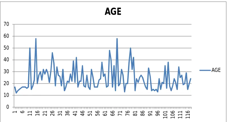
-The patients were from 12 to 58 years of age
-mean age was 24 years.
Our study excluded children less than 12 years and adults more
than 60 years.
FIG.1 (a)
0 10 20 30 40 50 60 70
1 6 11 16 21 26 31 36 41 46 51 56 61 66 71 76 81 86 91 96
101 106 111 116
AGE
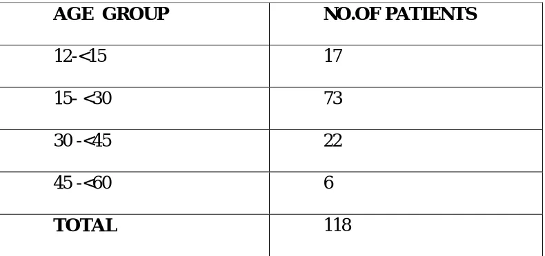
[image:66.612.122.518.327.539.2]The age group wise distribution was as below:
AGE GROUP NO.OF PATIENTS
12-<15 17
15- <30 73
30 -<45 22
45 -<60 6
[image:67.612.130.506.121.583.2]TOTAL 118
Table.1
Fig 1(b)
14%
62% 19% 5%
NO.OF PATIENTS
[image:67.612.116.511.123.309.2]Sex incidence
In emergency appendicectomy group a total number of 73 patients
were studied. Of them 36 were male and 37 were females (1:1 ratio).
In the delayed appendicectomy group a total number of 45
patients were studied. Of them 21 male and 24 were females (1:1.1
ratio)
Fig2a Fig 2b
47% 53%
DELAYED APPENDICECTOMY
MALE FEMALE
49% 51%
EMERGENCY APPENDICECTOMY
CLINICAL SCORING
The Alvarado scoring system was used
The range of values were between 6-9
The median was 7
Fig.3
0 2 4 6 8 10
0 20 40 60 80 100 120 140
ALVARADO SCORE
PATTERN OF DISTRIBUTION
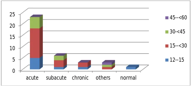
The pattern of distribution of histopathological diagnoses in
Emergency appendicectomy group was as follows:
Male patients
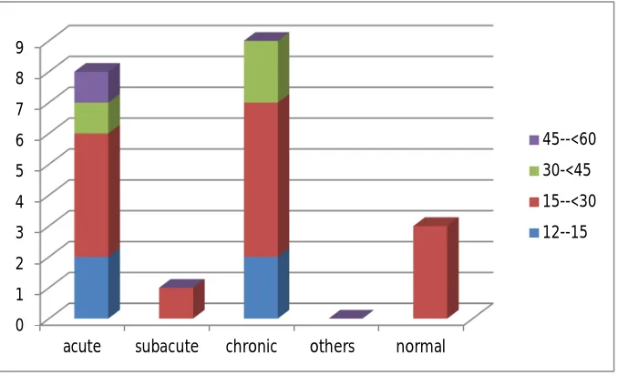
Acute appendicitis-23
Subacute appendicitis-6
Chronic appendicitis-3
Others-3(Appendicular perforation-1, Acute suppurative
appendicitis-1, Associated Meckel’s diverticulitis-1,)
Normal- 1
The age wise distribution of the various histopathological
diagnoses is as below:
0 5 10 15 20 25
acute subacute chronic others normal
45--<60
30-<45
15--<30
[image:70.612.154.483.526.673.2]Female patients
Acute appendicitis-16
Subacute appendicitis-8
Chronic appendicitis-9
Other -1(Appendicular perforation)
Normal -3
The age wise distribution of the above is as depicted:
Fig.5
0 2 4 6 8 10 12 14 16
acute subacute chronic others normal
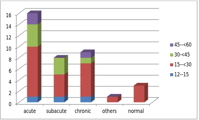
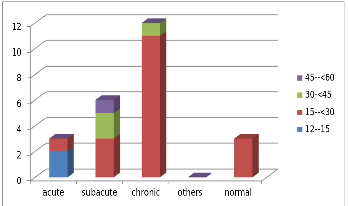
[image:71.612.143.485.384.590.2]The pattern of distribution of histopathological diagnoses in
Delayed appendicectomy group was as follows:
Male patients
Acute appendicitis-8
Sub acute appendicitis-1
Chronic appendicitis-9
Others-nil
Normal -3
The age wise distribution of the above is as depicted:
0 1 2 3 4 5 6 7 8 9
acute subacute chronic others normal
[image:72.612.145.484.458.664.2]Female patients
Acute appenditis-3
Subacute appendicitis-6
Chronic appendicitis-12
Others-nil
Normal-3
The age wise distribution of the above is as follows:
Fig.7
0 2 4 6 8 10 12
acute subacute chronic others normal
[image:73.612.154.498.384.588.2]Histopathology Analysis
In both the groups of patients the following criteria were
analysed:
Serosal congestion.
Luminal obstruction.
Mucosal ulceration.
Type of infiltrates.
Eosinophilic infiltration in muscularis.
Other associated pathologies.
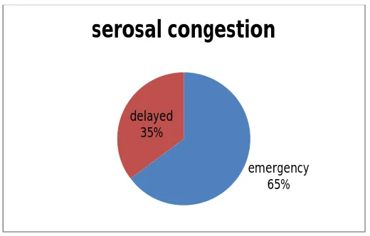
Serosal Congestion
Emergency delayed Total
present 57 31 88
[image:74.612.113.515.440.551.2]absent 16 14 30
Fig.8
Luminal obstruction
Luminal obstruction was noted both in the emergency and
delayed appendicectomy group.
Emergency Delayed Total
present 39 27 66
absent 34 18 52
[image:75.612.154.414.78.244.2]73 45 118
Table.3
emergency 65% delayed
35%
Luminal Obstruction is represented as below:
Fig.9
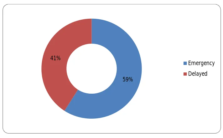
Mucosal inflammation
The following chart depicts the mucosal inflammation pattern in
Emergency appendicectomy group of patients:
MALE FEMALE TOTAL
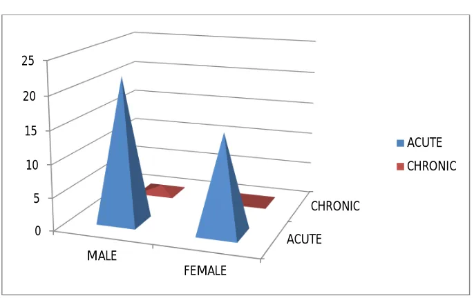
ACUTE 22 15 37
[image:76.612.134.490.118.339.2]CHRONIC 1 0 1
Table.4
59% 41%
[image:76.612.115.512.518.653.2]Mucosal inflammation
Types of mucosal infiltrate:
Type of mucosal/submucosal infiltrates:
A) neutrophils- 49
B) Lymphocytes- 33
C)neutrophil and lymphocytes- 18
D)neutrophil and eosinophil- 8
[image:77.612.142.485.114.330.2]E)normal pattern- 10
Table.5
ACUTE CHRONIC 0
5 10 15 20 25
MALE
FEMALE
TYPES OF MUCOSAL/SUBMUCOSAL INFILTRATES
Fig.10
42%
28% 15%
7% 8%
A) neutrophils- B)
[image:78.612.122.508.118.391.2]pattern-EOSINOPHILIC INFILTRATION IN MUSCULARIS:
Eosinophilic Infiltration of the muscularis along with the
neutrophils was found in a group of patients with acute appendicitis.
The results are as below:
Total no of patients diagnosed with acute appendicitis : 51
Patients with eosinophilic infiltration along with neutrophils :8
eosinophil 16%
others 84%
A patient with Gangrenous Appendicitis
During our study we came across one patient each with Meckel’s
diverticulitis and Oxyuriasis vermicularis.
Two patients presented with appendicular perforation with
histopathology showing tip perforation.
One patient presented with ileocaecal intussusception which was
reduced and appendicectomy done.
We did not get any reports of carcinoids, benign tumors of the
appendix, endometriosis, Crohn’s disease or tuberculous lesions in our
DISCUSSION
Our Study included patients with appendicitis of age group of 12
to 59 years.
The mean age of incidence was 24 years. This correlates with
other studies which quote an age range of 15-24 to have the highest
incidence.
The male: female sex distribution in our study was 1:1, in contrast
to the .other studies showing a ratio of 3:2
A diagnosis of acute appendicitis was given in 51of the 118
patients studied, of whom 40 had undergone emergency appendectomy
and 11 had a delayed appendectomy.
In the emergency group 57.5% were males and 42.5% were
females.
In the delayed group 72.5% were males and 27.5% were females.
Alvarado scoring has a sensitivity of 87.5% and our study patients had
The negative appendicectomy rate in our study was 8.4% ,more in
female patients and was common in the age group of 15-<30 years.
This increased pattern in females suggests the role of diagnostic
laparoscopy and watchful observation in female patients .It also suggest
the possibilities of other diagnoses in them.
The importance of routine histopathological analysis of the
appendix retrieved during surgery has been stressed by many authors in
the context of analysing the type of lesion, its correlation with clinical
features and for the diagnosis of any other lesions in the appendix. This
is followed in our setup.
Serosal congestion was earlier considered as early form of
appendicitis.
Presently it is regarded as periappendicitis and presence of
it in the absence of mucosal/submucosal inflammation is interpreted as
significant.
It indicates other causes of peritonitis like pelvic
inflammatory disease, perforation of bowel etc., which might need
further management. In our study too we observed similar pattern in a
Serosal congestion was present in 65% of emergency
appendicectomy and 35% of delayed appendicectomy patients in our
study.
The presence of such high rate of serosal congestion in the
latter group suggests that the inflammation has not resolved and the
intervention by surgery was appropriate.
Lymphoid follicles are regarded the most common agent to cause
appendiceal luminal obstruction which foreruns most of the acute
appendicitis. This occurs due to bacterial and viral invasion of the
submucosal lymphoid follicles resulting in their hypertrophy.
Fecoliths are considered the next common etiological agent.
In contrast to this in our study fecolith was the commonest agent
causing luminal obstruction.
In studies done to follow the resolution of appendicitis after
nonsurgical treatment, using ultrasonogram ,the response was delayed as
sonographically documented ,when there is an appendiculolith.
Since our group of patients are presenting commonly with fecolith
the chance of resolution of the condition can be delayed if managed
Mucosal Inflammation
According to Howie, the histological features of mucosal
inflammatory changes include
1. Presence of neutrophils in the lumen of the appendix.
2. Focus of ulceration of the mucosa with neutrophilic
invasion of the adjacent stroma
3. Lack of involvement of deeper layers.
This was mentioned by other authors as ‘endoappendicitis’, ‘acute
focal appendicitis’ and ‘limited acute appendicitis’.
The mucosal inflammation is related to the severity of the
symptomatology as denoted by elevated Alvarado scores in our study.
This also correlates with the study of Piper et al.
Mucosal inflammation was found mostly in emergency
appendicectomy group relating it to the severity of the clinical scoring
and hence our intervention.
A lower incidence of mucosal inflammation in the delayed
the conservative management and also that the severity scoring had a
positive relation with the same.
Eosinophilic infiltration of the muscularis apart from its
presence in mucosal and submucosal layers has been discussed as
Eosinophil Edema reaction by Aravindan et al and has been oserved in
this study too.The condition is described as another etiological event in
onset of appendicitis and attributed to allergy. However its correlation to
parasitic infestation remains unclear.
LIMITATIONS OF THE STUDY
Being an observational study, patients could not be
ascertained to particular study group and there is a possibility of
observer’s bias.
The duration of observation in the delayed appendicectomy
group is not fixed.
The reporting variation among different pathologist has not
CONCLUSION
The study states that histopathological analysis in
appendicectomy is absolutely necessary in guiding further management.
However the role of conservative management remains selective in our
BIBLIOGRAPHY
1. Rosai J: Ackerman’s Surgical Pathology. 8th ed. St. Louis,
Mosby-YearBook, 1996, pp. 711-716
2. Norman.S.Williams et al.Bailey &Love’s SHORT
PRACTICE OF SURGERY.25TH Edition. 2008 Edward Arnold
(Publishers) Ltd.Pg 1204-18
3. Townsend.et.al.Sabiston TEXTBOOK OF SURGERY The
Biological basis of Modern Surgical Practice.18th Edition.,2007
4. Xharra S, et al. Correlation of serum C-reactive protein,
white blood count and neutrophil percentage with histopathology
findings in acute appendicitis. World J Emerg Surg 2012 Aug
6;7(1):27
5. Alvarado A. A practical score for the early
diagnosis of acute appendicitis. Ann Emerg Med.1986 May;15(5):
557-64.
6. Aravindan KP, Vijayaraghavan D, Manipadam MT. Acute
eosinophilic appendicitis and the significance of eosinophil - Edema
7. Lien WC et al Appendicolith delays resolution of
appendicitis following nonoperative management. J Gastrointest Surg.
2012 Dec;16(12):2274-9.
8. Usha Rani Singh,et al. Eosinophils, mast cells, nerves and
ganglion cells in appendicitisIndain Journal of Surgery 2008 Dec;
70(5): 231-4.
9. Isik B,et al Appendiceal Enterobius vermicularis infestation
in adults. Int Surg. 2007 Jul-Aug;92(4):221-5.
10. Saidi HS, Chavda SK Use of a modified Alvorado score in
the diagnosis of acute appendicitis. East Afr Med J. 2003
Aug;80(8):411-4.
11. Nautiyal H et al. Combined use of modified Alvarado score
and USG in decreasing negative appendicectomy rate. Indian J
Surg.2010 Feb; 72(1):42-8.
12. Pieper R, Kager L, Näsman P .Clinical significance of
mucosal inflammation of the vermiform appendix. Ann Surg. 1983
Mar;197(3):368-74.
13. Wilms IMHA, de Hoog DENM, de Visser DC, Janzing
HMJ Appendectomy versus antibiotic treatment for acute appendicitis
14. Jones AE, Phillips AW, Jarvis JR, Sargen K. The value of
routine histopathological examination of appendicectomy specimens.
BMC Surg. 2007 Aug 10;7:17.
15. Marudanayagam R, Williams GT, Rees BI. Review of the
pathological results of 2660 appendicectomy specimens. J
Gastroenterol. 2006 Aug;41(8):745-9.
16. Mussack T,et al Chronic appendicitis as an independent
clinical entityChirurg. 2002 Jul;73(7):710-5.
17. Akbulut S, et al. Unusual histopathological findings in
appendectomy specimens: a retrospective analysis and literature review.
World J Gastroenterol. 2011 Apr 21;17(15):1961-70.
18. HOWIE JG. TOO FEW APPENDICECTOMIES?
Lancet.1964 Jun 6;1(7345):1240–1242.
19. CAMPBELL JS, FOURNIER P, DASILVA T. When is the
appendix normal? A study of acute inflammations of the appendix
apparent only upon histologic examination.Can Med Assoc J.1961 Nov
18;85:1155–1157
20. TOULOUKIAN RJ, TRAINER TD. SIGNIFICANCE OF
FOCAL INFLAMMATION OF THE APPENDIX.Surgery.1964
21. CONWAY DJ, CAMPBELL JS. ACUTE FOCAL
APPENDICITIS--REALITY OR RED HERRING.Med Serv J
Can.1965 Mar;21:177–182.
22. Crabbe MM, Norwood SH, Robertson HD, Silva JS.
Recurrent and chronic appendicitis. Surg Gynecol Obstet 1986;163: