CLONIDINE AS AN ADJUVANT TO
BUPIVACAINE IN SUPRACLAVICULAR
BRACHIAL PLEXUS BLOCK
Dissertation submitted to
THE TAMIL NADU DR. M.G.R. MEDICAL UNIVERSITY
In partial fulfillment of the regulations for the award of the degree of
M.D. BRANCH - X
ANAESTHESIOLOGY
K.A.P.V. GOVERNMENT MEDICAL COLLEGE, TIRUCHIRAPPALLI THE TAMIL NADU DR. M.G.R. MEDICAL UNIVERSITY
CHENNAI, INDIA
This is to certify that the dissertation entitled “COMPARISON OF DEXMEDETOMIDINE WITH CLONIDINE AS AN ADJUVANT IN
SUPRACLAVICULAR BRACHIAL PLEXUS BLOCK” is the bonafide original
work of Dr.K.HEMALATHA in partial fulfillment of the requirements for M.D. Branch-X (anaesthesiology) Examination of the Tamil Nadu Dr. M.G.R. Medical
University to be held in April 2013.
Prof. Dr. N. JOTHI MD, DA
Professor & Head of the Department, Department of Anaesthesiology, K.A.P.V. Govt. Medical College, Trichy – 1.
Prof Dr. A. Karthikeyan M.D., DEAN
I Dr.K.HEMALATHA, solemnly declare that dissertation titled, “COMPARISON OF DEXMEDETOMIDINE WITH CLONIDINE AS AN
ADJUVANT IN SUPRACLAVICULAR BRACHIAL PLEXUS BLOCK” is a bonafide work done by me at K.A.P.V. Government Medical College, during
2010-2013 under the guidance and supervision of my Professor & Head of the
department of Anaesthesiology Prof. Dr. N. JOTHI, M.D.,D.A .
The dissertation is submitted to the Tamilnadu Dr. M.G.R. Medical University,
towards the partial fulfillment of requirement for the award of M.D. Degree
(Branch – X) in Anaesthesiology.
Place: Trichy
ACKNOWLEDGEMENT
I owe my thanks to Prof. Dr. A. Karthikeyan, M.D., the Dean, K.A.P.V. Govt. Medical College and Annal Gandhi Memorial Government Hospital, for allowing me to avail the facilities needed for my dissertation work.
I am grateful to Prof. Dr. N. Jothi, M.D., DA Prof. and Head of the Department of Anaesthesiology, K.A.P.V. Govt. Medical College for permitting me to do the study and for his encouragement.
I express my gratitude to Prof. Dr. R. Selvakumar, M.D.,DA, DNB Associate Professor, Department of Anaesthesiology, K.A.P.V Govt medical college for his valuable assistance and guidance.
I am thankful to Prof. Dr.P. Maheshwari, M.D.,DA, Associate professor, Department of Anaesthesiology, for his valuable assistance and guidance.
I am extremely grateful to Prof. Dr. B.Vijayakumar, M.D.,DA Former Retired Professor and Head of the Department of Anaesthesiology , K.A.P.V. Govt. Medical College for his help and guidance.
I express my sincere thanks to all of my assistant professors for their unlimited encouragement, guidance and help during this study.
I thank all my colleagues who helped me and shared their knowledge about this study. Last but not least, my sincere thanks to all the patients who co-operated for this study, without whom this study could not have been undertaken.
SL. NO
TITLE PAGE NO
1 INTRODUCTION 1
2 AIM OF THE STUDY 4
3 REVIEW OF LITERATURES 5
4 METHODOLOGY 49
5 OBSERVATION AND RESULTS 57
6 DISCUSSION 68
7 CONCLUSION 73
8 SUMMARY 74
9 BIBLIOGRAPHY 77
INTRODUCTION
Brachial plexus blocks provides a useful alternative to general anaesthesia for
upper limb surgeries. They achieve near ideal operating conditions by producing good
muscular relaxation, maintaining stable haemodynamics and associated sympathetic
block. The sympathetic block reduces the postoperative pain, vasospasm and edema.
Although various anaesthetic agents have been used, bupivacaine is a better
choice due to its longer duration of action of 3 to 8hrs. However it has certain
disadvantages like delayed onset, patchy or incomplete analgesia etc,
Various adjuvants like neostigmine, midazolam, opioids etc,(1-4) have been
tried to improve the onset of block, quality of block, prolong the duration of block and
postop analgesia. But these were associated with side effects.
Clonidine an alpha-2 agonist which had been used as antihypertensive
initially has sedative, sympatholytic and analgesic properties. It is also known to have
anti nociceptive action and enhances the effect of local anasthetics when given
intrathecally, epidurally and in peripheral nerve blocks. This effect is produced by
modulating pain pathways through presynaptic alpha-2 adrenergic receptors. It also
produces sedation through its action on pontine locus ceruleus where highest number
of alpha-2 receptors are present.
Dexmedetomidine, the next recent highly potent alpha-2 agonist, is also a
dexmedetomidine is its high selectivity for alpha-2 receptors and its ability to produce
sedation and analgesia while still maintaining patient arousability and respiratory
function.
So the present study has been undertaken as randomized single blinded
manner to compare the onset time, duration and analgesic efficacy of clonidine with
dexmedetomidine when added as adjuvant to bupivacaine(0.25%) for brachial plexus
OBJECTIVES
• This study of adding clonidine (1µg/kg) or dexmedetomidine (1µg/kg) to
bupivacaine (0.25%) in brachial plexus block for surgeries involving the upper
limb has the following objectives:
• To compare the
o Onset of sensory and motor blockade
o Duration of sensory and motor blockade
o Sedation score intra and postoperatively
o Haemodynamic variables (HR,BP,SPO2)
o No of rescue analgesics in postoperative 24hrs
AIM OF THE STUDY
To compare the effectiveness of clonidine and dexmedetomidine as adjuvant
in brachial plexus block by supraclavicular approach for prolongation of sensory
REVIEW OF LITERATURE
HISTORY(5-7)
1858 – theory of pain as a separate and distinct sense was formulated by Mortiz
S.Schiff.
1885 – the first brachial plexus block was performed by William Stwart Halsted
and Alfred Hall – idea of injecting cocaine into nerve trunk in 1885, less than a
year after Koller demonstrated the anaesthetic properties of cocaine on the eye of a
patient.
1897 – Crile used a similar trchnique in which the plexus was exposed under local
anaesthesia. Just behind the sternocleidomastoid, cocaine was injected into the
nerve trunks under direct vision which was done as a therapeutic measure in a 12yr
old boy who developed tetanic spasms following a compound fracture of the
forearm, after which it was used to provide anaesthesia for surgeries in the upper
limb.
EVOLUTION OF SUPRACLAVICULAR BRACHIAL PLEXUS BLOCK:
1911 – 1912 – KULENKAMPFF performed the first percutaneous supraclavicular
approach. He pointed out that above the clavicle the plexus lies under the skin as it
1922 – LABAT injected at three separate points, first beneath the deep fascia
towards the first rib, second towards the chassaignac’s tubercle and third towards
the lateral margin of the first rib behind the clavicle.
1926 – LIVINGSTON proved that the plexus and the artery are separated by a
fascial investment.
1940 – PATRICK chose to deposit the anaesthetic along the plexus in its course
over the first rib where 60-70ml of solution was injected during 5-6 insertions. The
technique became the ‘standard technique’ of supraclavicular block, subsequently
reffered to many as ‘classical supraclavicular technique’.
1942 – KNIGHT modified Patrick’s technique by using three needle insertions.
1944 – MURPHEY used a single injection technique by using the lateral border of
anterior scalene muscle as the landmark.
1949 – BONICA AND MOORE followed the ‘walking the rib’ method using
Kulenkampff and Patrick’s technique which was used over the subsequent twenty
years.
1958 – LOOKMAN like Livingston realized the fascial investment of the plexus
and identified that it lies between the anterior and the middle scalene muscles, but
1964 – WINNIE showed that the relation of the plexus and the subclavian artery to
the midpoint of the first rib is not constant, but there is a constant relationship
between the scalene muscles, plexus and the first rib. The plexus between the two
scalene muscles always inserted on the first rib. He inserted needle between the
two muscles in the direction of the space between them. Once paresthesia is
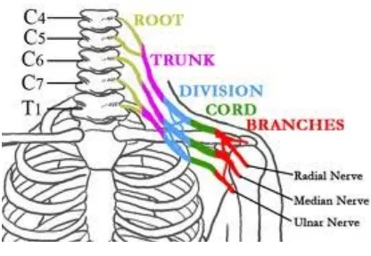
ANATOMY OF BRACHIAL PLEXUS(8-11)
Knowledge of the formation of brachial plexus and of its distribution is
essential to the intelligent and effective use of brachial plexus anesthesia for
surgeries of the upper limb. Close familiarity with the vascular, muscular and
fascial relationships of the plexus throughout its formation and distribution is
equally important.
While travelling from the intervertebral foramina to the upper limb, the fibres that
constitute the plexus are composed of roots, trunks, divisions and terminal nerves.
FORMATION OF BRACHIAL PLEXUS:
The plexus is formed by the anterior primary rami of the 5th to 8th cervical
nerves, together with the bulk of the 1st thoracic nerve (C – 8 and T -1). In addition
there might be a contribution above from 4th to 5th cervical root and another below
from 2nd to 1st thoracic nerve. Occasionally the plexus is may also be derived from
Fig. 1: Formation of brachial plexus
Fig.2: Relation of brachial plexus
[image:14.612.126.490.111.391.2] [image:14.612.201.409.484.628.2]
Roots:
Represents the anterior primary divisions of lower four cervical and first
thoracic nerves. They emerge from the intervertebral foramina and fuse above the
first rib to form the trunks.
Trunks:
The roots combine above the first rib to form the three trunks of the plexus.
C5 and C6 unite to form the upper trunk. C8 and T1 unite behind the scalenus
anterior to form the lower trunk and C7 continues as a sole contributor to form the
middle trunk.
Divisions:
As the trunks pass above the first rib and below the clavicle, each one of
them divides into anterior and posterior divisions.
Cords:
The fibres as they emerge from under the clavicle, again combine to form
the three cords. The anterior divisions of the upper and the middle trunks forms the
lateral cord, lateral to the axillary artery. The medial cord is formed by the anterior
division of the lower trunk which descends medial to the axillary artery .The
posterior cord is formed by the posterior divisions of all three trunks, behind the
The medial and lateral cords supply the flexor surface of the upper extremity,
whereas nerves from the posterior cord supply the extensor surface.
Major terminal nerves:
Each of these cords gives rise to one of the major nerves of the upper
extremity and then terminate as a major nerve. The lateral cord gives off the
lateral head of the median nerve and the medial cords give off the medial head of
the median nerve and continue as major terminal nerves. The lateral cord
terminates as musculocutaneous nerve and the medial cord as the ulnar nerve.
Posterior cord branches off as axillary nerve and continues as radial nerve.
In summary, conveniently it can be considered that the brachial plexus begins
with five nerves (C5 to T1) and terminates as five nerves (musculocutaneous,
axillary, radial, median and ulnar nerves) with its intermediate portions displaying
into 2 sets of three, which reunite and give rise to three cords. These three cords
gives off three lateral branches before becoming the major terminal branches of the
plexus.
Distribution of the brachial plexus:
These are divided into those that arise above the clavicle the
Supraclavicular branches:
From roots
1. Nerve to scalene and longus colli muscle – C5,6,7,8
2. Branch to phrenic nerve – C5
3. Dorsal scapular nerve – C5
4. Long thoracic nerve of Bell – C5,6,(7)
From trunks:
1. Nerve to subclavius muscle – C5,C6
2. Suprascapular nerve – C5,C6
Infraclavicular branches:
They branch from cords but their fibres may be tracked back to spinal nerves.
Lateral cord:
1. Lateral pectoral nerve – C5,6,7
2. Musculocutaneous nerve – C5,6,7
3. Lateral root of median nerve – C5,6,7
Medial cord:
1. Medial pectoral nerve – C8,T1
3. Ulnar nerve – C7,T1
4. Medial root of median nerve – C8,T1
5. Medial cutaneous nerve of arm – C8,T1
Posterior cord:
1. Upper subscapular nerve – C5,6
2. Thoracodorsal nerve - C6,7,8
3. Lower subscapular nerve – C5,6
Sympathetic contribution to brachial plexus:
The segmental preganglionic sympathetic contributions are variable, but
generally extend more caudal. The highest contribution is T2 with T1 contributing
very rarely, while lowest may be as far as T8, T9 or even T10. The postganglionic
contributions are from grey rami communicants from the sympathetic chain.
RELATIONS OF THE BRACHIAL PLEXUS
Roots: Lies between the scalenus anterior and medius above the second part of the
subclavian artery.
Trunks: The upper and the middle trunks lie above the subclavian artery as they
stream across the 1st rib, but the lower trunk lies behind the artery and may groove
Divisions: At the lateral border of the first rib, the trunks bifurcate into divisions,
which are situated behind the clavicle.
Cords: The cords are found at the apex of the axilla and become grouped around
the axillary artery.
The interscalene sheath:
As the roots C5 – T1 emerge in the groove between the transverse process and
the tubercles, they lie in the fibro-fatty space between the two layers of fibrinous
sheath. Posterior sheath from the posterior tubercle covers the front of the scalenus
medius and anterior sheath from the anterior tubercle covers the posterior aspect of
scalenus anterior. The sheath extends into the axilla around the plexus.
Significance of this space is that the local anaesthetic can be injected into this
sheath to produce block either by interscalene, subclavian perivascular or axillary
approach.
TECHNIQUE OF BRACHIAL PLEXUS BLOCK
Surgical anesthesia for the upper extremity and shoulder can be obtained by
blocking the brachial plexus at various sites. There are various approaches that can
be used for this blockade as follows
1.Interscalene approach
a.Classic approach
b. Plumb – bob technique
c.Subclavian perivascular technique
3.Axillary approach
4.Infraclavicular approach
Complications:
1. Intravascular injection
2. Pneumothorax
PATHOPHYSIOLOGY OF PAIN
Definition of Pain:
The Taxonomy Committee of International Association for the study of Pain
(IASP) defines pain as "An unpleasant sensory and emotional experience
associated with potential tissue damage or described in terms of such damage".
Postoperative pain is defined as a form of acute pain caused by surgical trauma
with an inflammatory reaction and with initiation of an afferent neuronal barrage.
Physiology of pain:
The spinal cord conveys signal from the brain to the nerves located
throughout the body. Nerves coming from the spinal cord and leading to all parts
of the body enter and leave the spinal cord along its entire length. There are 31
pairs of spinal nerves that leave the spinal cord through the intervertebral
foraminae. The peripheral nerves includes motor and sensory nerves. Sensory
nerves are that which receive and transmit sensory stimuli to Substantia gelatinosa.
Motor nerves are those which lead to the muscles and stimulate movement of
Various mechanisms are:
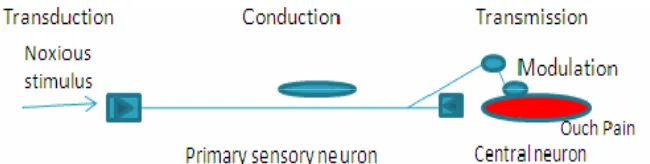
Nociception refers to the perception of a noxious stimulus by the brain.(13)
The components include transduction, modulation, transmission and perception
(fig.4).
[image:22.612.114.439.218.300.2]
Fig. 3: Process of Nociception
Peripheral sensitization.
Central sensitization
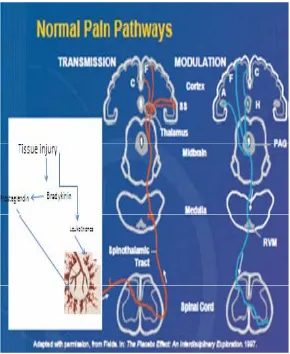
Pathways of pain:
Pain is conducted along neuron pathways that transmits the noxious stimuli
Fig. 4: Normal pathways of pain
A first order neuron, arising in the cell body in dorsal root ganglion of the
spinal cord transmits pain from a peripheral receptor to the dorsal horn of the
spinal cord.
A second-order neuron is located in the dorsal horn of the spinal cord and
sends axons that crosses the midline to ascend as the spinothalamic tract to
[image:23.612.158.448.101.455.2]
A third-order neuron in the thalamus projects its fibers to the post central
gyrus through the internal capsule.
Physiological responses to pain:
It has been found that uncontrolled pain in post operative period produce
physiological effects like altered stress response to surgery, increased
catecholamines, deep vein thrombosis, higher incidence of pulmonary
complications, and ultimately increasing the morbidity.(15-17)
Peripheral α2 receptors:
α2 adrenoceptors are located on primary afferent terminals, on neurons in
the superficial laminae of the spinal cord, and within several brainstem nuclei
implicated in analgesia, supporting the possibility of analgesic action at peripheral,
spinal and brainstem sites.
Clonidine enhances both sensory and motor blockade from peripheral nerve
injection and epidural/spinal injection of local anaesthetics. It also blocks the
conduction of C and A gamma fibres and increases the potassium conductance in
isolated neurons and intensifies conduction block of local anaesthetics.
ASSESSMENT OF PAIN
It is important in an effective postoperative pain management. Numerous
pain assessment scales are there to quantify pain.(21,22) The intensity of pain should
communicate and expressed what he feels like. A number of self-assessment scales
are available.
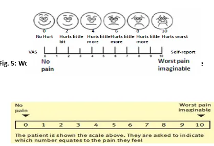
Visual analogue scale (VAS) :
VAS is the most commonly used method to assess pain which was first
described in 1966. It is a very simple and used in pain research. It consists of a 10
cm line with two anchor points starting from ‘no pain’ to ‘worst pain imaginable’
which is self assessed by patient (fig.5). It is the position of the mark on the line
that measures how much pain the subject experiences.
Fig. 5: Wong‐ Baker Faces pain rating scale and Visual Analogue scale
[image:25.612.74.492.396.681.2]
Others are:
• Facial expressions:
This is a pictogram of six faces with different expressions from smiling
or happy to tearful which can also be used(fig.5).
• Numerical rating scale (NRS):
This is similar to the visual analogue scale with the two anchors starting
from ‘no pain’ to ‘worst pain as from 0 to 10, assessed by patient (Fig.
6).
• Verbal rating scale (VRS)
The preoperative personality assessment is also helpful in assessing the
patient’s psychological background and his psycho reactions to surgery and the
pain that follows it. The VRS and NRS are the other mostly used assessment tools
in the clinical setting while the VAS scale is primarily used as a research too.
METHODS OF ACHIEVING PAIN RELIEF:
“Pain relief has always been bought at a Price” – Bromage
Number of factors are there that contribute to effective postoperative
patient education, regular pain assessment tools and use of balanced analgesia to
meet the needs of special patient groups.
Following any surgery, the pain after the tissue damage is usually self
limiting. It persists at the most for the first 24 hrs and mostly subsides in 4 days
time.44 The post operative pain is dull in nature, aggravated by mobility and
relieved by rest to that part. The acute pain of surgery is strongly accompanied by
emotional elements of fear, anxiety, and depression of previous experience of pain.
The goals of effective and appropriate pain management are to:
Facilitate rapid recovery and return of full function.
Reduce morbidity.
Improve the quality of life for the patient.
Allow early discharge from the hospital.
Methods adopted for providing post operative pain relief include: 45-50
Pharmacological methods: -
i. Balanced (multimodal) analgesia
ii. Opioids
iii. Non-opioids (NSAIDS)
iv. Adjuvants
v. Patient controlled analgesia
vi. Regional analgesia
Continuous Peripheral Nerve Blockade (CPNB)
Infiltration blocks
II. Non pharmacological methods: - This includes
A. Transcutaneous Electrical Nerve Stimulation (TENS)
B. Acupuncture
C. Cryotherapy
D. Heat Therapy
Regional analgesia:
Central neuraxial block involves either intermittent or continuous
administration of local anaesthetics in order to interrupt sensory transmission.49
The important draw back of this technique is the accompanying motor and
sympathetic blockade which can increase the incidence of post operative
complications. Extradural block offers complete pain relief, permits effective
coughing & better ventilation. But the total spinal, accidental dural punctures are
more with inexperience hands.
Peripheral nerve blocks are being increasingly used since they may
provide more selective but still excellent postoperative analgesia with reduced
need for opioids over an extended period. Peripheral nerve blocks (PNBs) avoid
the side effects associated with central neuraxial blockade, such as hypotension
and wide motor blockade with reduced mobility and proprioception, and
PHARMACOLOGY
Local Anaesthetic Drugs:
Local anesthetics are drugs that produce reversible conduction
blockade of nerve impulses along the central and peripheral pathways after
regional anaesthesia. They produce autonomic, sensory and motor blockade of
the area innervated by the affected nerve. Removal of the local anaesthetic
results in complete return of nerve conductions with no structural domage to the
nerve fibres.
They have similar configurations with one aromatic lipophilic part
(Benzene ring) and one hydrophilic part (quaternary ring) connected by an
BUPIV B by A.F. Chemis hydroch
it is ava
bupivac mixture The mo VACAINE Bupivacain .Ekenstam
stry: It is
hloride, a ailable as caine appe e. olecular fo E HYDRO ne hydroch
m in 1957.
1-n-butyl-chiral drug
racemic m
ears to be
rmula is C
Fig. OCHLORI hloride is -DL-piperi g possessin mixture of less toxic
C₁₈N₂OH₂₈
7: Bupiva
IDE
a aminoa
idine-2 car
ng an asym
f R & S e
c than the
₈HCl
caine Hyd
amide loca rboxylic a mmetrical enantiomer e commerc rochloride al anaesth acid-2,6 dim carbon ato
rs. The S
[image:30.612.206.405.531.684.2]
Physiochemical properties:
1. Molecular weight: of chloride salt is 325 and of its base form is 288.
2. pKa: 8.1
3. Protein binding: 95%
4. Solubility: The base is sparingly soluble and the hydrochloride salt is
readily soluble in water.
5. Partition coefficient: 28
6. T1/2: 210min
7. Clearance: 0.47L/min
8. Moderate onset and long action.
Mechanism of action: It acts on the cell membrane of the axon producing
electrical stabilization. The permeability to sodium ions necessary for impulse
propagation is prevented. Thus resting membrane potential is maintained and
depolarization is inhibited.
The local anaesthetics block the sodium conductance by the following
1. Local anaesthetics in the cationic form act on the receptors within the
sodium channels, on the cell membrane and block it. They reach the
sodium channels via the lipophilic pathway directly across the lipid
membrane or via the axoplasmic opening. This mechanism is responsible
for 90% of nerve blocking effect of amide local anaesthetic.
2. The second mechanism is by membrane expansion, which is a non
specific action in contrast to more specific drug receptor interaction.
Available concentrations:
• 0.25% and 0.5%
• 0.25% and 0.5% soluble in isotonic saline – isobaric
• 0.5% soluble in 8% dextrose – hyperbaric
These doses may be repeated in three to four hours but the maximum dose is
400mgs. Addition of a vasoconstrictor produces very little increase in duration
of action, but significantly reducing the peak blood levels decreasing the
Actions:
CNS effects are characterized by excitation or depression. The intial
manifestation may be nervousness, dizziness, blurring of vision or tremors
followed by drowsiness, convulsions and unconsciousness. In CVS, it produces
reduction in the maximum rate of depolarization in the purkinji fibres and
ventricular muscles. Action potential duration and effective refractory period is
also decreased. The rate of recovery is also slower with bupivacaine. Therefore
there is incomplete restoration of V-max between action potential particularly at
higher heart rates. Hence,it is highly arrythmogenic. If excessive plasma level
is reached, it produces respiratory depression.
Pharmacodynamics:
Onset – 4-6min
Peak effect – 15-20min
Average duration – 3.5-5hrs, for nerve blocks – 5-6hrs
the toxic levels. Local irritant effects on the nerve tissue has been noted but
permanent nerve damage has not been found in clinical dosage.
Pharmacokinetics:
It reaches the blood within 5 min, but the plasma are related to the dosage
administered. In plasma, it binds avidly to alpha 1 acid glycoprotein to the
extent of 70-95%. Conversely its unbound active form is one third that of
lidocaine.
Metabolism and elimination:
As it is an amide it is primarily metabolised in the liver by n-dealkylation to
pipecolyloxylidine. It crosses the placental barrier, but the lowest level of
placental diffusion has been reported (umbilical vein to maternal ratio – 0.31 to
0.44). No effect on fetus has been reported so far.
10% of the drug is excreted unchanged in urine within 24hrs and 5% as
Adverse reactions:
Might be due to inadvertent iv injection or slow metabolic degradation,
producing its effects on CNS & CVS. CNS effects include nervousness,
dizziness, tremors or blurring of vision followed by convulsion and
unconsciousness. CVS manifestations include myocardial depression,
hypotension and cardiac arrest. Allergic reactions such as utricaria,
bronchospasm and hypotension can also occur.
Treatment is symptomatic maintaining ventilation and circulation with
oxygen, controlled ventilation and iv fluids and vasopressors for circulation.
Diazepam (0.1 – 0.2mg/kg) or thiopentone (2-3mg/kg) can be used for
convulsions and corticosteroids for allergic reactions. For ventricular fibrillation
or tachycardia, amiodarone (5mg/kg) or defibrillation (2-5joule/kg) can be used.
Cardiovascular collapse/CNS ratio:
The CC/CNS ratio for bupivacaine is 3.5± 0.5or findings indicating that three
times the drug was required to induce irreversible cardiovascular collapse as
was needed to produce convulsions. It has also been suggested that some of the
CLONI Clonidi for its centrall sympath addition Chemis I (2,6-dic IDINE HY ine hydroc hypotensiv
ly acting a
hetic nerv
n to sympa
stry:
It is an im
chlorophen YDROCH chloride is ve proper alpha adre vous syste atholysis. midazoline nylamino) Fig HLORIDE
s an imida
rties was f
energic ago
m activity
derivative
-2-imidazo
g. 8:Clonid
E
azoline de
first introd
onist that
y. It also
e existing
oline hydr
dine Hydro
erivative w
duced in E
lowers th
causes sed
as a mes
rochloride.
ochloride
which was
Europe in
he BP by d
dation and
omeric co
.
initially u
n 1966. It
Physiochemical properties:
Molecular weight is 266.56
It is odourless, bitter, white and crystalline substance soluble in water.
Available as 1ml ampoule containing 150µgs.
It should be stored below 25⁰C.
Clonidine is a centrally acting partial α2 adrenergic agonist having a
selectivity ratio of 220:1 in favor of α2 receptors. There are three subtypes of α2
receptors α2a, α2b, α2c. Of these sedation, analgesia and sympatholysis are
mediated via α2a receptors, vasoconstriction and anti-shivering via α2b
receptors and a startle response may reflect activation via α2c receptors. This
startle response includes a physical movement away from the stimulus and
breathing changes. It acts on the α2 receptors which are found densely in the
locus ceruleus in pons and also stimulates the α adrenergic inhibitory neurons in
the medullary vasomotor centre and inhibits sympathetic outflow from the CNS
to the peripheral tissues.
It modifies the potassium channels in the CNS and hyperpolarises the
Neuraxially it inhibits spinal substance P release and thereby inhibiting the
nociceptive neuronal firing. Its acts on α2 afferent terminals situated in the
superficial lamina of the spinal cord and brain stem nuclei and produces
analgesic effects after neuraxial administration. It also acts on the central
thermoregulatory system and decreases the threshold to cold stimuli.
Pharmacological effects:
IV clonidine causes a transient rise in BP by α2 agonistic action on the
vascular smooth muscle of the skin and mucosa, followed by a decrease in BP
due to its central α2 action.
Pharmacokinetics:
Well absorbed orally and 100% bioavailable.
Peak plasma concentration is achieved within 60 – 90min.
Plasma half life 9 – 12hrs.
Plasma protein binding – 20 – 40%.
Metabolism – minor pathways with major metabolite, P-hydroxyclonidine
A transdermal delivery system is available in which the drug is released
constantly for about a week.
Dosage regimen:
Oral : 3-5µg/kg
Intranasal : 2-4µg/kg
Intramuscular: 2µg/kg
Intravenous : 1-3µg/kg
Spinal : 50-100g
Epidural : 1-2µg/kg
Transdermal : 3mg/day
Precautions:
• In patients with renal insufficiency, lower dose is needed.
• Sudden withdrawal produces hypertensive crisis, hence discontinued
gradually over 2-4 days.
• Use with caution in patients with cerebrovascular or coronary disease.
• If a patient with β blocker is on continuous epidural, those drugs should
Contraindications:
• Known hypersensitivity to clonidine.
• Bradyarrythmias or AV block.
• Cardiovascular disease/hemodynamic instability.
Interactions:
• Clonidine potentiates the CNS depressive effect of alcohol , barbiturates
or other sedatives.
• Narcotics also potentiate the hypotensive effects of clonidine.
• Tricyclic anti-depressants antagonizes the hypotensive effects of
clonidine.
• Drugs with negative chronotropic effect can potentiate bradycardia.
• Epidural clonidine prolongs the duration of epidural local anaesthetics,
opoids, neostigmine, etc,.
Uses:
• Premedication:
• This blunts stress response during laryngoscopy or intubation.
Intra-operative hemodynamic stability, intravenous and inhaled anaesthetic
drug requirement is reduced.
• Epidural block:
Solely or in combination provides excellent analgesia in labor.
Indicated for treatment of intractable pain which is unresponsive to
opoids or reflux sympathetic dystrophy or neuropathic pain.
• Spinal anaesthesia:
Combined with local anaesthetics, it improves the quality and
duration of block, minimizes tourniquet pain and prevents shivering.
• Caudal anaesthesia:
Clonidine with LAs ,increases the duration of anaesthesia and
analgesia.
• Perpheral nerve block:
Clonidine prolongs the duration of anesthesia and analgesia with
• Bier’s block:
Enhances the tolerance of tourniquet.
• Decreasing MAC of sevoflurane:
Oral clonidine in a dose of 4µg/kg given 2hrs before induction
decreases the MAC values of sevoflurane for LMA insertion.
• Post-Op Nausea and Vomiting (PONV):
Oral clonidine of 4µg/kg before surgery enhances the anti-emetic
effect of propofol when compared with midazolam.
• Other uses:
Protection against myocardial ischemia. Used to diagnose
pheochromocytoma by administering 0.3mg of clonidine which will
decrease concentration of catecholamines in normal patients but not in
pheochromocytoma. At a dose of 75µg IV, it stops shivering by
thermoregulatory control. Used to treat opoid and alcohol withdrawal
Side effects:
• CVS: Orthostatic symptoms, palpitation & tachycardia/bradycardia.
Syncope, congestive heart failure, Raynaud’s phenomenon, and
electrocardiographic changes (sinus node arrest, AV block, junctional
bradycardia, and arrhythmias) have been reported but rarely.
CNS: Nervousness, agitation, mental depression, insomnia occur
commonly and behavioral changes, visual and auditory hallucinations,
restlessness, anxiety and delirium have also been reported but rarely.
• Others include rash, pruritis, utricaria, alopecia, nausea and vomiting,
anorexia and malaise and mild abnormalities in liver function tests.
• Decreased sexual activity, loss of libido, impotence, nocturia and urinary
retention, thrombocytopenia, weight gain, gyenacomastia, transient
elevation of blood glucose and serum creatine phosphokinase have also
Over d No spe fluids a not the DEXM
over α
1 respons anxioly Chemis Dexm and is 1H-imi dosage: ecific anti are enough
BP and he
MEDETOM
It is a hig
1 of 1620: se to anae
ysis. stry: medetomid chemical idazole m dote. Sup h. Yohimb eart chang MIDINE ghly select
:1. It decr
esthesia an dine hydr lly descri monohydr pportive tr bine partia es.
tive α
2 ago reases the
nd surgery
rochloride ibed as (+ rochloride
Fig. 9: De
reatment w
ally reverse
onist with
sympathe
y, and als
e is the S +)-4-(S)-e.
exmedetom
with atrop
es the ana
h a selectiv
etic tone a
o causes S-enantiom [1-(2,3-di midine pine, ephe algesia and
vity ratio o
and attenua
analgesia,
mer of m imethylph
edrine and
d sedation,
of α
2recep ates the st
[image:44.612.184.426.537.686.2]hyl]-
Physiochemical properties:
• Molecular weight:236
• White powder which is freely soluble in water.
• Pka:7.1
• available as a clear, isotonic solution with pH of 4.5 – 7
• Supplied in one or two ml ampoules containing 100µ of
dexmedetomidine in water.
• Solution is preservative free.
Mechanism of action:
α2 receptors are found in the central and peripheral nervous
systems. They are also found in the platelets and many other organs
including the liver, kidney, pancreas, and eye. Stimulation of these
receptors in the spinal cord and brain inhibits neuronal firing causing
hypotension, bradycardia, sedation and analgesia . The response in other
organs include decreased secretions, decreased bowel motility,
glomerular filtration is increased, inhibition of rennin release with
decrease in the intraocular pressure and decreased insulin release from
the pancreas. It differs from clonidine in its selectivity for α2a subtype
receptor causing more effective sedation and analgesia.
Majority of patients who received dexmedetomidine were
sedated, but easily arousable, which is a unique feature not found in
other sedative.
Pharmacodynamics:
Does not have any direct effect on the heart. It produces a
biphasic response in CVS , where a bolus of 1µ/kg results in a transient
rise in BP with a reflux fall in heart rate, especially in younger patients
which is found to be due to stimulation of β2 receptors in the vascular
smooth muscles and can be reduced by a slow infusion. This was due to
inhibition of central sympathetic outflow. The norepinephrine release is
decreased due to stimulation of alpha 2 receptors resulting in fall in BP
and HR. These effects may be observed in the post-operative period and
The respiratory depression caused by dexmedetomidine has
been reported to be much lower than other sedatives.
Pharmocokinetics:
It undergoes almost complete hydroxylation via direct
glucuronidation and cytochrome P450 metabolism in the liver.
Metabolites are excreted in the urine (95%) and in the feces (4%).
Its elimination half life is 2hrs, hence making it necessary to decrease
the dose in patients with renal failure and hepatic failure.
Uses:
It produces anxiolytic, sedative, analgesic and sympatholytic effects,
hence used for premedication in patients in whom pre-operative stress is
undesirable.
Found as an effective premedication before iv regional anaesthesia, as it
reduces sympathoadrenal responses, patient anxiety, and opioid
For intra-operative period, it is used in the dose of 0.2 – 0.7µ/kg/hr like
clonidine, it attenuates the stress induced sympathoadrenal responses to
laryngoscopy, intubation and surgery as well as provides increased
hemodynamic stability.
Potentiates the effects of all intra-operative anaesthetics, whatever the
method of administration may be.
Dexmedetomidine should not be used in intracranial pathologies till its
safety is proved by further studies.
Also provides analgesia during the post-operative period. Also reduces
the post-operative analgesic requirements by 50% in cardiac patients
and the need for rescue midalozam for sedation was reduced by 80%.
However it may lack amnestic properties since a small no of patients
were able to recall their ICU stay. It seems to have very few respiratory
side effects, it can be used safely in the extubated patients,
spontaneously breathing patients. Like clonidine, it is also associated
Clonidine on epidural/subarchnoid administration produces analgesia
partly by causing spinal acetylcholine and nitric oxide release similar to
which dexmedetomidine also produces dose-dependent analgesia with
similar potency, but it is less dependent on acetylcholine-NO mechanism
than clonidine.
In ICU it is used as sedative, analgesic and anxiolytic as it does not
produce respiratory depression due to its non-opioid mechanism of
analgesia. It is generally initiated in a loading infusion dose of 1µ/kg
over 10min, and can be maintained by an infusion of between 0.2 –
0.7µ/kg/hr must be administered, diluted in 0.9% saline for infusion.
Patients who receive dexmedetomidine in ICU were observed to be alert
and arousable when stimulatd from sedation and rapidly return to their
sleep like state.
Caution:
In patients with pre-existent bradycardia and conduction problems, in
those with reduced ejection fraction (<30%) and in those who are
All the effects of dexmedetomidine can be antagonized by α2 antagonist,
atipamezole which is a drug that reverses sedation and sympatholysis of
dexmedetomidine and has a half life of 1.5 – 2hrs.
Side effects:
It crosses the placenta and hence it is not safe to be administered in
pregnancy and in children. Common adverse effects include nausea,
hypertension, hypotension , hypoxia, bradycardia, atrial fibrillation, and
AV blocks. These adverse effects can be reduced by omitting or
Review of literature
1. A study was conducted on seven healthy volunteers,where three
brachial plexus procedures were administered using bupivacaine 0.25%
and epinephrine 1µg/kg a randomized double blinded cross over
fashion. (a) control treatment: this group received a local analgesic
with 0.9% sodium chloride and saline. (b)intramuscular treatment: this
group received local injection with 0.9% NaCl and im clonidine 2µg/kg
and (c) block treatment: this group received local analgesic with
clonidine 2µg/kg for block and an im injection of saline. The onset and
duration of complete sensory and motor blockade was evaluated in the
four nerve regions of forearm and hand. Additionally, sedation score,
BP, HR and plasma clonidine concentration were determined. The
median duration of sensory block was 270min (range 0 – 600) for the
block treatment compared to 0min (range 0 – 400) for im treatment
(P<0.05) and 0min (range0 – 100) for control treatment (P<0.05%).
Similar results were obtained in motor blockade. Administration of
clonidine resulted in sedation and decrease in HR and BP whatever be
for the block compared to the im treatment and it was concluded that
adding clonidine to bupivacaine and epinephrine prolongs and enhances
the blockade.
2. Eledjam jj , Deschodt et al, Canadian journal of anaesthesia 1991:
This study compares the effect of adding alpha 2 agonist,
clonidine and epinephrine to bupivacaine. In this study, 60 patients
were randomly allocated into two groups, 30 patients receiving 150mics
of clonidine and 30 patients receiving 200mics of epinephrine. No
difference was found in the clonidine group in the onset of sensory and
motor blockade compared to adrenaline. The duration of motor block
was prolonged in clonidine group (580.4± 38.7 vs 290.6± 34.5)
compared to adrenaline group. Block produced by adding clonidine was
longer (994.2±34.2 vs 728 .3±5.8min) and superior to adrenaline.
3. Dorothee M,Gaumann et al,Anaesthesia and Analgesia 1992:
This study concluded that the enhancing effect of low dose clonidine
(500M) on lignocaine (500M) induced inhibition of C-fibre action
approximately thousand fold lower concentration prolongs the action of
lignocaine in peripheral nerve block.
4. Maze and Tranquil et al anaesthesiology 1999:
Alpha 2 adrenergic agonistic role in clinical anesthesia. This study
concluded that clonidine has analgesic activity like a potent opoid, as
anxiolytic and sedative as benzodiazepines, and sympatholysis.
5. Henri Iskandar et al Anaesthesia analgeais 2000: The analgesic effect of
clonidine as an analgesic for shoulder arthroscopy. This study reported
that clonidine administered via an interscalene catheter enhanced
analgesia compared with systemic administration.
6. Wolfgang Erlacher et al, Canadian journal of anaesthesia 2001:
This study compared the effect of clonidine as adjuvant for
mepivacaine, bupivacaine and ropivacaine in axillary & perivascular
brachial plexus block. This study shows that the addition of clonidine
has different impact on each of the three local anaesthetics investigated
in terms of onset and duration of block. Mepivacaine group shows rapid
onset compared to ropivacaine and bupivacaine group. The mepivacaine
ropivacaine + clonidine (712 ± 82min) vs ropivacaine alone (702±
52min), bupivacaine + clonidine (972± 72min) vs bupivacaine alone
(728 v36min).
7. a.Duma,B.Urbanek et al, Br.J.anaesthesia 2005:
A randomized control study with clonidine as an adjuvant to
local anaesthetic in axillary approach of brachial plexus block,.
8. Cucchiaro G, Ganesh A et al, Anasthesia analgesia 2007:
The effects of clonidine on post-operative analgesia after peripheral
nerve blockade in children. This study concluded that adding clonidine
to ropivacaine and bupivacaine can extend the sensory block and
increase the duration of motor blocks.
9. A study by Brunett et al. showed that dexmedetomidine enhancs the
duration of bupivacaine anaesthesia and analgesia of sciatic nerve block
without any damage to the nerve.
10.Esmaoglu et al. added dexmedetomidine to levobupivacaine for
axillary block and showed that the onset time of both sensory and motor
11. Memis et al. in their study showed that addition of
dexmedetomidine to lignocaine for IVRA improves both the quality of
METHODOLOGY
This study was conducted on 50 patients undergoing upper
limb surgeries aged between 15 & 55 yrs under supraclavicular block in
Annal Gandhi Memorial Government Hospital attached to
K.A.P.Viswanatham Government Medical college, Trichy. Informed
written consent was obtained from each patient. Values were recorded
using a preset proforma. It was a bouble blinded study in which patients
were divided into two groups BD & BC comprising 25 each. Surgery
was done under supraclavicular approach of brachial plexus block.
Inclusion criteria:
1.ASA grade – I & II
2.Age between 15 & 55yrs
Exclusion criteria:
1.ASA grade – III & IV
2.Patients with complications like severe anaemia, hypovolemia,
3.Known case of hypersensitivity reaction to clonidine or
dexmedetomidine.
4.Bleeding disorders or on anticoagulant therapy
5.Local infection at the site of puncture.
Investigations required:
• Hb%, TC,DC,BT,CT
• Urine routine
• Serum urea,sugar, creatinine
• Chest x-ray, ECG
• HIV,HBsAg
Equipments for the procedure:
For the procedure:
A portable tray covered with sterile towel containing
Sterile syringes containing one 20ml and one 10ml.
Hypodermic needles of 5cms length, 22G.
Sponge holding forceps.
Towel and towel clips.
Sterile gauze pieces.
Fig. 10: Sterile tray containing drugs and equipments
[image:58.612.183.432.182.370.2]
[image:58.612.150.451.440.664.2]
For emergency resuscitation:
The anaesthesia machine, emergency oxygen source (E type cylinder),
pipeline oxygen supply, working laryngoscopes, appropriate size
endotracheal tubes and connectors.
Working suction apparatus with suction catheter.
Oropharyngeal airways.
Intravenous fluids.
Drugs: Thiopentone sodium, propofol, midazolam, succinylcholine,
glucopyllorate, atropine sulphate, adrenaline, deriphylline,
dexamethasone, hydrocortisone, calcium gluconate and sodium
bicarbonate.
Monitors:
Pulse oxymeter.
Non invasive blood pressure monitor by sphygmomanometer on the
Procedure:
• After obtaining ethical committee approval, informed consent was
obtained from the patients. Intravenous access was obtained,
anaesthesia machine checked, resuscitative equipments and drugs
were kept ready. Patients were allocated into the following two
groups.
• Group BD – received 35cc of 0.25% bupivacaine with
dexmedetomidine 1µg/kg.
• Group BC – received 35cc of 0.25% bupivacaine with clonidine
1µg/kg.
• Patient was made to lie in supine position, with the arms by the
side and the head is turned slightly to the opposite side.
• The interscalene groove and the midpoint of the clavicle were
identified.
• After strict aseptic precautions of the area, just above the midpoint
of the clavicle, the subclavian artery pulsation was felt and 1.5 to
2cms posterosuperior to it a skin wheel was raised with local
• A 22G, 5cms needle mounted on a 20ml syringe loaded with the
drug was inserted at the same point in a backward, inward and
downward direction. Either paresthesia was elicited or the first rib
was encountered while injection.
• If the first rib was encountered the needle was gently walked over
the first rib until paresthesia was elicited in the arm or hand, after
which the drug was injected following a negative aspiration of
blood.
• All the patients were monitored for anaesthesia and analgesia for
24hrs post-operatively.
• Sensory block was evaluated by eliciting temperature sensation
using spirit soaked cotton over the distribution of the ulnar and
median nerve, whereas a motor block was assessed by asking the
Parameters observed:
• Onset of sensory block:
o This was taken as abolishment of temperature sensation over
the distribution of the ulnar and median nerve and was
assessed every minute after performance of the block.
• Onset of motor blockade:
This was assessed every minute after the block using modified
Bromage scale. Onset of motor block was considered when there was
grade 1 motor blockade.
o Grade 0: Full flexion and extension of elbow,wrist and fingers.
o Grade 1: Weakness but able to move the arm.
o Grade 2: Unable to move the arm but able to move the fingers.
o Grade 3: Complete motor blockade
• Duration of sensory block:
o The pain was assessed using Visual Analogue Scale having
10cms length. Patients were explained about the visual
analogue scale as 0 as no pain and 10 as the worst possible
observed every 30 minutes till the surgery is over and hourly
thereafter till 6 hrs, 2nd hrly for next 6 hrs and thereafter every
3rd hrly till 24 hrs. Time at which VAS score was 5 was noted
and patient was given im NSAID.
• Duration of motor blockade:
o The time interval between the administration of local
anaesthetic and the return of complete motor function of the
forearm and hand.
• Vital parameters:
o The pulse rate, blood pressure, O2 saturation were monitored
every 15min till 1hr, 2nd hrly till 6 hrs and 6th hrly till 24hrs.
• Sedation score:
This was evaluated using Ramsay sedation score:
• IM injection of diclofenac sodium would be given as rescue
analgesic when patient complains of pain.
• Number of rescue analgesics in 24hrs of postoperative period
would also be noted.
• Qualitative data will be analysed by Fisher’s chi square test.
• P value of <0.05 would be considered statistically significant.
Patients in whom the block was unsuccessful, due to total
failure or missed dermatomes who needed intravenous
supplementation or general anaesthesia were excluded from the
RESULTS
50 ASA I & II of either sex aged 15-55yrs, posted for upper
limb surgeries under supraclavicular brachial plexus block were selected
for the study. The study was undertaken to evaluate the efficacy of
dexmedetomidine (1µ/kg) over clonidine(1µ/kg) as an adjuvant to
[image:65.612.68.547.357.471.2]bupivacaine (0.25%) for brachial plexus block.
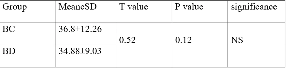
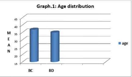
Table.1:Age Distribution
Group MeancSD T value P value significance
BC 36.8±12.26
0.52 0.12 NS BD 34.88±9.03
The minimum age of the patient was 15yrs and the max age was
55yrs. The mean age of the patient in group BC was
36.8±12.26Bwas34.88±9.
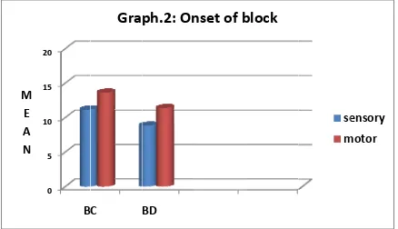
Table. Group BC BD was 1 analysi sensory 0.001) M E A N .2:Onset M 1 8
The m
1.14±1.13
is by stud
y block in 15 20 25 30 35 40 45 B of Senso Mean±SD 11.14±1.1 8.8±0.91 mean tim
3 and in
dent’s un
n group B
BC
Grap
ry block
D T v
3
8.0
me for ons
n group
npaired “t
BD was s
BD
ph.1:
Age
(in min)
value
05
set of sen
BD was
t” test sho
significan
e
distrib
P valu <0.001 nsory blo 8.8±0.9 owed tha ntly faster
bution
ue s
1 H
ock was i
1min. Th
t the time
r than gro
significan
HS
in group
he statist
e of onse
oup BC (
[image:66.612.75.508.69.323.2]Table. Group BC BD 13.6±1 by stud
test sh
signific M E A N .3: Onset M 1 1
The m
1.11 and dent’s unp howed th cantly fas 0 5 10 15 20 B
t of Moto
Mean±SD 13.6±1.11 11.36±0.9 mean time in group paired “t”
at the tim
ster than g
C
Gra
or block (
D T v
7.6 952
e for ons
BD was
”
me of onse
group BC
BD
ph.2:
On
(in min)
value
65
set of mo
s 11.36±
et of moto
C (p < 0.0
nset
of
b
P valu
<0.001
otor bloc
±0.952. T
or block i
001).
block
ue s
1 H
ck in gro
The statist
in group B
[image:67.612.72.510.70.322.2]
Table.4: Duration of Sensory block (in hours)
Group Mean±SD T value P value significance
BC 7.06±0.664
11.9 <0.001 HS
BD 9.3±0.667
Patients of both groups were observed for 24hrs. The time
was noted when the patient asked for rescue analgesics. The mean
duration of sensory block in group BC was 7.06±0.664 and in group BD
was 9.3±0.667hrs. The statistical analysis by student’s unpaired “t” test
showed that the time of duration of sensory block in group BD was
Table Group BC BD T 6.66±0 by stud group B M E A N .5: Durat M 6 8 The mea 0.657and dent’s unp
BD was s 0 2 4 6 8 10 12 14 B
tion of M
Mean±SD 6.66±0.65 8.5±0.692 an durat in group paired “t” significan BC
Grap
Motor bloD T v
57
9.6 2
tion of
BD was
” test sho
ntly longe
BD
ph.3:
Dur
ock (in ho
value
64
motor
s 8.5±0.6
owed that
er than gro
ration
of
ours)
P valu
<0.001
block
692hrs. T
t the durat
oup BC (
f
block
ue s
1 H
in grou
The statist
tion of m
(p < 0.001
[image:69.612.74.515.68.323.2]
Table.
No of 24hrs p
1st dose
2nd dos
In and 80 one an differe statisti P E R C E N T A G E
.6:No of r
f RA i post-op
e
se
n group B
0% require
nalgesic
nce in no
cally sign 0 10 20 30 40 50 60 70 80 B rescue an in BC 5(20% 20(80 BC 20%
ed 2 anal
dose and
o of rescu
nificant by BC
Graph
nalgesics %) %) of patien gesic dosd only 4
ue analge
y Chi-squ
BD
h.4:
Resc
in post-o BD 14(56 11() nts requir ses, where 44% requ esics requ
uare test (
cue
anal
op 24hrs
6%)
red one r
eas in gro
uired 2 a
uired by b
(p<0.018)
lgesics
:
P
0.01
rescue an
oup BD, 5
analgesic
both the
).
8
nalgesic d
[image:70.612.74.506.488.732.2]Table Sedatio score 2 3 Total Mean SD ‘p’ P E R C E N T A G E 7:Sedati
on G
N p 2 4 2 2 0 < 0 20 40 60 80 100 on score Group BC No patients 21 4 25 2 0.374 <0.001 (s BC 84 16
Grap
: C of % 84 16 100 ignificant BD 20 80ph.5:
Se
0
t)
dation
s
[image:71.612.68.544.112.705.2]
This was evaluated using Ramsay sedation score:
1. Anxious and alert.
2. Conscious and oriented.
3. Sedated, responding to verbal commands.
4. Responding only to mild physical stimulus.
5. Responding to moderate & severe physical stimulus.
6. Not arousable.
In group, sedation score corresponding to score 2 was observed in
84% of patients and sedation score of 3 in 16% of patients, whereas in
group BD, sedation score corresponding to 2 was observed in 20% of
patients and sedation score of 3 in 80% of patients. The difference in
sedation score between the two groups was found to be statistically
significant by student’s unpaired ‘t’ test. (p<0.001).
Hemodynamic variables:
Pulse rate, systolic BP, diastolic BP and oxygen saturation was recorded
Table 8: Pulse rate (beats/min)
Time of
assessment
BC BD T
value
P
value significance Mean SD Mean SD
0min 79.240 7.008 78.880 6.760 0.854 0.020 NS
5min 78.920 7.427 78.800 6.782 0.953 0.028 NS
15min 78.320 7.284 78.480 7.148 0.938 0.014 NS
30min 78.080 7.729 78.080 6.964 1.000 0.051 NS
60min 77.840 7.888 78.240 6.888 0.849 0.033 NS
2hrs 77.920 7.926 78.000 6.633 0.969 0.000 NS
6hrs 78.480 7.880 78.280 7.481 0.927 0.092 NS
12hrs 78.360 7.566 78.040 7.586 0.882 0.051 NS
24hrs 78.480 7.600 78.040 7.552 0.838 0.013 NS
In group BC the mean pulse rate ranged from 73.920±9.9 to
82.760±13.18beats/min and in group BD it ranged from 76.880±0.247 to
test sho
the 2 g
M E A N
owed that
groups(p>
50 60 70 80 90 100
0min
t there wa
>0.05).
5min 15m
T
G
as no sign
min 30min 60
TIME OF A
raph.6:
nificant d
0min 2hrs
SSESSMEN
Pulse
ra
difference
6hrs 12hrs
NT
ate
in pulse
24hrs
rate betw
BC BD
Table 9: Systolic BP(mmHg)
Time of
assessment
BC BD T
value
P
value significance Mean SD Mean SD
0min 123.44 8.515 120.480 7.922 0.209 0.067 NS
5min 123.28 8.244 120.320 7.993 0.204 0.042 NS
15min 123.28 8.163 119.920 7.884 0.145 0.016 NS
30min 123.04 8.023 118.880 8.167 0.075 0.073 NS
60min 122.72 7.547 119.040 7.961 0.100 0.002 NS
2hrs 122.16 6.479 119.120 7.748 0.139 0.067 NS
6hrs 121.2 7.141 119.760 7.557 0.492 0.013 NS
12hrs 121.92 7.604 120.000 7.483 0.373 0.003 NS
24hrs 122.48 7.644 120.080 7.449 0.266 0.033 NS
In group BC the mean systolic BP ranged from 122.56±9.05 to
124.08±7.77mmHg and in group BD it ranged from 116.24±7.53 to
test sh
betwee
M E A N
howed th
en the 2 g
100 110 120 130 140
0min
hat there
groups(p>
5min 15m
T
Gr
was no
>0.05).
min 30min 60
TIME OF A
raph.7:
S
significa
0min 2hrs
SSESSMEN
Systolic
ant differ
6hrs 12hrs
NT
BP
rence in
24hrs
systolic
BC BD
Table 10:Diastolic BP(mmHg)
Time of
assessment
BC BD T
value P
value
significance
Mean SD Mean SD
0min 75.92 4.949 76 7.348 0.964 0.064 NS
5min 75.84 5.064 75.76 7.242 0.964 0.099 NS
15min 75.52 5.009 75.52 6.514 1.000 0.013 NS
30min 74.56 5.116 75.28 6.680 0.671 0.012 NS
60min 74.16 4.652 74.88 6.809 0.664 0.059 NS
2hrs 74.4 4.796 74.96 6.761 0.737 0.106 NS
6hrs 74.48 5.009 75.44 6.795 0.572 0.180 NS
12hrs 75.08 5.212 75.68 7.134 0.736 0.122 NS
24hrs 75.6 5.196 75.68 7.134 0.964 0.158 NS
In group BC the mean diastolic BP ranged from 71.36±5.73
to75.92±4.94mmHg and in group BD it ranged from 71.04±6.19
to76.48±7.6mmHg. The statistical analysis by student’s unpaired “t” test
showed that there was no significant difference in diastolic BP between
M E A N
50 60 70 80 90 100
0min 5min 15m
Gr
in 30min 60
TIME
raph.8:
D
0min 2hrs
OF ASSES
Diastolic
6hrs 12hrs
SMENT
c
BP
24hrs
Table 11:Oxygen saturation(%)
Time of
assessment
BC BD T
value
P
value
significance
Mean SD Mean SD
0min 99.28 0.541 99.2 0.707 0.655 0.478 NS
5min 99.4 0.577 99.4 0.707 1 0.204 NS
15min 99.48 0.509 99.44 0.506 0.782 0.367 NS
30min 99.2 0.577 99.2 0.633 1 4.654 NS
60min 99.28 0.458 99.32 0.485 0.782 0.236 NS
2hrs 99.44 0.583 98.92 0.483 0.002 0.127 NS
6hrs 99.08 0.493 99.28 0.452 0.151 0.265 NS
12hrs 99.04 0.538 99.04 0.196 0.196 0.371 NS
24hrs 99.12 0.6 99.12 0.543 0.257 0.612 NS
In group BC the mean O2 saturation ranged from 99.04±0.53 to
99.04±0.19% and in group BD it ranged from 99.04±0.19 to
showed
the 2 g
Side ef bradyc and br patien gradua 4 patie M E A N
d that the
groups(p>
ffects:
Patients
cardia. In
radycardi
nt when f
ally resol
ents had a 50 60 70 80 90 100 110 0min
ere was no
>0.05).
were obs
n both the
ia. Only o
followed lved. an inadeq n 5min T
G
o signific served fo e groups, one patien by takin quate bloc 15minTIME OF AS
Graph.9:
cant differ
or side eff
there wa
nt had a m
ng chest X
ck and we 30min
SSSSMENT
Oxygen
rence in O
ffects such
as no inci
minimal p
X-Ray th
ere exclud 60min 2
T
n
saturat
O2 satura
h as hypo
DISCUSSION
Brachi