JOURNAL OFVIROLOGY,
0022-538X/99/$04.0010 July 1999, p. 6141–6146 Vol. 73, No. 7
Copyright © 1999, American Society for Microbiology. All Rights Reserved.
Frequency and Stability of Chromosomal Integration of
Adenovirus Vectors
AIRI HARUI,1SHINOBU SUZUKI,1STEFAN KOCHANEK,2ANDKOHNOSUKE MITANI1,3*
Department of Microbiology, Immunology and Molecular Genetics, UCLA School of Medicine,1and
Jonsson Comprehensive Cancer Center,3Los Angeles, California 90095-1747, and Center for
Molecular Medicine (ZMMK), University of Cologne, 50931 Cologne, Germany2
Received 20 November 1998/Accepted 22 March 1999
One of the limitations of adenovirus vectors is the lack of machinery necessary for their integration into host chromosomes, resulting in short-term gene expression in dividing cells. We analyzed frequencies of integration and persistence of gene expression from integrated adenovirus vectors. Both E1-substituted and helper-dependent adenovirus vectors achieved similar integration efficiencies of ;1023 to 1025 per cell, with the helper-dependent vector showing slightly higher efficiencies. In stable cell pools, gene expression of the integrated vector persisted for at least 50 cell divisions without selection. However, some stable cell clones showed changes in gene expression, which were accompanied by structural changes in the integrated vector DNA.
Recombinant adenovirus vectors are attracting increasing attention as in vivo gene transfer vehicles for human gene therapy (2, 31, 36). However, one of the limitations of E1-substituted adenovirus vectors currently used in most clinical gene therapy protocols is the relatively short-term expression of the transferred gene in vivo. E1-substituted vectors express viral antigens that induce a cytotoxic-T-lymphocyte-mediated immune response against the vector-transduced cells by the host, resulting in inflammation and short-term gene expression from the vector in vivo (3, 28, 32–35). To overcome this prob-lem, we and others have recently developed a helper-depen-dent (gutless) adenovirus vector by removing all the viral cod-ing sequences from adenovirus vector DNA (7, 11, 12, 15, 19). Such vectors are therefore expected to minimize the immune responses of the host that would cause rejection of the trans-duced cells (20, 24). However, even with this system, vector DNA is eventually lost in dividing cells because although ad-enoviruses exist as multiple episomal copies in the infected cell nuclei, they lack the machinery necessary for integration into host chromosomes. Furthermore, activation of T-helper cells and B cells in response to viral capsid proteins produces neu-tralizing antibodies that block the efficient readministration of vector (13, 37). Therefore, further improvement of an adeno-virus vector that replicates or efficiently integrates into host chromosomes is required to obtain long-term expression, even in dividing cells. Although it is known that a wild-type adeno-virus rarely integrates into the chromosomes of cells that are not permissive for viral DNA replication, there have not yet been any extensive investigations of how frequently replica-tion-incompetent adenovirus vectors integrate into host chro-mosomes. In this study, we analyzed frequencies of integration of E1-substituted and helper-dependent adenovirus vectors and stability of gene expression from the integrated vectors.
Integration efficiencies of E1-substituted and helper-depen-dent adenovirus vectors in cell lines.To compare the efficien-cies of integration of the E1-substituted and helper-dependent
adenovirus vectors, we rescued both types of vectors with the
b-geo marker gene, a fusion of the E. coli b-galactosidase (b-Gal) gene and the neomycin phosphotransferase II gene (neo) (9) that is driven by the SRapromoter (27) (SRab-geo) (Fig. 1). The SRab-geomarker gene cassette (19) was sub-cloned into an adenovirus transfer plasmid, pXCX2 (26). AdSRab-geo, an E1-substituted adenovirus vector (DE1 vec-tor), was rescued by cotransfection into the 293 cell line (Mi-crobix, Toronto, Ontario, Canada) of the plasmid with pJM17 (18). The virus was then plaque isolated, propagated, and pu-rified as described previously (10). The titer of the vector was 1.23109PFU/ml on 293 cells. A helper-dependent adenovirus
vector DNA was constructed as follows. First, an AvrII-SmaI fragment of pFG140 (18) encompassing the junction of the ligated right end (452 bp) and left end (1,009 bp) of Ad5 was subcloned into anXbaI site of the charomid 9-22 vector (23). This adenovirus sequence contains two inverted terminal re-peats and the packaging signal but does not encode any intact open reading frames of the parental human adenovirus type 5 (Ad5). Second, the SRab-geocassette was subcloned into a SmaI site of the plasmid. The helper-dependent AdxSRab -geo vector (DAd vector) was rescued by cotransfection into 293 cells of the resultant plasmid with a helper-virus DNA–termi-nal protein complex. AdHprt, an adenovirus vector with E1 and E3 deleted and with a nonfunctional genomic sequence from the mouseHprtlocus, was used as a helper virus. The vector was propagated and purified as described previously (12, 19). After three rounds of purification by a CsCl density gradient, the titer of the vector was measured in situ, using 5-bromo-4-chloro-3-indolyl-b-D-galactoside (X-Gal) as a
sub-strate, as previously described (17). The titer of the vector on the African green monkey cell line COS7 (American Type Culture Collection [ATCC], Rockville, Md.) was 2.4 3 109
b-Gal-transducing units (BTU)/ml, and that of the helper on 293 cells, as measured by a plaque assay, was 2.23107PFU/
ml. Therefore, the vector stock contained 0.9% helper virus contamination.
The human cell lines HeLa (ATCC), HT1080 (ATCC), and KB (ATCC), the African green monkey kidney cell lines CV-1 (ATCC) and Vero (provided by Harumi Kasamatsu), the baby hamster kidney (BHK) cell line (provided by Debi Nayak), the * Corresponding author. Mailing address: Department of
Microbi-ology, Immunology and Molecular Genetics, UCLA School of Medi-cine, Box 951747, Los Angeles, CA 90095-1747. Phone: (310) 267-2031. Fax: (310) 206-3865. E-mail: [email protected].
6141
on November 9, 2019 by guest
http://jvi.asm.org/
Chinese hamster ovary (CHO) cell line (provided by Owen N. Witte), and the mouse cell line NIH 3T3 (ATCC) were in-fected with the vectors at 10 PFU/cell for theDE1 vector or 10 BTU/cell for theDAd vector. At 48 h postinfection, the in-fected cells were diluted and replated onto 96-well plates at densities of 105, 104, and 103cells/plate. To avoid low plating
efficiencies at low cell densities, the total cell numbers in each plate were adjusted to 105 by adding uninfected cells. After
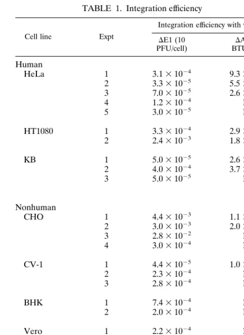
24 h, medium containing G418 was added. The final concen-tration of G418 was 500 mg/ml (HeLa, HT1080, BHK, and NIH 3T3 cells) or 1,000 mg/ml (KB, CHO, CV-1, and Vero cells). G418-containing medium was added again 1 week later, and the G418-resistant colonies were counted and integration efficiencies were calculated 4 weeks postinfection. The results are summarized in Table 1. In most of the cell lines, both vectors integrated efficiently at frequencies of 1024 to 1025,
but we occasionally observed a higher efficiency of 1023 in
HT1080 and KB cells. CHO cells showed relatively high inte-gration efficiencies, particularly with the DAd vector, which achieved levels above 1%. In the experiments with HeLa, KB, and CHO cell lines, in which both types of vectors were tested in parallel, integration frequencies were severalfold higher with theDAd vector than with theDE1 vector, but both vectors showed similar efficiencies in HT1080 cells.
The relation of the PFU titer of theDE1 vector to the BTU titer of theDAd vector was determined as follows. Infected HeLa cells were harvested 4 h postinfection, at which time viral DNA synthesis had not yet started, and total DNA was ex-tracted. The DNA was digested withHindIII and subjected to Southern hybridization with ab-Gal fragment (nucleotides 118 to 581 of ECLACZ; GenBank no. V00296) as a probe. Based on the intensity of the signal measured by a Storm Phospho-rImager (Molecular Dynamics, Sunnyvale, Calif.), the copy number of theDAd vector at 10 BTU/cell is not as high as that of theDE1 vector at 10 PFU/cell but corresponds to the copy number at 6.4 PFU/cell. Therefore, the integration efficiency of theDAd vector in Table 1 might be an underestimate, com-pared to that of theDE1 vector.
Integration efficiencies of the adenovirus vector that were assessed by G418 resistance could be affected by vector expres-sion levels of the selectable marker gene,b-geo(25), and/or by the ability of vector DNA to integrate into chromosomes. To distinguish between these two possibilities, the infected HeLa, HT1080, KB, and CHO cells were plated into 100-mm dishes and subjected to G418 selection to obtain pools of stably trans-duced cells. Cell extracts were prepared from these cell pools,
[image:2.612.111.491.70.233.2]and the expression levels of theb-geomarker gene were com-pared. The levels ofb-Gal activity showed no correlation to vector or cell types (data not shown), indicating that there are no significant differences in the levels of expression of SRab -geoonce it integrates into a host chromosome, regardless of the vector backbones used. These results suggest that more efficient integration by theDAd vector is mainly due to the stronger ability of theDAd vector to integrate into cellular FIG. 1. Structure of adenovirus vectors. H,HindIII sites used for Southern hybridization analysis of the structure of integrated vectors.
TABLE 1. Integration efficiency
Cell line Expt
Integration efficiency with vector
DE1 (10
PFU/cell) BTU/cell)DAd (10a
Human
HeLa 1 3.131024 9.331024 2 3.331025 5.531025 3 7.031025 2.631024 4 1.231024 NT 5 3.031025 NT
HT1080 1 3.331024 2.931024 2 2.431023 1.831023
KB 1 5.031025 2.631023 2 4.031024 3.731023 3 5.031025 NT
Nonhuman
CHO 1 4.431023 1.131022 2 3.031023 2.031022 3 2.831022 NT 4 3.031024 NT
CV-1 1 4.431025 1.031024 2 2.331024 NT 3 2.831024 NT
BHK 1 7.431024 NT
2 2.031024 NT
Vero 1 2.231024 NT
2 6.031025 NT
NIH 3T3 1 1.031025 NT
aNT, not tested.
on November 9, 2019 by guest
http://jvi.asm.org/
[image:2.612.310.551.387.718.2]chromosomes. The higher integration efficiencies of theDAd vector can be attributed to the nature of completely nonviral genomic sequences or to the lack of leaky expression of viral genes, some of which inhibit normal cellular machinery, unlike those in theDE1 vector.
Although wild-type adenovirus does not integrate into the chromosomes of the permissive cells because its infection leads to lytic infection, it can integrate in nonpermissive cells (e.g., hamster cells infected with Ad12) (for a review, see reference 6). In addition, adenovirus mutants that code for a tempera-ture-sensitive DNA-binding protein yield stable cell clones at nonpermissive temperatures (8). Chromosomal integration of an E1-substituted adenovirus vector in rat and simian cells has previously been reported (30). At a multiplicity of infection (MOI) of 200, the integration efficiencies were 0.4% for Rat2 cells and 0.75% for CV-1 cells. More recently, stable integra-tion of E1-substituted adenovirus vector with theb-galmarker gene in mouse NIH 3T3, human A549, and primary human cells has been reported (38). Although the integration effi-ciency was 15% in these cell lines when ionizing radiation was used, the efficiency without radiation was not reported.
In our study, theDE1 andDAd vectors were used to
inves-tigate the frequencies of integration of adenovirus vectors in different cell types (Table 1). Human epidermoid cell lines such as HeLa and KB are known to be permissive for viral DNA replication of wild-type Ad5. BHK and Vero cells are semipermissive, and CV-1 and CHO cells are nonpermissive (16). Most of the cell lines infected by theDE1 vector showed similar integration efficiencies of ;1023 to 1025 (Table 1),
suggesting that factors determining cellular permissiveness do not affect the integration of adenovirus vectors. Under these conditions (MOI of 10), only up to 10 viral DNA molecules entered per cell, in contrast to approximately 106DNA
mole-cules per cell transferred by the calcium phosphate transfec-tion method, in which 2.2 to 6.4% of DNA is internalized into the nucleus (21). Our results indicate that the ability of ade-novirus DNA to integrate into host chromosomes seems ex-tremely high compared to that of naked plasmid DNA, even though the end is protected by the terminal protein.
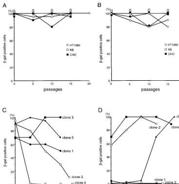
[image:3.612.123.490.73.449.2]Sustained gene expression from integrated adenovirus vec-tor.To analyze the persistence of gene expression from inte-grated adenovirus vectors, the pools and clones of stably trans-duced cells, obtained as described above, were cultured without G418 selection. Expression ofb-Gal in these cells was FIG. 2.b-Gal expression from the integrated vectors over passages of the cells infected withDE1 vector (A and C) andDAd vector (B and D) without G418 selection. The stable cell pools (A and B) and HeLa clones (C and D) were maintained without G418 selection and stained by X-Gal every five passages; the percentages of X-Gal-positive cells were determined by microscopic observation. Two independent pools were analyzed for each cell line.
VOL. 73, 1999 NOTES 6143
on November 9, 2019 by guest
http://jvi.asm.org/
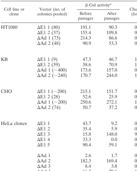
detected by X-Gal staining at every five passages (Fig. 2). In most of the stable cell pools,b-Gal expression did not diminish over at least 15 passages, which corresponds to approximately 50 cell divisions (Fig. 2A and B). There were no significant differences in the time-course profiles of marker gene expres-sion between theDE1 andDAd vectors. For more quantitative analysis, the levels ofb-Gal enzymatic activity were also mea-sured in triplicate (with the luminescentb-Gal gene reporter system 2; Clontech, Palo Alto, Calif.) before and after the series of passages without G418 (Table 2). The amount of protein in each sample was standardized by the Bradford method (Bio-Rad protein assay; Bio-Rad, Hercules, Calif.). Each cell pool showed different patterns of stability ofb-Gal expression from the integrated vectors. In HT1080 cells, the levels ofb-Gal activity tended to decrease with both theDE1 (0.5- and 0.7-fold) andDAd (0.4- and 0.6-fold) vectors. In KB cells, the levels ofb-Gal activity remained the same for both theDE1 (1.0- and 1.2-fold) andDAd (0.9- and 1.4-fold) vec-tors. In CHO cells, the levels ofb-Gal activity from the inte-gratedDE1 vector (0.7- and 0.5-fold), but not from the DAd vector (1.1- and 0.7-fold), tended to decrease. We also ana-lyzed the persistence of gene expression of individual stable HeLa clones (Fig. 2C and D). In two of five HeLa cell clones with stably integratedDE1 vectors, the level ofb-Gal activity decreased from high to low over time (clones 2 and 4) (Fig. 2C). Interestingly, in HeLa clone 5, which was transduced by the DAd vector, the percentage of b-Gal-positive cells in-creased dramatically over time (Fig. 2D). This observation is in contrast to the expression of viral genes from integrated wild-type adenovirus, which is often shut off by methylation (4–6). Structure of the integrated vector.The structure of adeno-virus DNA integrated into host chromosomes is known to vary,
FIG. 3. Southern analysis of integrated vector DNA. DNA from HeLa cell clones infected with theDE1 (A, C, and E) orDAd (B, D, and E) vector was digested withHindIII and subjected to Southern hybridization with the right-end (A and B),neo(C and D), and full-length Ad5 DNA (E) probes. (A and C) Lanes 1 to 3,DE1 clones 1 to 3, respectively; lane 4, clone 4 before the passages without selection; lane 5, clone 4 after the passages; lane 6, clone 5 before the passages; lane 7, clone 5 after the passages; lane 8, AdSRab-geo DNA. (B and D) Lanes 1 to 4,DAd clones 1 to 4, respectively; lane 5, clone 5 before the passages without selection; lane 6, clone 5 after the passages. (E) Lane 1,DE1 clone 5; lane 2,DE1 clone 3; lane 3,DAd clone 3; lane 4,DAd clone 4, lane 5,
DE1 clone 1; lane 6,DE1 clone 2; lane 7, uninfected HeLa; lane 8, AdSRab-geo DNA.
Cell line or
clone colonies pooled)Vector (no. of
b-Gal activitya
Change (fold) Before
passages passagesAfter
HT1080 DE1 1 (80) 191.1 90.3 0.5
DE1 2 (37) 155.4 109.8 0.7
DAd 1 (73) 214.3 86.6 0.4
DAd 2 (48) 90.9 53.3 0.6
KB DE1 1 (9) 47.3 46.7 1.0
DE1 2 (59) 58.6 70.9 1.2
DAd 1 (;400) 173.6 157.0 0.9
DAd 2 (;240) 170.7 244.0 1.4
CHO DE1 1 (;200) 215.1 151.7 0.7
DE1 2 (28) 52.6 25.9 0.5
DAd 1 (;200) 250.6 272.1 1.1
DAd 2 (74) 50.7 37.2 0.7
HeLa clones DE1 1 43.7 9.2 0.2
DE1 2 35.4 5.9 0.2
DE1 3 15.8 148.0 9.4
DE1 4 33.3 0.0 0.0
DE1 5 90.4 59.1 0.7
DAd 1 2.6 1.7 0.7
DAd 2 182.3 169.4 0.9
DAd 3 8.4 3.8 0.5
DAd 4 66.3 70.1 1.1
DAd 5 8.3 98.7 11.9
aValues are in units (105) of enzyme per microgram of protein.
6144
on November 9, 2019 by guest
http://jvi.asm.org/
[image:4.612.54.295.70.371.2]depending on the cell line and the type of virus. Rat embryonic cells transformed by Ad5 usually contain only the left end of the genome, whereas cell lines transformed by temperature-sensitive adenovirus mutants at semi- or nonpermissive tem-peratures often contain multiple copies of all or most of the adenovirus genome (6). To analyze the structure of integrated adenovirus vectors, genomic DNA was extracted from stably transduced HeLa cell clones, digested withHindIII, and sub-jected to Southern hybridization. Fragments of theneogene, the right end of Ad5 (nucleotides 35032 to 35780 of ADRCOMPGEN; GenBank no. M73260 and M29978), and full-length Ad5 DNA were used as probes (Fig. 1). The right-end probe hybridized with the DNA fragment between the rightmostHindIII site in the adenovirus genome and aHindIII site to the right of the integration site, which produced a band representing a size unique to the integration site. InDE1 stable clones, the right-end probe hybridized with one band in three of five clones, indicating a single-copy integration of the right end of the vector. However, clones 2 and 5 had three and two bands, respectively, indicating multiple integrations of the right end in these clones (Fig. 3A). Similarly, the neo probe with
DE1 integrants produced a band representing a size unique to a left-end integration junction. Contrary to the results with the right-end probe, clones 1, 2, and 5 showed single-copy integra-tion, while clones 3 and 4 showed two integrants each (Fig. 3C). Clone 1 had one additional faint band hybridizing with the right-end probe. This faint band might indicate that in some cells the integrated vector is rearranged. Finally, the full-length adenovirus probe should produce eight bands specific for the full-length vector, with two additional bands corresponding to the junctions of integration sites at both ends of the vector (Fig. 1). All of the clones showed the bands common to AdSRab-geoviral DNA (Fig. 3E, lanes 1, 2, 5, and 6). These results indicate that all of the HeLaDE1 stable clones had at least one copy of full-length vector integrated into their chro-mosomes, which was often accompanied by additional integra-tion events. In the case of the stableDAd clones, all the inte-grants showed a single band with both the right-end and the neoprobes (Figs. 3B and D), suggesting that most of the stable cell clones had a single-copy integration. Clone 2 had two additional faint bands hybridizing with the right-end probe, suggesting rearrangement of integrated vectors in some cells. Theneoprobe should produce a 26-kb internal fragment en-compassing the SRab-geo cassette and the stuffer sequence consisting of repeats of a 2-kb fragment from pBR (Fig. 1). However, at least two clones (clones 3 and 5) (Fig. 3D) showed fragments smaller than those of other clones. Therefore, in these clones, an internal fragment was deleted before or after integration. The deletions might be associated with the nature of the repetitive sequences of the fragment. In CV-1 cell clones,DE1 vector showed patterns of integration similar to those in HeLa cells (i.e., a single full-length copy with an additional end sequence). On the other hand, DAd vector tended to integrate at multiple sites in many clones but, unlike in HeLa cells, no internal deletion was detected by Southern analysis (data not shown). It is not clear why theDE1 andDAd vectors show distinct patterns of integration.
We also examined the changes in the structure of integrated vector in three HeLa clones before and after passages without selection. InDE1 clone 4, the integrated vector signal disap-peared, as indicated by both probes after 15 passages, consis-tent with the decrease in levels ofb-Gal-positive cells from 95 to 0% (Fig. 3A and C, lanes 4 and 5). InDE1 clone 5, which had a moderate decrease inb-Gal-positive cells (from 90 to 70%), one of the two fragments hybridizing with the right-end probe disappeared (Fig. 3A, lanes 6 and 7), while the only band
that hybridized with theneoprobe remained unaltered before and after passages without selection (Fig. 3C, lanes 6 and 7). On the other hand, in the DAd stable clone 5, which had increased activity (from 1 to 100%), the integrated vector showed a rearranged pattern after 15 passages (Fig. 3B and D, lanes 5 and 6). This rearrangement might account for the increased b-Gal activity. Rearrangement of integrated viral DNA was also documented in hamster cells with stably inte-grated Ad12 (14).
In summary, although adenovirus vectors integrate into host chromosomes relatively efficiently, unlike retroviral integra-tion, most of the stable clones have an extra fragment(s) of the vector or deleted vector. Gene expression from the integrated vector is relatively stable. However, integrated vectors some-times become further rearranged, resulting in an altered level of gene expression. Adenovirus vectors can infect a variety of cell types at very high efficiencies. Our results suggest that it might be possible to use an adenovirus vector to establish stable cell clones in cells which are refractory to other gene delivery methods. Considering the somewhat unstable nature of integrated adenovirus vector DNA, selection strategies for the integrated DNA might be needed to obtain long-term expression. It is known that gene expression from retroviral vectors is shut off in some primary cell cultures. Therefore, it would be of interest to analyze the integration frequencies of adenovirus vectors and the longevity of gene expression, espe-cially in primary cultures such as cultures of hematopoietic cells, which are one of the main targets for human gene ther-apy. On the other hand, because one of the advantages of an adenovirus vector for human gene therapy is its rare chromo-somal integration, thereby circumventing potential insertional mutagenesis of cancer-related genes, it would be important to determine how frequently an adenovirus vector integrates in vivo. There are reports suggesting that stable integration of adenovirus vectors into the host chromosome occurs after in vivo gene transfer in animals (1, 22). Efficient production in transgenic mice by adenovirus gene transfer into fertilized eggs was also reported (29). Therefore, considering the high MOIs usually used for in vivo gene transfer, elucidation of adenovirus integration in vivo is a very important issue for evaluating the safety of adenovirus vectors for human gene therapy.
A.H. and S.S. contributed equally to this work.
We thank Harumi Kasamatsu, Debi Nayak, and Owen N. Witte for providing cell lines, Frank L. Graham for providing the pXCX2 and pJM17 plasmids, Izumu Saito for providing the charomid vector, Arnie Berk for critical discussion and for reading the manuscript, Wendy Aft for preparation of the manuscript, Amit Oberai for generating prelim-inary data, and Lena Kim for technical support.
This work was supported by NIH grant AI-42214.
REFERENCES
1.Brown, G. R., D. L. Thiele, M. Silva, and B. Beutler.1997. Adenoviral vectors given intravenously to immunocompromised mice yield stable transduction of the colonic epithelium. Gastroenterology112:1586–1594.
2.Crystal, R. G.1995. Transfer of genes to humans: early lessons and obstacles to success. Science270:404–410.
3.Dai, Y., E. M. Schwarz, D. Gu, W. W. Zhang, N. Sarvetnick, and I. M. Verma.
1995. Cellular and humoral immune responses to adenoviral vectors con-taining factor IX gene: tolerization of factor IX and vector antigens allows for long-term expression. Proc. Natl. Acad. Sci. USA92:1401–1405. 4.Doerfler, W.1991. Abortive infection and malignant transformation by
ad-enoviruses: integration of viral DNA and control of viral gene expression by specific patterns of DNA methylation. Adv. Virus Res.39:89–128. 5.Doerfler, W.1984. DNA methylation and its functional significance: studies
on the adenovirus system. Curr. Top. Microbiol. Immunol.108:79–98. 6.Doerfler, W.1982. Uptake, fixation, and expression of foreign DNA in
mammalian cells: the organization of integrated adenovirus DNA sequences. Curr. Top. Microbiol. Immunol.101:127–194.
7.Fisher, K. J., H. Choi, J. Burda, S. Chen, and J. M. Wilson.1996.
Recom-VOL. 73, 1999 NOTES 6145
on November 9, 2019 by guest
http://jvi.asm.org/
binant adenovirus deleted of all viral genes for gene therapy of cystic fibrosis. Virology217:11–22.
8.Fisher, P. B., L. E. Babiss, I. B. Weinstein, and H. S. Ginsberg.1982. Analysis of type 5 adenovirus transformation with a cloned rat embryo cell line (CREF). Proc. Natl. Acad. Sci. USA79:3527–3531.
9.Friedrich, G., and P. Soriano.1991. Promoter traps in embryonic stem cells: a genetic screen to identify and mutate developmental genes in mice. Genes Dev.5:1513–1523.
10. Graham, F. L., and L. Prevec.1995. Methods for construction of adenovirus vectors. Mol. Biotechnol.3:207–220.
11. Haecker, S. E., H. H. Stedman, R. J. Balice-Gordon, D. B. Smith, J. P. Greelish, M. A. Mitchell, A. Wells, H. L. Sweeney, and J. M. Wilson.1996. In vivoexpression of full-length human dystrophin from adenoviral vectors deleted of all viral genes. Hum. Gene Ther.7:1907–1914.
12.Kochanek, S., P. R. Clemens, K. Mitani, H.-H. Chen, S. Chan, and C. T. Caskey.1996. A new adenoviral vector: replacement of all viral coding sequences with 28 kb of DNA independently expressing both full-length dystrophin andb-galactosidase. Proc. Natl. Acad. Sci. USA93:5731–5736. 13. Kozarsky, K. F., D. R. McKinley, L. L. Austin, S. E. Raper, L. D.
Stratford-Perricaudet, and J. M. Wilson.1994.In vivocorrection of low density lipoprotein receptor deficiency in the Watanabe heritable hyperlipidemic rabbit with recombinant adenoviruses. J. Biol. Chem.269:13695–13702. 14. Kuhlmann, I., and W. Doerfler.1983. Loss of viral genomes from hamster
tumor cells and nonrandom alterations in patterns of methylation of inte-grated adenovirus type 12 DNA. J. Virol.47:631–636.
15. Kumar-Singh, R., and J. S. Chamberlain.1996. Encapsidated adenovirus minichromosomes allow delivery and expression of a 14 kb dystrophin cDNA to muscle cells. Hum. Mol. Genet.5:913–921.
16. Lucher, L. A.1995. Abortive adenovirus infection and host range determi-nants. Curr. Top. Microbiol. Immunol.199:119–152.
17. MacGregor, G. R., A. E. Mogg, J. F. Burke, and C. T. Caskey.1987. Histo-chemical staining of clonal mammalian cell lines expressingE. colib -galac-tosidase indicates heterogeneous expression of the bacterial gene. Somatic Cell Mol. Genet.13:253–265.
18. McGrory, W. J., D. S. Bautista, and F. J. Graham.1988. A simple technique for the rescue of early region I mutants into infectious human adenovirus type 5. Virology163:614–617.
19. Mitani, K., F. L. Graham, C. T. Caskey, and S. Kochanek.1995. Rescue, propagation, and partial purification of a helper virus-dependent adenovirus vector. Proc. Natl. Acad. Sci. USA92:3854–3858.
20. Morsy, M. A., M. Gu, S. Motzel, J. Zhao, J. Lin, Q. Su, H. Allen, L. Franlin, R. J. Parks, F. L. Graham, S. Kochanek, A. J. Bett, and C. T. Caskey.1998. An adenoviral vector deleted for all viral coding sequences results in en-hanced safety and extended expression of a leptin transgene. Proc. Natl. Acad. Sci. USA95:7866–7871.
21. Orrantia, E., and P. L. Chang.1990. Intracellular distribution of DNA internalized through calcium phosphate precipitation. Exp. Cell Res.190:
170–174.
22. Overturf, K., M. Al-Dhalimy, C. N. Ou, M. Finegold, R. Tanguay, A. Lieber, M. Kay, and M. Grompe.1997. Adenovirus-mediated gene therapy in a mouse model of hereditary tyrosinemia type I. Hum. Gene Ther.8:513–521. 23. Saito, I., and G. R. Stark.1986. Charomids: cosmid vectors for efficient
cloning and mapping of large or small restriction fragments. Proc. Natl. Acad. Sci. USA83:8664–8668.
24. Schiedner, G., N. Morral, R. J. Parks, Y. Wu, S. C. Koopmans, C. Langston, F. L. Graham, A. L. Beaudet, and S. Kochanek. 1998. Genomic DNA transfer with a high-capacity adenovirus vector results in improvedin vivo gene expression and decreased toxicity. Nat. Genet.18:180–183. 25. Soriano, P., C. Montgomery, R. Geske, and A. Bradley.1991. Targeted
disruption of the c-srcproto-oncogene leads to osteopetrosis in mice. Cell
64:693–702.
26. Spessot, R., K. Inchley, T. M. Hupel, and S. Bacchetti.1989. Cloning of the herpes simplex virus ICP4 gene in an adenovirus vector: effects on adeno-virus gene expression and replication. Virology168:378–387.
27. Takebe, Y., M. Seiki, J.-I. Fujisawa, P. Hoy, K. Yokota, K.-I. Arai, M. Yoshida, and N. Arai.1988. SRapromoter: an efficient and versatile mam-malian cDNA expression system composed of the simian virus 40 early promoter and the R-U5 segment of human T-cell leukemia virus type 1 long terminal repeat. Mol. Cell. Biol.8:466–472.
28. Tripathy, S. K., H. B. Black, E. Goldwasser, and J. M. Leiden.1996. Immune responses to transgene-encoded proteins limit the stability of gene expres-sion after injection of replication-defective adenovirus vectors. Nat. Med.
2:545–550.
29. Tsukui, T., Y. Kanegae, I. Saito, and Y. Toyoda.1996. Transgenesis by adenovirus-mediated gene transfer into mouse zona-free eggs. Nat. Biotech-nol.14:982–985.
30. Van Doren, K., D. Hanahan, and Y. Gluzman.1984. Infection of eucaryotic cells by helper-independent recombinant adenoviruses: early region 1 is not obligatory for integration of viral DNA. J. Virol.50:606–614.
31. Verma, I. M., and N. Somia.1997. Gene therapy—promises, problems and prospects. Nature389:239–242.
32. Yang, Y., S. E. Haecker, Q. Su, and J. M. Wilson.1996. Immunology of gene therapy with adenoviral vectors in mouse skeletal muscle. Hum. Mol. Genet.
5:1703–1712.
33. Yang, Y., F. A. Nunes, K. Berencsi, E. E. Furth, E. Gonczol, and J. M. Wilson.1994. Cellular immunity to viral antigens limits E1-deleted adeno-viruses for gene therapy. Proc. Natl. Acad. Sci. USA91:4407–4411. 34. Yang, Y., and J. M. Wilson.1995. Clearance of adenovirus-infected
hepato-cytes by MHC class I-restricted CD41CTLsin vivo. J. Immunol.155:2564– 2570.
35. Yang, Y., Z. Xiang, H. C. Ertl, and J. M. Wilson.1995. Upregulation of class I major histocompatibility complex antigens by interferon-gis necessary for T-cell-mediated elimination of recombinant adenovirus-infected hepatocytes in vivo. Proc. Natl. Acad. Sci. USA92:7257–7261.
36. Yeh, P., and M. Perricaudet.1997. Advances in adenoviral vectors: from genetic engineering to their biology. FASEB J.11:615–623.
37. Yei, S., N. Mittereder, K. Tang, C. O. Sullivan, and B. C. Trapnell.1994. Adenovirus-mediated gene transfer for cystic fibrosis: quantitative evalua-tion of repeatedin vivovector administration to the lung. Gene Ther.
1:192–200.
38. Zeng, M., G. J. Cerniglia, S. L. Eck, and C. W. Stevens.1997. High-efficiency stable gene transfer of adenovirus into mammalian cells using ionizing ra-diation. Hum. Gene Ther.8:1025–1032.