Copyright © 1998, American Society for Microbiology. All Rights Reserved.
Identification of a Human Immunodeficiency Virus Type 2 (HIV-2)
Encapsidation Determinant and Transduction of Nondividing
Human Cells by HIV-2-Based Lentivirus Vectors
ERIC POESCHLA,
1JAMES GILBERT,
2XINQIANG LI,
1SHIANG HUANG,
1ANTHONY HO,
1ANDFLOSSIE WONG-STAAL
1,2*
Departments of Medicine
1and Biology,
2University of California at
San Diego, La Jolla, California 92093-0665
Received 17 October 1997/Accepted 21 April 1998
Although previous lentivirus vector systems have used human immunodeficiency virus type 1 (HIV-1), HIV-2
is less pathogenic in humans and is amenable to pathogenicity testing in a primate model. In this study, an
HIV-2 molecular clone that is infectious but apathogenic in macaques was used to first define cis-acting regions
that can be deleted to prevent HIV-2 genomic encapsidation and replication without inhibiting viral gene
ex-pression. Lentivirus encapsidation determinants are complex and incompletely defined; for HIV-2, some
de-letions between the major 5
*
splice donor and the gag open reading frame have been shown to minimally affect
encapsidation and replication. We find that a larger deletion (61 to 75 nucleotides) abrogates encapsidation
and replication but does not diminish mRNA expression. This deletion was incorporated into a
replication-defective, envelope-pseudotyped, three-plasmid HIV-2 lentivirus vector system that supplies HIV-2 Gag/Pol
and accessory proteins in trans from an HIV-2 packaging plasmid. The HIV-2 vectors efficiently transduced
marker genes into human T and monocytoid cell lines and, in contrast to a murine leukemia virus-based vector,
into growth-arrested HeLa cells and terminally differentiated human macrophages and NTN2 neurons. Vector
DNA could be detected in HIV-2 vector-transduced nondividing CD34
1CD38
2human hematopoietic
progen-itor cells but not in those cells transduced with murine vectors. However, stable integration and expression of
the reporter gene could not be detected in these hematopoietic progenitors, leaving open the question of the
accessibility of these cells to stable lentivirus transduction.
Replication-defective retrovirus vectors are advantageous
for gene transfer because they permit permanent chromosomal
integration and stable gene expression. Following entry into
target cells, however, murine retrovirus and retrovirus vectors
require mitosis-dependent dissolution of the nuclear envelope
to achieve integration (36, 43). Therefore, these vectors can
stably transduce dividing cells but possess limited utility for
gene delivery to quiescent or postmitotic cells that are
impor-tant targets for gene therapy.
In contrast, lentiviruses infect nondividing cells (36). For
human immunodeficiency virus type 1 (HIV-1), this property
has been mapped to establishment of a stable preintegration
complex and to virion proteins that mediate transport of the
preintegration complex across an intact nuclear envelope (6,
17, 18, 22, 55). Accordingly, retrovirus vectors derived from
HIV-1 (35) can transduce growth-arrested and terminally
dif-ferentiated, postmitotic cells. Naldini et al. (45, 46) established
that these lentivirus-specific biological properties hold for
HIV-1-derived vectors and showed their capacity for in vitro
and in vivo gene delivery.
HIV-1 vectors have now been engineered to reduce both the
risks for recombination and the complement of genes needed
for transduction of neuronal cells (65). However, some safety
concerns remain incompletely explored, since the
determi-nants of the severe pathogenicity of HIV-1 in humans remain
uncertain and no animal model amenable to testing of disease
causation by the parental lentivirus exists (16). In addition,
some of the accessory genes of HIV-1 appear to be required
for targeting of some tissues in vivo—vif and vpr for
hepato-cytes, for example (25). In this regard, simian
immunodefi-ciency virus (SIV) with multiple nonstructural gene deletions
has been shown to cause disease in infant primates (2) and
more recently even in adult animals (51).
Safety of HIV-2 vectors can be tested in primates susceptible
to HIV-2/SIV pathogenicity (50). In addition, HIV-2 accounts
for less than 1% of human HIV infections worldwide and has
now been documented to be both less transmissible sexually
and less pathogenic in longitudinally studied West African
human populations (26, 39). In the most comprehensive,
pro-spective natural history study, all indices of virulence, including
HIV-related morbidity and CD4
1lymphocyte depletion, were
much lower for HIV-2-infected than HIV-1-infected subjects
in the same West African population; 5-year AIDS-free
sur-vival was 100% in the HIV-2 cohort (39). These considerations
prompted us to study the feasibility of a replication-defective
lentivirus vector system derived from HIV-2. To further
en-hance safety potential, we have used HIV-2
KR(57), a
molec-ular clone that was apathogenic following infection established
by high-dose intravenous challenge in pig-tailed macaques and
either delayed or prevented disease induction by subsequent
challenge with highly virulent HIV-2
EHO(38).
Genomic regions that determine mRNA encapsidation are
crucial to retrovirus vector system design but have received
very limited study for HIV-2. In the present work, we initially
focused on studying the effects of deleting the region between
the major splice donor (SD) and the gag start codon (following
convention, in this paper this segment of retroviral genomes is
designated “
c
”). In murine oncoretroviruses such as Moloney
murine leukemia virus (Mo-MuLV),
c
is relatively long (351
* Corresponding author. Mailing address: Department of Medicine
0665, University of California, San Diego, 9500 Gilman Dr., La Jolla,
CA 92093-0665. Phone: (619) 534-7957. Fax: (619) 534-7743. E-mail:
fwongstaal@ucsd.edu.
6527
on November 9, 2019 by guest
http://jvi.asm.org/
nucleotides [nt]), and deletions in
c
markedly attenuate
geno-mic mRNA encapsidation. In addition, attachment of
c
(and
more optimally, the
c9
segment, which includes
c
plus a
por-tion of gag) to heterologous test RNAs confers nearly wild-type
levels of encapsidation (3, 37). In HIV-1,
c
is considerably
shorter (44 nt), and a 21-nt deletion in the region also suffices
to greatly reduce or prevent HIV-1 encapsidation (34).
How-ever, requirements for HIV-1 encapsidation appear more
com-plex than for murine retroviruses: an important distinction is
that neither
c
nor
c9
from HIV-1 can confer efficient
encap-sidation when attached to heterologous test RNAs;
involve-ment of regions outside of
c
and
c9
in HIV-1 RNA packaging
have been suggested (4, 5, 10, 20, 27, 35, 40, 62). A complete
description of packaging determinants has not been achieved
for any lentivirus: interaction of multiple regions distributed
widely within the HIV-1 genome has been proposed (5).
HIV-2 packaging determinants are potentially even more
complex. For example, deletions in HIV-2
c
were reported to
variably increase or decrease HIV-2 genome encapsidation
without inhibiting infectivity and to produce an increase in
HIV-2 LTR expression in a transient proviral transfection
as-say (19). In one study of the closely related lentivirus SIV
mac,
the leader sequence upstream of the major 5
9
SD was reported
to be the principal packaging determinant (52). Recently,
de-letions within the
c
region of HIV-2 were reported to have
minimal effects on encapsidation or replication, while regions
in the 5
9
leader (that are also present in all spliced RNAs)
severely reduced gag/pol mRNA packaging (41); this finding
implies the existence of other genomic encapsidation signals,
as some means of discriminating the full-length mRNA from
spliced viral messages must be available to the packaging
ma-chinery. Another level of complexity stems from the functional
intron within the HIV-2 long terminal repeat (LTR) (9, 12, 61,
63), a situation which is unique in retroviruses and which
pro-vides another potential means of distinguishing genomic from
subgenomic mRNAs. In this study, regions of the HIV-2
ge-nome that, when deleted, prevent genomic encapsidation and
replication but not viral protein expression were identified.
These data were then used to construct an HIV-2-based
retrovirus vector system, and vectors were tested for the ability
to transduce dividing, growth-arrested, and terminally
differ-entiated human cells. Aphidicolin-arrested cells,
monocyte-de-rived macrophages, and a terminally differentiated postmitotic
human neuronal cell culture model (NTN2 neurons) were
transduced efficiently with these vectors. NTN2 neurons are a
polarized human neuronal cell system derived from NT2
ter-atocarcinoma cells by a 6-week process using retinoic acid and
several mitotic inhibitors (13, 14). Third-replate cells, used in
this study, are irreversibly postmitotic, morphologically
resem-ble primary neurons, express a number of neuron-specific
mark-ers, and can be maintained on a basement membrane matrix as
clumps of neurons that elaborate functional axons and
den-drites (13, 14, 29, 48, 49).
The ability to lentivirus vectors to transduce quiescent
he-matopoietic cells or their pluripotent precursors remains
un-certain. Indeed, a large body of literature suggests that resting
T cells (cells in G
0, which represent the majority of peripheral
blood T lymphocytes) cannot be productively infected with
HIV; arrest of reverse transcription at various stages and
vari-able rescuability of such intermediates by subsequent cycling
have been reported (55, 56, 58, 64). We compared the abilities
of HIV-2 vectors and Mo-MuLV vectors to target primitive
(CD34
1CD38
2) hematopoietic progenitor cells that are not
actively cycling. These cells have proven elusive in transduction
experiments using murine retrovirus vectors (1, 47).
Hemato-poietic stem cells possess dual properties of self-renewal and
multilineage differentiation (44). These rare cells are largely
quiescent and divide stochastically in vivo. Since no specific
assay exists for true stem cells, they have been operationally
defined by the CD34 antigen (expressed by 1 to 3% of bone
marrow cells) and lack of expression of the CD38 antigen in
combination with other lineage markers. A preponderance of
studies have indicated that human pluripotent
marrow-repop-ulating ability resides within the small fraction of total CD34
1cells that are CD38
2(23, 44, 47). A consensus stem cell
phenotype has been suggested by numerous studies: CD34
1CD38
2CD33
2HLA-DR
2Thy-1
loLin
2CD45RO
1rhoda-mine-123
dull(44, 47, 60). Since ex vivo manipulation and
ret-rovirus vector transduction may trigger cell cycling and
differ-entiation of CD34
1cells, we performed multiparameter flow
cytometry after transduction to sort these cells according to
surface expression of CD34 and CD38 as well as cell division
history and then assayed sorted subsets for the marker gene.
The results showed a preference of HIV-2 vectors over murine
vectors to establish proviral DNA in this cell population.
How-ever, the integration status of the vector DNA remains to be
determined.
MATERIALS AND METHODS
Plasmid construction. pE32, an infectious HIV-2KR molecular clone, was constructed by a series of ligations combining portions of the subgenomic viral plasmids KTM2 (57) and RTDSAC (a modification of RTSAC 57 which elimi-nates an extra SacI site flanking the 39 LTR) with pRc/CMV (Invitrogen). Briefly, a NotI site was introduced at nt 165 of the HIV-2 LTR within KTM2 by PCR-based mutagenesis. The NotI-SacI fragment of this plasmid (containing the 59half of the HIV-2 genome), the SacI-XbaI fragment of RTDSAC (containing the 39half of the genome), and the XbaI-NotI fragment of pRc/CMV were combined in one plasmid by three-part ligation; a second three-part ligation with KTM2 restored the full 59LTR, generating a full-length, infectious provirus. pE32Dcwas derived by overlapping PCR-based deletion mutagenesis of the illustrated 61 nt from KTM2, followed by substitution of the appropriate frag-ment into pE32 to generate pE32Dc. DNA sequencing verified the deletion.
We deleted 771 nt in the HIV-2 env gene that encompass the V3 loop by excising the two contiguous NsiI fragments in env from RTDSAC. In addition, PCR-based mutagenesis was used to terminate HIV-2 sequences with the stop codon of the nef gene (introducing an XbaI site that was joined in a separate ligation to the XbaI site of pRc/CMV), thereby replacing the HIV-2 39LTR with the bovine growth hormone polyadenylation signal. The SacI-PvuI fragment of this construct was then joined in a three-part ligation with pE32Dcto create pE41, an HIV-2 packaging plasmid that has deletions of thecregion, env, and the 39LTR. pE40 is identical to pE41 except that env and a portion of the 39U3 elements are intact.
lacZ vector L15.7 contains, 59to 39, the 59LTR, the leader andc, the first 373 nt of gag, the HIV-2 Rev response element (RRE), a simian virus (Sv40)-promoted lacZ gene cassette derived from pCH110 (Pharmacia), and the 39
LTR. To construct the nef/gfp fusion in vector pLGFP, an MluI linker was inserted at a unique Nco site in nef, followed by an in-frame insertion of a PCR-generated copy of the S65T mutant of green fluorescent protein (GFP) (21). Vector LACG is deleted in viral genes in the same manner as vector L15.7 but contains an internally promoted S65T mutant GFP reporter in reverse orientation to the HIV-2 LTR.
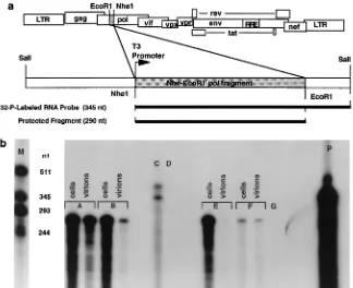
RNase protection assays.A 290-nt EcoRI-NheI fragment of HIV-2KRgag was cloned in antisense orientation to the T3 promoter, and a riboprobe was in vitro transcribed with 6.25mM [32P]UTP (800 Ci/mmol), 250mM each of the three other ribonucleoside triphosphates (rNTPs), placental RNase inhibitor, dithio-threitol (5 mM), 0.5 ng of plasmid template linearized at the SalI site, and T3 polymerase (see Fig. 3A). Virions were isolated for RNase protection by clearing supernatants with two low-speed centrifugations at 450 and 1,2003g, followed
by passage through a 0.2-mm-pore-size filter and ultracentrifugation at 50,0003 g for 90 min at 4°C. Riboprobes were mixed with various portions of RNase-free
DNase-treated total cellular RNA or virion RNA from each transfection; in each case, the fractions of cellular and virion RNA used were equal. RNA was isolated by the guanidium isothiocyanate method. Riboprobes and RNAs were co-etha-nol precipitated, heat denatured at 95°C for 5 min and annealed at 68°C for 10 to 20 min in a thermal block, digested at 37°C for 45 min with a mixture of RNase A and RNase T1, reprecipitated, and electrophoresed in a denaturing 8 M urea–5% polyacrylamide gel. The assay was linear over 5 orders of magnitude.
PCR and Southern blotting for detection of HIV-2 pol sequences.Genomic DNA from heavily transduced cells was prepared by proteinase K digestion and organic extraction. One microgram of this genomic DNA was added to all tubes except for the PCR mix negative control, with or without added genomic DNA from HIV-2-infected T cells as internal standards. Reactions were subjected to
on November 9, 2019 by guest
http://jvi.asm.org/
25 cycles of amplification in 100-ml PCR mixtures with Taq DNA polymerase, 1.5 mM MgCl2, 200mM dNTPs, and outer HIV-2 pol primers (cctacttctagagaagcct ggg and gtgcccatatatatcctgattcc), followed by transfer of 10ml of this reaction mixture to a 100-ml PCR mixture containing inner (cttaaggccccacctcctgagg and cttcttgccagattccctcc) primers and 40 cycles of amplification; 10ml of each product was subjected to electrophoresis in 1.5% agarose followed by overnight alkaline Southern transfer to a nylon membrane and hybridization to a randomly primed, internal32P-labeled HIV-2 pol probe.
Transfections and production of pseudotyped vectors.Lipofection of T-cell lines was performed with DOTAP (Boehringer Mannheim), using 30 mg of proviral DNA. COS-1 cells were electroporated at 250 V in a Bio-Rad Gene-Pulser. 293-T cells seeded the day before in Dulbecco modified Eagle medium (DMEM) supplemented with 10% fetal calf serum (FCS), glutamine, penicillin, and streptomycin were transfected by calcium phosphate coprecipitation using a 2:3:1 weight ratio of HIV-2 packaging, HIV-2 vector, and VSV-G expression plasmids with a total of 35 to 50mg of DNA per 75-cm2flask. Medium was replaced 8 to 16 h after transfection, and supernatant was harvested once or twice between 48 and 96 h. LZRNL vector was prepared similarly by calcium phosphate transfection of pHCMV-G in 293GPLZRNL cells (7). HIV-2 and LZRNL vector supernatants were precleared by two 10-min centrifugations at 435 and 1,2003g and filtered through a 0.45-mm-pore-size filter. p26 antigen in unconcentrated supernatants measured by antigen capture enzyme-linked im-munosorbent assay (Coulter) averaged 90 to 120 ng, with peak values greater than 400 ng/ml. To produce concentrated stocks of vesicular stomatitis virus G protein (VSV-G)-pseudotyped vectors, cleared, filtered supernatants were ultra-centrifuged at 50,0003g for 90 to 120 min at 4°C. The viral pellet was
resus-pended 4 h to overnight at 4°C in 50 mM Tris (pH 7.8)–130 mM NaCl–1 mM EDTA or DMEM with 1% fetal bovine serum. DNase treatment of vectors was performed with 50 U of RNase-free DNase I (Boehringer Mannheim) per ml for 2 h at 37°C.
Transductions and vector titrations. HeLa cells (53104per well) were seeded in 12-well plates and incubated for 4 to 16 h with vector supernatants supplemented with 4 to 6mg of Polybrene per ml. The medium was replaced, and staining was performed at 48 to 60 h by fixing cells in 1% formaldehyde–0.2% glutaraldehyde in phosphate-buffered saline (PBS) for 5 min, washing them twice with PBS, and quenching aldehydes with a 0.1 M glycine rinse, followed by incubation for 2 to 8 h in 5-bromo-4-chloro-3-indolyl-b-D-galactopyranoside
(X-Gal) staining medium (0.4 mg of X-Gal per ml, 2 mM MgCl2, 6 mM potassium ferrocyanide, and 6 mM potassium ferricyanide in PBS). Titers were calculated as the number of blue-staining foci divided by the dilution factor. For transduc-tion of U937 cells, 4mg of Polybrene per ml was used, and flow cytometric analysis of GFP expression was performed at 48 h (Becton Dickinson Immuno-cytometry Systems [BDIS], San Jose, Calif.).
Growth arrest.HeLa cells were arrested in G1/S phase with the DNA poly-merasea/dinhibitor aphidicolin (24), which was maintained at 20mg/ml during transduction and replenished daily until staining forb-galactosidase expression. Fluorescence-activated cell sorting (FACS) analysis of propidium iodide-stained cells at 24 h confirmed complete G1/S arrest of the aphidicolin-treated cells.
NTN2 neurons.Third-replate NTN2 neurons (Stratagene) were plated at 104 per well in 48-well plates (precoated with Matrigel basement membrane matrix) for 10 days to 2 weeks in neuron-conditioned medium containing mitotic inhib-itors (1mM cytosine arabinoside, 10mM fluorodeoxyuridine, and 10mM uri-dine), which was replenished every 2 days. The cells displayed prominent neurite extension by 24 to 36 h and remained viable for over 4 weeks. Vectors were diluted for titration in medium containing the mitotic inhibitors plus Polybrene (4mg/ml). Cells were stained for 4 h with X-Gal at 72 h after transduction. No background X-Gal staining of untransduced NTN2 cells was seen, even if cells were stained for 1 week. Heat treatment (56°C, 40 min), leaving the VSV-G expression plasmid pHCMV-G out of the producer cell transfection, or addition of zidovudine (10mM) eliminated transduction.
Monocyte-derived macrophages.Primary human macrophages were prepared from Ficoll-purified human peripheral blood mononuclear cells from normal donors by adherence to plastic as described previously (30). After initial adher-ence to fibronectin, cells were plated at 105per ml in plastic chamber slides (Nunc) in RPMI with 15% FCS and 5% autologous donor serum. The chambers were washed four times by vigorous pipetting with PBS to remove all nonadher-ent cells on days 4, 6, and 7 after plating and before transduction on day 10. The cells were.99% nonspecific esterase positive (Sigma kit 90-A1); 48 h after transduction with serial dilutions of GFP-encoding vector in the presence of Polybrene (4mg/ml), slides were washed with PBS and scored by epifluorescence microscopy.
CD341cell isolation, purification, PKH26-GL staining, and transduction. Human CD341cells were isolated from growth factor-mobilized peripheral blood as described previously (31). Briefly, five daily injections of granulocyte-macrophage and granulocyte colony-stimulating factors (GM-CSF and G-CSF; each at 5mg/kg/day) were administered; leukapheresis was performed 24 h after the last growth factor injection. CD341cells were purified from leukapheresis products by immunomagnetic separation (Isolex-300; Baxter Immunotherapy, Irvine, Calif.) and cryopreserved by controlled-rate freezing in RPMI with 10% dimethyl sulfoxide. Purity of the CD341cells was 94% by FACS analysis per-formed as described previously (31).
For PKH26-GL staining, 63106to 103106frozen CD341cells were thawed
at 37°C and washed twice with Iscove modified DMEM (Gibco-BRL). Cells were resuspended in serum-free medium and stained with PKH26-GL (Sigma, St. Louis, Mo.) according to the manufacturer’s instructions. Equivalent aliquots of CD341cells not treated with PKH26-GL were used for background control measurements in the subsequent FACS analyses. Four hours after PKH26-GL staining, 106cells were transduced with either Mo-MLV or HIV-2 DNase I (Boehringer Mannheim)-treated (50 U/ml for 60 min at 37°C) lacZ vectors at a multiplicity of infection (MOI) of 1.0 in a total volume of 1.5 ml of Iscove modified with 10% FCS supplemented with Polybrene (4mg/ml) and dNTPs (100
mM each), with or without cytokine stimulation (interleukin-3 [IL-3; 500 U/ml], IL-6 [500 U/ml], stem cell factor [40 ng/ml], GM-CSF [10 ng/ml], basic fibroblast growth factor [2.5 ng/ml], and erythropoietin [2.5 U/ml]). Cells were centrifuged in vector supernatants at 2,7003g for 30 min and incubated at 37°C for 16 to
24 h. Following transduction, the cells were washed five times and then cultured in the dark for a further 48 h with or without the above cytokines. Two aliquots of PKH26-GL-treated or untreated CD341cells were also cultured under the same conditions except for addition of the vectors.
CD34/38 labeling, three-color flow cytometry, and vector DNA detection in hematopoietic progenitor cells.At 48 h after transduction, PKH26-GL-stained or unstained CD341 cells were labeled with CD34-fluorescein isothiocyanate (BDIS) and/or CD38-Cy-chrome (Pharmigen, San Diego, Calif.) (31). Flow cy-tometric analysis and sorting of CD341cells were performed on a FACStarplus flow cytometer (BDIS) equipped with an argon-ion laser tuned at 488 nm. Data acquisition was performed with Lysis 2.0 (BDIS). Forward light scatter, orthog-onal light scatter, and three-color fluorescent signals were determined for each cell, and the list mode data files were analyzed with Cut-a-Cluster software (BDIS). Cells were sorted into Falcon tubes containing Tris-EDTA (pH 8.0), 100 mM NaCl, proteinase K (200mg/ml), and 1% sodium dodecyl sulfate. DNA was isolated by organic extraction and ethanol precipitation with glycogen as a carrier and amplified in parallel with a lacZ standard curve for 35 cycles (94, 60, and 72°C, 30 s each plateau) in 100-ml PCRs all prepared from the same mix con-taining 1.5 mM MgCl2, 200mM dNTPs, 0.5mM lacZ PCR primers (CCTTTG CGAATACGCCCACGCGATGGG and CGTACTGTGAGCCAGAGTTGCC CGGCGC), and 0.5 U of Taq polymerase. DNAs from each individual sort were adjusted to 200 cell equivalents per tube, and input DNA equivalence was assessed by PCR with humanb-globin gene primers (53) in parallel with a human genomic DNA standard curve; in addition, 100-ml aliquots of each DNase-treated vector supernatant were simultaneously extracted and amplified to assess completeness of DNase treatment. A 5-ml aliquot of each PCR product was subjected to electrophoresis in 1% agarose followed by overnight alkaline South-ern transfer and hybridization to a randomly primed, intSouth-ernal (32P)-labeled lacZ or humanb-globin probe under standard conditions.
RESULTS
Deletion in HIV-2
c
abrogates replication.
The
three-plas-mid system used in these studies (Fig. 1) was designed to
express HIV-2 proteins except Env in trans from an mRNA
that will not be encapsidated. Since the packaging
determi-nants of HIV-2 are not well defined, the effect of a mutation in
HIV-2
c
on encapsidation and expression was first examined.
HIV-2
c
is 70% longer than HIV-1
c
(75 versus 44 nt). To
maximally disrupt the potential HIV-2 encapsidation signal
while not interfering with splicing or gag translation, 61 nt were
deleted from this region (Fig. 1); the deletion extends from 11
nt downstream of the major SD to 3 nt upstream of the gag
start codon. As shown in Fig. 2, removal of the 61 nt blocked
replication of HIV-2
KRbut did not interfere with transient
expression. Transient transfection of a proviral plasmid
carry-ing the 61-bp
c
deletion alone (pE32
Dc
) or the
c
deletion plus
a truncated 3
9
LTR (pE40) into T-cell lines highly permissive
for the parental virus resulted in high but transient Gag protein
(p26) expression and marked transient syncytium formation
(HIV-2
KRis syncytium inducing in human T-cell lines).
How-ever, viral replication was abrogated (followed out to 6 months
as illustrated). In addition, transfer of 30 ml of supernatant
containing 210 ng/ml of p26 antigen from COS-1 cells
electro-porated with pE32
Dc
to a culture of 10
7CD4-LTR/
b
-gal
in-dicator cells (28) was negative for any
b
-galactosidase-express-ing cells when stained at 2, 7, and 33 days after transfer. In
contrast, 0.005 ng of viral antigen per ml from
pE32-trans-fected cells was detected in this assay. Potential transfer of
coding sequences by pseudotyped vectors was later examined
by a PCR-based assay (below).
on November 9, 2019 by guest
http://jvi.asm.org/
Deletion in HIV-2
c
prevents encapsidation.
Although these
experiments demonstrated that deletion of 61 of 75 bp in
c
permitted wild-type levels of viral gene expression while
pre-venting both HIV-2 replication and transmission of coding
sequences to target cells, they did not specifically measure the
effect of the deletion on HIV-2 genomic encapsidation. An
RNase protection assay was then used to directly compare
levels of intracellular HIV-2 genomic RNA and of HIV-2
vi-rion genomic RNA in supernatants of transfected cells. As
shown in Fig. 3b, the ratio of virion to cellular genomic mRNA
was markedly reduced by the
c
deletion alone (compare lanes
A and B) or the
c
deletion in combination with the env and 3
9
LTR deletions of pE41 (compare lanes E and F). These results
identify the
c
region of HIV-2 as a determinant of genomic
encapsidation.
HIV-2 expression by fully modified packaging plasmid.
In
addition to the
c
deletion, several other attenuating
modifica-tions were made in constructing the packaging plasmid (pE41)
used for packaging VSV-G-pseudotyped vectors (Fig. 1).
HIV-2 sequences were terminated precisely at the stop codon of the
nef gene (pE40 retained a short stretch of the 3
9
U3). The 3
9
LTR was replaced with the bovine growth hormone
polyade-nylation signal, and a 776-bp span of env that encompasses the
V3 loop was deleted. Figure 4 shows that high levels of p26
antigen were produced from transient transfection of these
modified expression plasmids; in either COS-1 or human 293-T
cells, 100 to 400 ng of p26 per ml was routinely generated.
Figure 3 (lane E) shows the comparatively negligible levels of
HIV-2 gag RNA present in pelleted virions from these cells.
Transduction of dividing and nondividing HeLa cells.
Three
HIV-2 vectors were used in this study (Fig. 1). HeLa cells were
transduced with VSV-G-pseudotyped lacZ-encoding HIV-2
vector L15.7 prepared by triple cotransfection in 293-T cells
(see Materials and Methods). lacZ titers scored 48 h after
transduction for unconcentrated and
ultracentrifuge-concen-FIG. 1. Schematic representation of the HIV-2 lentivirus vector system. (A) Nucleotides deleted from thecregion of HIV-2KR.; (B) the protein expression plasmid used for trans packaging and the VSV-G expression plasmid supplying the deleted env function; (C) HIV-2-based vectors. Abbreviations: BGH, bovine growth hormone; CMV, cytomegalovirus.
FIG. 2.cdeletion alone abrogates replication but preserves transient HIV-2 protein expression. HIV-2KRproviral plasmids pE32Dcand pE40 were trans-fected into Molt4-8 T cells. The ordinate cutoff is 10 pg of p26/ml, the limit of sensitivity of the assay.
on November 9, 2019 by guest
http://jvi.asm.org/
[image:4.612.102.495.71.385.2] [image:4.612.310.549.527.692.2]trated supernatants are shown in Table 1. When HeLa cells
were transduced as in the experiments reported in Table 1 but
subsequently allowed to proliferate for 2 weeks, staining for
b
-galactosidase expression yielded uniformly blue-staining
colonies of several hundred cells at titers 85 to 90% of those
scored at 48 h, indicating stable, clonal maintenance of the
transgene.
To examine the ability of vector L15.7 to transduce
nondi-viding cells and compare this ability with that of a conventional
retrovirus vector, HeLa cells were arrested in the G
1/S phase
with the DNA polymerase
a
/
d
inhibitor aphidicolin (24) (20
m
g/ml), which was maintained during transduction and
replen-ished daily until staining for
b
-galactosidase. FACS analysis of
propidium iodide-stained cells at 24 h confirmed complete
G
1/S arrest of the aphidicolin-treated cells (data not shown).
Cell counts also showed that no cell proliferation occurred
during the 4 days of aphidicolin exposure until X-Gal staining.
By 24 h into aphidicolin treatment, cells were transduced with
serial dilutions of Mo-MuLV lacZ retroviral vector LZRNL
(VSV-G) or HIV-2 vector L15.7(VSV-G) for 4 h in the
pres-ence of Polybrene (4
m
g/ml); 48 h after transduction, lacZ
titers were scored. Figure 5 shows that aphidicolin markedly
reduced the ability of the LZRNL vector to transduce HeLa
cells whereas the HIV-2 vector was only minimally affected.
That nondividing cells were transduced could be confirmed
visually also: aphidicolin-arrested HIV-2 vector-transduced
cells were uniformly present as clearly isolated single blue cells,
whereas nonarrested transduced cells had proliferated into
colonies of 4 to 16 blue-staining cells.
[image:5.612.137.461.68.332.2]Lack of transfer of coding sequences by vector.
To test for
transfer of coding sequences by pE41-packaged vector, 5
3
10
4log-phase HeLa cells were transduced at an MOI of 10 with
DNase-treated L15.7 vector, yielding
.
98% transduction as
assessed by X-Gal staining of 10% of the cells at 60 h. The
remaining 90% were expanded for 15 days (four passages) and
genomic DNA was prepared by proteinase K digestion, organic
extraction, and ethanol precipitation. As shown in Fig. 6, 1
m
g
of this genomic DNA was negative by a sensitive, nested PCR/
Southern blot assay for a 319-bp segment of pol, while
simul-taneous amplification in the same assay of the same amount of
FIG. 3. RNase protection assay comparing amounts of intracellular HIV-2 genomic mRNA and of virion genomic mRNA. RNAs were harvested from 23106 COS-1 cells, and from pelleted virions from the cell supernatants, 48 h after electroporation of 10mg of plasmid DNA. RNAs were treated with 20 U of RNase-free DNase I for 4 h at 37°C and analyzed as described in Materials and Methods. (a) Probe design and expected fragments. (b) Lane M,32P-labeled RNA markers in vitro transcribed from templates of known size. Plasmids electroporated were pE32 (wild-type full-length HIV-2; lanes A), pE32Dc(lanes B), and pE41 lanes E. Lanes F, separate transfection of pE32 (wild type). Results for cellular (lane C) and virion RNA (lane D) controls from COS-1 cells electroporated with a plasmid expressing only the probe sequence in sense orientation from the SV40 promoter are also shown. Lane P, free probe minus RNase (10% of the amount added to other samples to avoid overloading autoradiogram); unmarked lane just left of P, 100% of free probe added to other samples plus RNase; lane G, untransfected COS-1 cell RNA control. The sense transcript controls in lanes C and D indicate that substantial amounts of cellular RNA were not nonspecifically pelleted but the small amount of RNA measured in theDc(B, virions) and pE41 (E, virions) virion samples may in part represent cosedimented 0.2-mm-pore-size-filterable RNA-containing subcellular fragments in addition to encapsidated RNA.
FIG. 4. p26 antigen production at 48 h in supernatants of COS-1 cells elec-troporated with HIV-2 expression plasmids. Bars indicate standard errors.
on November 9, 2019 by guest
http://jvi.asm.org/
[image:5.612.325.531.612.707.2]this DNA spiked with genomic DNA from as few as five cells
from a chronically HIV-2-infected Molt4 T-cell line was
posi-tive. To this limit of sensitivity, therefore, VSV-G-pseudotyped
vectors generated using pE41 did not transfer HIV-2 coding
sequences to target cells. In addition, these transduced cells
also produced no detectable p26 antigen when assayed at 1 and
3 weeks after transduction. Finally, after continued passage for
3 weeks, 50 ml of filtered supernatant from 10
7log-phase cells
was transferred to 5
3
10
6CD4-LTR/beta-gal cells, which were
negative by X-Gal staining at 96 h.
Transduction of T and monocytoid cell lines.
GFP-express-ing HIV-2 vectors were constructed to study transduction of T
cells and monocytes. Vector LGFP employs internally encoded
HIV-2 tat transactivation of the HIV-2 LTR to promote
tran-scription of a Nef/GFP fusion protein. This fusion protein
contains the 5
9
nef myristoylation signal and localizes to
cyto-plasmic vesicles with the same distribution as the Nef protein
(data not shown). VSV-G-pseudotyped LGFP previously
ti-tered on HeLa cells was used to transduce a T-cell line (Molt4)
and a monocytoid cell line (U937). Flow cytometric analysis for
GFP expression 48 h after transduction is shown in Fig. 7.
[image:6.612.310.552.67.222.2]Transduction of human macrophages and NTN2 neurons.
NTN2 neurons and monocyte-derived macrophages were
plat-ed as describplat-ed in Materials and Methods and transducplat-ed with
HIV-2 vectors. NTN2 neurons were plated on a Matrigel
base-ment membrane matrix in the presence of mitotic inhibitors
for 10 days and transduced with vector L15.7. As shown in
Table 2, these postmitotic human neuronal cells (13, 14, 29, 48,
49) were efficiently transduced by the HIV-2 vector but not by
the control Mo-MuLV lacZ vector.
Background
b
-galactosidase staining was seen in human
monocyte-derived macrophages from some donors (data not
shown). Therefore, vector pLACG was used to transduce
mac-rophages. Because this vector contains an internally promoted
gfp gene in reverse orientation to the HIV-2 LTR, the RRE is
positioned downstream of the marker gene cassette and a
sep-arate polyadenylation signal is used for gfp. As shown in
Ta-ble 2, titers on human macrophages exceeding 10
5/ml were
achieved; in contrast, Mo-MuLV vector transduction of
mac-rophages was negligible.
CD34
1human hematopoietic progenitor cell transduction.
Purified human CD34
1hematopoietic progenitor cells
isolat-ed from growth factor-mobilizisolat-ed peripheral blood were
trans-duced at an MOI of 1.0 for 16 to 24 h with pretitered
Mo-MuLV LacZ vector (LZRNL) or HIV-2-based LacZ vector
L15.7 in the presence or absence of stimulatory cytokines (see
Materials and Methods); after washing and 48 h of subsequent
culture, the cells were further stratified according to CD38
ex-pression status and proliferation index. To distinguish dividing
and nondividing cells, cells were stained with the lipophilic
membrane-fluorescent tracking dye PKH26-GL prior to
trans-duction. PKH26-GL has been used to accurately track the
mi-totic history of hematopoietic cells since partitioning between
daughter cells reduces its fluorescence intensity by one-half
with each cell division; the dye does not exchange
spontane-ously between labeled and unlabeled cells and does not
iden-tifiably alter hematopoietic cell physical properties or function
in vivo (32, 59). The PKH26-GL staining profiles of CD34
1cells were not affected by exposure to either vector as analyzed
by flow cytometry (data not shown). Three-color FACS
anal-ysis showed that 2 to 5% of CD34
1cells maintained a CD38
2phenotype after 72 h in culture and 48 h posttransduction (data
not shown). Among these, 12 to 17% had undergone zero to
one cell division, while 83 to 88% of CD34
1CD38
2cells had
undergone more extensive cell divisions and had diminished
PKH26-GL content.
The two CD34
1CD38
2cell subsets with the highest and
lowest PKH26-GL staining, corresponding to cells with low
and high proliferative indices, were sorted and collected
sep-arately for DNA extraction. The samples were then analyzed
by PCR and Southern blotting for the presence of the lacZ
transgene to determine transduction by VSV-G-pseudotyped
HIV-2 lacZ and LNL lacZ vectors. Results are shown in Fig. 8.
In one experiment, the vector transduction and subsequent
cell culture were carried out in the presence or absence of a
stimulatory cytokine cocktail culture consisting of IL-3, IL-6,
GM-CSF, basic fibroblast growth factor, stem cell factor, and
erythropoietin.
b
-Globin sequences were amplified as internal
controls to verify equivalent DNA input. As shown in Fig. 8A,
Mo-MuLV vector DNA was observed in the PKH26
lo(actively
dividing) cells cultured in the presence of cytokines and to a
FIG. 5. Effect of mitotic arrest on an HIV-2 lentivirus vector compared to an Mo-MuLV retrovirus vector. HeLa cells were arrested in the G1/S phase by treatment with 20mg of aphidicolin (aphid.) per ml; cell cycle arrest (,0.1% G2/M) was verified by flow cytometry after propidium iodide staining.
FIG. 6. Assay for transfer of HIV-2 coding sequences. Nested PCR amplifi-cations followed by Southern blotting with an internal32P-labeled pol probe were performed with 1mg of genomic DNA (present in all tubes except PCR blank in lane 1) from HeLa cells transduced at a high MOI, yielding an efficiency of
.98% as described in the text. Lanes: 1, PCR without genomic DNA; 2 to 5, reactions containing 1 mg of genomic DNA from transduced cells; 6 to 12, reactions containing 1mg of the same DNA from the L15.7-transduced cells coamplified with various cell equivalents of genomic DNA prepared from HIV-2-infected T cells: lane 6, 1 cell; lane 7, 5 cells; lane 8, 50 cells; lane 9, 100 cells; lane 10, 500 cells; lane 12, 1,000 cells.
TABLE 1. HIV-2 vector transduction of dividing HeLa cells
aAssay Transduction
Unconcentrated Concentrated (107)
1
7.8
3
10
52.3
2
1.2
3
10
62.2
3
7.7
3
10
58.5
4
6.9
3
10
59.9
Mean
(8.6
6
2.3)
3
10
55.7
6
4.1
aValues are mean transducing units per milliliter6standard deviation. VSV-G-pseudotyped vector pL15.7 supernatants were cleared by two low-speed cen-trifugations and 0.45-mm-pore-size filtration and titered directly (unconcen-trated) and after two rounds of ultracentrifugation at 50,0003g (concentrated).
on November 9, 2019 by guest
http://jvi.asm.org/
[image:6.612.51.290.81.162.2]lesser extent in the PKH26
losubset derived from
nonstimu-lated cells. Most notably, no vector DNA was detected in cells
from the PKH26
hisubsets (not actively dividing). In contrast,
HIV-2 vector DNA could be detected in all four dividing and
nondividing cell populations. Based on the LacZ standards and
input total cell DNA, we estimate
.
1 copy of vector DNA per
cell. Figure 8B presents results of an experiment in which
cytokines were included in the in vitro cell culture. Again, the
Mo-MLV vector showed restricted capacity to transduce the
CD38
2cells, while HIV-2 vectors transduced both PKH26
hiand PKH26
losubsets efficiently (see the legend to Fig. 8). In
both of these experiments, DNase-treated vector supernatants
used for transduction were negative for the transgene
se-quences (Fig. 8A and B, lanes M and H), indicating that the
detected DNA was not carryover DNA. We did not detect
expression of the lacZ reported gene in these cells. It is not
clear whether this was due to lack of promoter activity in these
cells or lack of proviral DNA integration. Unfortunately, the
number of transduced cells was too low to allow us to examine
the latter issue.
DISCUSSION
We report a replication-defective, three-plasmid lentivirus
vector system derived from a parental lentivirus with
demon-strated apathogenicity in a standard animal model. (SIV-derived
FIG. 7. Flow cytometric analysis for GFP expression in a T-cell line (Molt4; A) and a monocytoid cell line (U937; B) 48 h after transduction with an HIV-2 GFP vector (MOI51.0). Solid lines, untransduced control cells; dashed lines, transduced cells. Fl., fluorescence.
on November 9, 2019 by guest
http://jvi.asm.org/
vectors have been reported, but HIV-1 virions were used to
package them [52]). Similar to 1-based vectors, the
HIV-2-based vectors can transduce both dividing and nondividing
cells at high efficiency. Consistent with a large body of
pre-vious studies, transduction by Mo-MuLV vectors in this
study was restricted to dividing cells. In addition to high
efficiency on dividing cell lines, HIV-2 vectors efficiently
trans-duced aphidicolin-arrested cells, primary macrophages, and
postmitotic NTN2 neurons. Concentration of the
VSV-G-pseu-dotyped vectors by ultracentrifugation was readily achieved.
HIV-1-based vectors in previous studies have included true
vectors, in which viral structural proteins are supplied fully in
trans (46), and simpler systems that employ modified HIV-1
proviruses in which the vector itself supplies one or more of the
viral structural proteins in cis. The HIV-2-based vector system
described here is of the former type and contains additional
deletions of 5
9
and 3
9
cis-acting regions illustrated in Fig. 1.
Safety issues will require extensive investigation for gene
therapy vectors derived from primate lentiviruses (16). A
three-plasmid HIV-1 vector system deleted in multiple
acces-sory genes has also been described and may lessen risk (66),
although some cell types may require more than Gag/Pol and
Rev. For example, hepatocytes in vivo were transduced
effi-ciently in vivo only if Vpr and Vif were supplied (25). In
con-trast to HIV-1 vectors, safety of HIV-2 vectors can be
ad-dressed by studies in primates susceptible to pathogenicity.
Although infection of macaques with HIV-2
KRcan be achieved,
the animals have remained free of symptoms or disease for
more than 2 years; matched animals infected with HIV-2
EHOrapidly developed AIDS; interestingly, prior infection with
HIV-2
KReither delayed or prevented disease induction by
HIV-2
EHO(38). Deletion of HIV-2 accessory genes (e.g., vpr,
vpx, vif, and nef) is under investigation. We have minimized risk
for recombination (42) by distributing viral functions to
sepa-rate DNAs, avoiding large regions of homologous sequence
overlap, and showing that packaging of the mRNA coding for
viral proteins is minimal. In addition, transfer of coding
se-quences to target cells was not detected.
The nature of the HIV-2 packaging signal has received
lim-ited study and produced conflicting results (19, 41, 52). Our
results suggest that, as for HIV-1, deleting most of the region
between the major SD and the gag start codon abrogates
rep-lication, prevents incorporation of HIV-2 genomes into viral
particles, and, in combination with the env deletion and
LTR modifications, prevents detectable transfer of coding
sequences to heavily transduced target cells. These results are
consistent with the results of McCann and Lever, who reported
that deletions of up to 40 of the 75 nt (53%) in HIV-2
c
reduced encapsidation by only 33 to 70% (41); a larger
dele-tion, such as that of the present study (61 nt, 83%), would
appear to be required to reduce HIV-2 genomic mRNA
en-capsidation to nonspecific levels. Additional env and LTR
de-letions make regeneration of wild type-HIV-2 impossible.
Sta-ble HIV-1 packaging lines using the native HIV-1 envelope
have been described (8, 15, 54).
[image:8.612.50.289.98.183.2]The CD38
2subset of human hematopoietic progenitors
contains cells capable of both multilineage differentiation and
long-term repopulating ability. Up to a third of CD38
2CD34
1cells remain in G
0/G
1phase after cytokine stimulation as used
in our study (1). Numerous studies have shown that
conven-tional murine leukemia virus-based vectors cannot transduce
this subset of hematopoietic precursors in vitro or permit
ef-ficient chimeric reconstitution of NOD/SCID mice with
trans-duced human CD34
1cells (33). Our results with HIV-2
vec-tors suggest that the ability of lentiviruses to infect nondividing
cells may extend to both cycling and noncycling CD38
2CD34
1human hematopoietic progenitor cells. A clear difference
between the murine vector and the lentivirus vector in the
infective phase up to the point of proviral DNA formation is
indicated by the experiments in Fig. 8. Expression of either
[image:8.612.52.546.557.666.2]FIG. 8. PCR analyses of transduced CD341cell subsets for lacZ andb-globin DNA (single-copy cellular gene DNA input control). Cells were transduced with DNase-treated vectors and sorted by flow cytometry into CD341CD382and PKH26hior PKH26lofractions before DNA extraction, PCR, and Southern blotting with an internal lacZ orb-globin probe. (A) Analysis of cells FACS sorted as CD341CD382and PKH26hior PKH26loafter transduction in the presence (1) or absence (2) of cytokines as detailed in Materials and Methods. (B) Cells were transduced with each DNase-treated vector in the presence of cytokines and similarly sorted. LacZ standards (lanes a to h): 0, 1, 4, 16, 64, 256, 1,024, and 4,096 copies of the lacZ gene;b-globin standards (lanes a to f): genomic DNA equivalent to 0, 1, 5, 50, 500, and 5,000 U937 cells.b-globin PCRs for both experiments were amplified simultaneously; the standard curve is shown in the bottom panel of B. P, PCR blank; M, DNase-treated Mo-MuLV (LZRNL) vector supernatant; H, DNase-treated HIV-2 lacZ vector supernatant.
TABLE 2. Comparative transduction of monocyte-derived
macrophages and NTN2 neurons by Mo-MuLV
and HIV-2 vectors
aCell type Transduction
Mo-MuLV vector LNL6-GFP
HIV-2 vector LACG
HeLa
(1.0
6
0.6)
3
10
6(1.0
6
0.8)
3
10
6MDM
,
1
3
10
1(5.4
6
1.1)
3
10
5Mo-MuLV vector LZRNL
HIV-2 vector L15.7
HeLa
(0.8
6
0.3)
3
10
6(1.1
6
0.2)
3
10
6NTN2
,
1
3
10
1(2.3
6
1.7)
3
10
5aValues are mean transducing units per milliliter6standard deviation from three experiments. HeLa cells and third-replate NTN2 neurons were transduced with pretitered HIV-2 vector pL15.7 and Mo-MuLV vector LZRNL; monocyte-derived macrophages (MDM) were transduced with HIV-2 vector pLAGC, and an Mo-MuLV GFP-encoding vector (LNL6-GFP) and scored by epifluorescence microscopy at 48 h. No background X-Gal staining of untransduced NTN2 cells occurred, even if cells were stained for 1 week. Heat treatment (56°C, 40 min), leaving the VSV-G expression plasmid pHCMV-G out of the producer cell transfection, or addition of 10mM zidovudine reduced transduction of macro-phages and NTN2 neurons to,1/ml.
on November 9, 2019 by guest
http://jvi.asm.org/
b
-galactosidase or GFP could not be detected in these cells,
and hence the completeness of the transduction process
re-mains uncertain. It is not clear if these results represent
tran-scriptional shutoff or lack of integration in these primitive
hematopoietic progenitors, since unintegrated retroviral DNA,
both that of lentiviruses and that of murine retroviruses, is
transcriptionally silent (11, 46). In lieu of reporter gene
expres-sion, inverse PCR assays to detect integrated proviruses were
performed but were insufficiently sensitive to verify integrated
proviral DNA from the low number of cells remaining after the
repeated sorting for CD38 negativity (data not shown).
Meth-odological constraints thus limit the conclusions that can be
drawn about the integration state from the human CD34
1cell
experiments because transgene expression could not be
de-tected and because the low numbers of cells obtainable after
two rounds of sorting prevented demonstration of viral-cellular
DNA junctions. Although Fig. 8 shows a clear difference
be-tween the abilities of the Mo-MuLV vector and the HIV-2
vector to generate proviral DNA in the CD38
2cells, it may be
that hematopoietic stem cells with both pluripotent
differenti-ation capacity and self-renewal capacity will harbor blocks to
lentivirus vectors analogous to those seen for HIV-1 in resting
G
0T cells (55, 56, 58, 64). To prove stable gene transfer to
functional, repopulating stem cells, hematopoietic
reconstitu-tion with vector-transduced cells in an in vivo model will be
required.
ACKNOWLEDGMENTS
This work was supported in part by NIH grants 1U19 AI3661203
(SPIRAT), 3K12DK01408-10S1, and 2P30AI3621404 (CFAR).
We thank T. Friedmann for supplying pHCMV-G and 293GPL
ZRNL cells and the UC San Diego Center for AIDS Research for
technical assistance.
REFERENCES
1. Agrawal, Y. P., R. S. Agrawal, A. M. Sinclair, D. Young, M. Maruyama, F.
Levine, and A. D. Ho. 1996. Cell-cycle kinetics and VSV-G pseudotyped retrovirus-mediated gene transfer in blood-derived CD341cells. Exp. He-matol. 24:738–747.
2. Baba, T. W., Y. S. Jeong, D. Pennick, R. Bronson, M. F. Greene, and R. M.
Ruprecht.1995. Pathogenicity of live, attenuated SIV after mucosal infection of neonatal macaques. Science 267:1820–1826.
3. Bender, M. A., T. D. Palmer, R. E. Gelinas, and A. D. Miller. 1987. Evidence that the packaging signal of Moloney murine leukemia virus extends into the
gag region. J. Virol. 61:1639–1646.
4. Berkowitz, R. D., and S. P. Goff. 1994. Analysis of binding elements in the human immunodeficiency virus type 1 genomic RNA and nucleocapsid pro-tein. Virology 202:233–246.
5. Berkowitz, R. D., M. L. Hammarskjold, C. Helga-Maria, D. Rekosh, and
S. P. Goff.1995. 59regions of HIV-1 RNAs are not sufficient for encapsida-tion: implications for the HIV-1 packaging signal. Virology 212:718–723. 6. Bukrinsky, M. I., S. Haggerty, M. P. Dempsey, N. Sharova, A. Adzhubel, L.
Spitz, P. Lewis, D. Goldfarb, M. Emerman, and M. Stevenson.1993. A nuclear localization signal within HIV-1 matrix protein that governs infec-tion of non-dividing cells. Nature 365:666–669.
7. Burns, J. C., T. Friedmann, W. Driever, M. Burrascano, and J. K. Yee. 1993. Vesicular stomatitis virus G glycoprotein pseudotyped retroviral vectors: concentration to very high titer and efficient gene transfer into mammalian and nonmammalian cells. Proc. Natl. Acad. Sci. USA 90:8033–8037. 8. Carroll, R., J.-T. Lin, E. J. Dacquel, J. D. Mosca, D. S. Burke, and D. C. St.
Louis.1994. A human immunodeficiency virus type 1 (HIV-1)-based retro-viral vector system utilizing stable HIV-1 packaging cell lines. J. Virol. 68: 6047–6051.
9. Chatterjee, P., A. Garzino-Demo, P. Swinney, and S. K. Arya. 1993. Human immunodeficiency virus type 2 multiply spliced transcripts. AIDS Res. Hum. Retroviruses 9:331–335.
10. Clever, J., C. Sassetti, and T. G. Parslow. 1995. RNA secondary structure and binding sites for gag gene products in the 59packaging signal of human immunodeficiency virus type 1. J. Virol. 69:2101–2109.
11. Coffin, J. M. 1992. Retroviral DNA integration. Dev. Biol. Stand. 76:141– 151.
12. Colombini, S., S. K. Arya, M. S. Reitz, L. Jagodzinski, B. Beaver, and F.
Wong-Staal. 1989. Structure of simian immunodeficiency virus regulatory
genes. Proc. Natl. Acad. Sci. USA 86:4813–4817.
13. Cook, D. G., M. S. Forman, J. C. Sung, S. Leight, D. L. Kolson, T. Iwatsubo,
V. M. Y. Lee, and R. W. Doms.1997. Alzheimer’s A beta(1-42) is generated in the endoplasmic reticulum/intermediate compartment of NT2N cells. Nat. Med. 3:1021–1023.
14. Cook, D. G., V. M. Lee, and R. W. Doms. 1994. Expression of foreign proteins in a human neuronal system. Methods Cell Biol. 43(Part A):289–303. 15. Corbeau, P., G. Kraus, and F. Wong-Staal. 1996. Efficient gene transfer by
a human immunodeficiency virus type 1 (HIV-1)-derived vector utilizing a stable HIV packaging cell line. Proc. Natl. Acad. Sci. USA 93:14070–14075. 16. Emerman, M. 1996. From curse to cure: HIV for gene therapy? Nat.
Bio-technol. 14:943.
17. Gallay, P., S. Swingler, C. Aiken, and D. Trono. 1995. HIV-1 infection of nondividing cells: C-terminal tyrosine phosphorylation of the viral matrix protein is a key regulator. Cell 80:379–388.
18. Gallay, P., S. Swingler, J. Song, F. Bushman, and D. Trono. 1995. HIV nuclear import is governed by the phosphotyrosine-mediated binding of matrix to the core domain of integrase. Cell 83:569–576.
19. Garzino-Demo, A., R. C. Gallo, and S. K. Arya. 1995. Human immunodefi-ciency virus type 2 (HIV-2): packaging signal and associated negative regu-latory element. Hum. Gene Ther. 6:177–184.
20. Geigenmuller, U., and M. L. Linial. 1996. Specific binding of human immu-nodeficiency virus type 1 (HIV-1) Gag-derived proteins to a 59HIV-1 ge-nomic RNA sequence. J. Virol. 70:667–671.
21. Heim, R., A. B. Cubitt, and R. Y. Tsien. 1995. Improved green fluorescence. Nature 373:663–664. (Letter.)
22. Heinzinger, N. K., M. I. Bukinsky, S. A. Haggerty, A. M. Ragland, V.
Kewal-ramani, M. A. Lee, H. E. Gendelman, L. Ratner, M. Stevenson, and M. Emerman.1994. The Vpr protein of human immunodeficiency virus type 1 influences nuclear localization of viral nucleic acids in nondividing host cells. Proc. Natl. Acad. Sci. USA 91:7311–7315.
23. Holyoake, T. L., and M. J. Alcorn. 1994. CD341positive haemopoietic cells: biology and clinical applications. Blood Rev. 8:113–124.
24. Huberman, J. A. 1981. New views of the biochemistry of eucaryotic DNA replication revealed by aphidicolin, an unusual inhibitor of DNA polymerase alpha. Cell 23:647–648.
25. Kafri, T., U. Blomer, D. A. Peterson, F. H. Gage, and I. M. Verma. 1997. Sustained expression of genes delivered directly into liver and muscle by lentiviral vectors. Nat. Genet. 17:314–317.
26. Kanki, P. J., K. U. Travers, S. Mboup, C. C. Hsieh, R. G. Marlink, N. A.
Gueye, T. Siby, I. Thior, M. Hernandez-Avila, J. L. Sankale, et al.1994. Slower heterosexual spread of HIV-2 than HIV-1. Lancet 343:943–946. 27. Kim, H. J., K. Lee, and J. J. O’Rear. 1994. A short sequence upstream of the
59major splice site is important for encapsidation of HIV-1 genomic RNA. Virology 198:336–340.
28. Kimpton, J., and M. Emerman. 1992. Detection of replication-competent and pseudotyped human immunodeficiency virus with a sensitive cell line on the basis of activation of an integratedb-galactosidase gene. J. Virol. 66: 2232–2239.
29. Kleppner, S. R., K. A. Robinson, J. Q. Trojanowski, and V. M. Lee. 1995. Transplanted human neurons derived from a teratocarcinoma cell line (NTera-2) mature, integrate, and survive for over 1 year in the nude mouse brain. J. Comp. Neurol. 357:618–632.
30. Kornbluth, R. S., P. S. Oh, J. R. Munis, P. H. Cleveland, and D. D. Richman. 1989. Interferons and bacterial lipopolysaccharide protect macrophages from productive infection by human immunodeficiency virus in vitro. J. Exp. Med. 169:1137–1151.
31. Lane, T. A., P. Law, M. Maruyama, D. Young, J. Burgess, M. Mullen, M.
Mealiffe, L. W. Terstappen, A. Hardwick, M. Moubayed, et al.1995. Har-vesting and enrichment of hematopoietic progenitor cells mobilized into the peripheral blood of normal donors by granulocyte-macrophage colony-stim-ulating factor (GM-CSF) or G-CSF: potential role in allogeneic marrow transplantation. Blood 85:275–282.
32. Lansdorp, P. M., and W. Dragowska. 1993. Maintenance of hematopoiesis in serum-free bone marrow cultures involves sequential recruitment of quies-cent progenitors. Exp. Hematol. 21:1321–1327.
33. Larochelle, A., J. Vormoor, H. Hanenberg, J. C. Wang, M. Bhatia, T.
Lapi-dot, T. Moritz, B. Murdoch, X. L. Xiao, I. Kato, D. A. Williams, and J. E. Dick.1996. Identification of primitive human hematopoietic cells capable of repopulating NOD/SCID mouse bone marrow: implications for gene ther-apy. Nat. Med. 2:1329–1337.
34. Lever, A., H. Gottlinger, W. Haseltine, and J. Sodroski. 1989. Identification of a sequence required for efficient packaging of human immunodeficiency virus type 1 RNA into virions. J. Virol. 63:4085–4087.
35. Lever, A. M. L. 1996. HIV and other lentivirus-based vectors. Gene Ther. 3: 470–471.
36. Lewis, P. F., and M. Emerman. 1994. Passage through mitosis is required for oncoretroviruses but not for the human immunodeficiency virus. J. Virol. 68: 510–576.
37. Linial, M. L., and A. D. Miller. 1990. Retroviral RNA packaging: sequence requirements and implications. Curr. Top. Microbiol. Immunol. 157:125– 152.
on November 9, 2019 by guest
http://jvi.asm.org/
38. Looney, D. J., J. McClure, S. Kent, A. Radaelli, G. Kraus, A. Schmidt, K.
Steffy, P. D. Greenberg, S. Hu, W. R. Morton, and F. Wong-Staal.1998. A minimally replicative HIV-2 live-virus vaccine protects M. nemestrina from disease after HIV-2287 challenge. Virology 242:150–160.
39. Marlink, R., P. Kanki, I. Thior, K. Travers, G. Eisen, T. Siby, I. Traore, C. C.
Hsieh, M. C. Dia, E. H. Gueye, et al.1994. Reduced rate of disease devel-opment after HIV-2 infection as compared to HIV-1. Science 265:1587– 1590.
40. McBride, M. S., and A. T. Panganiban. 1996. The human immunodeficiency virus type 1 encapsidation site is a multipartite RNA element composed of functional hairpin structures. J. Virol. 70:2963–2973.
41. McCann, E. M., and A. M. Lever. 1997. Location of cis-acting signals im-portant for RNA encapsidation in the leader sequence of human immuno-deficiency virus type 2. J. Virol. 71:4133–4137.
42. Miller, A. D. 1992. Retroviral vectors. Curr. Top. Microbiol. Immunol. 158: 1–24.
43. Miller, D. G., M. A. Adam, and A. D. Miller. 1990. Gene transfer by retro-virus vectors occurs only in cells that are actively replicating at the time of infection. Mol. Cell. Biol. 10:4239–4242. (Erratum, 12:443, 1992.) 44. Morrison, S. J., N. Uchida, and I. L. Weissman. 1995. The biology of
hematopoietic stem cells. Annu. Rev. Cell Dev. Biol. 11:35–71.
45. Naldini, L., U. Blomer, F. H. Gage, D. Trono, and I. M. Verma. 1996. Efficient transfer, integration, and sustained long-term expression of the transgene in adult rat brains injected with a lentiviral vector. Proc. Natl. Acad. Sci. USA 93:11382–11388.
46. Naldini, L., U. Blomer, P. Gallay, D. Ory, R. Mulligan, F. H. Gage, I. M.
Verma, and D. Trono.1996. In vivo gene delivery and stable transduction of nondividing cells by a lentiviral vector. Science 272:263–267.
47. Nienhuis, A. W. 1994. Gene transfer into hematopoietic stem cells. Blood Cells 20:141–147.
48. Pleasure, S. J., and V. M. Lee. 1993. NTera 2 cells: a human cell line which displays characteristics expected of a human committed neuronal progenitor cell. J. Neurosci. Res. 35:585–602.
49. Pleasure, S. J., C. Page, and V. M. Lee. 1992. Pure, postmitotic, polarized human neurons derived from NTera 2 cells provide a system for expressing exogenous proteins in terminally differentiated neurons. J. Neurosci. 12: 1802–1815.
50. Putkonen, P., L. Walther, Y. J. Zhang, S. L. Li, C. Nilsson, J. Albert, P.
Biberfeld, R. Thorstensson, and G. Biberfeld.1995. Long-term protection against SIV-induced disease in macaques vaccinated with a live attenuated HIV-2 vaccine. Nat. Med. 1:914–918.
51. Rasmussen, R., P. Sharma, Y. Hu, and R. Ruprecht. 1997. Presented at the 15th Annual Symposium on Non-Human Primate Models of AIDS, Seattle, Wash., Sept. 3 to 6.
52. Rizvi, T. A., and A. T. Panganiban. 1993. Simian immunodeficiency virus RNA is efficiently encapsidated by human immunodeficiency virus type 1 particles. J. Virol. 67:2681–2688.
53. Saiki, R. K., T. L. Bugawan, G. T. Horn, K. B. Mullis, and H. A. Erlich. 1986. Analysis of enzymatically amplified beta-globin and HLA-DQ alpha DNA with allele-specific oligonucleotide probes. Nature 324:163–166.
54. Srinivasakumar, N., N. Chazal, C. Helga-Maria, S. Prasad, M. L.
Hammar-skjold, and D. Rekosh.1997. The effect of viral regulatory protein expression on gene delivery by human immunodeficiency virus type 1 vectors produced in stable packaging cell lines. J. Virol. 71:5841–5848.
55. Stevenson, M., B. Brichacek, N. Heinzinger, S. Swindells, S. Pirruccello, E.
Janoff, and M. Emerman.1995. Molecular basis of cell cycle dependent HIV-1 replication. Implications for control of virus burden. Adv. Exp. Med. Biol. 374:33–45.
56. Stevenson, M., T. L. Stanwick, M. P. Dempsey, and C. A. Lamonica. 1990. HIV-1 replication is controlled at the level of T cell activation and proviral integration. EMBO J. 9:1551–1560.
57. Talbott, R., G. Kraus, D. Looney, and F. Wong-Staal. 1993. Mapping the determinants of human immunodeficiency virus 2 for infectivity, replication efficiency, and cytopathicity. Proc. Natl. Acad. Sci. USA 90:4226–4230. 58. Tang, S., B. Patterson, and J. A. Levy. 1995. Highly purified quiescent human
peripheral blood CD41T cells are infectible by human immunodeficiency virus but do not release virus after activation. J. Virol. 69:5659–5665. 59. Teare, G. F., P. K. Horan, S. E. Slezak, C. Smith, and J. B. Hay. 1991.
Long-term tracking of lymphocytes in vivo: the migration of PKH-labeled lymphocytes. Cell. Immunol. 134:157–170.
60. Terstappen, L. W., S. Huang, M. Safford, P. M. Lansdorp, and M. R. Loken. 1991. Sequential generations of hematopoietic colonies derived from sin-gle nonlineage-committed CD341CD382progenitor cells. Blood 77:1218– 1227.
61. Unger, R. E., M. W. Stout, and P. A. Luciw. 1991. Simian immunodeficiency virus (SIVmac) exhibits complex splicing for tat, rev, and env mRNA. Vi-rology 182:177–185.
62. Vicenzi, E., D. S. Dimitrov, A. Engelman, T. S. Migone, D. F. Purcell, J.
Leonard, G. Englund, and M. A. Martin.1994. An integration-defective U5 deletion mutant of human immunodeficiency virus type 1 reverts by elimi-nating additional long terminal repeat sequences. J. Virol. 68:7879–7890. 63. Viglianti, G. A., P. L. Sharma, and J. I. Mullins. 1990. Simian
immunode-ficiency virus displays complex patterns of RNA splicing. J. Virol. 64:4207– 4216.
64. Zack, J. A., S. J. Arrigo, S. R. Weitsman, A. S. Go, A. Haislip, and I. S. Chen. 1990. HIV-1 entry into quiescent primary lymphocytes: molecular analysis reveals a labile, latent viral structure. Cell 61:213–222.
65. Zufferey, R., D. Nagy, R. J. Mandel, L. Naldini, and D. Trono. 1997. Multiply attenuated lentiviral vector achieves efficient gene delivery in vivo. Nat. Biotechnol. 15:871–875.
66. Zufferey, R., D. Nagy, R. J. Mandel, L. Naldini, and D. Trono. 1997. Multiply attenuated lentiviral vector achieves efficient gene delivery in vivo. Nat. Bio-technol. 15:871–875.
on November 9, 2019 by guest
http://jvi.asm.org/