Loss of vaccinia virus A18R gene function results in an aberrant transcription profile termed promiscuous transcription, defined as transcription within regions of the genome which are normally transcriptionally silent late during infection. Promiscuous transcription results in an increase in the intracellular concentration of double-stranded RNA, which in turn results in activation of the cellular 2-5A pathway and subsequent RNase L-catalyzed degradation of viral and cellular RNAs. One of three hypotheses could account for promiscuous transcription: (i) reactivation of early promoters late during infection, (ii) random transcription initiation, (iii) readthrough transcription from upstream promoters. Transcriptional analysis of several viral genes, presented here, argues strongly against the first two hypotheses. We have tested the readthrough hypothesis by conduct-ing a detailed transcriptional analysis of a region of the vaccinia virus genome which contains three early genes (M1L, M2L, and K1L) positioned directly downstream of the intermediate gene, K2L. The results show that mutation of the A18R gene results in increased readthrough transcription of the M1L gene originating from the K2L intermediate promoter. A18R mutant infection of RNase L knockout mouse fibroblast (KO3) cells does not result in 2-5A pathway activation, yet the virus mutant is defective in late viral gene expression and remains temperature sensitive. These results demonstrate that the A18R gene product is a negative transcrip-tion elongatranscrip-tion factor for postreplicative viral genes.
Vaccinia virus, the prototypical member of the orthopoxvi-rus family, is unique among DNA viorthopoxvi-ruses in that it replicates in the cytoplasm of the infected cell (32). This replication strategy requires that the virus encode the majority of the enzymes necessary for macromolecular synthesis, including RNA poly-merase, associated transcription factors, and enzymes needed for DNA replication. Thus, vaccinia virus has served as a useful model system for understanding the basic mechanisms of RNA and DNA metabolism.
Vaccinia virus gene expression is controlled primarily at the level of transcription initiation (32). Vaccinia virus genes are expressed in a cascade which is divided into three gene classes, early, intermediate, and late. All three classes are transcribed by the same virus-encoded multisubunit RNA polymerase. Ini-tiation of early vaccinia virus transcription requires, in addition to the RNA polymerase, the early transcription factor vETF (11) and the RNA polymerase-associated protein RAP94 (1, 18). Early gene expression is initiated from the infecting virion immediately following infection and results in synthesis of the factors required for intermediate gene expression, which to date include the vaccinia virus capping enzyme (23, 49) and at least two additional factors: VITF-1, which is a 30-kDa subunit of RNA polymerase encoded by gene E4L (38), and VITF-2, which is a cellular protein (39). Expression of the intermediate genes A1L, A2L, G8R (27), the early gene H5R (29), and one other unidentified gene, VLTF-X (53), supplies the factors for
transactivating late gene expression. Intermediate and late gene expression are coupled to DNA replication (27); that is, intermediate and late gene expression is abolished in the pres-ence of DNA replication inhibitors such as hydroxyurea (HU) and cytosine-D-arabinoside. Many of the late viral proteins are components of the early transcription apparatus which is pack-aged in the virion for subsequent rounds of infection.
Termination of intermediate and late gene transcription is strikingly different from termination of early gene transcrip-tion. Early transcripts are homogeneous in size due to specific transcription initiation and termination signals. Termination is signaled by transcription of a highly specific sequence (T5NT)
(58) and results in a factor-dependent dissociation of the ter-nary elongation complex 30 to 50 nucleotides (nt) downstream of the termination signal, followed by polyadenylation of the nascent mRNA 39ends. The ternary complex responsible for elongation and termination of early transcripts contains the heterodimeric viral capping enzyme and a DNA-dependent ATPase, NPH-I (19). Transcription initiated from intermedi-ate and lintermedi-ate promoters reads through early transcription ter-mination signals and does not terminate at discrete sites. Thus, initiation at each intermediate or late promoter results in syn-thesis of a family of transcripts with homogeneous 59ends and heterogeneous 39ends (31). Since intermediate and late tran-scripts are heterogeneous in length, and since both DNA strands of the linear genome are utilized in transcription, sig-nificant amounts of double-stranded RNA (dsRNA) are formed late during infection.
Previous genetic experiments implicate the vaccinia virus A18R gene in regulation of viral transcription at late times during infection. The A18R protein is a DNA-dependent ATPase (5) and a DNA helicase with 39-to-59 directionality (44). Although the A18R protein is expressed during both early and late phases of infection and packaged in virions (43), the phenotype of A18R mutant infections is expressed only * Corresponding author. Mailing address: Department of Molecular
Genetics and Microbiology, University of Florida, Gainesville, FL 32610-0266. Phone: (352) 392-3128. Fax: (352) 392-3133. E-mail: condit@college.med.ufl.edu.
† Present address: Molecular Biology, Quidel Corporation, San Di-ego, CA 92121.
‡ Present address: Department of Molecular Pharmacology, Mem-phis, TN 38105-2794.
7012
on November 9, 2019 by guest
http://jvi.asm.org/
late during viral infection (35). Specifically, A18R mutant in-fections display an aberrant late transcription profile termed promiscuous transcription, characterized by transcription of regions of the viral genome that are normally transcriptionally silent late during infection, for example, the early gene D9R (4). Promiscuous transcription results in an increase in the intracellular concentrations of dsRNA which activates the cel-lular 2-5A pathway and hence the celcel-lular RNase L, resulting ultimately in a global degradation of viral and host mRNA and rRNA and an abortion of protein synthesis (4, 12).
One of three possible models could account for the promis-cuous transcription phenotype: (i) reactivation of early pro-moters late during infection, (ii) random, promoter-indepen-dent transcription initiation throughout the genome, or (iii) readthrough transcription from upstream gene promoters. Pre-vious genetic experiments favor the last of these hypotheses. Specifically, mutation of the A18R gene compensates for null mutation of gene G2R (17), which itself is implicated in control of transcription elongation. By itself, a G2R null mutation results in synthesis of intermediate and late viral mRNAs that are truncated at their 39ends, implying that the wild-type (wt) G2R gene product normally serves as a positive transcription elongation factor (8). The fact that mutation of the A18R gene compensates for loss of G2R function implies that the A18R mutation may have the effect of restoring or extending tran-scription of the abnormally 39-truncated mRNAs, an activity consistent with the readthrough hypothesis.
We present here a detailed transcriptional analysis of A18R mutant infections designed to distinguish among the three above-mentioned hypotheses. The results show that early pro-moters are not reactivated during A18R mutant infections and that random transcription of the viral genome does not occur, thus discrediting the first two hypotheses. The results show further that A18R mutant infections result in synthesis of longer than normal intermediate mRNAs which could account for promiscuous transcription, thus supporting the read-through hypothesis. Last, the results show that A18R muta-tions are lethal even in the absence of 2-5A pathway activation, implying that transcriptional readthrough compromises down-stream gene expression. Taken together, the data show that the wt A18R protein is a negative transcription elongation factor which either restricts intermediate and late transcription elon-gation or promotes termination.
MATERIALS AND METHODS
Cells and virus.The continuous African green monkey kidney cell line BSC40 and conditions for cell culture have been previously described (15, 16). KO3 cells are immortalized fibroblasts established from an RNase L knockout mouse (60). They were grown in Dulbecco’s modified Eagle medium supplemented with 10% fetal bovine serum and antibiotics. Wild-type vaccinia virus strain WR, the gene A18R temperature-sensitive mutant virus Cts23, and the conditions for their growth, infection, and plaque titration have been described previously (15, 16).
Plasmids. (i) G-less cassettes.PCFW10 (54), which contains the vaccinia virus 11K (gene F17R) late promoter placed upstream of the 375-nt G-less cassette in pC2AT19 (41), was obtained from Cynthia Wright (University of South
Caroli-na). pVGFG (14) contains the vaccinia virus growth factor (VGF, gene C11R) early promoter (10) placed upstream of the 375-nt G-less cassette in pC2AT19.
PG8G (14) contains the vaccinia virus gene G8R intermediate promoter (2) placed upstream of the 375-nt G-less cassette in pC2AT19. pK2G, which contains
the vaccinia virus gene K2L promoter placed upstream of the 375-nt G-less cassette in pC2AT19, was constructed as follows. Two complementary K2L
promoter-containing oligonucleotides were synthesized, 59phosphorylated, an-nealed, and ligated to pC2AT19 linearized with SacI and blunt ended with T4
DNA polymerase. The inserted oligonucleotide has the sequence 59AGTACT AACATAAAAATAAGGTTAATTATTAATACCATAAAATCAT 39, where the plain text represents K2L promoter, the first two nucleotides of the transla-tion initiatransla-tion ATG are underlined, and the italic text represents a ScaI site introduced for ease of identification of the desired clone. pK4G, containing the vaccinia virus gene K4L promoter, was constructed in the same fashion as pK2G except that the sequence of the inserted oligonucleotide was 59AGTACTGAG
TGAAGTGATATAGGATTATTCTTTTAACAAATAAAAT 39. The inserts in both pK4G and pK2G were sequenced to confirm their identity.
(ii) Riboprobe template clones.pGEM-VGF, pGEM-30K, and pGEM-11K, clones used for synthesis of riboprobes specific for the 59ends of standard early, intermediate, and late genes, respectively, were kindly provided by Bernard Moss (2). pGEM-M2L, which contains the vaccinia virus M2L coding sequence (lack-ing the 39-terminal 27 nt), was provided by Richard Moyer.
Riboprobes.Riboprobes were synthesized as described by the supplier (Pro-mega), using as a template for T7 or SP6 RNA polymerase either linearized plasmid DNA or PCR products (8) obtained by amplification of desired regions of wt vaccinia virus DNA. In the latter case, the downstream primer contained the consensus T7 RNA polymerase promoter sequence, 59tgTAATACGACTC ACTATA 39, where uppercase letters represent the T7 RNA polymerase pro-moter and lowercase letters represent extra nucleotides necessary for efficient T7 RNA polymerase binding. Riboprobes synthesized from PCR-amplified vaccinia virus genomic DNA were purified on a 6% polyacrylamide–50% urea gel (Se-quagel; National Diagnostics, Atlanta, Ga.) to eliminate potential nonspecific hybridization.
Isolation of RNA.RNA was purified from infected cells essentially as de-scribed previously (4). Briefly, confluent BSC40 cells or KO3 cells (107cells in
100-mm-diameter dishes) were infected with wt or mutant virus at a multiplicity of infection (MOI) of 15 at 31°C (permissive temperature) or 40°C (nonpermis-sive temperature). At various times postinfection, cells were lysed with a guani-dine thiocyanate-containing buffer and total cellular RNA was purified by cen-trifugation through a CsCl cushion. Alternatively, total cellular RNA was purified by using RNeasy Total RNA purification columns as described by the supplier (Quiagen, Inc., Chatsworth, Calif.). Control experiments revealed that these two RNA preparations were indistinguishable in the analyses reported here (not shown).
Northern analysis.Purified RNA was denatured in formamide and electro-phoresed through 1.2% formaldehyde agarose gels as previously described (4). The RNAs were transferred to a GeneScreen membrane (New England Nuclear) and hybridized with antisense riboprobes as described by the manufacturer.
RNase protection analysis.RNase protection assay was done as described previously (8). Briefly, 2mg of total RNA was hybridized to 5 ng of [a-32
P]CTP-labeled riboprobe, digested with RNase A and RNase T1, and analyzed on a 6%
acrylamide–8 M urea gel. When necessary, conditions of probe excess were determined by titrating the probe versus a constant amount of cellular RNA.
Drug swap assay.Vaccinia virus genes were identified as early, intermediate, or late genes by using a drug swap assay performed essentially as described by Baldick and Moss (3). Confluent BSC40 cells (107cells in 100-mm-diameter
dishes) were infected with wt or mutant virus at an MOI of 15, and viral DNA replication was blocked by addition of HU (Sigma Biochemical, St. Louis, Mo.) at a final concentration of 10 mM, added at the end of the 30-min adsorption period. At 3 h postinfection, HU was removed and replaced with cycloheximide (CHX) at a final concentration of 100mg/ml. After various times, total RNA was purified from the cells as described above.
In vitro transcription.Infected cell extracts for transcription were prepared by lysolethicin permeabilization of infected cell monolayers as described previously (14). Transcription was assayed by incubation of extracts with DNA templates containing vaccinia virus promoter-driven G-less cassettes in the presence of ATP, UTP, [a-32P]CTP, and 39-O-Me-GTP at 30°C for 30 min. Labeled RNA
products were analyzed by electrophoresis on 4% polyacrylamide urea gels.
RT-PCR analysis of vaccinia virus-expressed RNA.RNA was extracted from virus-infected BSC40 cells and purified on Quiagen RNeasy Total RNA purifi-cation columns as described above. For reverse transcription (RT)-PCR, the eluted RNA was DNase treated and repurified as described in the protocol for the Invitrogen RNA kit. Purified DNA-free RNA (1mg) was incubated at 42°C for 1 h with 200 U of Moloney murine leukemia virus reverse transcriptase (Promega) in a 20-ml reaction containing 50 mM Tris-HCl (pH 8.3), 75 mM KCl, 3 mM MgCl2, 10 mM dithiothreitol, 1 mM deoxynucleoside triphosphates, 25
pmol of experimental primer (antisense to the M1L gene 59region), 6 pmol of control primer (antisense to the K2L 39region), and 20 U of RNasin. Control reactions lacking either reverse transcriptase or template RNA were performed. For PCR amplification, 2ml of the 20-ml RT mixtures was added to 23ml of PCR mixture consisting of 13 PCR buffer (Promega), 0.2 mM deoxynucleoside triphosphates, 1.5 mM MgCl2, 6 pmol of each experimental primer (antisense to
the M1L 59region and sense to the K2L 39region), 1.25 pmol of each control primer (antisense to the K2L 39region and sense to the K2L 59region), 0.25mCi of [a-32P]dCTP, and 1.25 U of Taq polymerase (Promega). Cycling conditions
(determined empirically) were an initial denaturation step of 2 min at 94°C, followed by 30 cycles of 94°C for 30 s, 51°C for 30 s, and 72°C for 3 min, followed by a final extension at 72°C for 5 min. Seven microliters of the reaction was loaded on 0.8% nondenaturing agarose gels. The gels were dried, and the RT-PCR products were detected by autoradiography and quantified by analysis on a Molecular Dynamics PhosphorImager.
One-step growth experiment.One-step growth experiments were performed as previously described (24). Briefly, cells were infected with wt or Cts23 virus at an MOI of 6 and incubated at 31 or 40°C. At various time postinfection, virus was harvested and the yield from each time point was quantified by plaque titration at 31°C.
on November 9, 2019 by guest
http://jvi.asm.org/
Protein pulse-labeling analysis.Pulse-labeling of proteins in virus-infected cells was done as described previously (15). Briefly, cells were infected with wt or Cts23 at an MOI of 15 or mock infected. At various times postinfection, cells were metabolically labeled with [35S]methionine for 15 min. Cells were lysed on
the dishes by addition of sodium dodecyl sulfate (SDS)-polyacrylamide gel elec-trophoresis (PAGE) sample buffer, and solubilized proteins were analyzed by SDS-PAGE. The gels were Coomassie blue stained, dried, and autoradio-graphed.
RESULTS
Promiscuous transcription is context sensitive.The
pheno-type of infection with A18R mutants is characterized by pro-miscuous transcription (originally called aberrant transcrip-tion) (4). Promiscuous transcription is defined as transcription within regions of the genome, for example, early genes D9R and G2R, which are normally transcriptionally silent late dur-ing wt infection. One of three hypotheses could account for promiscuous transcription: reinitiation at early promoters, pro-moter-independent random transcription throughout the ge-nome, or readthrough transcription from upstream intermedi-ate or lintermedi-ate promoters. To distinguish among these hypotheses, we extended our analysis of transcription in A18R mutant-infected cells by analyzing transcription of representatives of each of the three viral gene classes: early (gene C11R), inter-mediate (gene G8R), and late (gene F17R).
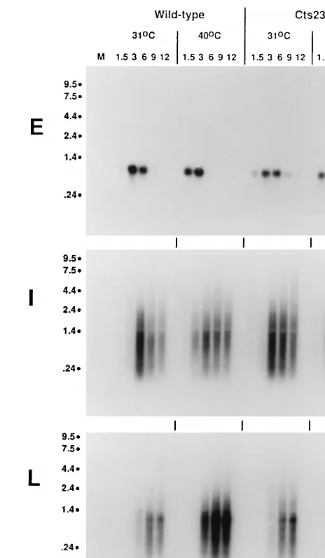
Promoter utilization in A18R mutant-infected BSC40 cells was assessed by RNase protection (Fig. 1). The riboprobes used contain antisense RNA sequence from both upstream and downstream of the previously determined mRNA 59ends. The sizes of the protected fragments shown in Fig. 1 corre-spond precisely to the mRNA 59ends previously determined for each gene tested (2). In the A18R mutant (Cts23) infection, protected fragments for early and intermediate promoters ap-pear with similar kinetics and in similar amounts compared to the wt infection. The early C11R promoter turns on at 1.5-h postinfection, peaks at 3 h, and decreases at late times. The intermediate G8R promoter turns on at 3 h, peaks at 6 h, and stays on throughout the time course. Compared to wt infection,
in the A18R mutant infection the late F17R promoter turns on at a similar time at 31 and 40°C (6 h postinfection), but the signal is reduced in intensity and decays prematurely at 40°C. Most importantly, the early promoter is not utilized at late times in the A18R mutant infection at 40°C, thereby contra-dicting the hypothesis that promiscuous transcription repre-sents reactivation of early promoters late during infection. Significantly, we have also used RNase protection to measure the promoter activity of a promiscuously transcribed early gene, M2L, and found that this promoter is also not utilized at late times in an A18R mutant infection at 40°C (data not shown).
To determine whether random initiation occurs throughout the virus genome during A18R mutant infections, we per-formed Northern blot analysis using the same three standard early, intermediate, and late gene riboprobes (Fig. 2). The kinetics of mRNA synthesis observed are similar to those
[image:3.612.309.541.70.468.2]de-FIG. 1. Promoter utilization in wt- and Cts23-infected BSC40 cells. BSC40 cells were infected with wt or Cts23 virus at an MOI of 15 and incubated at 31 or 40°C. Total RNA was extracted from infected cells at various times postin-fection, indicated in hours above the lanes. RNA was hybridized to uniformly labeled antisense riboprobes specific for the 59end of an early (E), intermediate (I), or late (L) gene. After RNase digestion, the protected fragments were analyzed by denaturing PAGE and autoradiography. P, unhybridized probe digested with RNase; M, mock-infected cell RNA.
FIG. 2. Northern blot analysis of RNA synthesized in wt- and Cts23-infected BSC40 cells. The total RNA was purified from infected BSC40 cells as described in the legend to Fig. 1. RNA was fractionated on formaldehyde-agarose gels, transferred to GeneScreen membranes, and probed with uniformly labeled an-tisense RNA riboprobes specific for an early (E), intermediate (I), or late (L) gene. Lanes M contain uninfected cell RNA. Sizes are denoted at the left in kilobases.
on November 9, 2019 by guest
http://jvi.asm.org/
[image:3.612.53.286.71.254.2]tected by RNase protection. Importantly, the early C11R mRNA signal disappears at late times, confirming that the early promoter does not reactivate late during infection. In Cts23-infected cells at 40°C at late times, some G8R transcripts are shorter than normal, indicating the expected RNase L-catalyzed breakdown of RNAs. Consistent with the RNase protection analysis described above, the late F17R gene seems to be poorly transcribed in A18R mutant infections at 40°C. Most importantly, promiscuous transcription is not observed with either the early or the late gene probe at late times postinfection, thereby contradicting the hypothesis that pro-miscuous transcription represents completely random initia-tion within untranscribed regions of the genome.
A compilation of all results obtained to date concerning transcription in A18R mutant-infected cells reveals that miscuous transcription is context sensitive. Specifically, pro-miscuous transcription has been observed within the D9R and G2R genes (4) but not within the C11R and F17R genes (Fig. 2). Interestingly, inspection of the vaccinia virus genetic map (21) reveals that the C11R and F17R genes are unusual in that there are no other known promoters in the same transcrip-tional orientation within 14 kb upstream from either gene. By contrast, D9R and G2R each lie 3 to 5 kb downstream from intermediate or late promoters driving transcription of up-stream genes. These observations provide further support for the hypothesis (referred to below as the readthrough hypoth-esis) that promiscuous transcription results from transcrip-tional readthrough from upstream intermediate or late gene promoters.
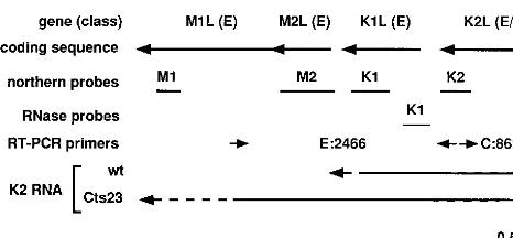
Promiscuous transcription in the region from K2L to M1L.
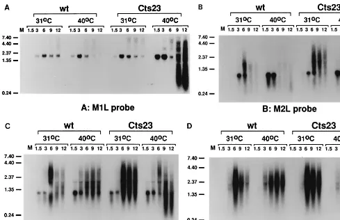
To test the readthrough hypothesis, we focused attention on a region spanning the vaccinia virus genes M1L through K2L (Fig. 3). Previous Northern blot analysis from other laborato-ries using exclusively early viral RNA shows that M1L, M2L, K1L, and K2L are each expressed early during infection. Fur-thermore, Northern blot analysis by Smith et al. (45) revealed transcription within the K2L region at late times. Whether any of the four genes contains intermediate or late promoters is unknown. Previous data from our lab showed that the M2L gene was promiscuously transcribed (6). We hypothesize that promiscuous transcription in the M2L region results from readthrough transcription from an upstream intermediate or late promoter. As an initial test of this hypothesis, we con-ducted Northern blot analysis within the region (Fig. 4), using antisense riboprobes specific for M1L, M2L, K1L, and K2L
(Fig. 3). Northern blot analysis of M1L (Fig. 4A) and M2L (Fig. 4B) shows that early M1L transcripts (1.6 kb) and early M2L transcripts (800 bp) appear at 1.5 h postinfection and decease at late times in both wt- and Cts23-infected BSC40 cells. The sizes of the early transcripts are consistent with previous results from other laboratories (46, 47). At late times, the M1L and M2L genes are transcriptionally silent in wt infections at either 31 or 40°C. At late times in Cts23 infections at 31°C, the M1L gene is transcriptionally silent, while heter-ogeneous transcripts are observed within the M2L gene. Abun-dant transcription is observed in Cts23-infected cells at 40°C after 6 h postinfection in both the M1L and M2L genes. Some of these transcripts have a shorter than normal chain length resulting from RNA degradation catalyzed by RNase L. These results show that both the M1L and M2L genes are promiscu-ously transcribed in the absence of A18R activity. The low level of transcription observed at late times in Cts23-infected cells within the M2L gene may indicate that the A18R gene product is not fully active at permissive temperatures. Nevertheless, promiscuous transcription observed at 31°C in Cts23-infected cells is not sufficiently robust to either affect the M1L gene or activate RNase L. Northern blot analysis of the K1L gene (Fig. 4C) reveals a 1.1-kb early transcript, consistent with published experiments (46). Transcription is detected at late times in both wt and Cts23 infections at both temperatures. Some of the late transcripts in Cts23-infected cells at 40°C are shorter than normal, resulting from RNase L-catalyzed RNA degradation. Northern blot analysis of the K2L gene (Fig. 4D) reveals two barely detectable early transcripts of 1.5 and 2.1 kb, consistent with published experiments (45). At late times, the K2L gene is transcribed in both wt and Cts23 infections. Northern blot analysis of the K3L gene (data not shown) is very similar to the K1L analysis, indicating that the K3L gene is transcribed at both early and late times. Northern blot analysis of the K4L gene (data not shown) reveals transcription only at late times in both wt and Cts23 infections. Since intermediate and late vaccinia virus RNAs are normally heterogeneous in size and may read through into downstream genes, we cannot deter-mine the origin of any of the late transcripts detected in this region from Northern blot analysis alone. Nevertheless, if the readthrough hypothesis is correct, we predict that promiscuous transcription of the M1L and M2L genes results from readthrough transcription originating from K1L, K2L, K3L, or K4L. Detailed analysis of transcription initiation is required to further investigate this hypothesis.
[image:4.612.55.288.69.177.2]Characterization of gene class in the region from M1L to K2L. RNase protection analysis was used in an attempt to characterize the promoter type for each of the genes in the M1L–K2L region. Published S1 nuclease analysis shows that M1L contains an early promoter, consistent with Northern analysis (47). The published S1 nuclease analysis of the M1L gene also indicates the presence of a weak late promoter. This late promoter must be very weak, since late transcription oc-curs at only a very low level within the M1L gene, as revealed by Northern analysis. We did not attempt to analyze transcrip-tion of the M1L gene further. Antisense riboprobes for RNase protection analysis of the M2L, K1L, and K2L genes were designed to measure simultaneously both gene-specific tran-scription and upstream readthrough trantran-scription. The viral sequence contained in each probe spans the 59 end of each gene, so that protection of only a fraction of the viral se-quences in the probe reveals a gene-specific 59end. Each probe also contained nonviral sequence, 6 nt at the 59end and 11 nt at the 39end, which permits differentiation between undigested full-length probe and digested probe in which all of the viral sequences are protected by upstream readthrough transcripts. FIG. 3. Diagram describing the M1L through K2L region of the vaccinia
virus genome. From the top down, the drawing shows the identity and class (Fig. 4 to 6) of each gene, the coding sequence with arrows representing transcrip-tional orientation, the positions of Northern and RNase protection probes, the positions of primers used for RT-PCR (E:2466, experimental primer set, 2,466-bp product; C:862, control primer set, 862-bp product), and an interpre-tation of the transcriptional analysis in Fig. 4 to 8, showing extended readthrough transcription from the K2L promoter into the M1L gene in a Cts23 infection. E, early; I, intermediate.
on November 9, 2019 by guest
http://jvi.asm.org/
Riboprobes specific for M2L, K1L, and K2L were used in RNase protection analysis of RNA extracted at different times after infection of BSC40 cells at 31 or 40°C with either wt virus or Cts23 (data not shown). Consistent with Northern blot anal-ysis, the RNase protection analysis showed that each of these three genes contains an early promoter. The data showed fur-ther that neifur-ther M2L nor K1L contains a postreplicative pro-moter and therefore that any intermediate or late transcription through these genes represents readthrough from upstream genes. Last, the results showed that K2L contains a postrepli-cative promoter and is also transcribed by readthrough from an upstream postreplicative promoter. Detailed transcriptional analysis of the K1L and K2L 59regions is presented below.
To further confirm the gene class of K1L, we performed RNase protection analysis on RNA purified from cells treated with a drug swap protocol designed to distinguish intermediate from late transcription. Vaccinia virus early, intermediate, and late RNAs can be distinguished by analysis of RNA from cells infected in the presence of the DNA replication inhibitor HU, followed by a shift to drug-free medium or medium supple-mented with the protein synthesis inhibitor CHX (3). Since intermediate and late gene transcription is coupled to DNA replication, only early genes are expressed in the presence of HU. Early gene expression includes synthesis of intermediate transcription factors; thus, when the HU block is removed, intermediate and ultimately late transcription proceeds and early gene expression is shut off. If CHX is added at the time HU is removed, early transcription continues and additional
[image:5.612.55.544.70.386.2]transcription is limited to intermediate genes since synthesis of late transcription factors encoded by intermediate genes is inhibited. RNase protection analysis of the K1L and K2L 59 regions by using RNA from the drug swap protocol is shown in Fig. 5. With the K1L-specific riboprobe (Fig. 5A), very weak 59-protected fragments are detected in infected cells in drug-free medium at early times (lanes 4 and 5) and disappear at late times (lane 16). This 59-protected fragment is also de-tected in cells infected in the presence of CHX only (lane 17). Lanes 6 to 10 show that the 59-protected fragment is observed in the presence of HU but decreases when HU is removed and postreplicative gene expression is allowed to proceed in the absence of drug. Lanes 11 to 15 show that the 59-protected fragment accumulates if, after removal of the HU block, late gene expression is inhibited by the addition of CHX. These results show that the 59-protected fragment has the character-istics of an early transcript. While the drug swap experiments do not formally rule out the possibility that the K1L 59 -pro-tected fragment is also expressed as an intermediate transcript, precise mapping of this 59 end relative to a sequence ladder (not shown) shows that it originates approximately 20 nt up-stream of the K1L translation start site, within a sequence that does not contain the requisite TAAA intermediate transcrip-tion initiatranscrip-tion signal. We conclude that the K1L 59-protected fragment represents an exclusively early transcript and there-fore that the K1L gene contains an early promoter. RNase protection with the K1L riboprobe also reveals readthrough transcription. The readthrough transcripts are detected neither
FIG. 4. Promiscuous transcription in the region from M1L through K2L in Cts23-infected BSC40 cells. The total RNA was purified from infected BSC40 cells as described in the legend to Fig. 1. Equal amounts of RNA were fractionated on formaldehyde-agarose gels, transferred to GeneScreen membranes, and probed with uniformly labeled antisense RNA riboprobes specific for the M1L (A), M2L (B), K1L (C), and K2L (D) genes. Lanes M contain uninfected cell RNA. Sizes are denoted at the left in kilobases.
on November 9, 2019 by guest
http://jvi.asm.org/
at early times (lanes 4 and 5) nor in the presence of CHX (lane 17). The readthrough transcripts are present in drug-free me-dium late during infection (lane 16); they appear after the HU block is removed without added CHX (lanes 7 to 10) and also following the drug swap (lanes 11 to 15). These results show that the readthrough transcript has the characteristics of an intermediate RNA. It is noteworthy that the signal represent-ing readthrough transcription is more intense in the absence than in the presence of CHX following removal of HU (com-pare lanes 10 and 15), suggesting that these transcripts may have late as well as intermediate character.
RNase protection with the K2L riboprobe in the drug swap protocol (Fig. 5B) also reveals both a 59-protected fragment and a readthrough transcript. Consistent with Northern blot analysis of the K2L gene, the 59-protected fragment has char-acteristics consistent with a weak early transcript, notably a weak signal in the presence of CHX (lane 17). The K2L 59 -protected fragment also has characteristics of an intermediate transcript in that it is present at late times in the absence of inhibitors (lanes 8 to 10 and 16) and also following the drug swap (lanes 13 to 15). In this case, mapping of the K2L 59end (not shown) places it near the A4 stretch in the sequence
TAAAATCATG proximal to the K2L translation start site (underlined). This sequence contains the requisite TAAA in-termediate promoter initiation consensus, and thus the K2L gene may contain a compound early-intermediate promoter. The K2L readthrough transcript is absent early and in CHX-treated cells (lanes 4, 5, and 17) but is present late and during the drug swap (lanes 8 to 10 and 16) and thus has character-istics of an intermediate transcript. Sequence analysis and pre-liminary transcription analysis (not shown) indicate that readthrough into the K2L gene may arise from the K4L pro-moter. Once again it is noteworthy that the signals represent-ing both the K2L 59 end and the readthrough transcript are more intense in the absence than in the presence of CHX following removal of HU (compare lanes 10 and 15), suggest-ing that these transcripts may have late as well as intermediate character.
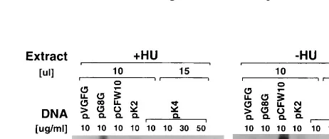
To confirm that K2L and K4L contain intermediate promot-ers, we analyzed these promoters in an in vitro transcription assay. In this assay, extracts from vaccinia virus-infected, HU-treated cells are used to transcribe a template containing an
intermediate promoter fused to a G-less cassette. HU treat-ment prevents intermediate and late gene transcription and therefore prevents synthesis of late and early viral transcription factors (14). Thus, HU-treated extracts contain only early gene products, including intermediate gene transcription factors, and they are capable of initiating transcription at intermediate but not early or late vaccinia virus promoters. We cloned each of the candidate K2L and K4L promoter sequences upstream of the G-less cassette in pC2AT19. Results of in vitro
transcrip-tion directed by these templates are shown in Fig. 6. Control experiments with standard early (pVGFG DNA, C11R pro-moter), intermediate (pG8G DNA, G8R propro-moter), and late (pCFW10 DNA, F17R promoter) templates (lanes 1 to 3 and 9 to 11) shows that early, intermediate, and late transcripts are detected with the extracts made in the absence of HU, (2HU extracts), while only intermediate transcripts are detected with extracts made in the presence of HU (1HU extracts). In ad-dition, the intermediate template is transcribed more effi-ciently in the1HU extracts. In vitro transcription with the pK2 template shows that, like the G8R control, the K2L promoter is transcribed more efficiently with 1HU extracts than with 2HU extracts, confirming that the K2L promoter has
inter-FIG. 5. Drug swap RNase protection analysis. BSC40 cells were infected with wt virus in the absence (lanes 3 to 5) or presence (lanes 6 to 15) of HU; 3 h postinfection, medium was removed and replaced with drug-free medium (lanes 6 to 10) or medium containing CHX (lanes 11 to 15). RNA was purified at various times postinfection, indicated above the lanes in hours. RNA was hybridized to uniformly labeled antisense riboprobes specific for the 59end of K1L (A) or K2L (B). After RNase digestion, the protected fragments were analyzed by denaturing PAGE and autoradiography. Lanes DME (lane 16) and CHX (lane 17) contain RNA extracted at 7 h postinfection from cells infected in drug-free medium and in the presence of CHX, respectively. P (lane 1), undigested probe; PR(lane 2), unhybridized
[image:6.612.60.546.70.228.2]probe digested with RNase; M, mock infection; FL, full-length probe; RT, readthrough transcript protected fragment; 59, mRNA 59-end-protected fragment.
FIG. 6. In vitro transcription of templates containing K2L and K4L promoter sequences. Transcription-competent extracts were made from cells infected with wt virus in the presence of 10 mM HU (1HU) or in the absence of drug (2HU). Transcription was done with 10ml (lanes 1 to 5 and 9 to 13) or 15ml (lanes 6 to 8 and 14 to 16) of extract. Reactions contained 10, 30, or 50mg of pVGFG (early), pG8G (intermediate), pCFW10 (late), pK2, or pK4 DNA per ml, as indicated. Reaction products were separated on a 7% polyacrylamide gel, which was dried and autoradiographed.
on November 9, 2019 by guest
http://jvi.asm.org/
[image:6.612.312.543.560.658.2]mediate character. In vitro transcription with the pK4 template shows that it is transcribed very weakly in a high concentration of1HU extract, suggesting that K4L contains a weak inter-mediate promoter. In summary, both the K2L and the K4L promoters have intermediate characteristics in the in vitro transcription analysis.
The results from detailed transcription analysis of the M1L through K2L region are summarized in Fig. 3. We conclude that M1L, M2L, and K1L are exclusively early genes and that K2L contains a compound early-intermediate promoter. In addition, the K4L promoter also has intermediate character. Furthermore, intermediate transcription reads through from upstream genes into both the K1L and K2L genes. Most of the readthrough into K1L probably derives from the K2L interme-diate promoter, while most of the readthrough into the K2L gene probably arises from the K4L promoter. We hypothesize that during an A18R mutant infection, transcription initiated from the K2L intermediate promoter reads through into the downstream early M1L and M2L genes, resulting in the pro-miscuous transcription phenotype.
RT-PCR analysis of the readthrough transcription from
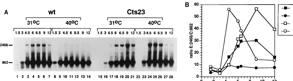
K2L into M1L. We used RT-PCR as a direct measure for
transcription which extends from the K2L gene into M1L or M2L. Analysis of the size of nascent transcripts in A18R mu-tant infections is complicated by the fact that promiscuous transcription induces RNase L-catalyzed RNA breakdown. However, intermediate gene transcription begins at 3 h postin-fection (Fig. 1 and 2) whereas RNA degradation is not evident until 7.5 h postinfection (reference 4 and data not shown). We therefore attempted to measure readthrough transcription by RT-PCR during the interval between the initiation of interme-diate transcription and induction of RNase L (Fig. 7). RNA was extracted from BSC40 cells infected with wt or Cts23 at various times postinfection and analyzed by RT-PCR using two sets of primers simultaneously (Fig. 7A). The experimental primer set amplified a 2466-nt readthrough transcript (E:2466) extending from K2L to M1L, whereas the control primer set amplified an 862-nt transcript from within the K2L gene. Both transcripts were quantified by phosphorimage analysis, and the ratio of readthrough transcripts to control transcripts (E:2466/ C:862) was determined. As expected, the 862-nt control tran-script appears early after infection, persists throughout the experiment, and is present in similar amounts regardless of the virus or temperature used. Surprisingly, the long 2,466-nt tran-script is observed in wt-infected cells at 31°C (lanes 1 to 7), even though the 2-5A pathway is not induced at this
temper-ature. At 40°C, these readthrough transcripts are reduced in abundance in the wt infection (lanes 8 to 14). These results indicate that the incubation temperature affects the steady-state level of readthrough transcription and also activation of the 2-5A pathway in wt-infected cells. The amount of readthrough transcription observed in Cts23-infected cells at 31°C (lanes 15 to 21) is slightly increased relative to the wt infection at 31°C (lanes 1 to 7), consistent with the previous suggestion that the Cts23 mutant is slightly defective even under permissive conditions (Fig. 4B). Most importantly, at 40°C after induction of intermediate transcription but before induction of the 2-5A pathway, the transcripts which extend from K2L into M1L are much more abundant in the Cts23 infection (lanes 23 to 26) compared to the wt infection (lanes 8 to 12). At later times, the Cts23 readthrough transcripts disappear (lanes 27 and 28) due to activation of 2-5A pathway. (The 862-nt control transcript is presumably small enough to be a poor target for RNase L in vivo.) In summary, at 40°C, more readthrough transcription is detected in the Cts23 infec-tion preceding RNA breakdown, which supports the hypothe-sis that the A18R mutation causes extended readthrough tran-scription from intermediate promoters.
Readthrough transcription in the RNase L knockout cell
line KO3. To prove conclusively that the A18R mutation
[image:7.612.61.540.72.205.2]causes readthrough transcription, we investigated the A18R mutant phenotype in mouse KO3 cells, which are derived from an RNase L knockout mouse and which therefore lack RNase L activity (60). Preliminary experiments demonstrated that infection of KO3 cells with Cts23 at 40°C does not cause rRNA breakdown (data not shown). Interestingly, Cts23 infection of normal control cells from the parental mouse at 40°C also revealed no rRNA breakdown, and therefore differences ob-served during vaccinia virus infection of KO3 cells compared to BSC40 cells cannot be attributed solely to a lack of RNase L. Nevertheless, KO3 cells provide a method for analyzing the A18R mutant phenotype in the absence of RNA degradation. In KO3 cells, we expect to observe abnormally long transcripts in A18R mutant infections in regions where promiscuous tran-scription occurs. Northern blot analysis using an M2L ribo-probe was performed with RNAs extracted at various times from virus-infected KO3 (Fig. 8). The results show that the discrete 800-nt M2L early RNA is expressed similarly in Cts23-and wt-infected KO3 cells at both permissive Cts23-and nonpermis-sive temperatures. In addition, larger heterogeneous read-through transcripts appear at late times in both Cts23- and wt-infected KO3 cells at both temperatures. Most importantly,
FIG. 7. Readthrough transcription of the M1L gene in Cts23-infected BSC40 cells detected by RT-PCR analysis. (A) Total RNA was purified from infected BSC40 cells as described in the legend to Fig. 1. RNA was DNase treated and analyzed by RT-PCR using primers extending from M1L into K2L, generating a 2,466-nt-long product (E:2466 in Fig. 3). Internal control primers measured a K2L RNA of 862 nt (C:862 in Fig. 3). (B) RT-PCR signals from panel A were quantified by phosphorimage analysis, and the ratio of E:2466 signal to C:862 signal was plotted as a function of time postinfection.
on November 9, 2019 by guest
http://jvi.asm.org/
the readthrough transcripts observed in Cts23-infected KO3 cells at 40°C include a large population of transcripts longer than 4.4 kb which are not observed under any other condition of infection. This result is consistent with readthrough tran-scription from upstream genes into the M2L region in A18R mutant-infected KO3 cells. Northern blot analysis using M1L, K2L, D9R, and G2R riboprobes show the similar results (data not shown and reference 40).
A18R mutant phenotype in KO3 cells.KO3 cells provide an
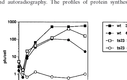
opportunity to study whether the A18R gene is essential under circumstances where the 2-5A pathway is not activated. Cts23 is temperature sensitive on KO3 cells in a plaque assay (data not shown). A one-step growth experiment was done to quan-tify the growth of wt and Cts23 on KO3 cells (Fig. 9). The wt virus grown at 31 or 40°C shows a burst size of between 80 and 800 PFU per cell, with maximum yield occurring after 48 h postinfection. Growth of Cts23 at 31°C is identical to wt growth. Cts23 does not grow on KO3 cells at 40°C. This result shows that the A18R gene is essential even in the absence of 2-5A pathway induction.
Protein pulse-labeling studies were done to examine the pattern of gene expression in wt- and Cts23-infected KO3 cells (Fig. 10). KO3 cells were infected with wt or Cts23; at various times postinfection, cells were pulse-labeled with [35
S]methi-onine, and the labeled proteins were analyzed by SDS-PAGE and autoradiography. The profiles of protein synthesis
ob-served in wt-infected cells at 31 and 40°C and in Cts23-infected cells at 31°C are identical and representative of the normal pattern of vaccinia virus gene expression observed on BSC40 cells. Specifically, early viral proteins are expressed concomi-tant with shutoff of host cell protein synthesis, followed by expression of intermediate and late viral proteins, which per-sists throughout the experiment. In a Cts23 infection at the nonpermissive temperature, host shutoff and early viral protein synthesis appear normal, and late protein synthesis is initiated at a normal time, but synthesis of late proteins is reduced in amount throughout the duration of the experiment. Thus, the A18R mutant virus is defective in late protein synthesis on KO3 cells.
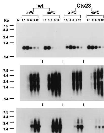
[image:8.612.53.288.71.196.2]The defective late protein synthesis phenotype seen in Cts23-infected KO3 cells could be due to a deficiency in viral mRNA metabolism. To test this hypothesis, we used Northern blot analysis to determine kinetics of mRNA synthesis, as well as the size and quantity of the steady-state mRNAs synthe-sized. Total cellular RNA extracted from KO3 cells infected with wt or Cts23 was hybridized with antisense riboprobes specific for the early (C11R), intermediate (G8R), or late (F17R) gene (Fig. 11). Early C11R mRNAs in wt- and Cts23-infected cells are expressed at the same times postinfection, are expressed in the same quantities, and appear as discrete bands, regardless of the incubation temperature. Intermediate G8R mRNAs in wt- and Cts23-infected KO3 cells are ex-pressed at the same time postinfection, are exex-pressed in similar quantities, and appear as smears diagnostic of the expected 39-end heterogeneity. In the Cts23 infection done at 40°C, there is an increase in intermediate transcripts larger than 4.4 kb, consistent with Northern analysis of the M2L gene (Fig. 8). At 31°C, late F17R mRNA synthesis in Cts23-infected KO3 cells is similar to that in the wt infection at 31°C. Under these conditions, the F17R mRNA appears as a characteristic smear, superimposed on a relatively discrete transcript peculiar to this late gene (Fig. 2). In Cts23-infected cells at 40°C, late F17R mRNA synthesis is initiated at the appropriate time postinfection, but the quantities of late mRNA are significantly decreased relative to all other conditions of infection. In summary, syn-thesis of steady-state late mRNA in A18R mutant-infected KO3 cells is reduced in quantity, consistent with the defective late protein synthesis phenotype described above.
FIG. 8. Readthrough transcription of the M2L gene in Cts23-infected KO3 (RNase L2) cells. Total RNA was purified from infected KO3 cells as described
[image:8.612.310.543.73.233.2]in the legend to Fig. 1. RNA was analyzed by Northern blot analysis using a uniformly labeled antisense riboprobe specific for the M2L gene (Fig. 3). Sizes are denoted at the left in kilobases.
FIG. 9. One-step growth of wt and Cts23 in KO3 cells. KO3 cells were infected at an MOI of 6 with either wt or Cts23 and incubated at 31 or 40°C. Samples were taken at various times postinfection, and virus yields were deter-mined by plaque titration at 31°C.
FIG. 10. Protein synthesis in wt- and Cts23-infected KO3 cells. KO3 cells were infected with wt or Cts23 at an MOI of 15, incubated at 31 or 40°C, and pulse-labeled for 15 min with [35S]methionine at the times postinfection
indi-cated above the lanes in hours. Lane M, mock infection. Labeled proteins were electrophoresed on SDS–10% polyacrylamide gels and autoradiographed.
on November 9, 2019 by guest
http://jvi.asm.org/
[image:8.612.60.282.550.690.2]DISCUSSION
The experiments described here were done to refine our understanding of the effects of the vaccinia virus A18R gene on postreplicative viral transcription. Previous research had shown that mutations in the A18R gene cause promiscuous transcription, that is, transcription from regions of the genome which are normally transcriptionally silent late during infec-tion. Our results discredit two possible explanations for pro-miscuous transcription. Specifically, the data show that (i) the early VGF and M2L promoters do not reactivate at late times postinfection and (ii) random transcription throughout the ge-nome does not occur. Our detailed analysis of transcription within the M1L through K2L region of the viral genome pro-vides positive support for the only remaining explanation for promiscuous transcription. Specifically, both RT-PCR analysis conducted in virus-infected BSC40 cells and Northern analysis conducted in RNase L knockout KO3 cells show that in A18R mutant infections transcription initiated from the K2L inter-mediate promoter yields longer than normal transcripts which read through into the downstream early M1L gene. In sum-mary, these results show that late during a wt virus infection, the A18R gene product limits elongation by the viral RNA polymerase and thus has the properties of a negative transcrip-tion elongatranscrip-tion factor.
In the course of our characterization of the A18R mutant, we have carried out a detailed transcription analysis of gene class in the region spanning the M1L and K2L genes which both confirms and extends previous analysis of individual genes within this region. Our results confirm that M1L, M2L, K1L, and K2L are expressed early during infection. While published Northern analysis of M1L transcription, like our own, shows little or no late transcription through this gene, the published
S1 nuclease mapping indicates the presence of a very weak late M1L promoter a short distance upstream from the early M1L promoter (47). Our experiments do not address the existence of this late M1L promoter. Also consistent with our results, published Northern analysis of the K2L gene revealed late transcriptional activity (45) which we can now state represents both initiation from a complex early-intermediate promoter and readthrough from the upstream K4L promoter. Ours is the first transcriptional analysis of the K4L gene, and the results suggest that the K4L gene contains an intermediate promoter. Perhaps the most important outcome of this transcription anal-ysis is the discovery of two new intermediate genes, K2L and K4L.
The discovery of two new intermediate genes, K2L and K4L, provides additional insight into intermediate promoter struc-ture. Published analysis of the five known intermediate genes (2, 25, 50) shows that intermediate promoters are approxi-mately 30 nt in length and contain two critical regions, an upstream 14-bp core element that is A/T rich, separated by 10 or 11 bp from a 4-bp initiator element which contains the sequence TAAA (Fig. 12). We have shown here that the 38-nt sequences upstream from both the K2L and K4L translation initiation codons have intermediate promoter activity in vitro and that in vivo, an mRNA 59 end with characteristics of an intermediate RNA maps within 10 nt upstream of the K2L translation initiation codon. Importantly, we have not mapped with absolute precision the 59end of either the K2L or K4L in vivo mRNA. Nevertheless, inspection of the K2L and K4L upstream regions reveals sequence which matches precisely the initiator TAAA and which closely approximates the A/T-rich core. Closer inspection of these sequences, allowing for inclu-sion of 10 or 11 bp in the spacer region, reveals deviations from previously characterized critical residues in the G8R interme-diate promoter which could both increase and decrease pro-moter activity (2). For example, both propro-moters contain a potentially inhibitory deviation from a consensus AAA in the region from217 to219, while both contain potentially stim-ulatory deviations from the G8R sequence in the220 and223 regions.
[image:9.612.316.538.68.149.2]The discovery that K2L and K4L are intermediate genes provides some additional insight into the functional organiza-tion of the intermediate gene class. Zhang et al. (59) have provided evidence that the intermediate gene class contains a minimum of 13 genes. The five previously characterized inter-mediate genes, three late transcription factors (27), a virion RNA helicase (42), and a single-stranded DNA binding pro-tein (37), all map within the central conserved region of the virus genome and are all implicated in nucleic acid metabo-lism. By contrast, while the function of K4L is unknown, K2L
FIG. 11. Northern blot analysis of RNA synthesized in wt- and Cts23-in-fected KO3 cells. The total RNA was purified from inCts23-in-fected KO3 cells as described in the legend to Fig. 1. RNA was analyzed by Northern blot analysis using uniformly labeled antisense RNA riboprobes specific for an early (E), intermediate (I), or late (L) gene. Lanes M contain uninfected cell RNA.
underlined. Translation initiation ATGs are italicized.
on November 9, 2019 by guest
http://jvi.asm.org/
[image:9.612.66.272.69.332.2]encodes SPI3, a serine protease inhibitor homolog which plays a role in virus-induced cell fusion (48), and both K2L and K4L map in the variable left terminus of the genome. Importantly, any vaccinia gene which has been previously classified as a late gene and not specifically tested for intermediate gene activity is potentially an intermediate gene. In summary, our observa-tions support the idea that the intermediate gene class may in fact be relatively large and include genes with a wide variety of functions.
Our phenotypic analysis of the A18R mutant infection of KO3 cells provides fresh insight into the primary consequences of readthrough transcription on the viral infection. Interpreta-tion of prior phenotypic analysis of A18R mutant infecInterpreta-tions, done exclusively on BSC40 cells, was complicated by the fact that readthrough transcription from converging promoters causes an accumulation of dsRNA, which triggers the 2-5A pathway, which in turn activates RNase L and causes a global degradation of mRNA and rRNA and a cessation of protein synthesis (4, 12, 35). Thus, it was unclear whether in the ab-sence of RNase L activity, the A18R gene would be essential and whether readthrough transcription would have deleterious effects on the infection. Significantly, we have found that in RNase L knockout KO3 cells, the A18R mutant virus is tem-perature sensitive with respect to virus growth and that steady-state viral late mRNAs and late viral proteins are present in reduced amounts. Several possible explanations exist for the observed defect in late gene expression on KO3 cells. First, it is formally possible that the A18R gene product is directly involved in the initiation of late viral transcription. We feel that this possibility is unlikely since in vitro experiments from our lab (data not shown) and from other labs (29, 53) have failed to demonstrate any role of A18R in initiation of late viral transcription. Second, since readthrough transcription from converging intermediate promoters should still cause accumu-lation of dsRNA in KO3 cells, the dsRNA-dependent protein kinase pathway (30) may be activated at intermediate times, thus inhibiting synthesis of late viral transcription factors, which in turn could cause defective synthesis of late mRNAs. Third, it is possible that formation of dsRNA results in inter-ference with translation of late transcription factors from in-termediate mRNAs, also affecting late mRNA synthesis. Fourth, readthrough transcription could result in direct inter-ference with initiation of transcription from downstream genes, a phenomenon previously documented in studies of transcription in mammalian cells (20). Unfortunately, the de-crease in late mRNA synthesis in A18R mutant-infected cells has so far made it difficult to determine whether the A18R mutation affects readthrough transcription from late as well as intermediate promoters. In any case, the phenotypic analysis of A18R mutant infections on KO3 cells emphasizes the impor-tance of restricting readthrough transcription from intermedi-ate promoters during a normal vaccinia virus infection.
In both eucaryotic and procaryotic systems, a variety of neg-ative transcription elongation factors have been identified. Vir-tually all of these factors have termination factor activity, de-fined experimentally as the release of nascent transcripts from a ternary elongation complex, and many are helicases and/or nucleic acid-dependent ATPases. The Escherichia coli factor Rho is the most extensively studied termination factor (26, 51, 52). Rho is an RNA-dependent ATPase and an RNA-DNA helicase which is thought to bind nascent RNA and to trans-locate in the 59-to-39 direction along the RNA in an ATP-dependent fashion, finally causing the dissociation of the ter-nary elongation complex by unknown mechanisms. Drosophila factor 2, a double-stranded DNA-dependent ATPase which lacks detectable helicase activity (55, 56), can cause the release
of RNA polymerase II transcripts in an ATP-dependent man-ner (57). Recently it has been shown that in vaccinia virus, the ATP-dependent step in early transcription termination is cat-alyzed by a single-stranded DNA-dependent ATPase, the product of gene D11L, which also lacks detectable helicase activity (19). Thus, it is clear that transcription termination in several systems requires the participation of a factor which can bind single- or double-stranded RNA or DNA and hydrolyze ATP. The precise mechanism of action of these factors is not known; however, it seems reasonable that translocation or helicase activities of these factors within an elongation com-plex may destabilize the comcom-plex. We have shown here that the A18R protein is a negative transcription elongation factor. We have shown previously that the A18R protein, a member of the DExH helicase superfamily II (28), possesses both DNA-de-pendent ATPase and DNA helicase activities. The ATPase activity of A18R is stimulated by both single- and double-stranded DNA but not by RNA (5). The helicase activity is restricted exclusively to DNA-DNA hybrids, it is capable of separating only hybrids containing less than 25 bp, and it dis-plays 39-to-59 directionality (44). Thus, based on prior bio-chemical analysis of the A18R protein, based on the A18R mutant analysis presented here, and by analogy with other known transcription termination factors, we propose that A18R serves as a termination factor for intermediate (and perhaps late) transcription in vivo.
Our experiments imply that 39-end formation during pos-treplicative vaccinia virus transcription is a factor-mediated event but provide no information about potential cis-acting elements in either RNA or DNA that might be required for termination. In fact, termination of vaccinia virus postreplica-tive transcription resembles termination of transcription in metazoan cells in that it occurs at a large number of sites, generating extreme 39-end heterogeneity (22, 31, 36). Thus, if specific nucleic acid sequences or structures mediate postrep-licative vaccinia virus transcription termination, these elements must be both abundant and inefficient.
Prior genetic and biochemical experiments suggest that the A18R protein does not act alone but rather acts as part of a larger complex containing the viral RNA polymerase, the viral transcription factors G2R and H5R (9), and perhaps other factors as well. H5R is a 35-kDa DNA binding phosphoprotein (7, 33, 34) which has late transcription factor activity in vitro (29) and which interacts directly with G2R and either directly or indirectly with A18R. The precise role of H5R in stimulat-ing late transcription has not been determined, and no virus mutants in H5R exist. G2R is a novel 26-kDa protein which interacts either directly or indirectly with A18R as well as undergoing a direct interaction with H5R. G2R mutants cause synthesis of 39-truncated intermediate and late vaccinia virus RNAs (8) and also function as extragenic suppressors of A18R mutants (17). Thus, G2R behaves like a positive transcription elongation factor whose function serves to balance A18R ac-tivity. G2R could function independently of A18R, or it could be a positive regulator of A18R activity. Experiments with the antipoxvirus drug isatin-b-thiosemicarbazone (IBT) suggest that RNA polymerase interacts with A18R, since IBT induces promiscuous transcription (4), and IBT resistance maps to the second-largest subunit of the RNA polymerase (13). Biochem-ical experiments designed to elucidate the precise activities of A18R, G2R, and H5R in an elongating RNA polymerase com-plex are under way.
ACKNOWLEDGMENTS
We thank Jackie Lewis for technical support. We thank Carman Sancho for communication of unpublished data.
on November 9, 2019 by guest
http://jvi.asm.org/
5. Bayliss, C. D., and R. C. Condit. 1995. The vaccinia virus A18R gene product is a DNA-dependent ATPase. J. Biol. Chem. 270:1550–1556.
6. Bayliss, C. D., and R. C. Condit. 1993. Unpublished data.
7. Beaud, G., and R. Beaud. 1997. Preferential virosomal location of under-phosphorylated H5R protein synthesized in vaccinia virus-infected cells. J. Gen. Virol. 78:3297–3302.
8. Black, E. P., and R. C. Condit. 1996. Phenotypic characterization of mutants in vaccinia virus gene G2R, a putative transcription elongation factor. J. Vi-rol. 70:47–54.
9. Black, E. P., N. Moussatche, and R. C. Condit. 1998. Characterization of the interactions among vaccinia virus transcription factors G2R, A18R, and H5R. Virology 245:313–322.
10. Broyles, S. S., J. Li, and B. Moss. 1991. Promoter DNA contacts made by the vaccinia virus early transcription factor. J. Biol. Chem. 266:15539–15544. 11. Broyles, S. S., L. Yuen, S. Shuman, and B. Moss. 1988. Purification of a
factor required for transcription of vaccinia virus early genes. J. Biol. Chem.
263:10754–10760.
12. Cohrs, R. J., R. C. Condit, R. F. Pacha, C. L. Thompson, and O. K. Sharma. 1989. Modulation of ppp(A29p)nA-dependent RNase by a temperature-sensitive mutant of vaccinia virus. J. Virol. 63:948–951.
13. Condit, R. C., R. Easterly, R. F. Pacha, Z. Fathi, and R. J. Meis. 1991. A vaccinia virus isatin-b-thiosemicarbazone resistance mutation maps in the viral gene encoding the 132-kDa subunit of RNA polymerase. Virology
185:857–861.
14. Condit, R. C., J. I. Lewis, M. Quinn, L. M. Christen, and E. G. Niles. 1996. Use of lysolecithin-permeabilized infected-cell extracts to investigate the in vitro biochemical phenotypes of poxvirus ts mutations altered in viral tran-scription activity. Virology 218:169–180.
15. Condit, R. C., and A. Motyczka. 1981. Isolation and preliminary character-ization of temperature-sensitive mutants of vaccinia virus. Virology 113:224– 241.
16. Condit, R. C., A. Motyczka, and G. Spizz. 1983. Isolation, characterization, and physical mapping of temperature-sensitive mutants of vaccinia virus. Virology 128:429–443.
17. Condit, R. C., Y. Xiang, and J. I. Lewis. 1996. Mutation of vaccinia virus gene G2R causes suppression of gene A18R ts mutants: implications for control of transcription. Virology 220:10–19.
18. Deng, L., and S. Shuman. 1994. A role for the H4 subunit of vaccinia RNA polymerase in transcription initiation at a viral early promoter. J. Biol. Chem. 269:14323–14328.
19. Deng, L., and S. Shuman. 1998. Vaccinia NPH-I, a DExH-box ATPase, is the energy coupling factor for mRNA transcription termination. Genes Dev.
12:538–546.
20. Eggermont, J., and N. J. Proudfoot. 1993. Poly(A) signals and transcriptional pause sites combine to prevent interference between RNA polymerase II promoters. EMBO J. 12:2539–2548.
21. Goebel, S. J., G. P. Johnson, M. E. Perkus, S. W. Davis, J. P. Winslow, and
E. Paoletti.1990. The complete DNA sequence of vaccinia virus. Virology
179:247–266.
22. Hagenbuchle, O., P. K. Wellauer, D. L. Cribbs, and U. Schibler. 1984. Termination of transcription in the mouse alpha-amylase gene Amy-2a oc-curs at multiple sites downstream of the polyadenylation site. Cell 38:737– 744.
23. Harris, N., R. Rosales, and B. Moss. 1993. Transcription initiation factor activity of vaccinia virus capping enzyme is independent of mRNA guanyly-lation. Proc. Natl. Acad. Sci. USA 90:2860–2864.
24. Hassett, D. E., and R. C. Condit. 1994. Targeted construction of tempera-ture-sensitive mutations in vaccinia virus by replacing clustered charged residues with alanine. Proc. Natl. Acad. Sci. USA 91:4554–4558. 25. Hirschmann, P., J. C. Vos, and H. G. Stunnenberg. 1990. Mutational analysis
of a vaccinia virus intermediate promoter in vivo and in vitro. J. Virol.
64:6063–6069.
26. Jin, D. J., R. R. Burgess, J. P. Richardson, and C. A. Gross. 1992. Termi-nation efficiency at rho-dependent terminators depends on kinetic coupling between RNA polymerase and rho. Proc. Natl. Acad. Sci. USA 89:1453– 1457.
B. N. Fields, D. M. Knipe, P. M. Howley, R. M. Chanock, J. L. Melnick, T. P. Monath, B. Roizman, and S. E. Strauss (ed.), Fields virology. Lippincott-Raven, New York, N.Y.
33. Nowakowski, M., W. Bauer, and J. Kates. 1978. Characterization of a DNA-binding phosphoprotein from vaccinia virus replication complex. Virology
86:217–225.
34. Nowakowski, M., J. Kates, and W. Bauer. 1978. Isolation of two DNA-binding proteins from the intracellular replication complex of vaccinia virus. Virology 84:260–267.
35. Pacha, R. F., and R. C. Condit. 1985. Characterization of a temperature-sensitive mutant of vaccinia virus reveals a novel function that prevents virus-induced breakdown of RNA. J. Virol. 56:395–403.
36. Proudfoot, N. J. 1989. How RNA polymerase II terminates transcription in higher eukaryotes. Trends. Biochem. Sci. 14:105–110.
37. Rochester, S. C., and P. Traktman. 1998. Characterization of the single-stranded DNA binding protein encoded by the vaccinia virus I3 gene. J. Vi-rol. 72:2917–2926.
38. Rosales, R., N. Harris, B. Y. Ahn, and B. Moss. 1994. Purification and identification of a vaccinia virus-encoded intermediate stage promoter-spe-cific transcription factor that has homology to eukaryotic transcription factor SII (TFIIS) and an additional role as a viral RNA polymerase subunit. J. Biol. Chem. 269:14260–14267.
39. Rosales, R., G. Sutter, and B. Moss. 1994. A cellular factor is required for transcription of vaccinia viral intermediate-stage genes. Proc. Natl. Acad. Sci. USA 91:3794–3798.
40. Sancho, C., Y. Xiang, and R. C. Condit. 1997. Unpublished data. 41. Sawadogo, M., and R. G. Roeder. 1985. Factors involved in specific
tran-scription by human RNA polymerase II: analysis by a rapid and quantitative in vitro assay. Proc. Natl. Acad. Sci. USA 82:4394–4398.
42. Shuman, S. 1992. Vaccinia virus RNA helicase: an essential enzyme related to the DE-H family of RNA-dependent NTPases. Proc. Natl. Acad. Sci. USA
89:10935–10939.
43. Simpson, D. A., and R. C. Condit. 1994. The vaccinia virus A18R protein plays a role in viral transcription during both the early and the late phases of infection. J. Virol. 68:3642–3649.
44. Simpson, D. A., and R. C. Condit. 1995. Vaccinia virus gene A18R encodes an essential DNA helicase. J. Virol. 69:6131–6139.
45. Smith, G. L., S. T. Howard, and Y. S. Chan. 1989. Vaccinia virus encodes a family of genes with homology to serine proteinase inhibitors. J. Gen. Virol.
70:2333–2343.
46. Smith, K. A., V. Stallard, J. M. Roos, C. Hart, N. Cormier, L. K. Cohen, B. E.
Roberts, and L. G. Payne.1993. Host range selection of vaccinia recombi-nants containing insertions of foreign genes into non-coding sequences. Vaccine 11:43–53.
47. Tamin, A., E. C. Villarreal, S. L. Weinrich, and D. E. Hruby. 1988. Nucle-otide sequence and molecular genetic analysis of the vaccinia virus HindIII N/M region encoding the genes responsible for resistance to alpha-amanitin. Virology 165:141–150.
48. Turner, P. C., and R. W. Moyer. 1995. Orthopoxvirus fusion inhibitor gly-coprotein SPI-3 (open reading frame K2L) contains motifs characteristic of serine proteinase inhibitors that are not required for control of cell fusion. J. Virol. 69:5978–5987.
49. Vos, J. C., M. Sasker, and H. G. Stunnenberg. 1991. Vaccinia virus capping enzyme is a transcription initiation factor. EMBO J. 10:2553–2558. 50. Vos, J. C., and H. G. Stunnenberg. 1988. Derepression of a novel class of
vaccinia virus genes upon DNA replication. EMBO J. 7:3487–3492. 51. Walstrom, K. M., J. M. Dozono, S. Robic, and P. H. von Hippel. 1997.
Kinetics of the RNA-DNA helicase activity of Escherichia coli transcription termination factor rho. 1. Characterization and analysis of the reaction. Biochemistry 36:7980–7992.
52. Walstrom, K. M., J. M. Dozono, and P. H. von Hippel. 1997. Kinetics of the RNA-DNA helicase activity of Escherichia coli transcription termination factor rho. 2. Processivity, ATP consumption, and RNA binding. Biochem-istry 36:7993–8004.
53. Wright, C. F., A. E. Hubbs, S. K. Gunasinghe, and B. W. Oswald. 1998. A vaccinia virus late transcription factor copurifies with a factor that binds to a
on November 9, 2019 by guest
http://jvi.asm.org/
viral late promoter and is complemented by extracts from uninfected HeLa cells. J. Virol. 72:1446–1451.
54. Wright, C. F., and B. Moss. 1989. Identification of factors specific for tran-scription of the late class of vaccinia virus genes. J. Virol. 63:4224–4233. 55. Xie, Z., and D. Price. 1998. Unusual nucleic acid binding properties of factor 2,
an RNA polymerase II transcript release factor. J. Biol. Chem. 273:3771–3777. 56. Xie, Z., and D. Price. 1998. Drosophila factor 2, an RNA polymerase II transcript release factor, has DNA-dependent ATPase activity. J. Biol. Chem. 272:31902–31907.
57. Xie, Z., and D. H. Price. 1996. Purification of an RNA polymerase II tran-script release factor from Drosophila. J. Biol. Chem. 271:11043–11046.
58. Yuen, L., and B. Moss. 1987. Oligonucleotide sequence signaling transcrip-tional termination of vaccinia virus early genes. Proc. Natl. Acad. Sci. USA
84:6417–6421.
59. Zhang, Y., J. G. Keck, and B. Moss. 1992. Transcription of viral late genes is dependent on expression of the viral intermediate gene G8R in cells infected with an inducible conditional-lethal mutant vaccinia virus. J. Virol.
66:6470–6479.
60. Zhou, A., J. Paranjape, T. L. Brown, H. Nie, S. Naik, B. Dong, A. Chang, B.
Trapp, R. Fairchild, C. Colmenares, and R. H. Silverman.1997. Interferon action and apoptosis are defective in mice devoid of 29,59 -oligoadenylate-dependent RNase L. EMBO J. 16:6355–6363.