cently described ME/76Neo BHK cell line [A. A. Khromykh and E. G. Westaway, J. Virol. 71:1497–1505, 1997]) was used for rescue and propagation of KUN viruses defective in the RNA polymerase gene (NS5). A new in-fectious full-length KUN virus cDNA clone, FLSDX, prepared from our previously described cDNA clone pAKUN (A. A. Khromykh and E. G. Westaway, J. Virol. 68:4580–4588, 1994) and possessing;105-fold higher specific infectivity than that of pAKUN, was used for preparation of defective mutants. Deletions of the predicted RNA polymerase motif GDD (producing FLdGDD) and of one of the predicted methyltransferase motifs (S-adenosylmethionine [SAM] binding site, producing FLdSAM) were introduced separately into FLSDX. Transcription and transfection of FLdGDD and FLdSAM RNAs into repBHK cells but not into normal BHK cells resulted in their replication and the recovery of defective viruses able to replicate only in repBHK cells. Reverse transcription-PCR and sequencing analyses showed retention of the introduced deletions in the ge-nomes of the recovered viruses. Retention of these deletions, as well as our inability to recover viruses able to replicate in normal BHK cells after prolonged incubation (for 7 days) of FLdGDD- or FLdSAM-transfected repBHK cells, excluded the possibility that recombination had occurred between the deleted defective NS5 genes present in transfected RNAs and the functional NS5 gene present in the repBHK cells. An RNA with a point mutation in the GDD motif (FLGVD) was also complemented in transfected repBHK cells, and defective virus was recovered by day 3 after transfection. However, in contrast to the results with FLdGDD and FLdSAM RNAs, prolonged (4 days or more) incubation of FLGVD RNA in normal BHK cells allowed recovery of a virus in which the GVD mutation had reverted via a single base change to the wild-type GDD sequence. Overall, these results represent the first demonstration of trans-complementation of defective flavivirus RNAs with deleteri-ous deletions in the flavivirus RNA polymerase gene NS5. The complementation system described here may prove to be useful for the in vivo complementation of deletions and mutations affecting functional domains or the essential secondary structure in any of the other flavivirus nonstructural proteins.
The genome of the Australian flavivirus Kunjin (KUN) con-sists of single-stranded RNA of positive polarity comprising 11,022 nucleotides (14) with one long open reading frame coding for three structural (C, prM, and E) and seven non-structural (NS; NS1 to NS5) proteins (9). We have been fo-cusing our studies on the components of the flavivirus replica-tion complex using KUN virus as a model for many years (5–7, 34). Previously we partially purified a functionally active KUN replication complex and showed that it was devoid of structural proteins and contained most of the NS proteins (7). Earlier we proposed a model for flavivirus RNA replication based on the recycling role of double-stranded (ds) RNA, the main template for RNA synthesis (5). Colocalization of NS1 protein and ds RNA by immunogold electron microscopy was shown in den-gue virus-infected cells (24), suggesting a role for NS1 protein in RNA replication. Recent data on effects of mutations in yellow fever (YF) virus NS1 protein on synthesis of viral RNA also suggest its involvement in RNA replication (25). The in-volvement of NS3 and NS5 proteins in RNA replication has been implied because of the presence of conserved helicase (NS3) and RNA polymerase (NS5) motifs, experimental in vitro data on the nonspecific RNA-dependent RNA polymer-ase (RDRP) activity of purified dengue virus NS5 with
inhibi-tion of this RDRP activity by anti-NS5 antibodies (31), binding
of Japanese encephalitis NS3 and NS5 proteins to the 39
un-translated region (UTR) (4), and blocking of the exchange of ds RNA templates during in vitro RDRP assays for dengue virus by anti-NS3 and anti-NS5 antibodies (2). Recently we showed colocalization in KUN virus-infected cells of NS1 and NS3 with ds RNA by immunofluorescence (IF) and immuno-gold electron microscopy analyses and that virtually all the NS but no structural proteins were coprecipitated by antibodies to ds RNA (34). Taken together, these data indicate involvement of nearly all the NS proteins in flavivirus RNA replication.
In extension of our studies of the roles of individual com-ponents of the replication complex, we decided to explore the use of our stable full-length KUN virus cDNA clone (14) and our recently developed BHK cell line persistently expressing KUN virus replicon RNA deficient in the structural genes (15) for mutagenesis and complementation analyses of individual KUN virus NS proteins. The NS5 gene was chosen as a first target because it contains several highly conserved domains characteristic of RDRPs of positive-strand RNA viruses (3, 13, 18, 27, 28). Within these domains the sequence motif GDD (KUN virus NS5 residues 665 to 667) (9) was of particular interest because of the demonstrated importance of this se-quence for the functional activities of RNA polymerases of oth-er positive-strand RNA viruses, including encephalomyocardi-tis virus (29), poliovirus (12), and hepatiencephalomyocardi-tis C virus (23). In addition to the RNA polymerase domains, two other conserved domains characteristic for methyltransferases were identified at
* Corresponding author. Mailing address: Sir Albert Sakzewski Vi-rus Research Centre, Royal Children’s Hospital, Herston Rd., Bris-bane, QLD 4029, Australia. Phone: (617) 1568. Fax: (617) 3253-1401. E-mail: [email protected].
7270
on November 9, 2019 by guest
http://jvi.asm.org/
the amino terminus of flavivirus NS5 by computer-assisted analysis (19). No experimental data on the importance of these domains for functional activity and/or viral replication are available for any of the members of Flaviviridae. It was there-fore of interest to determine whether mutations or deletions in the described motifs in NS5 would have an effect on virus RNA replication in vivo and furthermore whether they could be complemented by functionally active NS5 supplied in trans. In contrast to the extensive number of complementation studies with RNA polymerase and other NS genes of other positive-stranded RNA viruses, such as poliovirus (reviewed in refer-ences 17 and 35; see also referrefer-ences 10, 26, and 32) and al-phaviruses (see, for example, references 11 and 21), only one publication describes successful complementation of a defec-tive flavivirus NS gene, the NS1 gene of YF virus (22).
In this report, we present the first direct demonstration in vivo of the deleterious effects on RNA replication of a deletion and a point mutation in the conserved RNA polymerase motif (GDD) and of a deletion in one of the methyltransferase do-mains (S-adenosylmethionine [SAM] binding site) in the flavi-virus NS5 gene. We also show for the first time that the rep-lication of these defective mutated RNAs was complemented in trans by transfection into a BHK cell line persistently ex-pressing KUN virus replicon RNA.
MATERIALS AND METHODS
Cells.BHK21 cells were grown in Dulbecco’s modification of minimal essen-tial medium (DMEM; Gibco BRL) supplemented with 10% fetal bovine serum (FBS) at 37°C in a CO2incubator.
RT and PCR amplification. All reverse transcription (RT) reactions were performed with Superscript II RNase H2reverse transcriptase (Gibco BRL) essentially as described by the manufacturer by using 100 to 200 ng of purified KUN virion RNA or 1mg of total cell RNA and appropriate primers. PCR amplification after RT of a 6,895-bp DNA fragment was performed with an Expand High Fidelity PCR kit (Boehringer Mannheim) and with a 1/25 to 1/10 volume of RT product as follows. The PCR mixture (50 ml) containing all necessary components except the enzyme mixture (3 parts Taq polymerase and 1 part Pwo polymerase) was preheated at 95°C for 5 min and then the enzyme mixture was added and the following cycles were performed: 10 cycles of 95°C for 15 s and 72°C for 6 min, followed by 6 cycles of 95°C for 15 s and 72°C for 6 min, with an automatic increase in the extension time (at 72°C) of 20 s in each subsequent cycle. All PCRs with Pfu DNA polymerase (Stratagene) were per-formed essentially as described by the manufacturer.
Construction of the plasmids.Plasmids FLSD and FLSDX, shown in Fig. 1,
were obtained from the previously described stable KUN virus full-length cDNA clone pAKUN (14) by replacement of the original cDNA fragments with those obtained by RT and PCR amplification of purified KUN virus RNA (see the previous section) with existing unique restriction sites, which were also incorpo-rated into the primers for PCR amplification. A KUN virus replicon plasmid, C20DXrep, was prepared by replacing SphI at position 2467 and XhoI at position 11021 in C20rep (15) with the fragment from the full-length cDNA clone FLSDX (Fig. 1). The dicistronic replicon construct C20DXrepNeo used for generation of replicon-expressing BHK cells (repBHK) was prepared from C20DXrep by clon-ing an internal ribosomal entry site-neomycin transferase gene cassette into the 39UTR 25 nucleotides downstream of the polyprotein termination codon (sim-ilar toDME/76Neo [15]).
Deletion or mutation of the GDD motif and deletion of the SAM binding motif in the KUN virus NS5 gene (see Fig. 3A and B) were initially introduced into an intermediate plasmid, pBSNS5wt, containing the full-length NS5 gene in the pBluescript IIKS vector (Stratagene) by PCR-directed mutagenesis with high-fidelity Pfu DNA polymerase (8) and appropriate primers (Table 1) to obtain pBSNS5dGDD, pBSNS5GVD, and pBSNS5dSAM, respectively. In order to later distinguish between mutated RNAs and RNAs with deletions in comple-mentation experiments, new restriction sites were incorporated into the individ-ual primers used for the introduced mutation and deletions (Table 1; see Fig. 3B). After confirmation of the introduced mutation and deletions by restriction digestion with appropriate enzymes, fragments of the NS5 gene containing the corresponding mutation or deletions were first transferred into the C20DXrep plasmid and then into the FLSDX plasmid (containing full-length KUN virus cDNA) to obtain FLGVD, FLdGDD, and FLdSAM, respectively (see Fig. 3B). The mutation and deletions in the resulting FLGVD, FLdGDD, and FLdSAM plasmids were confirmed by restriction digest and sequencing analyses.
RNA transcription and transfection and determination of specific infectivity.
RNA transcripts were prepared with SP6 RNA polymerase from the plasmid DNAs FLGVD, FLdGDD, and FLdSAM, linearized with XhoI, and electropo-rated into BHK21 cells, essentially as described previously (14, 15). Briefly,;10
mg of in vitro-transcribed RNAs were electroporated into 23106BHK21
(normal BHK) or repBHK cells in 400ml in a 0.2-cm-electrode-gap cuvette (Bio-Rad) with a Bio-Rad Gene Pulser apparatus. To determine specific infec-tivity, BHK cells were electroporated with 10-ml serial 10-fold dilutions of the RNA transcripts (starting from 1mg) and incubated in DMEM–10% FBS in 60-mm-diameter culture dishes for 6 h to allow cells to attach. Then cells were overlaid with DMEM–5% FBS in 1.5% agarose and stained with crystal violet after 4 to 5 days of incubation at 37°C.
Preparation of BHK cells persistently expressing the C20DXrepNeo replicon.
BHK21 cells persistently expressing KUN virus replicon RNA C20DXrepNeo (repBHK cells) were established by G418 (Geneticin) selection as described previously for preparation ofDME/76Neo cells (15).
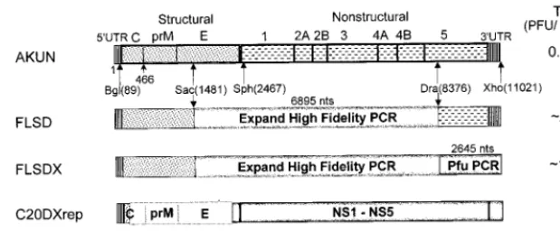
[image:2.612.145.455.74.212.2]Immunofluorescence and Northern blot analyses. Replication of mutated RNAs in transfected cells was monitored by IF analysis with mouse monoclonal antibodies to KUN virus E protein, designated 3.91D, 10A1, and 3.67G (1) (generously provided by Roy Hall, University of Queensland, Brisbane, Austra-lia), as described elsewhere (16). Dual-IF analysis with anti-E and anti-NS3 antibodies was performed essentially as described previously (34). Northern blot FIG. 1. Construction and specific infectivities of the full-length KUN virus cDNA clones and the structures of KUN virus replicon RNAs. Schematic representations of the full-length constructs and of the constructs with deletions (replicon) show consecutive replacements of the cDNA fragments in the AKUN clone (stippled boxes) with analogous fragments obtained by RT-PCR from KUN virion RNA (shaded boxes) as described in Materials and Methods. PFU titers on the right-hand side of the figure are averages (of results from three experiments) obtained after electroporation of the transcribed RNAs into BHK21 cells and determined by plaque assay (see Materials and Methods); the titer of purified wild-type KUN virus RNA was;105to;106PFU/mg of RNA. Arrows marked Bgl(89), Sac(1481), Sph(2467),
Dra(836), and Xho(11021) indicate restriction enzyme sites used in replacement cloning, with the numbers in parentheses representing nucleotide numbers in the KUN virus sequence (9, 14). An Expand High Fidelity PCR kit (Boehringer Mannheim) was used to obtain the indicated cDNA fragment of 6,895 nucleotides (nts) in the FLSD and FLSDX constructs, and Pfu PCR in FLSDX indicates that the cDNA fragment of 2,645 nucleotides was obtained with Pfu DNA polymerase (Stratagene). C20DXrep and C20DXrepNeo constructs were prepared as described in the Materials and Methods. Open boxes represent the deleted part of the genome (see reference 15). Ires, internal ribosomal entry site of encephalomyelitis virus RNA; Neo, neomycin transferase gene.
on November 9, 2019 by guest
http://jvi.asm.org/
hybridization of 2 to 5mg of total RNA isolated from transfected or infected cells was performed as described previously (15), with (as the hybridization probe) a
32P-labelled cDNA fragment representing 977 nucleotides of the KUN virus prM
and E genes (nucleotides 521 to 1498 of KUN virus cDNA) (9, 14).
Treatment of secreted mutant viruses prior to infectivity assays.The culture fluid recovered from cells after transfection with mutated RNAs was filtered through 0.45-mm-pore-size filters (Sartorius) and treated with RNase A (20mg per ml) for 20 min at 37°C in order to ensure the absence of particulate cellular material and of free RNA before attempting to transmit virus infections.
RESULTS
Improvement of the specific infectivity of the KUN virus full-length cDNA clone and of the transfection efficacy of the KUN virus replicon RNA. The specific infectivity of RNA transcribed from our previously described stable full-length KUN virus cDNA clone pAKUN was relatively low (1 to 5
PFU per 10mg of RNA) (Fig. 1) (14). For mutagenesis and
complementation experiments, it was desirable to significantly improve the specific infectivity. Therefore, we replaced the
SacII-DraIII (;7 kb) fragment in the pAKUN clone (Fig. 1) with the corresponding cDNA fragment obtained by RT of purified KUN virion RNA and PCR amplified with an Expand High Fidelity PCR kit (Boehringer Mannheim), using appro-priate primers (see Materials and Methods). RNA transcribed from the resulting cDNA clone (FLSD) had a specific
infec-tivity of;23103PFU per 1mg, compared to only 1 to 5 PFU
per 10 mg for AKUN RNA (Fig. 1). We then commenced
replacing the rest of the genome using PCR with high-fidelity
Pfu DNA polymerase (Stratagene) (8). Thus, a
2,645-nucleo-tide fragment covering most of the NS5 gene and the 39UTR
was inserted in FLSD cDNA to produce FLSDX (Fig. 1), which
resulted in a total 104- to 105-fold improvement of the original
specific infectivity, now equivalent to;104 PFU/mg of RNA
(Fig. 1). Further replacement of the 1,392-nucleotide fragment covering C, prM, and part of E did not noticeably improve the specific infectivity of the resulting FLBSDX RNA (data not shown). The most infectious FLSDX clone was therefore used in all further mutagenesis experiments.
In order to improve the efficiency of transfection of KUN virus replicon RNA, we transferred a fragment from SphI at position 2467 to XhoI at position 11021 from the FLSDX clone into our replicon clone C20rep (15) to obtain the C20DXrep
construct (Fig. 1). Electroporation of 5 to 10mg of C20DXrep
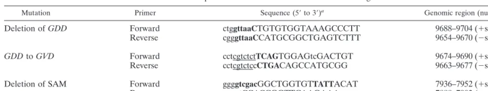
RNA resulted in its successful transfection and replication in ;80% of cells, as judged by IF analysis with anti-NS3 antibod-ies at 24 h after electroporation (Fig. 2B), which was about eight-fold more efficient than transfection with the same amount of C20rep RNA (Fig. 2A).
Preparation of the KUN virus replicon-expressing BHK cell line.Recently we described the preparation of a BHK cell line persistently expressing the KUN virus replicon RNA ME/
76Neo suitable for use in complementation experiments (15). To ensure maximum complementation efficiency, we estab-lished a new BHK cell line (repBHK) persistently expressing the replicon RNA C20DXrepNeo, a derivative of C20Dxrep that was constructed as described in Materials and Methods. Continuous passaging of these cells in the presence of 1 mg of G418 per ml showed persistent replication of replicon RNA in virtually 100% cells for at least 6 months (68 passages) after transfection, as judged by IF analysis with anti-NS3 antibodies (data not shown). Removing the selection pressure for at least nine passages did not have any noticeable effect on the pro-portion of positive cells and the intensity of fluorescence (Fig. 2C), although the cells grew better in the absence of G418 (data not shown). Importantly for the subsequent complemen-tation experiments (see below), repBHK cells were completely negative in IF with anti-E antibodies (Fig. 2D).
In order to detect possible interference of KUN virus repli-cation in repBHK cells by the replicating C20DXrepNeo RNA, we compared the levels of production of virus after infection of
normal BHK cells and of repBHK cells (passage 30) using;1
multiplicity of infection per cell of wild-type KUN virus. Some delay or inhibition of KUN virus replication was observed in the first 25 h after infection in repBHK cells compared with the
rate of replication in normal BHK cells (yields, 13 107 and
73107PFU/ml, respectively), but by 45 h KUN virus
produc-tion in repBHK cells (1.43108PFU/ml) was apparently
sim-ilar to if not better than that in normal BHK cells (63107
PFU/ml). Encouraged by the efficient replication of C20DXrep Neo RNA and the lack of any continuing interference with KUN virus replication, we commenced the complementation experiments described below using this newly prepared rep-BHK cell line.
Complementation of the mutated virion RNA FLGVD by replicon RNA.The GDD RNA polymerase motif in NS5 was mutated to GVD in the KUN virus full-length cDNA clone in plasmid FLSDX (see the previous section) as described in Materials and Methods (see Fig. 3A and B). In order to ensure that the introduced GVD mutation had not affected the open reading frame or the efficiency of translation, the mutated NS5 (NS5GVD) and native NS5 (NS5wt) mRNAs (prepared from the intermediate pBS plasmids) (see Materials and Methods and Fig. 1) were translated in rabbit reticulocyte lysates. We observed synthesis of similar amounts of predominantly full-size NS5 protein from both NS5GVD and NS5wt RNAs (Fig. 3C) plus some smaller products detected previously in similar assays which appeared to result from internal initiation of translation (33). Replication of FLGVD RNA was observed by 3 days posttransfection in repBHK cells but not in normal BHK cells, as judged by IF analysis with anti-E antibodies (results not shown) or by Northern blot analysis with a
on November 9, 2019 by guest
http://jvi.asm.org/
[image:3.612.49.550.81.175.2]and E-specific probe (Fig. 4A, lanes 2 and 3). When filtered and RNase-treated culture fluid from repBHK cells collected at 3 days after transfection was used for infection, replication of FLGVD RNA was again observed only in repBHK and not in normal BHK cells by 2 days after infection (Fig. 4A, lanes 4 and 5). Thus, an apparently lethal mutation in FLGVD RNA was successfully complemented in repBHK cells. However, a longer incubation of normal BHK cells after transfection of FLGVD RNA resulted in accumulation in culture fluid by 6 days posttransfection of a virus able to replicate after infection of fresh normal BHK cells (data not shown). RT-PCR analysis of RNA isolated from these transfected cells confirmed the presence of KUN virus-specific RNA at day 6 but not at day 3 (Fig. 4B, lanes 2 and 3). Sequencing analysis of a PCR frag-ment showed that one of the changed bases (T) in the mutant Val codon (GTC) had back mutated, resulting in restoration of the wild-type GDD amino acid sequence (Fig. 4C). Interest-ingly, the adjacent second mutated nucleotide (C) remained unchanged (Fig. 4C), thus confirming that the recovered RNA was indeed derived from the initially transfected FLGVD RNA.
Complementation of the KUN virus genome with a deletion of the GDD motif in the NS5 gene.In order to eliminate the possibility of reversion of the mutated GDD motif to a wild-type sequence, as was observed with the FLGVD RNA (see the previous section), we prepared RNA with a coding deletion of 4 amino acids including the GDD motif (FLdGDD) (Fig. 3A and B). In vitro translation analysis of NS5dGDD mRNA
tran-scribed from the intermediate pBSNS5dGDD plasmid (see Materials and Methods) showed efficient translation of full-length NS5dGDD protein (Fig. 3C), indicating that the intro-duced deletion did not affect either the open reading frame or the translational efficiency of the resulting mRNA.
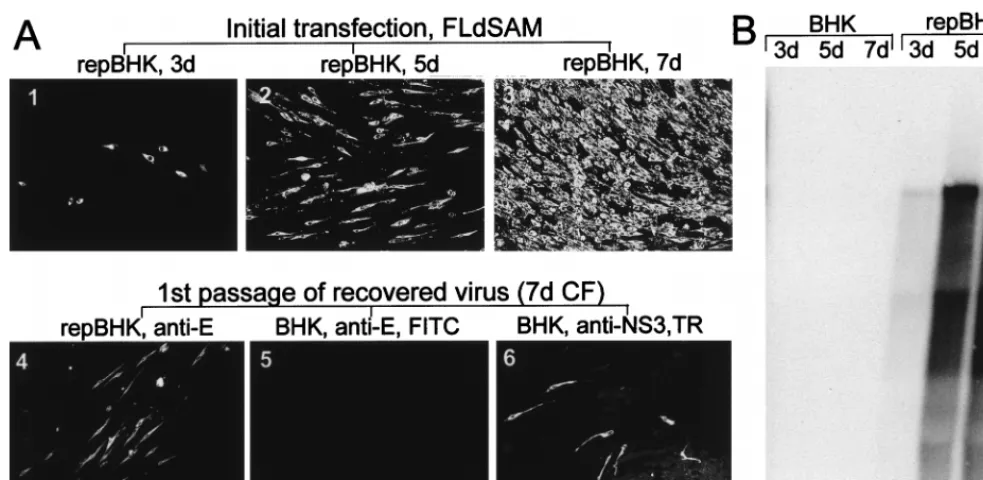
[image:4.612.110.493.68.364.2]When FLdGDD RNA was transfected into repBHK and nor-mal BHK cells in parallel experiments, replication of FLdGDD RNA was detected in repBHK cells but not in normal BHK cells; approximately 50 and 100% of repBHK cells were posi-tive by IF analysis with anti-E antibodies at 3 and 5 days, re-spectively (Fig. 5A, photos 1 and 2), indicating replication and secondary spread of the mutated genomic RNA after 3 days. Replication and secondary spread were confirmed by the ob-served accumulation of FLdGDD RNA in transfected repBHK cells detected by Northern blot analysis with a radiolabelled prM- and E-specific probe (Fig. 5B, lanes repBHK). Impor-tantly, no evidence of FLdGDD replication in normal BHK cells was detected at 5 days (Fig. 5A, photo 3, 5B, lane 5d BHK) or as late as 7 days (data not shown) after transfection. Infection of fresh repBHK cells with culture fluid collected at 5 days after productive transfection with FLdGDD RNA re-sulted in replication of defective FLdGDD virus in 100% of repBHK cells by 42 h after infection, as detected by IF with anti-E antibodies (Fig. 5A, photo 4). The infectious titer was determined by similar IF analysis with anti-E antibodies by using serial dilutions of the day 5 culture fluid assayed on repBHK cells. The number of IF foci decreased linearly with
FIG. 2. Improvement of transfection efficacy of KUN virus replicon RNA and establishment of a replicon-expressing BHK cell line (repBHK) shown by IF analysis. (A and B) IF-positive BHK21 cells with anti-NS3 antibodies at 24 h after electroporation with the original C20rep RNA and with C20DXrep RNA of improved efficiency, respectively. (C) IF analysis with anti-NS3 antibodies of BHK cells transfected with C20DXNeo RNA (constructed as described in Materials and Methods) and maintained for 38 passages as repBHK cells in medium supplemented with 1 mg of G418 per ml, followed by nine passages in medium without G418. (D) IF analysis of repBHK cells (passage 33) with anti-E monoclonal antibodies. This figure and subsequent figures were prepared by scanning all the original data (slides, autoradiograms, etc.) on an Arcus II scanner (Agfa) with FotoLook software (Agfa) for a Macintosh computer at a resolution of 150 lines per in., followed by adjustment of the brightness and the contrast of some images, assembling of the montages with Microsoft PowerPoint 97 software, and printing of the images on an Epson Stylus Color 400 printer at a resolution of 720 dots per in. on Epson photo-quality ink-jet or Epson photo-quality glossy paper.
on November 9, 2019 by guest
http://jvi.asm.org/
the dilutions of culture fluid, and the titer was ;5 3 105
infectious units per ml. Recently we showed that KUN virus replicon RNA can be packaged by KUN virus structural pro-teins expressed in trans from another expression vector (16). Therefore, it was possible for the KUN virus replicon RNA present in repBHK cells to be packaged by structural proteins expressed from the complemented FLdGDD RNA. Further-more, it was theoretically possible for FLdGDD RNA to be able to replicate in normal BHK cells, if single cells were simultaneously infected with two particles, one containing rep-licon RNA and the other containing FLdGDD RNA. We therefore performed dual-IF analysis at 42 h after infection of normal BHK cells with the virus recovered as described above
[image:5.612.63.275.72.387.2]days) (data not shown), indicating that no spread of the defec-tive FLdGDD virus had occurred. Importantly, virus able to replicate and spread in normal BHK cells was never detected in the culture fluid of FLdGDD-transfected repBHK cells even after prolonged incubation, or after two to three passages of secreted FLdGDD virus on repBHK cells (data not shown). These results exclude the presence of any replication-compe-tent (recombinant) virus in the stock of defective FLdGDD virus and thus strongly indicate that no detectable recombina-tion between the deleted NS5 gene (coded in FLdGDD RNA) and the functional native NS5 gene (present in repBHK cells)
FIG. 3. Mutagenesis of KUN virus NS5 gene. (A) Schematic representation of the NS5 gene with motifs indicated; (B) Nucleotide and amino acid sequences of the region with a mutation and the regions with deletions. Numbers represent amino acid positions in the KUN virus NS5 gene (9). The filled box in panel A shows the boundaries of the region encompassing two proposed methyltrans-ferase domains that include the SAM binding site (VIDLLGCGRGGW) (19), which is shown in boldface type in panel A. The hatched box in panel A shows boundaries of the region which includes a number of motifs proposed to be involved in RNA polymerase activity (3, 13, 18, 27, 28), including the RNA polymerase active site GDD shown in boldface type in panels A and B. R1 and F1 indicate the primers used for RT-PCR amplification (Fig. 4B). Dashed re-gions in panel B represent deleted nucleotides and corresponding amino acids. Boxed letters in panel B show new restriction sites introduced into the sequence during mutagenesis as described in Materials and Methods. (C) Autoradiogram of a sodium dodecyl sulfate–10% polyacrylamide gel containing electrophoresed samples of the [35S]methionine- and [35S]cysteine-labelled proteins translated in
rabbit reticulocyte lysates programmed with the native and mutated NS5 RNA transcripts produced by T7 RNA polymerase from the intermediate plasmids containing the corresponding NS5 cDNA sequences in the pBluescript IIKS vector (see Materials and Methods). The arrow shows the position of the full-length NS5 protein. The KUN virus lane represents a [35S]methionine-labeled
KUN virus-infected Vero cell lysate; dots identify NS5 and NS3. Numbers on the left represent Bio-Rad low-range prestained-protein standards. wt, wild type.
FIG. 4. Complementation in KUN virus replicon-expressing BHK (repBHK) cells of full-length KUN virus RNA with a GDD-to-GVD mutation in the NS5 gene. (A) Northern blot analysis of total RNA isolated from repBHK cells (lanes 2 and 4) or normal BHK cells (lanes 3 and 5) at 3 days after transfection with FLGVD RNA (p0) and at 2 days after infection with 3-day-posttransfection culture fluid (p1), with a radiolabelled cDNA probe representing 977 nucleotides of KUN virus prM and E genes (see Materials and Methods). Lane 1 contains mock-transfected repBHK cells, and lane 6 contains 10 ng of control FLGVD RNA transcribed in vitro. An arrowhead indicates the position of RNA of about 11 kb, determined relative to migration in the same gels of ethidium bromide-stained lDNA digested with BstEII (New England Biolabs). (B) RT-PCR analysis with F1 and R1 primers (Fig. 3A) of total RNA isolated from FLGVD RNA-transfected normal BHK cells. Lane 1 contains lDNA digested with
BstEII (New England Biolabs). Lanes 2 and 3 contain RNA samples isolated
from normal BHK cells at 3 and 6 days (3d and 6d), respectively, after electro-poration with FLGVD RNA. Lane 4 contains the control DNA fragment of 2.5 kb obtained by PCR amplification of FLGVD cDNA. (C) Comparison of the nucleotide and deduced amino acid (boldface italic letters above the nucleotides) sequences in the GDD motif of wild-type (wt) and GVD mutated cDNAs with that of revertant (rev) cDNA. The sequence of the rev cDNA was obtained by automatic cycle sequencing of the purified 2.5-kb RT-PCR fragment shown in lane 3 in panel B with appropriate primers and by using an ABI PRISM Dye Terminator Cycle Sequencing Ready Reaction Kit (Perkin-Elmer, Brisbane, Australia). Boldface underlined letters represent nucleotides that either were targeted for mutation (AT in the wt sequence), mutated (TC in the GVD sequence), or reverted after replication of FLGVD RNA in BHK cells (AC in the rev sequence). Boxed letters show the SalI recognition site introduced by the
GVD mutation.
on November 9, 2019 by guest
http://jvi.asm.org/
ever occurred in these experiments. Our results are compara-ble to those of Lindenbach and Rice (22), who also did not find any evidence for recombination between the functional YF NS1 gene expressed from a Sindbis vector and a deleted YF NS1 gene in YF genomic RNA in their trans-complementation experiments. Taken together, these results demonstrate that the deletion of the putative RNA polymerase GDD motif in the NS5 gene of genomic KUN virus RNA can be efficiently complemented in trans by wild-type NS5 expressed from KUN virus replicon RNA persistently replicating in repBHK cells.
Complementation of the KUN virus genome with a deletion in the methyl transferase motif in the NS5 gene.The methyl-transferase motif of flaviviruses identified by Koonin (19) by computer-assisted analysis consists of two conserved domains in NS5: domain 1, containing the SAM binding site (VIDL GCGRGG, KUN virus NS5 amino acids 78 to 87) (Fig. 3A), and domain 2, containing DTLLCD (KUN virus NS5 amino acids 150 to 155). There are no published experimental data showing a requirement of these motifs in flavivirus replication. To address this issue, we deleted the more conserved domain, the SAM binding site, from the KUN virus full-length clone FLSDX without disrupting the NS5 open reading frame (FLdSAM) (Fig. 3B) and examined the effect of this deletion on the replication of transcribed RNA after transfection into normal BHK cells. No replication of FLdSAM RNA was de-tected either by IF analysis with anti-E antibodies (data not shown) or by Northern blotting with an E-specific probe (Fig. 6B, BHK lanes) up to 7 days after transfection of normal BHK cells. Furthermore, no replicated RNA was detected after transfer of culture fluids collected at 5 and 7 days after
transfection into fresh normal BHK cells (data not shown). We showed that the deletion did not affect translation of NS5 protein, because when we translated NS5dSAM in a rabbit reticulocyte lysate using RNA prepared from the intermediate plasmid pBSNS5dSAM (see Materials and Methods), there were no significant differences in the size and in the amount of translated NS5dSAM protein from those of NS5wt protein (Fig. 3B). Thus, the absence of replication of FLdSAM RNA in transfected normal BHK cells implies an important role for the SAM binding motif in viral RNA replication and indicates that it may be associated with the RNA capping reaction (19). In order to study whether the defective NS5 protein with the deleted SAM binding site could be complemented in trans by functional NS5 protein, FLdSAM RNA was transfected into repBHK cells. Replication of FLdSAM RNA was detected but at a significantly lower rate than in the complementation
ex-periments with FLdGDD RNA. Only;1 to;2% of cells were
positive by IF with anti-E antibodies at 3 days after
transfec-tion,;40% were positive at 5 days, and 100% were positive at
[image:6.612.68.523.70.324.2]7 days (Fig. 6A, photos 1 to 3). Northern blot results also showed that accumulation of FLdSAM RNA in repBHK cells was slower than that of FLdGDD RNA (compare exposure times and lanes 1 and 3 in Fig. 5B with lanes 4 and 5 in Fig. 6B). The difference in levels of efficiency of complementation and replication between these two RNAs became even more evident when we compared the proportion of anti-E-positive repBHK cells assayed at 42 h after infection with that of rep-BHK cell culture fluids collected at 5 days after transfection with FLdGDD RNA (;100%) (Fig. 5A, photo 4) and at 7 days after transfection with FLdSAM RNA (;10%) (Fig. 6A, photo
FIG. 5. Complementation of full-length KUN virus RNA with a GDD deletion in the NS5 gene (construct FLdGDD). Results of IF analysis with anti-E antibodies (A) and Northern blot analysis with the E-specific probe (B) for detection of replicating FLdGDD RNA are shown. Photos 1 and 2 in panel A and the corresponding repBHK lanes in panel B demonstrate replication of FLdGDD RNA in repBHK cells at 3 and 5 days (3d and 5d), respectively, after electroporation. Photo 3 in panel A and the BHK lanes in panel B indicate the absence of replication of FLdGDD RNA as late as 5 days after transfection into normal BHK cells. Photo 4 in panel A shows results of IF analysis with anti-E antibodies of repBHK cells at 2 days after infection with the complemented FLdGDD virus recovered at 5 days after transfection of repBHK cells with FLdGDD RNA. Photos 5 and 6 show results of dual-IF analysis with anti-E (fluorescein isothiocyanate [FITC] stain; photo 5) and anti-NS3 (Texas Red [TR] stain; photo 6) antibodies of normal BHK cells infected for 2 days with complemented FLdGDD virus recovered at 5 days after transfection with FLdGDD RNA. CF, culture fluid. The control lane in panel B contains 10 ng of in vitro-transcribed FLdGDD RNA. The arrow indicates the position of RNA of about 11 kb, determined as described in the legend to Fig. 4. The Northern blot was exposed to X-ray film for 3 h.
on November 9, 2019 by guest
http://jvi.asm.org/
4). Likewise, the titer of defective FLdSAM virus in the day 7 culture fluid, determined by IF assay of infected fresh repBHK cells with anti-E antibodies (as described in the previous
sec-tion), was;23 104 infectious units per ml, about 25 times
lower than the corresponding titer of the day 5 FLdGDD
culture fluid (;53105infectious units per ml) (see the
pre-vious section). When the day 7 culture fluid from FLdSAM-transfected repBHK cells was used to infect normal BHK cells, dual-IF analysis with anti-NS3 and anti-E antibodies detected a few anti-NS3-positive and no anti-E-positive cells after 2 days in culture (Fig. 6A, photos 5 and 6). When these infected cells were incubated longer (4 days), some single isolated anti-E-positive cells were detected (results not shown), probably aris-ing from the slowly replicataris-ing FLdSAM RNA in cells doubly infected with both FLdSAM and C20DXrepNeo particles (see the previous section). Moreover, no virus able to replicate and spread was detected by IF analysis of normal BHK cells in-fected with secreted defective virus recovered after two pas-sages on repBHK cells (data not shown). The possibility of recombination occurring between replicon RNA and FLdSAM RNA was thus apparently excluded.
Structures of FLdGDD and FLdSAM virion RNAs.In or-der to confirm that the viruses recovered after transfection of FLdGDD and FLdSAM RNAs in repBHK cells retained the introduced deletions, we isolated RNAs from filtered and RNase A-treated culture fluids (collected at 5 days for FLdGDD and 7 days for FLdSAM) for restriction mapping and sequence analysis. The RNAs were reverse transcribed with a primer complementary to the C-terminal sequence of the NS5 gene (primer a) (Fig. 7A), and the resulting cDNAs were then PCR amplified with pairs of primers in the SAM and GDD regions (primer pairs c and d and a and b, respectively) (Fig. 7A). The
predicted products were 772 bp for the FLdSAM RT-PCR and 817 bp for the FLdGDD RT-PCR (lanes 2 in Fig. 7B and C, respectively), as was found for the fragments amplified with the same primers from plasmids containing FLdSAM and FLdGDD cDNAs (lanes 3 in Fig. 7B and C, respectively) or wild-type FLSDX cDNA (Fig. 7B and C, lanes 4). PCR am-plification from the parallel control reactions mixtures lacking reverse transcriptase produced no products (Fig. 7B and C, lanes 1). Because of the presence in culture fluid from comple-mentation experiments of two types of particles with either encapsidated wild-type (replicon) or deleted (FLdGDD or FLdSAM) RNA (Fig. 5A and 6A), and because the primer used for RT did not distinguish between these two RNAs (primer a) (Fig. 7A), amplification of both deleted and wild-type fragments was anticipated. Thus, restriction digest of gel-purified PCR fragments with SalI and HpaI restrictases dem-onstrated that indeed a mixed population of RNAs with partially deleted and wild-type NS5 genes was present in the RNA isolated from the culture fluid collected after transfec-tion with FLdSAM or FLdGDD RNA in repBHK cells (Fig. 7A, B, lanes 6 to 8, and C, respectively). A noticeably high percentage of undigested RT-PCR fragment (;50% in
FLdGDD particles and;80% in FLdSAM particles [lanes 6 in
[image:7.612.52.544.69.309.2]Fig. 7B and C, respectively]) probably represents a higher proportion of encapsidated replicon RNA in the particles se-creted from transfected repBHK cells than was expected from the results of dual-IF analysis with anti-NS3 antibodies (de-tecting both mutated full-length and nonmutated replicon RNAs) and anti-E antibodies (detecting only mutated full-length RNA) (Fig. 5A and 6A, photos 5 and 6). However, it must be noted that the IF assay was performed at 2 days after infection with recovered complemented viruses (see legend to
FIG. 6. Complementation of full-length KUN virus RNA with a deletion of the SAM binding site in the NS5 gene (construct FLdSAM). Results of IF analysis with anti-E antibodies (A) and Northern blot analysis with the E-specific probe (B) for detection of replicating FLdSAM RNA are shown. Photos 1 to 3 in panel A and the corresponding repBHK lanes in panel B demonstrate replication of electroporated FLdSAM RNA in repBHK cells at 3, 5, and 7 days (3d, 5d, and 7d, respectively). BHK lanes in panel B show the absence of replication of FLdSAM RNA at 3, 5, and 7 days after transfection into normal BHK cells. Photo 4 in panel A shows results of IF analysis with anti-E antibodies of repBHK cells at 2 days after infection with complemented FLdSAM virus recovered at 7 days after transfection of repBHK cells with FLdSAM RNA. Photos 5 and 6 show results of dual-IF analysis with anti-E (fluorescein isothiocyanate [FITC] stain; photo 5) and anti-NS3 (Texas Red [TR] stain; photo 6) antibodies of normal BHK cells infected with complemented FLdSAM virus recovered at 7 days after transfection of repBHK cells with FLdSAM RNA and immunostained 2 days later. CF, culture fluid. The arrow in panel B indicates the position of RNA of about 11 kb, determined as described in the legend to Fig. 4. The Northern blot was exposed to X-ray film for 24 h, compared to 3 h for the blot in Fig. 5B.
on November 9, 2019 by guest
http://jvi.asm.org/
Fig. 5 and 6). This period allowed the defective viruses to spread in repBHK cells, which resulted in detection of
ex-pressed FLdGDD RNA by anti-E antibodies in ;100%
(FLdGDD) or ;20% (FLdSAM) of infected repBHK cells
(photos 4 in Fig. 5A and 6A, respectively), while replicon RNA could not escape from normal cells. Thus, in FLdGDD- and
FLdSAM-infected normal BHK cells, only;1% or fewer cells
were replicon positive (anti-NS3 positive) (photos 6 in Fig. 5A and 6A, respectively). In addition, a high percentage of undi-gested RT-PCR fragment may have been due to the prefer-ential RT-PCR amplification of replicon (wild-type) cDNA over mutated (FLdGDD and FLdSAM) full-length RNAs because of the possible effects of introduced deletions on the RNA secondary structure. In order to demonstrate the pres-ence of the introduced deletions in the defective FLdSAM and FLdGDD genomes by sequencing analysis, we cloned their PCR fragments separately into the pGEM-T vector (Promega) and recombinant plasmids containing the inserts with the
ap-propriate restriction digest patterns were sequenced across the deleted regions. The results confirmed the retention of the deleted sequences (Fig. 3B) in the genomes of the defective viruses (data not shown).
Overall, the results described in the last four sections clearly demonstrate that our established repBHK cell line, which ex-presses KUN virus NS proteins from the persistently replicat-ing KUN virus replicon RNA, was successfully used to com-plement defective KUN virus genomes with deletions or a mutation in NS5, the putative RNA polymerase gene. It is also reasonable to assume from these results that this repBHK cell line can be used with a high probability of success for trans-complementation of KUN virus (or other flavivirus?) genomes with lethal deletions and mutations in any of the other NS genes.
DISCUSSION
A complementation system allowing trans rescue of defec-tive KUN virus RNAs with deleterious deletions in an NS protein has been developed and involves transfection of these RNAs into a BHK cell line persistently expressing a KUN virus replicon (repBHK cells). Complementation by the replicon permits recovery of defective viruses able to replicate only in repBHK cells and not in normal BHK cells. Thus, deletions in an RNA polymerase motif (GDD) or a methyltransferase mo-tif (SAM binding site) in the NS5 gene were rescued and cor-responding defective viruses were recovered. This is the first report on successful trans-complementation of defined func-tional motifs in any of the flavivirus NS proteins. However, a successful trans-complementation of a large deletion with no assigned function in the flavivirus NS1 gene was recently dem-onstrated by the recovery of defective YF virus after transfec-tion of YF RNA with the corresponding deletransfec-tion in cells ex-pressing NS1 protein from a noncytopathic Sindbis replicon (22).
In order to facilitate detection of possibly inefficient comple-mentation of mutated full-length KUN virus RNAs in repBHK cells, we improved the specific infectivity of RNA transcribed
from the parental cDNA by;105-fold by replacing 87% of the
genome in the FLSDX clone with the cDNA fragments ob-tained after RT-PCR of purified virion RNA with high-fidelity polymerases. We then also prepared a new replicon construct, C20DXrep, for use as a helper in complementation by
trans-ferring a fragment coding for the NS region and the 39UTR
from the FLSDX plasmid into our recently prepared C20rep replicon plasmid (15). About 80% of cells were successfully
transfected by C20DXrep RNA, representing an;5- to
;10-fold improvement in transfection efficiency of the parental C20rep RNA (see Results). The neomycin resistance gene in-serted in C20DXrep allowed establishment of the repBHK cell line persistently expressing C20DXrepNeo RNA for comple-mentation assays (see Results and Fig. 2).
Use of a single cell line (repBHK) with persistently replicat-ing KUN virus replicon RNA should have an advantage for a large number of proposed trans-complementation experi-ments, compared with the alternative use of a number of cell lines or expression constructs, each expressing an individual NS protein. Persistent replication of replicon RNA should ensure continuous expression and correct processing of all the seven NS proteins and their intermediates in a functionally active form and therefore provide a quick, reliable, and uni-versal system for complementing any defective NS genes. Rep-lication of complemented defective full-length genomes in rep-BHK cells can be easily distinguished from the helper replicon RNA either by IF analysis with anti-E antibodies or by
North-FIG. 7. Determination of the structure of the defective genomes. (A) Sche-matic representation of the KUN virus genome in the vicinity of the NS5 gene and the details of the RT-PCR protocol. SAM and GDD represent deleted motifs (Fig. 3). a, b, c, and d represent primers used for RT and PCR and correspond to the published KUN virus plus-sense sequence (9, 14). a, nucleo-tides 10378 to 10398 (minus sense); b, nucleonucleo-tides 9576 to 9597 (plus sense); c, nucleotides 8372 to 8400 (minus sense); d, nucleotides 7606 to 7621 (plus sense). Lines marked SalI and HpaI indicate the positions of new sites in the defective genomes introduced into their cDNAs during construction (Fig. 3B). Numbers indicate the predicted sizes of the fragments obtained by PCR amplification and restriction digestion. 4B, NS4B. (B and C) Results of RT-PCR and restriction digest analyses of the RNAs isolated from the culture fluid collected at 7 days after transfection of FLdSAM RNA and at 5 days after transfection of FLdGDD RNA, respectively. The primer for RT was a for both reactions; the primer pairs for PCR were a and b for FLdGDD samples and c and d for FLdSAM samples (see panel A). Lanes 1 in both panels B and C contain PCR products from the parallel control reaction lacking reverse transcriptase (RT2). Lanes 2 contain the PCR products obtained from the RT reactions performed with RNAs from the defective viruses (V) FLdSAM (B) and FLdGDD (C). Lanes 3 contain the PCR products obtained after amplification of the plasmid DNAs (Pl) FLdSAM (B) and FLdGDD (C). Lanes 4 contain the PCR products obtained from the parental FLSDX plasmid DNA with primers specific for SAM (B) and GDD (C). Lanes 6 and 7 contain restriction digests of the corresponding purified PCR fragments shown in lanes 2 to 4 with SalI (B) or HpaI (C) restrictases. Lanes 5 in both panels B and C contain a 100-bp molecular size marker (M) (Progene, Brisbane, Australia). Arrows show the sizes (in thousands) of the undigested and digested DNA fragments. wt, wild type.
on November 9, 2019 by guest
http://jvi.asm.org/
[image:8.612.50.290.68.305.2]Importantly, either deletion in the KUN virus NS5 gene re-sulted in the complete loss of ability of the mutated full-length RNAs to replicate in transfected normal BHK cells.
Although replicon RNA present in repBHK cells was en-capsidated into a small proportion of secreted transmissible particles (Fig. 5A and 6A, photos 6), interpretation of comple-mentation results was not compromised. Double infection of the same normal BHK cell with both types of encapsidated particles occurred with very low frequency (compare results in photos 5 and 6 in Fig. 5A and 6A) and was associated with a relatively high titer of defective viruses accumulated in the culture fluid. Moreover, no further spread of defective viruses in these cells (except by division of cells containing both de-fective and replicon RNAs) was detected even after prolonged (4 days) incubation. Importantly, no recombination apparently occurred between NS5 genes with deletions (in FLdGDD and FLdSAM RNAs) and the functional NS5 gene (in replicon RNA). This conclusion is based on (i) the absence of free virus spread in normal BHK cells after infection with the defective viruses recovered either at 5 to 7 days posttransfection of repBHK cells or after two to three passages on repBHK cells, as detected by IF analysis; (ii) a linear decrease in the number of IF-positive foci in repBHK cells infected with serial dilu-tions of the defective viruses; and (iii) retention of the intro-duced deletions in the recovered defective viral genomes as confirmed by RT-PCR, restriction digestion, and sequencing analysis. Significantly, although it occurs in other positive-strand RNA viruses of vertebrates such as alphaviruses, picor-naviruses, and coronaviruses (for a review, see reference 20), to the best of our knowledge, recombination has never been reported for any member of the Flavivirus genus. Moreover, in the similar study reporting the trans-complementation of the YF NS1 protein (22), no recombination was detected even after three serial passages of defective virus.
Attempted complementation of the point mutation in FLGVD appeared to be successful early after transfection in repBHK cells, but after more than 3 days of incubation in normal BHK cells, a revertant (GDD) virus was recovered. The reversion was unexpected, because lethal point mutations in-volving more conservative substitutions of the first D in the
GDD motif (e.g., with E, H, N, or Q) of poliovirus 3Dpolwere
stable for at least 5 days after transfection of the mutated full-length cDNAs (12). The emergence of the viable revertant virus from the nonviable mutant must mean that limited rep-lication which was sufficient to produce a back mutation oc-curred early after transfection. Similar results relating to the emergence of viable viruses from nonviable mutants were re-cently described for N-terminal mutants of the Sindbis virus RNA polymerase nsP4 (30). Alternatively, an error producing a rare miscopied (wild-type) RNA molecule which finally gave rise to a revertant virus in transfected normal cells may have been introduced during its transcription from cDNA by SP6
lieve that this efficient, quick, reliable, and generally applicable complementation system represents a major advance in flavi-virus molecular genetics and should provide a powerful tool for studying the functional roles of flavivirus NS proteins in RNA and virus replication.
ACKNOWLEDGMENTS
We are grateful to Roy Hall for providing KUN virus anti-E mono-clonal antibodies.
This work was supported by the National Health and Medical Re-search Council of Australia.
REFERENCES
1. Adams, S. C., A. K. Broom, L. M. Sammels, A. C. Hartnett, M. J. Howard,
R. J. Coelen, J. S. Mackenzie, and R. A. Hall. 1995. Glycosylation and antigenic variation among Kunjin virus isolates. Virology 206:49–56. 2. Bartholomeusz, A. I., and P. J. Wright. 1993. Synthesis of dengue virus RNA
in vitro: initiation and the involvement of proteins NS3 and NS5. Arch. Virol.
128:111–121.
3. Bruenn, J. A. 1991. Relationships among the positive-strand and double-strand RNA viruses as viewed through their RNA-dependent RNA poly-merases. Nucleic Acids Res. 19:217–226.
4. Chen, C.-J., M.-D. Kuo, L.-J. Chien, S.-L. Hsu, Y.-M. Wang, and J.-H. Lin. 1997. RNA-protein interactions: involvement of NS3, NS5, and 39noncoding regions of Japanese encephalitis virus genomic RNA. J. Virol. 71:3466–3473. 5. Chu, P. W., and E. G. Westaway. 1985. Replication strategy of Kunjin virus: evidence for recycling role of replicative form RNA as template in semicon-servative and asymmetric replication. Virology 140:68–79.
6. Chu, P. W., and E. G. Westaway. 1987. Characterization of Kunjin virus RNA-dependent RNA polymerase: reinitiation of synthesis in vitro. Virol-ogy 157:330–337.
7. Chu, P. W., and E. G. Westaway. 1992. Molecular and ultrastructural analysis of heavy membrane fractions associated with the replication of Kunjin virus RNA. Arch. Virol. 125:177–191.
8. Cline, J., J. C. Braman, and H. H. Hogrefe. 1996. PCR fidelity of Pfu polymerase and other thermostable DNA polymerases. Nucleic Acids Res.
24:3546–3551.
9. Coia, G., M. D. Parker, G. Speight, M. E. Byrne, and E. G. Westaway. 1988. Nucleotide and complete amino acid sequences of Kunjin virus: definitive gene order and characteristics of the virus-specified proteins. J. Gen. Virol.
69:1–21.
10. Collis, P. S., B. J. O’Donnell, D. J. Barton, J. A. Rogers, and J. B. Flanegan. 1992. Replication of poliovirus RNA and subgenomic RNA transcripts in transfected cells. J. Virol. 66:6480–6488.
11. De, I., S. G. Sawicki, and D. L. Sawicki. 1996. Sindbis virus RNA-negative mutants that fail to convert from minus-strand to plus-strand synthesis: role of the nsP2 protein. J. Virol. 70:2706–2719.
12. Jablonski, S. A., and C. D. Morrow. 1995. Mutation of the aspartic acid residues of the GDD sequence motif of poliovirus RNA-dependent RNA polymerase results in enzymes with altered metal ion requirements for ac-tivity. J. Virol. 69:1532–1539.
13. Kamer, G., and P. Argos. 1984. Primary structural comparison of RNA-dependent polymerases from plant, animal and bacterial viruses. Nucleic Acids Res. 12:7269–7283.
14. Khromykh, A. A., and E. G. Westaway. 1994. Completion of Kunjin virus RNA sequence and recovery of an infectious RNA transcribed from stably cloned full-length cDNA. J. Virol. 68:4580–4588.
15. Khromykh, A. A., and E. G. Westaway. 1997. Subgenomic replicons of the flavivirus Kunjin: construction and applications. J. Virol. 71:1497–1505. 16. Khromykh, A. A., A. N. Varnavski, and E. G. Westaway. 1998. Encapsidation
of the flavivirus Kunjin replicon RNA by using a complementation system
on November 9, 2019 by guest
http://jvi.asm.org/
providing Kunjin virus structural proteins in trans. J. Virol. 72:5967–5977. 17. Kirkegaard, K. 1992. Genetic analysis of picornaviruses. Curr. Opin. Genet.
Dev. 2:64–70.
18. Koonin, E. V. 1991. The phylogeny of RNA-dependent RNA polymerases of positive-strand RNA viruses. J. Gen. Virol. 72:2197–2206.
19. Koonin, E. V. 1993. Computer-assisted identification of a putative methyl-transferase domain in NS5 protein of flaviviruses and lambda 2 protein of reovirus. J. Gen. Virol. 74:733–740.
20. Lai, M. C. 1992. RNA recombination in animal and plant viruses. Microbiol. Rev. 56:61–79.
21. Li, M.-L., H.-L. Wang, and V. Stollar. 1997. Complementation of and inter-ference with Sindbis virus replication by full-length and deleted forms of the nonstructural protein, nsP1, expressed in stable transfectants of HeLa cells. Virology 227:361–369.
22. Lindenbach, B. D., and C. M. Rice. 1997. trans-complementation of yellow fever virus NS1 reveals a role in early RNA replication. J. Virol. 71:9608– 9617.
23. Lohman, V., F. Ko¨rner, U. Herian, and R. Bartenschlager. 1997. Biochem-ical properties of hepatitis C virus NS5B RNA-dependent RNA polymerase and identification of amino acid sequence motifs essential for enzymatic activity. J. Virol. 71:8416–8428.
24. Mackenzie, J. M., M. K. Jones, and P. R. Young. 1996. Immunolocalization of the dengue virus nonstructural glycoprotein NS1 suggests a role in viral RNA replication. Virology 220:232–240.
25. Muylaert, I. R., R. Galler, and C. M. Rice. 1997. Genetic analysis of the yellow fever NS1 protein: identification of a temperature-sensitive mutation which blocks RNA accumulation. J. Virol. 71:291–298.
26. Novak, J. E., and K. Kirkegaard. 1994. Coupling between genome
transla-tion and replicatransla-tion in an RNA virus. Genes Dev. 8:1726–1737.
27. Poch, O., I. Sauvaget, M. Delarue, and N. Tordo. 1989. Identification of four conserved motifs among the RNA-dependent polymerase encoding ele-ments. EMBO J. 8:3867–3874.
28. Rice, C. M., E. M. Lenches, S. R. Eddy, S. J. Shin, R. L. Sheets, and J. H.
Strauss.1985. Nucleotide sequence of yellow fever virus: implications for flavivirus gene expression and evolution. Science 229:726–733.
29. Sankar, S., and A. G. Porter. 1992. Point mutations which drastically affect the polymerisation activity of encephalomyocarditis virus RNA-dependent RNA polymerase correspond to the active site of Escherichia coli DNA polymerase I. J. Biol. Chem. 267:10168–10175.
30. Shirako, Y., and J. H. Strauss. 1998. Requirement for an aromatic amino acid or histidine at the N terminus of Sindbis virus RNA polymerase. J. Vi-rol. 72:2310–2315.
31. Tan, B.-H., J. Fu, R. J. Sugrue, E.-H. Yap, Y.-C. Chan, and Y. H. Tan. 1996. Recombinant dengue type 1 virus NS5 protein expressed in Escherichia coli exhibits RNA-dependent RNA polymerase activity. Virology 216:317–325. 32. Teterina, N. L., W. D. Zhou, M. W. Cho, and E. Enrenfeld. 1995. Inefficient
complementation activity of poliovirus 2C and 3D proteins for rescue of lethal mutations. J. Virol. 69:4245–4254.
33. Westaway, E. G., and A. P. Schrader. Unpublished data.
34. Westaway, E. G., J. M. Mackenzie, M. T. Kenney, M. K. Jones, and A. A.
Khromykh.1997. Ultrastructure of Kunjin virus-infected cells: colocalization of NS1 and NS3 with double-stranded RNA, and of NS2B with NS3, in virus-induced membrane structures. J. Virol. 71:6650–6661.
35. Wimmer, E., C. U. T. Hellen, and X. Cao. 1993. Genetics of poliovirus. Annu. Rev. Genet. 27:353–436.