0022-538X/95/$04.0010
Copyrightq1995, American Society for Microbiology
Effect of Different Donor Cells on Human Immunodeficiency
Virus Type 1 Replication and Selection In Vitro
ALEXANDER I. SPIRAANDDAVID D. HO*
The Aaron Diamond AIDS Research Center, New York University School of Medicine, New York, New York 10016
Received 15 July 1994/Accepted 17 October 1994
We sought to determine the effects of different host cells on human immunodeficiency virus type 1 (HIV-1) infection in vitro. First, 17 primary viruses of various phenotypes were examined for replicative capacity in peripheral blood mononuclear cells (PBMC) from 10 healthy donors. While the range of infection was variable over a 40-fold range, it was substantially less than that previously reported (L. M. Williams and M. W. Cloyd, Virology 184:723–728, 1991). In particular, no donor cells demonstrated total resistance to HIV-1 infection. We next cocultured PBMC from an HIV-1-infected patient with stimulated PBMC from three healthy donors to determine the effect of host cells on selection for a particular HIV-1 quasispecies. By using DNA sequencing, it was found that the dominant quasispecies (AD30-15) after culture was nearly identical in the cells of different donors. Furthermore, after 6 months in vivo, the patient developed a dominant proviral population in PBMC that was most closely related to the quasispecies preferentially selected in vitro, although this quasispecies was only a minor fraction of the sequences present earlier in PBMC. In subsequent biological characterizations, it was found that AD30-15 grew much better in PBMC and macrophages than did other related quasispecies. Hence, we conclude that the primary mechanism of in vitro selection for a particular HIV-1 variant in this case is mediated by the phenotypic properties of the virus and is less dependent on host cell origin. The findings reported here have important practical implications for studies of HIV-1 replication in primary cells derived from healthy donors.
Human immunodeficiency virus type 1 (HIV-1) exists in the infected host not as a single viral species but as a population of related variants known as quasispecies (4, 11, 14). This varia-tion provides the virus with a wide range of genotypes and, consequently, phenotypic properties (31, 36). These differing properties include replication kinetics, ability to induce syncy-tium formation, and tropism for macrophages and CD4-positive T-cell lines.
A substantial research effort has been devoted to the study of what happens to a viral population when it is transmitted from one individual to another. In both horizontal (38) and vertical (30, 35) transmission of HIV-1, it appears that a specific viral variant is preferentially selected by an unknown mechanism. This selection is strong enough that a wide array of viral quasispecies in the donor usually narrows to a single or few predominant species in the recipient. In horizontal trans-mission of HIV-1, it has been shown that the acute serocon-verter usually harbors a single dominant species that is typically macrophage tropic and non-syncytium inducing (38). There also appears to be strong pressure to conserve gp120 sequences during horizontal transmission (37, 38). As gp120 functions are implicated in syncytium formation and macrophage and T-cell line tropism (13, 17), it is apparent that HIV-1 phenotype may play an important role in viral selection during transmission.
The use of in vitro coculturing of infected patient peripheral blood mononuclear cells (PBMC) with stimulated healthy donor PBMC is a common method of obtaining HIV-1 iso-lates. This method, however, does not equally amplify each viral quasispecies in the input proviral mixture. It has been shown that ‘‘to culture is to disturb’’ (25) or, better put, ‘‘to discern’’ (33). In other words, the process of in vitro culturing
selects for a more restricted viral mixture than that originally present in patient PBMC. The mechanisms behind this in vitro selection, as well as those influencing viral transmission in vivo, are not fully understood. One possibility is that in vitro cocultivation of PBMC releases the virus from immune system selective pressures, such as neutralizing antibodies (25). Con-versely, the effects of cytotoxic T lymphocytes may be amplified in vitro, thereby selecting for viruses with the ability to elude the cytotoxic T-lymphocyte response (32). While there is a restriction in the overall viral quasispecies after culture, this change is relevant to the in vivo situation. Kuiken et al. (19) have shown that the HIV-1 genotypic changes seen over 3 to 9 months in vivo were first observed by cocultivation for 2 weeks in vitro. One possibility is that the same factors that cause selection in vivo also work in vitro, but at a highly accelerated rate. Also relevant is the effect of donor cells on in vivo and in vitro selection. Donor cell origin, in particular, the HLA type present, has been suggested as a factor that might influence selection (2, 22, 23, 27), in part because of the finding that significant amounts (up to one-fifth of the level of Gag proteins in the virion) of HLA molecules of cellular origin can be found on the viral surface (1). In addition, it has been postulated that the existence of HIV-1-infected long-term survivors and mul-tiply exposed uninfected persons may be in part related to particular HLA types that are protective (2, 22, 23, 27, 28). Other in vitro studies have shown that PBMC from some donors are more susceptible than others to HIV-1 infection, with differences ranging up to 1,000-fold, and that some viruses demonstrate very little tropism for PBMC from certain donors (5, 34). A limitation of these studies, however, is the use of viral stocks first grown in T-cell lines. T-cell line-adapted variants differ greatly from primary HIV-1 isolates (6, 26, 27) and may not reflect the pattern of infection of primary HIV-1 isolates or clones in healthy donor cells.
To further elucidate the role of the host cell in HIV-1 replication and selection in vitro, we undertook several
exper-* Corresponding author. Mailing address: The Aaron Diamond AIDS Research Center, New York University School of Medicine, 455 First Ave., 7th Floor, New York, NY 10016. Phone: (212) 725-0018. Fax: (212) 725-1126.
422
on November 9, 2019 by guest
http://jvi.asm.org/
iments. The effect of donor cells upon viral selection was determined by culturing PBMC from HLA class I- and class II-distinct healthy donors with cell-free viral stocks prepared in PBMC cultures. These stocks were either primary isolates or biologically cloned viruses from primary isolates and contained viruses with a range of biological phenotypes. Viral growth was measured by determining the kinetics of viral antigen expres-sion in vitro. We also determined the effects of the host cell upon selection for a particular viral quasispecies by coculturing infected patient PBMC with cells from three healthy donors and characterized the quasispecies that grew following ture by DNA sequencing. The virus obtained following cocul-ture was also compared with the virus present in the patient 6 months later.
MATERIALS AND METHODS
Culture of healthy donor PBMC with primary HIV-1.Blood was obtained from 10 HIV-1-seronegative individuals (A, B, C, J, AL, JL, LK, LF, YX, and JS), and PBMC were isolated by standard Ficoll-Hypaque (Pharmacia) density centrifugation and stimulated with phytohemagglutinin for 48 h as previously described (7, 10). Two hundred fifty 50% tissue culture infectious doses (TCID50), measured by endpoint dilution culture with PBMC, of each viral stock
was added in duplicate to 23106donor PBMC in a final volume of 1.0 ml of
RPMI 1640 medium supplemented with 10% heat-inactivated fetal calf serum, penicillin (250 U/ml), streptomycin (250mg/ml), 10 mM HEPES (N-2-hydroxy-ethylpiperazine-N9-2-ethanesulfonic acid) buffer, and interleukin-2 (10 U/ml). After 24 h, cells were washed three times and resuspended in a final volume of 1.5 ml of supplemented medium. One-half of the culture supernatant was changed on days 4, 7, 10, and 14, and those from days 7 and 14 were tested for HIV-1 p24 antigen by immunoassay (Abbott Laboratories). Each viral stock (described in detail below) was obtained by propagating culture supernatants that contained HIV-1 with PBMC obtained from a single donor for a period of 4 days. The infectivity of these stocks was measured by limiting dilution, and titers were calculated by the method of Reed and Muench (12). The HLA types of healthy donors were determined by a commercial laboratory (Blood Systems Laboratories).
Coculture of patient PBMC with three sets of healthy donor PBMC.Blood was obtained from patient AD30, a 46-year-old male diagnosed with HIV-1 infection approximately 5 years prior to this study. At the time of this study, he was in good physical condition, with a CD41lymphocyte count of 300/mm3
. His illnesses were limited to Kaposi’s sarcoma, which remained minor and stable. He had never been on antiretroviral therapy for HIV-1 and was taking trimethoprim-sulfamethoxazole as a prophylaxis for pneumocystis pneumonia. His fresh PBMC were isolated via Ficoll-Hypaque as described above and cocultured in triplicate with 23106stimulated PBMC from healthy donors A, B, and C (see above) in
an identical manner to that described above. Supernatant p24 antigen levels were measured on days 4, 7, 10, and 14, and the remaining cells on day 14 were frozen at2808C.
Biological cloning of HIV-1.Infectious culture supernatants were used in endpoint dilution cultures to yield biological clones of HIV-1 in a manner previously described (8). Briefly, viral stocks were serially diluted 10-fold and cocultured with 23106stimulated PBMC from healthy donors. Cells were
washed 16 h later, and supernatants were then monitored for p24 antigen production. Virus present in the last p24-positive well was propagated by a single short-term (4-day) culture in stimulated PBMC, and titers were determined on PBMC obtained from a single donor to determine its TCID50.
In vitro characterization of viral phenotype.The biological properties of viral stocks were analyzed for their replicative capacity in monocytes/macrophages, PBMC, and selected T-cell lines (H9, HPB-ALL, and MT-2) and assayed for
their ability to induce syncytia in MT-2 cells. Monocytes were separated from PBMC by adherence to plastic and cultured in the absence of growth factors for 5 days, yielding a cell population that was highly enriched for macrophages, as judged by cell morphology under light microscopy (7). Cultures containing approximately 23106cells were inoculated with 500 TCID
50of each viral stock
and washed twice 24 h later. Culture supernatants were assayed for HIV-1 p24 antigen levels on day 0 and designated days thereafter. MT-2 cell cultures were evaluated by light microscopy for the presence of syncytia on the same days.
DNA extraction and PCR.Cells were washed twice in phosphate-buffered saline (PBS), and high-molecular-weight DNA was isolated by a standard guanidinium thiocyanate procedure (U.S. Biochemical). HIV-1-specific se-quences were amplified from 0.5 to 1mg of genomic DNA by nested PCR. Outer primers for HIV-1 env were 59-CCAATTCCCATACATTA TTGT-39 (corre-sponding to positions 6848 to 6868 of the HXB2 genome) and 59-ATAGTGCT TCCTGCTGCTCCCAAGAACC-39 (positions 7648 to 7633). Inner primers were 59-GTTGGATCCCAGTCTAGCAGAAGAAGA-39(positions 6994 to 7020) and 59-ACTTCTAGAATTGTCCCTCAT-39(positions 7659 to 7628). BamHI and XbaI restriction sites were incorporated into the inner primer set (as underlined) to facilitate subsequent cloning. PCR mixtures contained 100 mM Tris (pH 8.3), 50 mM KCl, 0.01% gelatin, 0.2 mM each of the four deoxynucleo-side triphosphates, 2.0 mM MgCl2, and 100 ng of each of the appropriate primers
in a final volume of 100ml. Five microliters of the first-round amplification product was then used as a template for the second round of amplification. Cycling consisted of 2 cycles of 2 min at 958C, 30 s at 508C, and 2 min at 728C and 30 cycles of 1 min at 958C, 30 s at 558C, and 1 min at 728C with a final extension at 728C for 7 min. Products of approximately 625 bp were visualized by agarose gel electrophoresis.
Cloning, sequencing, and genetic analysis.PCR products were precipitated with ethanol, washed, and digested with BamHI and XbaI in appropriate buffer (New England Biolabs). M13mp19 was digested in a similar manner. Following phenol-chloroform extraction, the mixture was ligated overnight at 158C and used to transform competent Escherichia coli JM101 (29). After a 24-h incuba-tion, clear plaques were selected and grown. Single-stranded DNA was extracted, purified, and sequenced with Sequenase 2.0 (U.S. Biochemical). Sequencing primers included the M13240 sequencing primer and a second, internal primer (59-TCCTCAAGGAGGGGACCCAGA-39; positions 8004 to 8025 of HXB2). An unrooted phylogenetic tree was created by using the CLUSTAL V program. Sequences were aligned by using PHYLIP, and genetic distances were deter-mined by the method of Kimura (16).
RESULTS
All donor cells tested support replication of a panel of HIV-1 isolates or clones representing a spectrum of primary viruses.
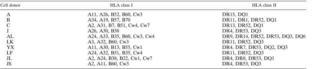
In initial experiments, stimulated cells from 10 different donors (Table 1) were tested for their ability to support the replication of viruses of various phenotypes (Table 2). Two hundred fifty
TCID50of each viral stock was used to infect duplicate wells
that contained donor cells. These viruses, representing a range of phenotypes (Table 2), were either patient isolates or bio-logical clones derived from primary cell cultures and previously characterized (7, 8). The extent of HIV-1 replication was determined by measuring supernatant p24 antigen levels on days 7 and 14 of culture. The results from day 14 for primary isolates are shown in Table 3, and those for biological clones are shown in Table 4. All cultures showed evidence of HIV-1 replication, with a minimum p24 value of 7 ng/ml on day 14 (donor JL and virus N70-2) and a maximum value of 574 ng/ml (donor JS and virus A144). While all donor cells were
suscep-TABLE 1. HLA class I and II determinations in healthy donors
Cell donor HLA class I HLA class II
A A11, A26, B52, B60, Cw3 DR15, DQ1
B A34, A19, B57, B70 DR11, DR1, DR52, DQ1
C A2, A31, B7, B51, Cw4, Cw7 DR13, DR52, DQ1
J A26, A30, B38 DR4, DR53, DQ3
AL A24, A33, B35, B60, Cw3, Cw4 DR9, DR14, DR52, DR53, DQ3, DQ6
LK A3, A32, B60, Cw3 DR11, DR52, DQ3
YX A11, A30, B13, B55, Cw1 DR4, DR7, DR53, DQ2, DQ3
LF A24, A32, B51, B35, Cw4 DR11, DR52, DQ3
JL A2, A24, B38, B22, Cw1, Cw7 DR4, DR8, DR53, DQ1
JS A2, A11, B60, Cw3 DR4, DR53, DQ3
on November 9, 2019 by guest
http://jvi.asm.org/
[image:2.612.58.566.614.727.2]tible to infection, there was a significant range of variation. For example, with HIV-1 isolate A144, the maximum level of replication was seen in cells from donor JS and the minimum in cells from donor J. The maximum range of viral replication was 1.6 orders of magnitude for isolate VS, but in general, the range was less than 20-fold and often less than 10-fold between different donors (Tables 3 and 4).
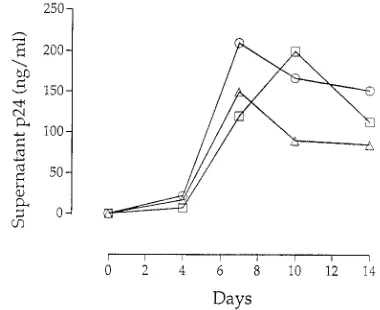
In vitro cocultivation with distinct donor cells selects for a similar quasispecies.PBMC from patient AD30 were cultured with PBMC from donors A, B, and C in triplicate (Fig. 1). Supernatant p24 antigen expression for each set of donor cells was determined by calculating the mean of triplicate cocultures
(Fig. 2). Among each triplicate set of donor cells, the levels of HIV-1 replication were similar (data not shown); as shown in Fig. 2, cells from each donor supported viral replication to comparable levels.
[image:3.612.54.559.82.264.2]Amino acid sequences from the C2-V4 region of gp120 were determined for viruses from uncultured AD30 PBMC, and these were designated by the prefix AD30 (Fig. 3). Although no clones were identical, quasispecies fell into several related groups. For example, AD30-4, -5, -10, and -15, while individ-ually distinct, form their own related grouping. Three clones contained stop codons within the first 15 amino acids of the fragment sequenced (data not shown).
TABLE 2. Summary of known properties of HIV-1 clones and isolates used in this studya
Virus Phenotypeb Infectivity
c
MT-2 HPB-ALL MOLT-4 H9 CEM
Biological clones
NYBC B SI 11 NT 2 2 2
J5H 3 SI 11 1111 11 1 1
J5H 5 SI 11 1111 1 2 2
J5H 9 NSI 2 1 1 1 1
J5H 10 SI 11 1111 1111 1111 1111
N70 1 NSI 2 2 11 2 2
N70 2 NSI 2 2 2 2 2
Patient isolates
A-1 NSI 2 2 NT 2 NT
A-2 SI 11 2 NT 2 NT
B-1 NSI 2 2 NT 2 NT
B-2 SI 11 6 NT 2 NT
C-1 NSI 2 2 NT 2 NT
D-1 NSI 2 2 NT 2 NT
a
Data are taken from previously published works. Biological clones are described in reference 8, and patient isolates are described in reference 7. b
SI, syncytium inducing; NSI, non-syncytium inducing. Viruses were tested in MT-2 cells (7, 8). c
[image:3.612.61.554.475.710.2]Infection was determined by measuring HIV-1 p24 antigen in culture supernatants, as previously described (7, 8).1111, the highest level of infection observed; 2, no observed infection. NT, not tested.
TABLE 3. p24 antigen levels of cultures from HIV-1 viral isolates
Donor Day p24 antigen (pg/ml)
a
A-1 A-2 B-1 B-2 D-1 C-1 VS A144
A 7 5,100 19,000 36,500 174,500 30,000 NT 94,500 8,950
14 118,000 254,000 147,000 232,000 97,000 NT 215,000 176,000
B 7 6,500 81,300 37,500 26,000 NT NT 4,400 12,000
14 244,000 222,000 248,000 485,000 NT NT 17,400 152,000
C 7 NT NT NT NT 5,000 2,200 30,000 47,500
14 NT NT NT NT 132,000 179,000 68,000 64,000
J 7 1,200 5,000 4,000 9,400 10,800 7,900 63,850 18,100
14 58,000 63,000 114,000 88,000 135,000 91,000 60,000 45,000
AL 7 6,900 60,000 20,500 93,000 55,500 24,000 13,300 86,900
14 126,000 210,000 172,000 405,000 143,000 203,000 19,300 158,000
LK 7 NT NT NT NT NT NT 1,920 3,500
14 NT NT NT NT NT NT 10,000 58,000
LF 7 NT NT NT NT NT NT 2,100 8,400
14 NT NT NT NT NT NT 422,000 250,000
JS 7 NT NT NT NT NT NT NT 1,080
14 NT NT NT NT NT NT NT 574,000
JL 7 NT NT NT NT NT NT 2,500 12,000
14 NT NT NT NT NT NT 26,000 73,500
YX 7 NT NT NT NT NT NT 1,500 12,850
14 NT NT NT NT NT NT 10,000 269,000
Day 14 range (fold) 4.2 4.0 2.2 5.5 1.5 2.2 42.2 12.8
a
p24 supernatant antigen levels of cultures on days 7 and 14 following inoculation of healthy donor cells with primary HIV-1 isolates. Data are means of duplicate infections. NT, not tested.
on November 9, 2019 by guest
http://jvi.asm.org/
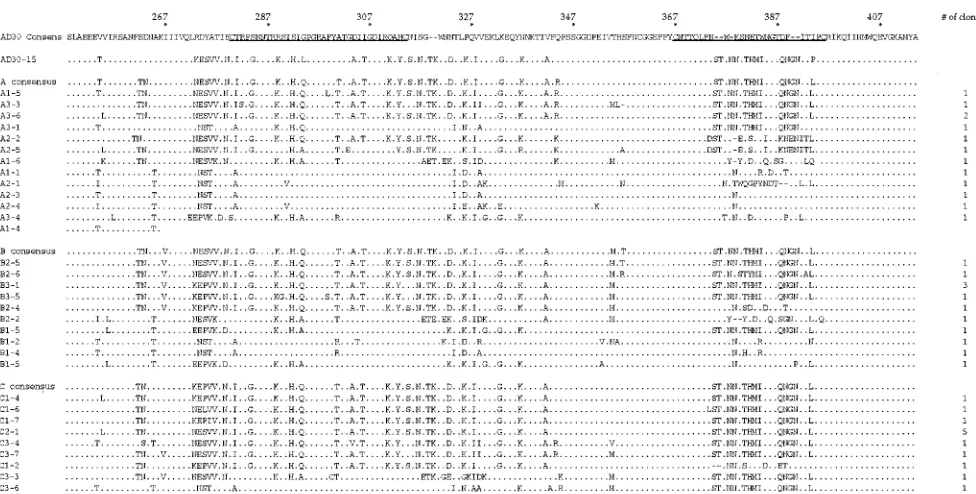
Following coculture, C2-V4 gp120 sequences were found to be more homogeneous than those present in uncultured PBMC (Fig. 4), a result consistent with those of others (19, 20, 25). Upon visual examination, most sequences appeared to be related to clone AD30-15, a minor variant in the original patient PBMC (Fig. 3). While related to the other clones in the patient, AD30-15 was distinct in the V4 region of gp120 (amino acids 367 to 396 of the AD30 consensus); it contained several short insertions. Although AD30-15-like sequences dominated after coculture with cells from different donors in vitro, there were differences in the level of selection for this variant (Fig. 4). Cocultures with cells from donor C appeared to be the most selective, with all 13 clones most closely related to AD30-15 (median amino acid identity, 97%). Cocultures with cells from donor B yielded 9 of 12 clones that were closely
related to AD30-15 (median identity, 93%). Cocultures with cells from donor A yielded 7 of 13 clones that were most related to AD30-15 (median identity, 93%). Additional clones found in cocultures were more related to other clones from patient AD30, most of them to AD30-7, -11, and -12.
[image:4.612.58.558.83.324.2]We next examined the amino acid sequences of clones to look for distinguishing characteristics that might be associated with enhanced viral growth. All cysteines that define the V3 and V4 loops were conserved (residues 280, 314, 367, and 396). Of note, AD30-15 as well as clones related to AD30-15 that dominated following coculture contained two extra N-linked glycosylation sites, one at the beginning of the V4 loop (residue 374) and one at the end of the V4 loop (residue 391). The V4 loop sequences of AD30-15 were unique compared with all others (Fig. 3 and 4), and these N-linked glycosylation sites
FIG. 1. Schematic outline of coculture experiment. Donor cells (106) were
added to 23106cells from donor A, B, or C in triplicate and designated by suffix
1, 2, or 3. Cultures were kept for 14 days, with one-half of the supernatant changed and p24 antigen levels measured every 3 to 4 days. DNA was isolated from uncultured PBMC at this initial time point and 6 months later and from cultured PBMC. Subsequently, the C2-V4 region of gp120 was amplified by PCR, cloned, and sequenced.
FIG. 2. Supernatant p24 antigen levels from cocultures. Data are means of triplicate wells for cells from each donor: donor A (E), donor B (h), and donor C (Ç).
TABLE 4. p24 antigen levels of cultures from HIV-1 biologic clones
Donor Day
p24 antigen (pg/ml)a
NYBC
B J5H-3 J5H-5 J5H-9 J5H-10 N70-1 N70-2 JR CSF JR FL
A 7 56,250 26,500 21,100 18,500 79,150 100,000 33,700 100,000 100,000
14 240,000 42,000 93,000 97,000 176,000 295,000 257,000 200,000 390,000
B 7 NT 21,200 10,000 2,305 33,800 13,000 1,320 21,300 3,170
14 NT 25,000 17,000 8,000 181,500 181,500 165,500 120,000 17,000
C 7 30,000 14,300 12,600 4,500 70,600 100,000 17,500 100,000 23,850
14 430,000 53,000 20,000 10,000 56,500 171,000 30,000 119,000 132,000
J 7 9,500 150,000 152,000 190,000 346,000 150,000 100,000 83,850 260,000
14 105,000 100,000 81,000 95,000 115,000 150,000 85,000 122,000 211,000
AL 7 129,000 140,000 98,500 183,000 183,000 100,000 23,000 188,500 6,000
14 310,000 137,000 109,000 153,000 153,000 75,000 18,500 120,000 58,850
LK 7 NT 3,380 2,050 9,420 9,420 10,000 6,730 10,000 2,470
14 NT 32,000 53,000 53,900 53,900 100,000 66,000 375,000 30,000
LF 7 NT 9,750 11,000 15,000 15,000 11,300 1,830 10,000 1,965
14 NT 85,100 47,250 142,000 142,000 284,000 60,000 528,000 500,000
JS 7 NT 10,000 11,000 12,000 12,000 NT 1,155 11,000 NT
14 NT 149,500 142,500 200,700 200,700 NT 219,000 453,500 NT
JL 7 NT 100,000 50,150 100,000 100,000 87,500 6,500 NT NT
14 NT 73,000 47,000 111,000 111,000 182,000 7,000 NT NT
YX 7 NT 7,060 11,200 33,900 33,900 35,400 3,950 45,450 2,950
14 NT 56,000 48,000 135,900 135,000 16,000 10,000 29,000 27,000
Day 14 range (fold) 4.1 6.0 8.4 25.1 3.7 18.4 36.7 18.2 29.4
a
p24 supernatant antigen levels of cultures on days 7 and 14 following inoculation of healthy donor cells with biological clones. Data are means of duplicate infections. NT, not tested.
on November 9, 2019 by guest
http://jvi.asm.org/
[image:4.612.337.527.544.699.2]often occurred simultaneously with insertions in V4. In addi-tion, the tip of the V3 loop (residues 294 to 297) showed an unusual sequence following coculture. While most clones from the patient had the typical GPGR motif (21), AD30-15 con-tained an LPGR motif (Fig. 3), and many clones that predom-inated following coculture had a QPGR motif (Fig. 4). This QPGR motif was also present in clones AD30-5 and AD30-10, which, as mentioned above, are closely related to AD30-15. It has also been reported that the ability of HIV-1 to induce syncytia may correlate with the presence of two basic amino acids in the V3 loop, at positions 290 and 304 (13) in our numbering system. On this basis, all these viruses are predicted to have the non-syncytium-inducing phenotype.
Evolution of virus present in AD30’s PBMC over time correlates with the virus that grew in vitro. It has been suggested that the genotype of the virus obtained following coculture may predict evolution of the virus in vivo (18, 22); hence, we sought to compare our in vitro culture results with the virus present in vivo at a subsequent time in patient AD30. PBMC were obtained from patient AD30 6 months after the initial blood draw. During this time, the patient remained
healthy, without noticeable decline in CD4 T-cell levels, and he did not receive antiretroviral therapy. DNA was again isolated directly from his PBMC without coculture, and sequences of the C2-V4 region of gp120 were determined (Fig. 5). Seven of eight sequences were highly related to the AD30-15 clone from the earlier time point, whereas one sequence was most highly related to the group of clones AD30-7, -11, and -12.
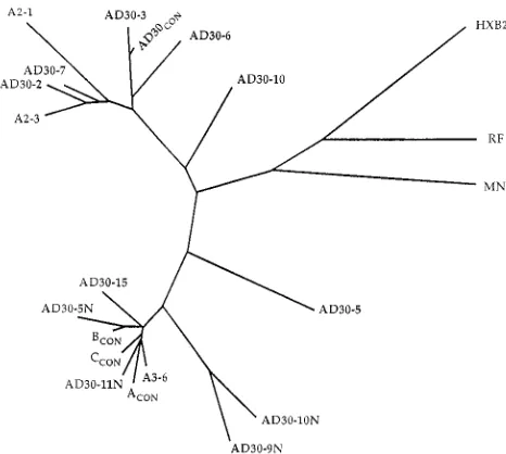
The relationships between sequences found in AD-30 PBMC at two time points and those found in cocultures (Fig. 3 to 5) have been analyzed by the neighbor-joining method with CLUSTAL V software to create an unrooted phylogenetic tree (Fig. 6). These results support the data presented above as the sequences from the second time point cluster with the AD30-15 clone from the first time point, as well as many sequences from cocultures.
[image:5.612.62.554.60.174.2]AD30-15-related viruses demonstrate increased replication kinetics in vitro. We next sought to determine whether AD30-15 and related variants demonstrated any differences in growth ability in vitro compared with that of other variants. The supernatant from day 14 of the C2 coculture, which was homogeneous in the C2-V4 region by sequencing and closely
FIG. 3. Deduced amino acid sequences from uncultured AD30 PBMC. The consensus sequence is at the top, with V3 and V4 loops underlined. Individual clones are indicated by the prefix AD30 and a suffix designating the number of each clone. Identity (.) and a lack of an amino acid (-) are indicated. The number of clones with the same sequence is shown at the right.
FIG. 4. Deduced amino acid sequences from cultured AD30 PBMC. Each sequenced clone is identified by the coculture from which it arose and a number. Other identifications are the same as in the legend to Fig. 3. The number of clones with the same sequence is shown at the right.
on November 9, 2019 by guest
http://jvi.asm.org/
[image:5.612.61.549.459.706.2]related to AD30-15, was defined as the AD30-15 pool. In addition, we obtained three other viruses for comparison by biological cloning of supernatant from day 3 of the original A1 coculture, identified as S4, S7, and S9. Sequence determination indicated that biologic clones S4 and S9 were identical in the C2-V4 region to clone A1-1, which is closely related to the AD30 consensus, while clone S7 was identical in C2-V4 to clone A3-6 (data not shown).
Supernatants from biologic clones were obtained and prop-agated for 5 days in PBMC, titers were determined, and the supernatants were used to infect PBMC, macrophages, and T-cell lines (H9, HPB-ALL, and MT-2). While all viruses grew well in primary PBMC and macrophages, none exhibited any evidence of growth or syncytium formation in any T-cell line tested. However, differences in the growth kinetics of these variants in macrophages and PBMC were observed (Fig. 7). While AD30-15 grew slightly better in PBMC (Fig. 7a), an especially pronounced difference was noticeable for its growth in macrophages (Fig. 7b). Compared with other biological clones from patient AD30, the virus that grew during coculture demonstrated the best replication kinetics in primary PBMC and macrophages in vitro.
DISCUSSION
We have examined the effects that different donor cells may have on the replication of and selection for a particular HIV-1 strain. In our first set of experiments, we obtained results different from those reported by Williams and Cloyd (34), who found that certain donor cells exhibited a high level of resis-tance to infection by certain strains of HIV-1. Our data indicate that the range of infection between different donor cells to a particular HIV-1 isolate or clone is, at most, 1.6 orders of magnitude (40-fold) and generally less than 1 order of magnitude (Tables 3 and 4). This is less than the reported range of 3 orders of magnitude (34). Furthermore, of the 122 virus-host cell combinations we tested, none exhibited com-plete resistance to infection.
We account for the discrepant results obtained in this report and previous studies in several ways. Our technique to assay for growth involves measuring the kinetics of viral replication following a standard inoculum, while Williams and Cloyd used endpoint dilution of a viral stock in PBMC from a particular donor (34). In part, this may account for some differences. However, a more important difference may be our use of viruses that were derived from short-term, low-passage-num-ber cultures in primary PBMC. Previous experiments used viruses that were first expanded in T-cell lines, a process which is selective for viral variants with properties quite different from primary viruses (18, 27). Tropism for T-cell lines has been associated with many features, including specific changes in the
V2 and V3 regions of gp120 (8, 13, 17) as well as increased sensitivity to soluble CD4 (9, 26, 27). Results obtained with a virus that has been passaged in T-cell lines may therefore reflect the properties of an extreme variant of HIV-1, rather than one which is found in patients. Our results indicate that with viruses passaged for a short time in primary PBMC, no host cells demonstrate resistance to infection by a particular virus; all donor cells appear to be infectible by HIV-1.
Nevertheless, there are some differences in replication ki-netics, up to 40-fold on day 14, among cells from different donors. However, the virus that grows to the highest level in one set of PBMC does not necessarily do so in another donor’s PBMC (Tables 3 and 4). Factors that have not been controlled
for in these experiments include the percentage of CD41cells
[image:6.612.66.546.64.177.2]in PBMC from different donors and the degree of activation of cells following stimulation. It is arguable that differences in replication kinetics may decrease even further if these param-eters are taken into account. Other factors that might account for the variation between donors include the effects of host cell-derived molecules on the viral surface that may influence infection. These molecules may vary in structure and surface density. In addition, CD4, the receptor for HIV-1, is not expressed at equivalent levels in different individuals and could therefore influence the extent and rapidity of infection by
FIG. 5. Deduced amino acid sequences from uncultured AD30 PBMC 6 months after the initial time point. Identifications are the same as in the legends to Fig. 3 and 4, except that these clones are designated with the suffix N.
FIG. 6. Unrooted phylogentic tree that represents genetic distances between sequenced clones. Sequence identifications are the same as in Fig. 3 to 5, with HIVRF, HIVHXB2, and HIVMNincluded as reference markers. This tree was
constructed as described in Materials and Methods.CON, consensus sequence.
on November 9, 2019 by guest
http://jvi.asm.org/
[image:6.612.317.550.482.691.2]HIV-1. While these factors are important in our experiments, they appear to exert effects that are lower in magnitude than previously thought. These results not only have strong practical implications for investigators doing quantitative cultures and other assays based on the use of primary PBMC but also have relevance to the pathogenesis of HIV-1 infection. It has been reported that certain persons either resist infection despite multiple exposures to HIV-1 or remain healthy despite long-term infection (2, 22, 24, 28). Our results here suggest that these clinical situations are not the consequence of an intrinsic resistance of certain donor cells to HIV-1 infection. This conclusion is supported by recent observations made in our
laboratory that the CD41T cells from long-term survivors are
readily susceptible to HIV-1 infection in vitro (3).
We also examined the process by which the selection of HIV-1 variants occurs in vitro. While much has been reported on the selection of viral variants during in vitro culturing (19, 20, 25) and in vivo transmission (30, 35, 38), the mechanisms behind this selection have not been identified. In the case we studied here, our results indicate that this selection is primarily related to viral factors and mostly independent of the host cell. The most important factor appears to be viral growth proper-ties in PBMC and macrophages (Fig. 7). However, the effects
of the particular donor cell population cannot be discounted. It is apparent that cells from donor C selected for an AD30-15-like virus uniformly, while those from donors A and B were clearly less selective. Nevertheless, given the results for this one case, it appears that the primary influence behind selection for a particular variant is intrinsic to the virus, with the origin of the host cell playing a secondary role. Because we studied only one patient, further studies are required to confirm the generality of our findings.
These results have general implications for experimental studies that use primary PBMC in assays for drug resistance, virus neutralization, and growth kinetics. There has been considerable concern regarding the validity of PBMC-based neutralization assays with primary viruses (15, 24). In the few studies that have addressed the issue of PBMC infectibility, variation ranges of up to 1,000-fold were reported (5, 34), but most studies have simply ignored this potential experimental variable. Our data demonstrate that while infectibility differ-ences should be taken into account when designing such experiments, they are far less than previously reported (5, 34). It has been stated that ‘‘to culture is to disturb,’’ meaning that cultured virus does not adequately reflect the viral popu-lation present (25). This notion has led some to minimize the importance of results obtained from studies done with cultured viruses. However, given our current findings and those of others (19, 20), it appears that the clause ‘‘to culture is to select’’ would be more accurate. Indeed, the HIV-1 selected by in vitro cultivation may represent a viral population that is more biologically relevant.
ACKNOWLEDGMENTS
We are grateful to Yunzhen Cao and Ruth Connor for technical assistance; Lin Qi Zhang for help with phylogenetic tree analysis; Charles Farthing for identifying the patient; and Ruth Connor, Eric Delwart, Richard Koup, and particularly John Moore for critical reading of the manuscript.
This work was supported by NIH grants AI24030, AI25541, AI27742, AI30358, AI32427, and AI27665, the Ernst Jung Foundation, and the Aaron Diamond Foundation. A.I.S. was supported by an NIH Medical Scientist Training Program Fellowship (GM07308) at the New York University School of Medicine and by a grant from the Pediatric AIDS Foundation.
REFERENCES
1. Arthur, L. O., J. W. Bess, Jr., R. C. Sowder II, R. E. Beneviste, D. L. Mann,
J.-C. Chermann, and L. E. Henderson.1992. Cellular proteins bound to immunodeficiency viruses: implications for pathogenesis and vaccines. Sci-ence 258:1935–1938.
2. Buchbinder, S., D. Mann, L. Louie, F. Villinger, M. Katz, and S. Holmberg. 1993. Healthy long-term positives (HLPs): genetic cofactors for delayed HIV disease progression, p. 46. In IXth International Conference on AIDS, vol. I.
3. Cao, Y., L. Qin, L. Zhang, and D. D. Ho. Virological and immunological characterization of long-term survivors of HIV-1 infection. Submitted for publication.
4. Cichiutek, K., H. Merget, S. Norley, R. Linde, W. Kreuz, M. Gahr, and R.
Kurth.1992. Development of a quasispecies of human immunodeficiency virus (HIV-1). Proc. Natl. Acad. Sci. USA 89:7365–7369.
5. Cloyd, M. W., and B. E. Moore. 1990. Spectrum of biological properties of human immunodeficiency virus (HIV-1) isolates. Virology 174:103–116. 6. Connor, R. I., and D. D. Ho. 1994. Human immunodeficiency virus type 1
variants with increased replicative capacity develop during the asymptomatic stage before disease progression. J. Virol. 68:4400–4408.
7. Connor, R. I., H. Mohri, Y. Cao, and D. D. Ho. 1993. Increased viral burden and cytopathicity correlate temporally with CD41T-lymphocyte decline and clinical progression in human immunodeficiency virus type 1-infected indi-viduals. J. Virol. 67:1772–1777.
8. Connor, R. I., D. W. Notermans, H. Mohri, Y. Cao, and D. D. Ho. 1993. Biological cloning of functionally diverse quasispecies of HIV-1. AIDS Res. Hum. Retroviruses 9:541–546.
[image:7.612.84.276.69.430.2]9. Daar, E. S., X. L. Li, T. Moudgil, and D. D. Ho. 1990. High concentrations of recombinant soluble CD4 are required to neutralize primary human FIG. 7. Growth kinetics of four biologically cloned viruses, S4 (Ç), S7 (É), S9
(h), and AD30-15 pool (E), in PBMC (a) and macrophages (b). Two hundred fifty TCID50of biologically cloned virus was cocultured with donor cells, and
culture supernatants were monitored for p24 antigen expression. Data are means of duplicate infections, with error bars showing the standard errors of the means.
on November 9, 2019 by guest
http://jvi.asm.org/
immunodeficiency virus type 1 isolates. Proc. Natl. Acad. Sci. USA 87:6574– 6578.
10. Daar, E. S., T. Moudgil, R. D. Meyer, and D. D. Ho. 1991. Transient high levels of viremia in patients with primary human immunodeficiency virus type 1 infection. N. Engl. J. Med. 324:961–964.
11. Domingo, E., J. J. Holland, and P. Ahlquist. 1988. RNA genetics. CRC Press, Boca Raton, Fla.
12. Dulbecco, R. 1988. End-point method-measurement of the infectious titer of a viral sample, p. 22–25. In R. Dulbecco and H. S. Ginsberg (ed.), Virology, 2nd ed. J. B. Lippincott, Philadelphia.
13. Fouchier, R. A. M., M. Groenink, N. A. Kootstra, M. Tersmette, H. G.
Huisman, F. Miedema, and H. Schuitemaker.1992. Phenotype-associated sequence variation in the third variable domain of the human immunodefi-ciency virus type 1 gp120 molecule. J. Virol. 66:3183–3187.
14. Goodenow, M., T. Huet, W. Saurin, S. Kwok, J. Sninsky, and S.
Wain-Hobson.1989. HIV-1 isolates are rapidly evolving quasispecies: evidence for viral mixtures and preferred nucleotide substitutions. J. Acquired Immune Defic. Syndr. 2:344–352.
15. Hanson, C. V. 1994. Measuring vaccine induced HIV neutralization: report of a workshop. AIDS Res. Hum. Retroviruses 10:645–648.
16. Higgins, D. G., and P. M. Sharp. 1989. CLUSTAL: a package for performing multiple sequence alignments on a microcomputer. Gene 73:237–244. 17. Hwang, S. S., T. J. Boyle, H. K. Lyerly, and B. R. Cullen. 1991. Identification
of the envelope V3 loop as the primary determinant of cell tropism in HIV-1. Science 253:71–74.
18. Kabat, D., S. L. Kozak, K. Wehrly, and B. Chesebro. 1994. Differences in CD4 dependence for infectivity of laboratory-adapted and primary patient isolates of human immunodeficiency virus type 1. J. Virol. 68:2570–2577. 19. Kuiken, C. L., J.-L. de Jong, E. Baan, W. Keulen, M. Tersmette, and J.
Goudsmit.1992. Evolution of the V3 envelope domain in proviral sequences and isolates of human immunodeficiency virus type 1 during transition of the viral biological phenotype. J. Virol. 66:4622–4627.
20. Kusumi, K., B. Conway, S. Cunningham, A. Berson, C. Evans, A. K. N.
Iversen, D. Colvin, M. V. Gallo, S. Coutre, E. G. Shpaer, D. V. Faulkner, A. deRonde, S. Volkman, C. Williams, M. S. Hirsch, and J. I. Mullins.1992. Human immunodeficiency virus type 1 envelope gene structure and diversity in vivo and after cocultivation in vitro. J. Virol. 66:875–885.
21. LaRosa, G. J., K. Weinhold, A. T. Profy, A. J. Langolis, G. R. Dreesman,
R. N. Boswell, P. Shadduck, D. P. Bolognesi, T. J. Matthews, E. A. Emini, and S. D. Putney.1990. Conserved sequence and structural elements in the HIV-1 principal neutralizing determinant: further clarifications. Science
253:1146.
22. Mann, D. L., M. Carrington, M. O’Donnell, T. Miller, and J. Goedert. 1992. HLA phenotype is a factor in determining rate of disease progression and outcome in HIV-1 infected individuals. AIDS Res. Hum. Retroviruses
8:1345–1346.
23. Mann, D. L., C. Murray, M. O’Donnell, W. A. Blattner, and J. J. Goedert. 1990. HLA antigen frequencies in HIV-1 related Kaposi’s sarcoma. J. Acquired Immune Defic. Syndr. 3(Suppl. 1):51–55.
24. Matthews, T. J. 1994. Dilemma of neutralization resistance of HIV-1 field isolates and vaccine development. AIDS Res. Hum. Retroviruses 10:631– 632.
25. Meyerhans, A., R. Cheynier, J. Albert, M. Seth, S. Kwok, J. Sninsky, L.
Morfeldt-Månson, B. Asjo¨, and S. Wain-Hobson.1989. Temporal fluctua-tions in HIV quasispecies in vivo are not reflected by sequential HIV isolations. Cell 58:901–910.
26. Moore, J. P., L. C. Burkly, R. I. Connor, Y. Cao, R. Tizard, D. D. Ho, and
R. A. Fisher.1993. Adaptation of two primary human immunodeficiency virus type 1 isolates to growth in transformed T cell lines correlates with alterations in the responses of their envelope glycoproteins to soluble CD4. AIDS Res. Hum. Retroviruses 9:529–539.
27. Moore, J. P., J. A. McKeating, Y. Huang, A. Ashkenazi, and D. D. Ho. 1992. Virions of primary human immunodeficiency virus type 1 isolates resistant to soluble CD4 (sCD4) neutralization differ in sCD4 binding and glycoprotein gp120 retention from sCD4-sensitive isolates. J. Virol. 66:235–243. 28. Plummer, F. A., K. Fowke, N. J. D. Nagelkerke, J. Simonsen, J. Bwayo, and
E. Ngugi.1993. Evidence of resistance to HIV among continuously exposed prostitutes in Nairobi, Kenya, p. 23. In IXth International Conference on AIDS, vol. I.
29. Sambrook, J., E. F. Fritsch, and T. Maniatis. 1989. Molecular cloning: a laboratory manual, 2nd ed. Cold Spring Harbor Laboratory Press, Cold Spring Harbor, N.Y.
30. Scarlatti, G., T. Leitner, E. Halapi, J. Wahlberg, P. Marchisio, M. A.
Clerici-Schoeller, H. Wigzell, E. M. Fenyo, J. Albert, M. Uhlen, and P. Rossi.
1993. Comparison of variable region 3 sequences of human immunodefi-ciency virus type 1 from infected children with the RNA and DNA sequences of the virus populations of their mothers. Proc. Natl. Acad. Sci. USA
90:1721–1725.
31. Tersmette, M., R. E. Y. de Goede, B. J. M. Al, I. N. Winkel, R. A. Gruters,
H. T. Cuypers, H. G. Huisman, and F. Miedema.1988. Differential syncyti-um-inducing capacity of human immunodeficiency virus isolates: frequent detection of syncytium-inducing isolates in patients with acquired immuno-deficiency syndrome (AIDS) and AIDS-related complex. J. Virol. 62:2026– 2032.
32. Walker, C. M., D. J. Moody, D. P. Stites, and J. A. Levy. 1986. CD81 lymphocytes can control HIV infection in vitro by suppressing virus replica-tion. Science 234:1563–1566.
33. Weiss, R. A. 1993. How does HIV cause AIDS? Science 260:1273–1279. 34. Williams, L. M., and M. W. Cloyd. 1991. Polymorphic human gene(s)
determines differential susceptibility of CD4 lymphocytes to infection by certain HIV-1 isolates. Virology 184:723–728.
35. Wolinsky, S., C. Wike, B. Korber, C. Huto, W. Parks, L. Rosenblum, K.
Kunstman, M. Furtado, and J. Mun˜oz. 1992. Selective transmission of human immunodeficiency virus type 1 variants from mothers to infants. Science 255:1134–1137.
36. York-Higgins, D., C. Cheng-Mayer, D. Bauer, J. A. Levy, and D. Dina. 1990. Human immunodeficiency virus type 1 cellular host range, replication, and cytopathicity are linked to the envelope region of the viral genome. J. Virol.
64:4016–4020.
37. Zhang, L. Q., P. MacKenzie, A. Cleland, E. C. Holmes, A. J. Leigh Brown,
and P. Simmonds.1993. Selection for specific sequences in the external envelope protein of human immunodeficiency virus type 1 upon primary infection. J. Virol. 67:3345–3356.
38. Zhu, T., H. Mo, N. Wang, D. S. Nam, Y. Cao, R. A. Koup, and D. D. Ho. 1993. Genotypic and phenotypic characterization of HIV-1 in patients with primary infection. Science 261:1179–1181.