0022-538X/95/$04.0010
Copyrightq1995, American Society for Microbiology
The Hemagglutinin Envelope Protein of Canine Distemper Virus
(CDV) Confers Cell Tropism as Illustrated by CDV
and Measles Virus Complementation Analysis
LULI BAR-LEV STERN,
1MICHAEL GREENBERG,
1JONATHAN M. GERSHONI,
2AND
SHMUEL ROZENBLATT
1*
Department of Molecular Microbiology and Biotechnology
1and Department of
Cell Research and Immunology,
2Tel-Aviv University, Tel Aviv, Israel 69978
Received 30 June 1994/Accepted 23 November 1994
Measles virus (MV) and canine distemper virus (CDV) are morbilliviruses that cause acute illnesses and
several persistent central nervous system infections in humans and in dogs, respectively. Characteristically, the
cytopathic effect of these viruses is the formation of syncytia in permissive cells. In this study, a vaccinia virus
expression system was used to express MV and CDV hemagglutinin (HA) and fusion (F) envelope proteins. We
found that cotransfecting F and HA genes of MV or F and HA genes of CDV resulted in extensive syncytium
formation in permissive cells while transfecting either F or HA alone did not. Similar experiments with
heterologous pairs of proteins, CDV-F with MV-HA or MV-F with CDV-HA, caused significant cell fusion in
both cases. These results indicate that in this expression system, cell fusion requires both F and HA; however,
the functions of these proteins are interchangeable between the two types of morbilliviruses. Human-mouse
somatic hybrids were used to determine the human chromosome conferring susceptibility to either MV and
CDV. Of the 12 hybrids screened, none were sensitive to MV. Two of the hybrids containing human
chromo-some 19 formed syncytia following CDV infection. In addition, these two hybrids underwent cell fusion when
cotransfected with CDV-F and CDV-HA (but not MV-F and MV-HA) glycoproteins by using the vaccinia virus
expression system. To discover the viral component responsible for cell specificity, complementation
experi-ments coexpressing CDV-HA with MV-F or CDV-F with MV-HA in the CDV-sensitive hybrids were performed.
We found that syncytia were formed only in the presence of CDV-HA. These results support the idea that the
HA protein is responsible for cell tropism. Furthermore, while the F protein is necessary for the fusion process,
it is interchangeable with the F protein from other morbilliviruses.
Canine distemper virus (CDV) and measles virus (MV) are
members of the genus Morbillivirus within the family
Paramyxoviridae. Both CDV and MV are associated with acute
illnesses and several persistent central nervous system
infec-tions, some of them fatal (17, 22, 28, 37). Moreover, a transient
immunodeficiency develops during the course of the acute
disease, and consequently it has been suggested that measles
can serve as a model for studying immune restoration (13).
The pathogenesis of a morbillivirus infection begins with
viral attachment to the surface of the host cell. In nature, CDV
and MV are specific to their hosts, i.e., dogs and humans,
respectively; nevertheless, in vitro both viruses can infect cell
lines of human or simian origin. After attachment, fusion of
the virion envelope with the cellular plasma membrane takes
place (18). These processes are elicited by two virus-encoded
glycoproteins of the lipid envelope. The major glycoprotein,
hemagglutinin (HA), possesses hemagglutination activity and
is responsible for the attachment of the virus to the host cell.
The second glycoprotein, the fusion (F) protein, mediates the
fusion of the cellular and viral membranes and subsequently
causes syncytium formation (24, 31). For the paramyxoviruses
in general, there has been some controversy about the role of
HA in the fusion process. For example, it has been found by
some that the F protein is sufficient to cause cell fusion (2, 14,
29), whereas others have shown that induction of cell fusion
required both F and HA glycoproteins (9, 16, 25, 32, 35, 39).
In this study, a vaccinia virus transient-expression system was
used to further define the significance of the HA glycoprotein
in the fusion process of morbilliviruses. Different combinations
of F and HA proteins from MV and CDV were coexpressed in
cell lines and in somatic hybrids to determine whether
func-tional complementation in the fusion process can take place.
Our results support the idea that the HA glycoprotein plays a
crucial role in determining cell specificity.
MATERIALS AND METHODS
Cells and viruses.Human epithelial cells (HeLa and HeLa T4), murine fibro-blasts (LA-9), and simian kidney cells (CV-1 and Vero) were used in these experiments. Mouse-human hybrid cell lines (Table 1) (33) were a generous gift from Lester Shulman, Shiba Medical Center, Tel-Hashomer, Israel. Cells were grown in Dulbecco’s modified Eagle’s medium supplemented with 10% fetal calf serum, 2 mM glutamine, 60 mg of penicillin per liter, and 100 mg of streptomycin per liter. The Edmonston MV strain and the Onderstepoort CDV strain were routinely grown in CV-1 cells. A vaccinia virus encoding T7 polymerase (vTF7-3) was provided by B. Moss, National Institutes of Health, Bethesda, Md. (11).
Construction of recombinant plasmids.Initially, to isolate the full-length F and HA genes from both CDV and MV, two cDNA libraries were constructed. These were screened with specific 20-mer oligonucleotides corresponding to previously reported sequences of CDV and MV F and HA genes (1, 4, 7, 30). All four cDNA clones were isolated in their entirety. For expression of proteins in the vTF7-3 system, we cloned genes into the multiple-cloning site of the vector pTM-1 (a gift from O. Elroy-Stein, Tel-Aviv University) downstream of the T7 promoter (10, 26). Four constructs were prepared by PCR technology. In each case, NcoI sites were introduced at the 59ends of the coding regions of the gene of interest, replacing the original ATG translation initiation codon. This was done by digesting the PCR products once with NcoI and then once at a conve-nient restriction site downstream of the 59end (keeping the PCR fragments as short as possible) and finally cloning these fragments into the remainder of the 39 region of each gene. Taq polymerase was purchased from Cetus, and the reactions were carried out as specified by the manufacturers of the Hybaid Intelligent Heating Block (Hybaid, Middlesex, United Kingdom). The sequences of all the PCR portions cloned in the recombinant plasmids were confirmed, and * Corresponding author. Phone: 972-3-6407509. Fax: 972-3-6409407.
1661
on November 9, 2019 by guest
http://jvi.asm.org/
the clones were further tested by in vitro coupled transcription-translation reac-tions (Promega TNT; technical bulletin no. 126).
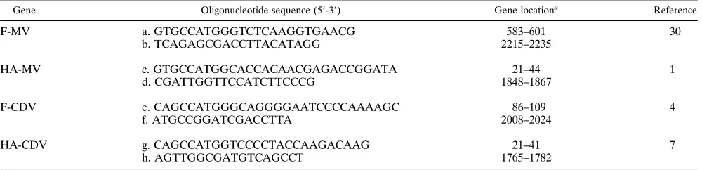
(i) pTM-F-MV.Oligonucleotides a and b corresponding to the 59and 39ends of the F gene (Table 2) were used as primers in PCR. The resulting fragment was digested with NcoI and BamHI and ligated with a pTM-1 clone containing the full-length F cDNA from which the corresponding fragment (plus regions from the polylinker) was removed. Next, the pTM-1 derivative was digested with
BamHI plus HpaI, and a 1,500-bp fragment was removed from the gene and
replaced by a corresponding BamHI-HpaI fragment isolated from the original F-MV cDNA clone.
(ii) pTM-HA-MV.Oligonucleotides c and d (Table 2) generated a PCR frag-ment which was digested with NcoI plus NheI and subcloned into a pTM-1 clone containing the full-length cDNA HA gene digested with the same enzymes (replacing the corresponding 59fragment that was removed).
(iii) pTM-F-CDV.Oligonucleotides e and f were used in PCR to form a DNA fragment which was then digested with NcoI plus EcoRI. The resulting 570-bp fragment was ligated into a NcoI-EcoRI window created in pTM-1. The clone was subsequently digested with EcoRI and was ligated with a portion of the 39 end of the F-CDV gene cloned from the original cDNA.
(iv) pTM-HA-CDV.Oligonucleotides g and h were used in PCR to produce a DNA fragment that was digested with NcoI plus XhoI and cloned into the
NcoI-XhoI sites of the pTM-1 polylinker. The construct was then digested with XhoI plus SalI, and the 39region from the original HA-CDV gene was cloned, replacing the existing fragment.
vTF7-3 infection–pTM-1 transfection assays.In the vaccinia virus T7 expres-sion system (vvT7), cells were first infected with vTF7-3 (a vaccinia virus encod-ing T7 polymerase) and then transfected with the recombinant plasmids carryencod-ing the genes of interest under the control of the T7 promoter. Confluent 35-mm dishes of cells were infected with vTF7-3 at a multiplicity of infection of 50 for 45 min and then transfected with 3 to 5mg of DNA by using Lipofectin or Transfectase (Gibco-BRL) as specified by the manufacturer (3).
Fusion assays.Cells were cotransfected by the method described above with 3 to 5mg each of various DNA combinations of F and HA plasmids. At 24 h postinfection, the cells were fixed in methanol, stained with Giemsa, and pho-tographed.
RESULTS
Coexpression of F and HA proteins in CV-1 cells mediates
cell fusion.
To study fusion in morbilliviruses, the recombinant
vvT7 expression system was used. The cDNA clones encoding
the F and HA proteins of MV and CDV were cloned into the
pTM-1 vector. CV-1 cells were infected with vTF7-3 and
trans-fected with either pTM-F-MV or pTM-HA-MV. The F and
HA recombinant proteins were detected by
immunoprecipita-tion. HA is accumulated to better than 10% of the total
[
35S]methionine incorporated during the course of an
[image:2.612.59.556.84.227.2]experi-ment. The relative amount of F protein was found to be
ex-tremely small compared with that of HA protein (data not
shown). However, in both cases the amount of recombinant
protein was found to be sufficient for biological activity, as
illustrated in the following experiments. To establish the
pres-ence of functional recombinant proteins, the cultures were
examined. Transfecting either F or HA alone under various
experimental conditions (e.g., at pH 5 to 8 or in a coincubation
with uninfected CV-1 cells) had no effect. On the other hand,
introduction of both F and HA genes into CV-1 cells resulted
in extensive syncytium formation. Similarly, syncytia were
formed when F and HA genes of CDV were coexpressed in
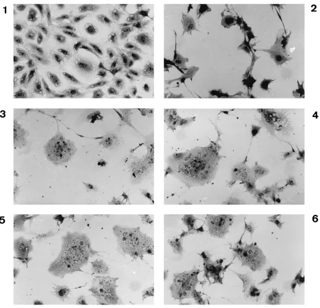
CV-1 cells (Fig. 1). A comparison of panels 2 to 6 in Fig. 1 with
panels 7 and 8 illustrates that in the absence of fusion the
vaccinia virus-infected cells undergo extensive contraction.
This might reflect progression of two simultaneous
phenom-ena: (i) cytopathic effect elicited by vaccinia virus alone and (ii)
TABLE 1. Ability of mouse-human hybrids to undergo cell fusion following either MV/CDV infection or viral glycoprotein coexpression
Cell line Human chromosome(s) Viral infection a
Coexpression in vv/T7 system
MV CDV MVgpb CDVgp
157BnpT-4 3, 11, 14, X 2 2 2 2
AIM-15a-A1 1, 2, 11, 12, 13, 14, 15, 17, 18, 20, 21, X 2 2 2 2
AIM-15a-A2 1, 2, 11, 13, 14, 15, 17, 18, 21, X 2 2 2 2
AIM-15a-A3 1, 2, 5, 11, 13, 14, 15, 17, 18, 21, X 2 2 2 2
AIM-15a-B5 1, 2, 11, 13, 14, 15, X 2 2 2 2
AIM-15a-C2 1, 2, 7, 11, 12, 13, 14, 15, 17, 18, 21, X 2 2 2 2
AIM-15a-C3 1, 2, 7, 11, 12, 13, 14, 15, 17, 18, 20, 21, X 2 2 2 2
AIM-15a-C5 1, 2, 7, 11, 13, 14, 15, 17, 18, 20, 21, X 2 2 2 2
IL-115 7, 11, 12, 15, 17, 19 2 1 2 1
Il-115-4d 11, 12, 17, 19 2 1 2 1
L-287-C 7, 11, 17, X 2 2 2 2
WAV-A17 21 2 2 2 2
aCells were infected at 1 PFU per cell. bgp, glycoprotein.
TABLE 2. Oligonucleotides used to clone viral glycoproteins genes
Gene Oligonucleotide sequence (59-39) Gene locationa Reference
F-MV a. GTGCCATGGGTCTCAAGGTGAACG 583–601 30
b. TCAGAGCGACCTTACATAGG 2215–2235
HA-MV c. GTGCCATGGCACCACAACGAGACCGGATA 21–44 1
d. CGATTGGTTCCATCTTCCCG 1848–1867
F-CDV e. CAGCCATGGGCAGGGGAATCCCCAAAAGC 86–109 4
f. ATGCCGGATCGACCTTA 2008–2024
HA-CDV g. CAGCCATGGTCCCCTACCAAGACAAG 21–41 7
h. AGTTGGCGATGTCAGCCT 1765–1782
a
Nucleotide number according to numeration in cited reference.
1662 BAR-LEV STERN ET AL. J. VIROL.
on November 9, 2019 by guest
http://jvi.asm.org/
[image:2.612.58.558.597.718.2]FIG. 1. Induction of fusion in CV-1 cells by coexpression of HA and F of MV and CDV. The photographs were taken at 24 h postinfection. 1, Mock-infected cells; 2, vTF7-3-infected cells; 3, vTF7-3-infected, pTM-HA-MV-transfected cells; 4, vTF7-3-infected, pTM-HA-CDV-transfected cells; 5, vTF7-3-infected, pTM-F-MV-transfected cells; 6, vTF7-3-infected, pTM-F-CDV-pTM-F-MV-transfected cells; 7, vTF7-3-infected, pTM-F-MV- and pTM-HA-MV-copTM-F-MV-transfected cells; 8, vTF7-3-infected, pTM-F-CDV- and pTM-HA-CDV-cotransfected cells. Cell fusion was observed as early as 8 h postinfection. Magnification,3110.
on November 9, 2019 by guest
http://jvi.asm.org/
expression of both proteins, leading to syncytium formation.
Such syncytium formation could be achieved in both human
and monkey cells (permissive for the viruses) but not murine
cells such as LA-9.
Functional complementation between MV and CDV
glyco-proteins in syncytium formation.
CDV and MV are closely
related morbilliviruses that exhibit immunological
cross-reac-tivity and share sequence homology, yet these viruses infect
different hosts in nature and their HA proteins are not highly
conserved (7, 17, 36). To determine whether the interaction
between F and HA in the fusion process is virus specific, we
determined if MV-HA could replace CDV-HA in syncytium
formation mediated by CDV-F. Cotransfection of MV-HA
and CDV-F resulted in syncytium formation in CV-1 cells (Fig.
2). Compared with the homologous systems, however, the
ex-tent and size of the heterologous F- and HA-mediated syncytia
were somewhat smaller as assessed by the smaller number of
nuclei per syncytium and the rate of their appearance. In a
reciprocal experiment, i.e., cotransfection with CDV-HA and
MV-F, considerable cell fusion in CV-1 cells was observed,
although to a lesser extent than that of the CDV-F and
CDV-HA pair. The results shown in Fig. 1 and 2 therefore
illustrate that both F and HA glycoproteins are necessary
for cell fusion and that interviral complementation is
possi-ble.
HA glycoprotein determines cell specificity in somatic
hy-brids.
To identify the cellular component involved in virus
[image:4.612.73.540.76.526.2]binding, several mouse-human hybrid cell lines containing
var-ious human chromosomes were used (Table 1). These cells
were tested for their ability to fuse following MV or CDV
FIG. 2. Functional complementation between MV-HA and CDV-F in CV-1 cells. The photographs were taken 24 h postinfection. 1, Mock-infected CV-1 cells; 2, vTF7-3-infected CV-1 cells; 3, vTF7-3-infected, pTM-F-MV- and pTM-HA-MV-cotransfected cells; 4, vTF7-3-infected, pTM-F-CDV- and pTM-HA-CDV-cotrans-fected cells; 5, vTF7-3-inpTM-HA-CDV-cotrans-fected, pTM-F-CDV- and pTM-HA-MV-cotranspTM-HA-CDV-cotrans-fected cells; 6, vTF7-3-inpTM-HA-CDV-cotrans-fected, pTM-F-MV- and pTM-HA-CDV-cotranspTM-HA-CDV-cotrans-fected cells. Magnification,3110.
1664 BAR-LEV STERN ET AL. J. VIROL.
on November 9, 2019 by guest
http://jvi.asm.org/
infection. None of the somatic hybrids fused as a result of MV
infection; however, following infection with CDV, two cell
lines, IL-115 and IL-115-4d, formed syncytia (Fig. 3). These
two cell lines are unique, because they are exclusive for human
chromosome 19. This finding therefore creates a concrete
dis-tinction between the two viruses used in this study. The
ques-tion arises which viral glycoprotein is responsible for the
se-lective nature of the somatic-hybrid infection. To address this
question, it was first necessary to confirm that syncytia could be
elicited by using the CDV recombinant glycoproteins. Indeed,
cell lines IL-115 and IL-115-4d continued to show selective
susceptibility to CDV HA- and F-induced syncytium formation
(Fig. 4) in contrast to their response to the MV glycoproteins.
Heterologous complementation experiments were then
performed. Figure 5 depicts such an experiment, in which
the IL-115 cell line was cotransfected with CDV-HA and
MV-F or with CDV-F and MV-HA. Syncytia can be detected
only for experiments in which the CDV-HA was expressed
(Fig. 5).
These results attribute a critical role to the HA glycoprotein
in determining cell tropism, while the F protein is a necessary
factor in the fusion process but is interchangeable with the F
protein from other morbilliviruses.
DISCUSSION
The function of the HA protein in viral attachment has been
well established for various paramyxoviruses by a variety of
methods (12, 15, 18, 24, 37). There is still some controversy,
however, about the possible role for HA in the fusion process.
Several reports have demonstrated that the F protein is
suffi-cient for membrane fusion when recombinant viral
glycopro-teins are used (2, 14, 29). Alternatively, it has been shown that
coexpression of the HA and F proteins was necessary (9, 16, 25,
FIG. 3. MV and CDV infection of mouse-human hybrid IL-115 cells. Photographs were taken at 24 h postinfection. 1, MV-infected cells (multiplicity of infection, ;0.5). 2, CDV-infected cells (multiplicity of infection,;0.5). Magnification,3220.
on November 9, 2019 by guest
http://jvi.asm.org/
32, 35, 39). Consequently, several models have been suggested,
substantiating the significance of the HA protein in triggering
cell fusion (18, 35). In the studies described here, using the
vvT7 expression system, we observed syncytium formation only
when F and HA proteins of either CDV or MV were
coex-pressed in permissive cells. It is generally believed that the
interaction between HA and F in the fusion process is
exclu-sively virus strain specific, and homologous pairs of F and HA
from identical viruses are required to produce cell fusion (14,
16, 18, 20, 23). In one instance, reported combinations of
functional F and HA proteins from different isolates of MV
exhibiting a 97% amino acid identity did not form syncytia (6).
By using the vvT7 expression system, functional
complemen-tation in cell fusion between the glycoproteins of far more
distant viruses, namely, CDV and MV, was tested. These
vi-ruses share only a 36% amino acid sequence identity for their
HA proteins and a 67% identity for their F proteins (4, 7).
Nonetheless, such interspecies complementation has been
il-lustrated.
Following the infection of mouse-human somatic hybrids
with CDV, two cell lines, IL-115 and IL-115-4d, were found to
be permissive for infection by CDV and permissive for
syn-cyium formation mediated by recombinant CDV
glycopro-teins. The sensitivity of hybrid cells to viruses has been
attrib-uted in many cases to the presence of specific receptors
encoded by the human chromosomes. The hybrid-cell analysis
technique has helped to identify these specific viral receptors
(8, 21, 33, 34). Therefore, noting the sensitivity of both distinct
hybrid cell lines to CDV, our data suggest that the CDV
receptor involved in specific recognition is encoded by human
chromosome 19. Because the chromosomal content of somatic
hybrid cells tends to be unstable, a Southern analysis of the
hybrid cell lines was also carried out with a specific probe from
the q arm of human chromosome 19. The results obtained
FIG. 4. Induction of fusion in mouse-human hybrid IL-115 cells by coexpression of HA and F glycoproteins. Photographs were taken at 24 h postinfection. 1, vTF7-3-infected, pTM-F-MV- and pTM-HA-MV-cotransfected cells; 2, vTF7-3-infected, pTM-F-CDV- and pTM-HA-CDV-cotransfected cells. Magnification,3220.
1666 BAR-LEV STERN ET AL. J. VIROL.
on November 9, 2019 by guest
http://jvi.asm.org/
confirmed the presence of human chromosome 19 exclusively
in the two cell lines that were identified. Because of the
simi-larity between the two morbilliviruses CDV and MV, it may
have been presumed that they share a common receptor.
Re-cently, the receptor for MV has been identified as the human
membrane cofactor protein, CD46, located on human
chromo-some 1 (8, 27). Naniche et al. reported that for murine
fibro-blasts, human CD46 is insufficient to support MV replication.
Interestingly, murine B cells or CHO cells can undergo
MV-dependent fusion following expression of CD46 (27). This
clearly illustrates that CD46 expression alone may not be
suf-ficient and that a requirement for an additional accessory
fac-tor may exist, as has also been suggested for human
immuno-deficiency virus (5, 19), ecotropic murine virus (38), and mouse
hepatitis virus (40). In conclusion, the results reported here
(note that IL-115 and IL-115-4d do not contain human
chro-mosome 1 [Table 1]) illustrate a marked difference in the
receptor requirements of CDV and MV.
Finally, a function for the HA of CDV was demonstrated.
This was made possible by the selective permissiveness of
IL-115 and IL-IL-115-4d to MV and CDV. In the ensuing
comple-mentation experiments in which both CDV-sensitive hybrid
lines were cotransfected with different combinations of
glyco-proteins, syncytium formation occurred only when the HA
recombinant glycoprotein of CDV was used in conjunction
with F protein. These results indicate that the HA protein
fulfills an essential function in determining cell specificity
whereas the source of the F protein is less important.
ACKNOWLEDGMENTS
We acknowledge Lester Shulman and Frank Ruddle for hybrid cell lines. We thank Bat-Sheva Bonne-Tamir for providing a probe for human chromosome 19 and Orna Elroy-Stein and Bernard Moss for gifts of vector and viruses. We also thank Bernard Moss for reviewing the manuscript.
This work was supported by the Oscar Lazar Fund for Biotechnol-ogy.
REFERENCES
1. Alkhatib, G., and D. J. Briedis. 1986. The predicted primary structure of the measles virus hemagglutinin. Virology 150:479–490.
2. Alkhatib, G., C. Richardson, and S. H. Shen. 1988. Intracellular processing, glycosylation, and cell-surface expression of the measles virus fusion protein (F) encoded by a recombinant adenovirus. Virology 175:262–270. 3. Ausubel, F. M., R. Brent, R. E. Kingston, D. D. Moore, J. G. Seidman, J. A.
Smith, and K. Struhl. 1988. Current protocols in molecular biology, p. 16.19.1–16.19.3. Greene Publishing Associates and John Wiley and Sons, Inc., New York.
4. Barrett, T., D. K. Clarke, S. A. Evans, and B. K. Rima. 1987. The nucleotide sequence of the gene encoding the F protein of canine distemper virus: a comparison of the deduced amino acid sequence with other paramyxovirus. Virus Res. 8:373–386.
5. Callebaut, C., B. Krust, E. Jacotot, and A. G. Hovanessian. 1993. T cell activation antigen, CD26, as a cofactor for entry of HIV in CD41cells. Science 262:2045–2050.
6. Cattaneo, R., and J. K. Rose. 1993. Cell fusion by the envelope glycoproteins of persistent measles viruses which caused lethal human brain disease. J. Virol. 67:1493–1502.
7. Curran, M. D., D. K. Clarke, and B. K. Rima. 1991. The nucleotide sequence of the gene encoding the attachment protein H of canine distemper virus. J. Gen. Virol. 72:443–447.
[image:7.612.76.536.74.388.2]8. Dorig, R. E., A. Marcil, A. Chopra, and C. D. Richardson. 1993. The human FIG. 5. Induction of fusion in mouse-human hybrid IL-115 cells with combinations of MV and CDV glycoproteins. Photographs were taken at 24 h postinfection. 1, vTF7-3-infected, pTM-F-CDV- and pTM-HA-CDV-cotransfected cells; 2, vTF7-3-infected, pTM-F-MV- and pTM-HA-CDV-cotransfected cells; 3, vTF7-3-infected, pTM-F-CDV and pTM-HA-MV-cotransfected cells. Magnification,3125.
on November 9, 2019 by guest
http://jvi.asm.org/
CD46 molecule is a receptor for measles virus (Edmonston strain). Cell
75:295–305.
9. Ebata, S. N., M. J. Cote, C. Y. Kang, and K. Dimock. 1991. The fusion and hemagglutinin-neuraminidase glycoproteins of human parainfluenza virus 3 are both required for fusion. Virology 183:437–441.
10. Elroy-Stein, O., T. R. Fuerst, and B. Moss. 1989. Cap-independent transla-tion of mRNA conferred by encephalomyocarditis virus 59sequence im-proves the performance of the vaccinia virus/bacteriophage T7 hybrid ex-pression system. Proc. Natl. Acad. Sci. USA 86:6126–6130.
11. Fuerst, T. R., E. G. Niles, F. W. Studier, and B. Moss. 1986. Eukaryotic transient-expression system based on recombinant vaccinia virus that syn-thesized bacteriophage T7 RNA polymerase. Proc. Natl. Acad. Sci. USA
83:8122–8126.
12. Gershoni, J. M., M. Lapidot, N. Zakai, and A. Loyter. 1986. Protein blot analysis of virus receptors: identification and characterization of the Sendai virus receptor. Biochim. Biophys. Acta 856:19–26.
13. Hilleman, M. R. 1994. Vaccinology, immunology, and comparative patho-genesis of measles in the quest for a preventative against AIDS. AIDS Res. Hum. Retroviruses 10:3–12.
14. Horvath, C. M., R. G. Paterson, M. A. Shaughnessy, R. Wood, and R. A.
Lamb.1992. Biological activity of paramyxovirus fusion proteins: factors influencing formation of syncytia. J. Virol. 66:4564–4569.
15. Hsu, M. C., A. Scheid, and P. W. Choppin. 1979. Reconstitution of mem-branes with individual paramyxovirus glycoproteins and phospholipid in cholate solution. Virology 95:476–491.
16. Hu, X., R. Ray, and R. W. Compans. 1992. Functional interactions between the fusion protein and hemagglutinin-neuraminidase of human parainflu-enza viruses. J. Virol. 66:1528–1534.
17. Kovamees, J., M. Blixenkrone-Moller, and E. Norrby. 1991. The nucleotide and predicted amino acid sequence of the attachment protein of canine distemper virus. Virus Res. 19:223–234.
18. Lamb, R. A. 1993. Paramyxovirus fusion: a hypothesis for changes. Virology
197:1–11.
19. Maddon, P. J., A. G. Dalgleish, J. S. McDougal, P. R. Clapham, R. A. Weiss,
and R. Axel.1986. The T4 gene encodes the AIDS virus receptor and is expressed in the immune system and the brain. Cell 47:333–348. 20. Malvoisin, E., and T. F. Wild. 1993. Measles virus glycoproteins: studies on
the structure and interaction of the haemagglutinin and fusion proteins. J. Gen. Virol. 74:2365–2372.
21. Miller, D. A., O. J. Miller, V. G. Dev, S. Hashmi, and R. Tantravahi. 1974. Human chromosome 19 carries a poliovirus receptor gene. Cell 1:167–170. 22. Morgan, E. M., and F. Rapp. 1977. Measles virus and its associated diseases.
Bacteriol. Rev. 41:636–666.
23. Morrison, T., C. McQuain, and L. McGinnes. 1991. Complementation be-tween avirulent Newcastle disease virus and a fusion protein gene expressed from a retrovirus vector: requirements for membrane fusion. J. Virol. 65: 813–822.
24. Morrison, T. G. 1988. Structure, function, and intracellular processing of paramyxovirus membrane proteins. Virus Res. 10:113–136.
25. Moscona, A., and R. W. Peluso. 1991. Fusion properties of cells persistently infected with human parainfluenza virus type 3: participation of
hemagglu-tinin-neuraminidase in membrane fusion. J. Virol. 65:2773–2777. 26. Moss, B., O. Elroy-Stein, T. Mizukami, W. A. Alexander, and T. R. Fuerst.
1990. New mammalian expression vectors. Nature (London) 348:91–92. 27. Naniche, D., G. Varior-Krishman, F. Cervoni, T. F. Wild, B. Rossi, C.
Rabourdin-Combe, and D. Gerlier.1993. Human membrane cofactor pro-tein (CD46) acts as a cellular receptor for measles virus. J. Virol. 67:6025– 6032.
28. Norrby, E., and M. N. Oxman. 1990. Measles virus, p. 1013–1044. In B. N. Fields and D. M. Knipe (ed.), Virology, 2nd ed. Raven Press, New York. 29. Paterson, R. G., S. W. Hiebert, and R. A. Lamb. 1985. Expression at the cell
surface of biologically active fusion and hemagglutinin/neuraminidase pro-teins of the paramyxovirus simian virus 5 from cloned cDNA. Proc. Natl. Acad. Sci. USA 82:7520–7524.
30. Richardson, C., D. Hull, P. Greer, K. Hasel, A. Berkovich, G. Englund, W.
Bellini, B. Rima, and R. Lazzarini.1986. The nucleotide sequence of the mRNA encoding the fusion protein of measles virus (Edmonston strain): a comparison of fusion proteins from several different paramyxoviruses. Vi-rology 155:508–523.
31. Rima, B. K. 1983. The proteins of morbilliviruses. J. Gen. Virol. 64:1205– 1219.
32. Sakai, Y., and H. Shibuta. 1989. Syncytium formation by recombinant vac-cinia viruses carrying bovine parainfluenza 3 virus envelope protein genes. J. Virol. 63:3661–3668.
33. Schnitzer, T. J., R. A. Weiss, D. K. Juricek, and F. H. Ruddle. 1980. Use of vesicular stomatitis virus pseudotypes to map viral receptor genes: assign-ment of RD114 virus receptor gene to human chromosome 19. J. Virol.
35:575–580.
34. Sommerfelt, M. A., B. P. Williams, P. R. Clapham, E. Solomon, P. N.
Goodfellow, and R. A. Weiss.1988. Human T cell leukemia viruses use a receptor determined by human chromosome 17. Science 242:1557–1559. 35. Tanabayashi, K., K. Takeuchi, K. Okazaki, M. Hishiyama, and A. Yamada.
1992. Expression of mumps virus glycoproteins in mammalian cells from cloned cDNAs: both F and HN proteins are required for cell fusion. Virology
187:801–804.
36. Taylor, J., S. Pincus, J. Tartaglia, C. Richardson, G. Alkhatib, D. Briedis, M.
Appel, E. Norton, and E. Paoletti.1991. Vaccinia virus recombinants ex-pressing either the measles virus fusion or hemagglutinin glycoprotein pro-tect dogs against canine distemper virus challenge. J. Virol. 65:4263–4274. 37. Vainionpa, R., R. Marusyk, and A. Salmi. 1989. The Paramyxoviridae:
as-pects of molecular structure, pathogenesis, and immunity. Adv. Virus Res.
37:211–232.
38. Wang, H., R. Paul, R. E. Burgeson, D. R. Keene, and D. Kabat. 1991. Plasma membrane receptors for ecotropic murine retroviruses require a limiting accessory factor. J. Virol. 65:6468–6477.
39. Wild, T. F., E. Malvoisin, and R. Buckland. 1991. Measles virus: both haem-agglutinin and fusion glycoproteins are required for fusion. J. Gen. Virol.
72:439–442.
40. Yokomori, K., M. Asanaka, S. A. Stohlman, and M. M. C. Lai. 1993. A spike protein-dependent cellular factor other than the viral receptor is required for mouse hepatitis virus entry. Virology 196:45–56.
1668 BAR-LEV STERN ET AL. J. VIROL.