FORMULATION AND EVALUATION OF
DICLOFENAC SOLID DISPERSION INCORPORATED
TOPICAL GEL
A Dissertation submitted to
THE TAMILNADU Dr. M.G.R. MEDICAL UNIVERSITY, CHENNAI
In partial fulfillment of the requirement for the award of the degree of
MASTER OF PHARMACY
(PHARMACEUTICS)
Submitted By
Reg. No: 26104206
Under the guidance of
Dr. S. TAMIZHARASI, M. Pharm., Ph.D.,
Head of the Department,
Department of Pharmaceutics
MAY – 2012
Head of the Department, Department of Pharmaceutics,
Nunda College of Pharmacy and Research Institute, Erode – 638 052.
CERTIFICATE
This is to certify that the work embodied in this thesis entitled “PREPARATION AND EVALUATION OF DICLOFENAC SOLID DISPERSION INCORPORATED TOPICAL GEL” submitted to The Tamil Nadu Dr. M.G.R. Medical University Chennai, was carried out by (Reg.No.26104206) in the Department of Pharmaceutics, Nandha College of Pharmacy, Erode-52 in partial fulfillment for the degree of MASTER OF PHARMACY
in Pharmaceutics under my direct supervision and guidance.
This work is original and has not been submitted in part or full for any other degree or diploma of any university.
Place: Erode Dr. S. TAMIZHARASI, M. Pharm., Ph.D.
The work presented in this thesis entitled “PREPARATION AND EVALUATION OF DICLOFENAC SOLID DISPERSION INCORPORATED TOPICAL GEL” was carried out by me in the Department of Pharmaceutics, Nandha College of Pharmacy and Research Institute, Erode, under the direct supervision and guidance of Dr. S. TAMIZHARASI M. Pharm., Ph.D., Nandha College of Pharmacy and Research Institute, Erode -52.
This work is original and has not been submitted in part or full for any other degree or diploma of any university.
Place: Erode Reg.No.26104206
Date: M. Pharm, II Year
S. NO TITLES Page No.
1 INTRODUCTION 1
2
LITERATURE REVIEW 24
3 DRUG PROFILE 31
4 POLYMER PROFILE 41
5 AIM AND OBJECTIVE 39
6
PLAN OF WORK 44
7 MATERIALS AND INSTRUMENTS 46
8
PREPARAION AND EVALUATION 48
9 RESULTS AND DISCUSSION 56
10
SUMMARY AND CONCLUSION 75
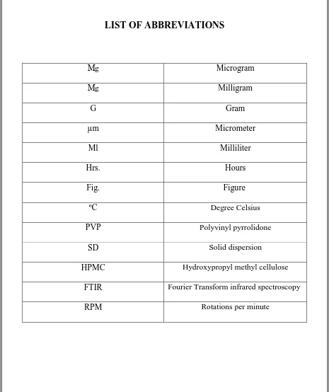
LIST OF ABBREVIATIONS
Μg Microgram
Μg Milligram
G Gram
µm Micrometer
Ml Milliliter
Hrs. Hours
Fig. Figure
ºC Degree Celsius
PVP Polyvinyl pyrrolidone
SD Solid dispersion
HPMC Hydroxypropyl methyl cellulose
FTIR Fourier Transform infrared spectroscopy
[image:5.595.65.530.76.630.2]First of all, I thank the God who is constantly showering his blessing on me. It is my great privilege pride and honor in expressing my humble thanks to my esteemed teacher and guide Prof. S. TamilzharasiM.Pharm, Ph.D., Department of Pharmaceutics, Nandha College Pharmacy, Erode for her valuable guidance, keen interest, inspiration and constant encouragement throughout the course of this investigation. Without her valuable advice and deep-rooted knowledge, this work would not have been a reality.
It is proud to express my sincere thanks to Dr.T.Sivakumar M.Pharm, Ph.D., Principal Nandha College of Pharmacy, with a deep sense of gratitude for his encouragement, co-operation , kind suggestion and providing the best facilities during this work .
I express my loyal thanks to Thiru.V.Shanmugam B.com.Chairman and Mr. Nandha Kumar Pradeep M.B.A., Secretary, Nandha College of Pharmacy, Erode for providing all the facilities to make this work a success.
I owe my warm and humble thanks to Dr.P.R.Radhika, M.Pharm.Ph.D. Prof. Dept. of Pharmaceutics, Mr. Jagadeeswaran, M. Pharm., Asst Prof. Dept. of Pharm. Analysis, Dr.Sengottuvelu, M.Pharm. Ph.D., and H.O.D. Asst Prof. Dept. of Pharmacology,
Dr.Duraisamy, M.Pharm.Ph.D., Asst Prof. Dept. of Pharmacognosy. Mr.Raja, M.Pharm., Ph.D., Asst Prof. Dept. of Pharmaceutics, Mrs. P. Amsa, M.Pharm,(Ph.D) Asst professor, Department of pharmaceutics, for their immense help throughout the work.
I express my deepest and special thanks to my batch-mates Mr. Praveen,
Mr.Tandava Krishna, Mr. Sanath, Mr. Sibu, Mr.Ravichand for their kind co-operation help and encouragement throughout my Post-graduation.
With no words I express my heartful and deep gratitude to my friends Mr. Sagar, Mr.Pavan, Mr.Murali, Mr. Bimal, Mr.Hemanth, Mr. Ramani Sudhir and Mr. Cyril.
Who always believed in me and stood with me in good and bad times.
and encouragement they always showered on me. I am very thankful to my juniors who have contributed directly or indirectly during my dissertation.
The completion of this dissertation is not only to fulfillment my dreams but also the dreams of my parents who have taken lots of pain for me in completion of higher studies. A word of thanks to all those, gentle people associate with this work directly or indirectly whose names I am unable to mention here.
Thank you to one and all ………
Place: Erode Reg. No: 26104206
Date M.Pharm.II Year,
Department of Pharmaceutics, Nandha college of Pharmacy, Erode. Page 1
INTRODUCTION
The skin is the largest organ of the body, accounting for more than 10% of body mass, and the one that enables the body to interact most intimately with its environment. The skin consists of four layers: the stratum corneum (nonviable epidermis), the remaining layers of the epidermis (viable epidermis), dermis, and subcutaneous tissues. There are also several associated appendages: hair follicles sweat ducts, apocrine glands, and nails.1
Many agents are applied to the skin either deliberately or accidentally, with either beneficial or deleterious outcomes and the mechanism was showed in Fig. 1. The use of topical products was evident in ancient times, and there are reports of systemic benefits of topical anti-infective and hormonal agents in the 1940s. Modern transdermal patch technology was introduced in the late 1970s. The main interests in dermal absorption assessment are in the application of compounds to the skin
For local effects in dermatology (e.g., corticosteroids for dermatitis)
For transport through the skin for systemic effects (e.g., nicotine patches for smoking cessation)
For surface effects (e.g., sunscreens, cosmetics, and anti-infective); To target deeper tissues (e.g., Non-steroidal anti-inflammatory agents [NSAIDs] for muscle inflammation)
Unwanted absorption (e.g., solvents in the workplace, agricultural chemicals, or allergens).
Fig. 1 Mechanism of drug through by transdermal delivery
[image:8.595.104.518.514.686.2]Department of Pharmaceutics, Nandha college of Pharmacy, Erode. Page 2
Avoid the problems of stomach emptying, pH effects, and enzyme deactivation associated with gastrointestinal passage
To avoid hepatic first-pass metabolism
To enable control of input, as exemplified by termination of delivery through removal of the device
Delivery of solutes through the skin is associated with various difficulties, such as
The variability in percutaneous absorption owing to site, disease, age, and species differences
The skin‘s ‗‗first-pass‘‘ metabolic effect
The reservoir capacity of the skin
Irritation and other toxicity caused by topical products
Heterogeneity and indelibility of the skin in both turnover and metabolism
Inadequate definition of bioequivalence criteria
An incomplete understanding of the technologies that may be used to facilitate or retard percutaneous absorption.
GROSS STRUCTURE AND FUNCTION OF THE SKIN
It needs to be emphasized that the protection, homeostatic, and sensing functions of the skin are both overlapping and integrated. For instance, barrier properties to a chemical entity involves resistance to its entry (barrier provided by stratum corneum), metabolism for that proportion of entity bypassing the stratum corneum (in viable epidermis), sensing of and attention to damage caused by entry (inflammatory mediator release in epidermis, with involvement of dermis), and removal of entity from site by dermal blood supply and distribution into those body organs specifically responsible for elimination of the entity by metabolism (liver) and excretion (kidney). Heat regulation occurs through the use of the subcutaneous fat pad, physiological regulation of blood flow to affect, for instance, heat loss by vasodilation, and cooling by perspiration.
A. The Epidermis
Department of Pharmaceutics, Nandha college of Pharmacy, Erode. Page 3 approximately 40 mm in diameter and 0.5 mm thick. The thickness varies, however, and may be a magnitude of order larger in areas such as the palms of the hand and soles of the feet, areas of the body associated with frequent direct and substantial physical interaction with the physical environment. Each stratum corneum cell is composed mainly of insoluble bundled keratins (_70%) and lipid (_20%) encased in a cell envelope, accounting for about 5% of the stratum corneum weight. The intercellular region consists mainly of lipids and desmosomes for coenocyte cohesion. The barrier function is further facilitated by the continuous desquamation of this horny layer with a total turnover of the stratum corneum occurring once every 2–3 weeks. The cells of the stratum corneum originate in the viable epidermis and undergo many morphological changes before desquamation. Thus the epidermis consists of several cell strata at varying levels of differentiation. The origins of the cells of the epidermis lie in the basal lamina between the dermis and viable epidermis. In this layer there are melanocytes, Langerhans cells, and two major keratinic cell types: the first functioning as stem cells having the capacity to divide and produce new cells; the second serving to anchor the epidermis to the basement membrane.
The basement membrane is 50–70 nm thick and consists of two layers—the lamina dense and lamina Lucida—which comprise mainly proteins, such as type IV collagen, laminin, nidogen, and fibronectin. Type IV collagen is responsible for the mechanical stability of the basement membrane, whereas laminin and fibronectin are involved with the attachment between the basement membrane and the basal keratinocytes.
B. The Dermis
Department of Pharmaceutics, Nandha college of Pharmacy, Erode. Page 4 cc_3 of skin, providing a vascular exchange area equivalent to that of the skin surface area. Skin blood vessels derive from those in the subcutaneous tissues, with an arterial network supplying the papillary layer, the hair follicles, the sweat and apocrine glands, the subcutaneous area, as well as the dermis itself. These arteries feed into arterioles, capillaries, venules, and, thence, into veins. Of particular importance in this vascular network is the presence of arterio venous anastomoses at all levels in the skin.
The lymphatic system is an important component of the skin in regulating its interstitial pressure, mobilization of defense mechanisms, and in waste removal. It exists as a dense, flat meshwork in the papillary layers of the dermis and extends into the deeper regions of the dermis. Also present in the dermis are a number of different types of nerve fibers supplying the skin, including those for pressure, pain, and temperature.
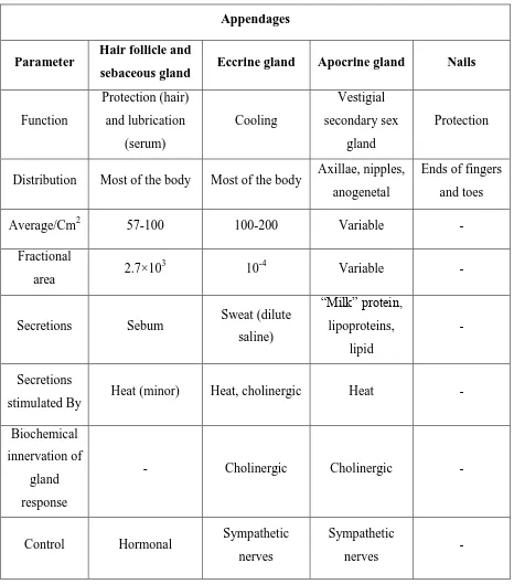
Fig. 2 T.S. of the Skin
[image:11.595.92.536.302.510.2]C. The Sub-cutis
Fig. 2 shows the deepest layer of the skin is the subcutaneous tissue or hypodermis. The hypodermis acts as a heat insulator, a shock absorber, and an energy storage region. This layer is a network of fat cells arranged in lobules and linked to the dermis by interconnecting collagen and elastin fibers. As well as fat cells (possibly 50% of the body‘s fat), the other main cells in the hypodermis are fibroblasts and macrophages. One of the major roles of the hypodermis is to carry the vascular and neural systems for the skin. It also anchors the skin to underlying muscle. Fibroblasts and adipocytes can be stimulated by the accumulation of interstitial and lymphatic fluid within the skin and subcutaneous tissue.
Department of Pharmaceutics, Nandha college of Pharmacy, Erode. Page 5 There are four skin appendages: the hair follicles with their associated sebaceous glands, eccrine sweat glands, apocrine sweat glands, and the nails showed in Fig 2. Each appendage has a different function as outlined in Table 1.
Appendages
Parameter Hair follicle and
sebaceous gland Eccrine gland Apocrine gland Nails
Function Protection (hair) and lubrication (serum) Cooling Vestigial secondary sex gland Protection
Distribution Most of the body Most of the body Axillae, nipples, anogenetal
Ends of fingers and toes
Average/Cm2 57-100 100-200 Variable -
Fractional
area 2.7×10
3
10-4 Variable -
Secretions Sebum Sweat (dilute
saline) ―Milk‖ protein, lipoproteins, lipid - Secretions
stimulated By Heat (minor) Heat, cholinergic Heat -
Biochemical innervation of
gland response
- Cholinergic Cholinergic -
Control Hormonal Sympathetic
nerves
Sympathetic
[image:12.595.66.532.151.680.2]nerves -
Department of Pharmaceutics, Nandha college of Pharmacy, Erode. Page 6 1, 2
Hydrogels are hydrophilic polymeric network of three dimensional cross linked structures that absorb substantial amount of water5. Cross linking facilitates insolubility in water because of ionic interaction and hydrogen bonding11. It also provides required mechanical strength and physical integrity to the Hydrogels. Thus, hydrogels can imbibe water nearly 10-20 times its molecular weight and hence become swollen. Some examples of Hydrogels include contact lenses, wound dressing, super absorbents.
BENEFITS
Biocompatible Can be injected Easy to modify
Timed release of growth factors and other nutrients to ensure proper tissue growth
Entrapment of microbial cells within polyurethane hydrogel beads with the advantage of low toxicity
Environmentally sensitive hydrogels have the ability to sense changes of pH, temperature or the concentration of metabolite and release their load as result of such a change.
Natural hydrogel materials are being investigated for tissue engineering, which include garose, methylcellulose, hylaronan, and other naturally derived polymers.
LIMITATIONS
High cost.
Low mechanical strength Difficult to load
Difficult to sterilize Nonadherent
Department of Pharmaceutics, Nandha college of Pharmacy, Erode. Page 7
CLASSIFICATION
1. On the basis of the nature of the cross linked junctions20
a. Chemically cross-linked networks having permanent junctions.
b. Physical networks have transient junctions arising from polymer chain entanglements or physical interactions
S. No Characteristics Natural origin Synthetic polymers
1 Preparation By using natural polymer By chemical polymerization 2 Advantages -Biocompatible
-Biodegradable
-Supports cellular activities
-Inherent bioactive properties absent
3 Disadvantages -Does not possess sufficient mechanical properties -May contain pathogen
-Evoke immune and responses
______________
4 Examples -Properties like collagen and gelatin
-Polysaccharides like alginate and agarose
-Acrylic acid
-Hydroxy-ethyl-methacrylate (HEMA)
-vinyl acetate
-Meth acrylic acid (MAA)
Preparation of hydrogels 1. Use of cross linkers
Copolymerization of monomers using multifunctional co-monomer, which acts as cross
linking agent, chemical initiator initiates the polymerization reaction which can be carried out in bulk, solution or suspension.
Cross linking of linear polymers by irradiation or by chemical compounds. Monomers
Department of Pharmaceutics, Nandha college of Pharmacy, Erode. Page 8 amines or a strong acidic and basic group like sulfonic acid and quaternary ammonium compounds.
Cross linkers incorporated are glutaraldehyde, calcium chloride and oxidized konjac
lucomannan (DAK). They impart sufficient mechanical strength to the polymers and thus prevent burst release of the medicaments.
2. Isotatic Ultra High Pressure (IUHP)
Suspension of natural biopolymers (e.g.-starch) are subjected to ultra-high pressure of 300-700 MP n for 5 or 20 minutes in a chamber which brings about changes in the morphology of the polymer (i.e. Gelatinization of starch molecules occur).Temperature in the chamber varies from 40 to 52OC30.
3. Use of Nucleophilic substitution reaction
A pH and temperature sensitive hydrogel viz. hydrogel of N-2-dimethylamino-ethyl-methacrylamide (DMAEMA) has been prepared using nucleophilic substitution reaction between methacyloyl chloride and 2-dimethylamino ethylamine31.
4. Use of gelling agent
Gelling agents like glycophosphate1-2propanediol, glycerol, trehalose, mannitol etc. have been used in the preparation of hydrogels. However, presence of negative charged moieties and turbidity are the problems associated with the method.
5. Use of irradiation and freeze thawing
Irradiation method is suitable as well as convenient but the processing is costly along with the poor mechanical strength of the product .Freeze thawing method imparts sufficient mechanical strength and stability to the hydrogels except that they are opaque in appearance with little swelling capacity. However, hydrogels prepared from microwave irradiation are more porous than conventional methods.
6. Synthesis of hydrogel in industry
Formulation of monomer along with initiators and additives lead to polymerization which forms the gel. The gel is dried, sieved and mixed with other additives and post treatment is done if needed.
DESIGN CRITERIA FOR HYDROGELS IN DRUG DELIVERY FORMULATIONS
Department of Pharmaceutics, Nandha college of Pharmacy, Erode. Page 9 shown in the Table 3: Design criteria for hydrogels Hydrogel formulation even designed with proper physical and transport properties, may still fail to show therapeutic effect when implanted in vivo due to a localized inflammatory response. Fibrous capsule formed around the delivery device gives rise to additional diffusion barriers that may limit drug release rates while increased proteolysis activity may increase rates of matrix and drug degradation. Thus, proper material selection, fabrication process and surface texture are important parameters in designing biocompatible hydrogel formulations for controlled release. Drug incorporation into hydrogel device can be achieved by one of the following methods.
1. Hydrogels Drug Uptake Release Mechanism
Inert hydrogels diffusion and/or gel swelling Hydrogel containing drug –binding ligands drug-polymer interaction and diffusion
2. In-situ Loading
Drug or drug polymer conjugates are mixed with polymer precursor solution and hydrogel network formation and drug encapsulation are achieved simultaneously. Here release of drugs occurs through diffusion, hydrogel swelling, reversible drug-polymer interaction, degradation of labile covalent bonds.
DRUG RELEASE MECHANISMS FROM HYDROGEL DEVICES
Hydrogels imbibe more water than 90% of their weight due to hydrophilicity, thus differing in their release mechanisms from hydrophobic polymers. Various models have been developed to predict the release of an active agent from a hydrogel device as a function of time. These models are based on the rate limiting step for controlled release and are divided into three categories viz.
Department of Pharmaceutics, Nandha college of Pharmacy, Erode. Page 10
DIFFUSION CONTROLLED
It is most widely applicable mechanism relating to drug release. Fick‘s law of diffusion is commonly used in modeling this release.
S. No Hydrogels Drug diffusion coefficients
1 Porous hydrogels-pore size >>> molecular dimensions of the drug
Related to porosity
2 Non- Porous hydrogels- porous gels with pore sizes comparable to the drug molecular size.34, 35
Decreases due to steric hindrance from polymer chains with in cross linked networks.
SWELLING CONTROLLED
It occurs when diffusion of drug is faster than hydrogel swelling. In this condition the modeling of drug involves moving boundary, where molecules are released at the interface of the rubbery and glassy phases of swollen hydrogels. Transition occurs from a glassy state where entrapped molecules remain immobile to a rubbery state where molecules rapidly diffuse. Release of small molecule drugs from HPMC hydrogel tablets are based on this mechanism. For example, Methocel matrices (a combination of methylcellulose and HPMC) from Dow chemical Company prepare swelling controlled drug delivery formulations.
CHEMICALLY CONTROLLED
It characterizes molecule release based on reactions occurring within a delivery matrix. Most commonly occurring reactions are-
Cleavage of polymer chains via hydrolytic or enzymatic degradation.
Reversible or irreversible reactions occurring between the polymer network and releasable drug.
It can be categorized on the basis of reactions occurring during drug release.
Purely–kinetic – controlled release
Polymer degradation (bond cleavage) is the rate determining step while diffusion contributes almost negligible to the drug release.
It is of two types viz.
Pendant chain (prodrugs)
Department of Pharmaceutics, Nandha college of Pharmacy, Erode. Page 11 In pendent chain systems, drugs are covalently linked to the hydrogel network device through cleavable spacers and drug release is controlled by the rate with which spacer bond cleavage occurs. In specific applications where a more targeted delivery approach is desired, it is advantageous to design enzymatically cleavable spacer bonds. In surface eroding systems, drug release is mediated by the rate of surface erosion of the polymer matrix. In hydrophobic polymer networks, surface erosion occurs when the rate of water transport into the polymer is much slower than the rate of bond hydrolysis. Nevertheless due to the inherently high water content of hydrogels, surface erosion occurs slowly in enzymatic degradation systems where the transport of enzyme into the gel is slower than the rate of enzymatic degradation. Models focusing on the release mechanisms are based on hydrolytic degrading polymers.
CHALLENGES OF HYDROGEL DEVICES
There are still many challenges associated with the modeling of drug delivery phenomena and release profiles related to complex hydrogel systems. Fundamental understanding of drug transport processes helps in developing a suitable mathematical model. Mass transport governs the translocation of drug from the interior to the surrounding environment of hydrogel devices. Factors affecting mass transport of encapsulated molecules are as follows.
Network cross linking density Extent of swelling
Gel degradation
Size and charge of the encapsulated molecules
Physical interactions between the encapsulated molecules and the polymer matrix Drug – ligand binding present within hydrogel devices.
APPLICATION OF HYDROGELS
Wound Healing – Modified polysaccharide found in cartilage is used in formation of hydrogels to treat cartilage defects. For example, the hydrogel of gelatin and polyvinyl alcohol (PVA) together with blood coagulants are formulated.
Department of Pharmaceutics, Nandha college of Pharmacy, Erode. Page 12 2. Industrial Applicability - Hydrogels are used as absorbents for industrial effluents like
methylene blue dye. Another example is adsorption of dioxins by hydrogel beads.
3. Tissue Engineering – Micronized hydrogels are used to deliver macromolecules phagosomes) into cytoplasm of antigen-presenting cells. This property is also utilized in cartilage repairing. Natural hydrogel materials used for tissue engineering include agarose, methylcellulose and other naturally derived products.
4. Drug Delivery in GI Tract – Hydrogel deliver drugs to specific sites in the GIT. Drugs loaded with colon specific hydrogels show tissue specificity and change in the pH or enzymatic actions cause liberation of drugs. They are designed to be highly swollen or degraded in the presence of micro flora.
5. Rectal Delivery – Hydrogels showing bio adhesive properties are used for rectal drug delivery. Miyazaki et al. explored the xyloglucan gel with a thermal gelling property as matrices for drug delivery.
6. Ocular Delivery – Chitoni et al. reported silicon rubber hydrogel composite ophthalmic inserts. Cohen et al. developed in-situ forming gelling system of alginate with high gluconic acid contents for the ophthalmic delivery of pilocarpine.
7. Transdermal Delivery – Swollen hydrogels can be used as controlled release devices in the field of wound dressing. Hydrogel based formulations are being explored for transdermal iontophoresis to obtainnicotine.
8. Subcutaneous Delivery – Hydrogel formulations for subcutaneous delivery of anticancer drugs are being prepared viz. cross-linked PHEMA was applied to cytarabine (Ara-c).implantable hydrogels are now leading towards the development of biodegradable systems which don‘t require surgical removal once the drug has been administered.
9. Novel Hydrogel For Controlled Drug Delivery – HYPAN is the novel hydrogel having properties useful controlled drug delivery. Physical network of crystalline clusters distinguishes HYPAN hydrogels from others.
10. Hydrogel For Gene Delivery – Modification of hydrogel composition leads to effective targeting and delivery of nucleic acids to specific cells for gene therapy. Hydrogel versatility has potential application in the treatment of many genetic and/or acquired diseases and conditions.
Department of Pharmaceutics, Nandha college of Pharmacy, Erode. Page 13 12. Tropical Drug Delivery – Instead of conventional creams, hydrogel formulation are
employed to deliver active components like Desonide, a synthetic corticosteroid used as an anti – inflammatory for better patient compliance.
13. Protein Drug Delivery – Interleukins conventionally administered as injection are now given as hydrogels which show better compliance and form in-situ polymeric network and release proteins slowly.
SOLID DISPERSION
Many potential drug candidates are characterized by a low oral bioavailability. Often poor drug dissolution / solubility rather than limited permeation through the epithelia of the gastrointestinal tract are responsible for low oral bioavailability. Thus aqueous solubility of any therapeutically active substance is a key property as it governs dissolution, absorption and thus the in-vivo efficacy. Drugs with low aqueous solubility have low dissolution rates and hence suffer from oral bioavailability problems3. Various techniques for the improvement of the dissolution rates of poorly water soluble drugs include micronization, inclusion complexation with cyclodextrins, conversion into amorphous drug and formation of solid dispersions with hydrophilic carriers2. Solid dispersion technique is widely used to increase the intrinsic solubility and dissolution and in turns oral bioavailability of poorly water soluble compounds. Solid dispersion formulation was developed by Chiou and Riegelman. Solid dispersion has traditionally been defined as the ―the dispersion of one or
more active ingredients in an inert excipient or matrix‖ where the active ingredients could exist in finely crystalline, solubilized or amorphous states.3
Department of Pharmaceutics, Nandha college of Pharmacy, Erode. Page 14
TYPES OF SOLID DISPERSION:
1. Binary solid dispersion: It consists of drug and a polymeric carrier.
2. Ternary solid dispersion: It consists of drug, a polymeric carrier and a surfactant.
Generally used surfactant is polysorbate 80 which plays an important positive role in dissolution of the solid dispersion both the binary and ternary solid dispersions enhanced the dissolution of poorly water soluble drugs. Moreover, the dissolution of ternary solid dispersion is found faster compared with that of binary solid dispersion 7.This was because of polysorbate 80, which improved the wettability and solubilized the non-molecularly dispersed or crystalline fraction of drug (ex. Ofloxacin).
3. Surface solid dispersion: Surface solid dispersion is formulated with polymers such as polyvinyl pyrrolidone, polyethylene glycol and polyvinyl pyrrolidone-vinyl acetate copolymer by fusion technique to improve its solubility. Preparation of surface solid dispersion, a technique that provides deposition of the drug on the surface of certain materials, can alter the dissolution characteristics of drug. Deposition of drug on the surface of an inert carrier leads to reduction in particle size of drug, therapy providing faster rate of dissolution.
SUITABLE PROPERTIES OF A CARRIER FOR SOLID DISPERSIONS
Following criteria should be considered during selection of carriers:
High water solubility – improves wettability and enhances dissolution High glass transition point – improve stability
Minimal water uptake (reduces TG)
Soluble in common solvent with drug –solvent evaporation Relatively low melting point –melting process
Capable of forming a solid solution with the drug-similar solubility parameters
First generation carriers
Crystalline carriers: Urea, Sugars, Organic acids.
Second generation carriers
Amorphous carriers: Poly ethylene glycol, Povidone, Poly vinyl acetate, Poly methacrylate, cellulose derivatives
Third generation carriers
Department of Pharmaceutics, Nandha college of Pharmacy, Erode. Page 15
SOLVENT SELECTION FOR SOLID DISPERSION SYSTEMS
In order to prepare solid dispersion, solvents should be selected on the basis of following criteria:
Dissolve both drug and carrier.
Toxic solvents to be avoided due to the risk of residual levels after preparation e.g. chloroform and dichloromethane
Ethanol is a less toxic alternative Water based systems preferable
Use of surfactants to create carrier drug solutions but care should be taken as they can reduce the glass transition point.
Class I Solvents (Solvents to be avoided)
Solvents in Class I should not be employed in the manufacture of drug substances, excipients and drug products because of their deleterious environmental effect Table 1. Class II Solvents (Solvents to be limited)
Solvents in Table 2 should be limited in pharmaceutical products because of their inherent toxicity.
Class III Solvents (Solvents with low toxic potential)
Solvents in class III (shown in table 3) may be regarded as less toxic and of lower risk to human health. Class III includes no solvents known as a human health hazard at level normally accepted in pharmaceuticals.
Class IV Solvents (Solvents for which no adequate toxicological data was found)
Some solvents may also be of interest to manufacturers of excipients, drug substances, or drug products for example Petroleum ether, isopropyl ether. However, no adequate
toxicological data on which to base a PDE was found. Table1. List of some Class I Solvents
Solvent Concentration LIMIT
(ppm) Concern Benzene Carbon tetrachloride 1,2-dichloroethane 1,1-dichloroethane 1,1,1-trichloroethane 2 4 5 8 1500 Carcinogen
Toxic and environmental hazards
Toxic Toxic
Department of Pharmaceutics, Nandha college of Pharmacy, Erode. Page 16 Table2. Class II Solvents in pharmaceutical products
PDE= Permitted Daily Exposure
USES OF SOLID DIPERSION:
Solid dispersion improves dissolvability in water of poorly water soluble drug in a pharmaceutical composition.
Drug is formulated with hydrophilic carrier (ex. poly ethylene glycol) as a solid dispersion to increase its aqueous solubility and dissolution. Then super disintegrant (ex.croscarmellose sodium) is used in tablet formulation to achieve rapid disintegration of tablets prepared by wet granulation method. Thus solid dispersion is used in preparing rapid disintegration oral tablets. These rapidly disintegrating tablets can be used as an alternative to parenteral therapy enabling patient for self-medication even without the aid of water.
Solvent PDE (mg/day) Concentration LIMIT
Department of Pharmaceutics, Nandha college of Pharmacy, Erode. Page 17 Solid dispersion is used as formulation vehicle to facilitate the preclinical safety and
early clinical studies on new chemical entities with very low aqueous solubility. A compound with extremely low or negligible aqueous solubility may significantly limit the dose range or exposure of the drug achievable in the preclinical and clinical studies when formulated via traditional means. In these cases, solid dispersion formulations may provide a means to rapidly assess the safety and efficacy profile of the drug substance that may be otherwise difficult to obtain.
In improving immunosuppressive therapy in lung transplant patients, dry powder formulation consisting of a solid dispersion (ex. Cyclosporine A) for inhalation is prepared. It can avoid many problems ex.
o With dry powder inhalation the use of local anesthesia and irritating solvents can be avoided.
o The higher deposition efficiency of dry powder inhalation compared with nebulization reduces the need for high metered doses. Solid dispersions are known for their dissolution rate enhancing properties of poorly soluble drugs such as Cyclosporine.
BENEFITS OF SOLID DISPERSION SYSTEM:
Solid dispersion systems can provide numerous additional benefits to oral drug therapy beyond improving bioavailability such as:
Solid dispersion formulations were demonstrated to accelerate the onset of action for drugs such as Non-steroidal anti-inflammatory drugs where immediacy of action is crucial to relieving acute pain and inflammation.
For anti-cancer drugs in particular, solid dispersion systems were shown to provide bioavailable oral dosage forms which could be substituted for standard injections to improve patient comfort and compliance.
Solid dispersion systems were also found to reduce food effect on drug absorption, thus increasing the convenience of drug therapy as the need for some drugs to be taken with food was eliminated.
Department of Pharmaceutics, Nandha college of Pharmacy, Erode. Page 18 Additionally, the improved absorption efficiency demonstrated for solid dispersion
systems allows for a reduction in the content of active agent per dose, thus decreasing the cost associated with these drug therapies.
Finally it was demonstrated the solid dispersion systems can be produced utilizing functional carriers that offer the added benefit of targeting the release of highly soluble forms of poorly water soluble drugs to an optimum site for absorption.
These benefits demonstrate the current contributions and future potential of solid dispersion systems toward improving drug therapies for a variety of important medical condition whose treatment involves poorly water soluble drugs.
METHOD OF PREPARATION:
1. Solvent evaporation method
2. Modified solvent evaporation method 3. Melting method
4. Melt-solvent method 5. Kneading method 6. Co-grinding method 7. Co-precipitation method
8. Co-precipitation with supercritical fluid 9. Spray drying method
10. Gel entrapment technique
[image:25.595.76.524.508.677.2]Department of Pharmaceutics, Nandha college of Pharmacy, Erode. Page 19
1. Solvent evaporation method:
Drug and carrier both are dissolved in organic solvent. After complete dissolution, the solvent is evaporated. The solid mass is ground, sieved and dried.
Ex. Solid dispersion of Ofloxacin with polyethylene glycol was prepared by solvent e vaporation method.
2. Modified solvent evaporation method:
Drug is dissolved in organic solvent at its saturation solubility with continued stirring for some time. Polymer is suspended in sufficient amount of water (up to wet mass of polymer). The drug solution is poured at once into polymer suspension. The entire solvent is evaporated. The mass obtained is dried.
3. Melting method:
Accurately weighed drug and carrier are mixed using glass mortar and pestle. The mixture is heated at or above the melting point of all the components to achieve a homogenous dispersion. It is then cooled to obtain a congealed mass. It is pulverized and sieved.
Ex. Albendazole and urea solid dispersion was prepared this method.
4. Melt-solvent method:
Accurately weighed drug is dissolved in organic solvent and the solution is incorporated into the melt of mannitol by pouring into it. It is suddenly cooled. The mass is kept in desiccator for complete drying. The solidified mass is crushed, pulverized and passed through sieve.
5. Kneading method:
A mixture of accurately weighed drug and carrier is wetted with solvent and kneaded thoroughly for some time in a glass mortar. The paste formed is dried and sieved.
Ex. furosemide and crospovidone solid dispersion was prepared by this method.
6. Co-Grinding method:
Accurately weighed pure drug powder and the carrier are physically mixed for some time using a blender at a specified speed. The mixture is then charged into the chamber of a vibration ball mill. A certain number of steel balls are added. The powder mixture is ground. Then the sample is collected and kept at room temperature in a screw capped glass vial until use.
Ex. chlordiazepoxide and mannitol solid dispersion was prepared by this method.
Department of Pharmaceutics, Nandha college of Pharmacy, Erode. Page 20 Accurately weighed carrier is dissolved in water and drug in organic solvent. After complete dissolution, the aqueous solution of carrier is then poured into the organic solution of the drug. The solvents are then heated and evaporated. The dispersion is pulverized with pestle and mortar, sieved and dried.
8. Co-precipitation with supercritical fluid:
Conventional methods for the preparation of solid dispersions include either the fusion or solvent processes, with supercritical fluid processing (SCP) emerging as an alternative solvent- evaporation method for formulating co precipitates of smaller particle size, lower residual organic solvent and better flow ability. A supercritical fluid exists as a single fluid phase above its critical temperature and pressure. Carbon dioxide is currently the most commonly used supercritical fluid because of its low critical temperature of carbon dioxide makes it attractive for processing heat labile pharmaceuticals. In the context of manufacturability, rate of cooling and solvent removal is stringently controlled, resulting in acceptable batch to batch variation.
A precipitation vessel with a nominal capacity of 50ml was loaded with a 7ml solution of pure drug or drug: polymer (carbamazepine: polyethylene glycol) in acetone. The supercritical carbon dioxide was added from the bottom of the chamber and when the liquid phase expanded, the formed particles were retained in the vessel by a suitable filter. During the co-precipitate formation, the pressure was fixed at 70bar and the temperature at 40oC.
9. Spray drying method:
Accurately weighed amount of drug with lipid carrier are dissolved in methanol to obtain a clear solution. This solution is then spray dried using a laboratory scale dryer. The sample is stored over silica gel in a vacuum desiccator.
10.Gel entrapment technique:
Department of Pharmaceutics, Nandha college of Pharmacy, Erode. Page 21
THE UNDERLYING PRINCIPLES FOR IMPROVING THE DISSOLUTION PROPERTIES OF DRUGS BY SOLID DISPERSION TECHNIQUES ARE:
Reducing particle size.
Altering the crystalline morphology.
Intimately mixing the drug with hydrophilic excipients .By altering the bulk drug according to these principles, drug particle surface area is increased, the thermodynamic barrier to dissolution imposed by the crystal lattice is eliminated and the wetting properties of the drug particles are enhanced.
MECHANISMS SUGGESTED BEING RESPONSIBLE FOR THE IMPROVED AQUEOUS SOLUBILITY / DISSOLUTION PROPERTIES OF SOLID DISPERSIONS
INCLUDES:
Reduction of the particle size of the incorporated drug: In solid dispersions, the particle size of the drugs was reduced, and the wettability and the dispersibility of the drugs were enhanced; therefore, drug dissolution was improved markedly.
Partial transformation of crystalline drug to the amorphous state. Formation of solid solution.
Formation of complexes.
Reduction of aggregation and agglomeration.
Improved wetting of the drug and solubilization of drug by the carrier at the diffusion layer.
EVALUATIONS OF SOLID DISPERSION:
It is highly acceptable, that often more than one of these phenomena determine the rate and extent of dissolution. Therefore, Bragg-Brentano powder diffractometry, differential scanning calorimetry, infrared spectroscopy, solubility and dissolution experiments are routinely used to study the relationship between dissolution and physicochemical state of solid dispersion.
Department of Pharmaceutics, Nandha college of Pharmacy, Erode. Page 22 Differential scanning calorimetry offers the possibility to evaluate the crystallinity of
both drug and polymer phase and to characterize polymorphic and amorphous phases. Differential scanning calorimetry can be used to study solid dispersion to determine whether the phase is monotectic or eutectic in nature. A monotectic system is one where little or no interaction occurs whereas a eutectic system is one where little or no interaction occurs.
Infrared spectroscopy reveals crystallographic changes of drug and polymer molecules, such as hydrogen bonds which are indicative of complex formation.
Equilibrium solubilities of the drug in aqueous polymer solutions of different polymer concentrations reveal the solubilization capacity of the polymer for the drug.
Micro –Raman spectroscopy with X- Ray Diffraction is used for characterization of the distribution, polymorphism and stability of drug in its solid dispersion in polyethylene glycol.
MARKETED PRODUCTS:
Commercial products based on solid solution /dispersion is:
Gris-PEG, a griseofulvin- PEG fusion method solid dispersion, was manufactured initially by Dorsey / Sandoz and reached the market in the mid- 1970s.Gris-PEG was developed as tablet product, and this led to two USP monographs for griseofulvin tablets. The solid dispersion form is referred to as ultra-micro size griseofulvin tablets USP and offers improved bioavailability and two- thirds reduced dosage compared to griseofulvin tablets USP. The griseofulvin tablets USP are manufactured from micronized drug substance using a conventional tableting approach. Griseofulvin solid dispersion tablets are currently marketed by a number of manufacturers and contain corn starch, lactose, magnesium stearate, PEG, and sodium lauryl sulfate as inactive ingredients.
Cesamet, a nabilone-PVP solvent method solid dispersion manufactured by Eli Lilly and Co. has been marketed internationally since 1982. Eli Lilly discontinued marketing Cesamet contain PVP and corn starch as inactive ingredients and is presented as a capsule product.
Solid dispersion formulation of Troglitazone (Rezulin) is marketed by Parke-Davis25. Solid solutions of lopinavir and ritonavir in poly vinyl pyrrolidone-vinyl acetate
Department of Pharmaceutics, Nandha college of Pharmacy, Erode. Page 23 to four tablets, tablets made with the solid solutions eliminate the need for refrigeration.
"Sporanox" (Janssen Pharmaceutical, Titusville, NJ) is a solid dispersion of itraconazole in hypromellose that has been layered onto sugar spheres.
Department of pharmaceutics, Nandha college of Pharmacy, Erode. Page 24
LITERATURE REVIEW
U. D. SHIVHARE et al.,[9]developed and evaluated diclofenac sodium gel using water soluble polyacrylamide polymerHigh molecular weights water soluble homopolymer of acrylamide are reported to possess very high viscosity in low concentration, transparency, film forming properties and are useful in formation of gel. The diclofenac sodium gels were prepared by using different concentration of polyacrylamide for topical drug delivery with an objective to increase transparency and spread ability. These preparations were further compared with marketedDiclofenac sodium gel. Spreadability and consistency of polyacrylamide gel containing diclofenac sodium (F9) were 6.5g.cm/sec and 5mm as compared to 5.5g.cm/sec and 10mmrespectively of marketed gel, indicating good spreadability and consistency of the preparedgel (F9). The transparency of prepared batch F9 was good as compared to the marketed gel. The percent drug release was 97.11 and 98.66 from F9 and marketed gel respectively. No irritation was observed by skin irritation test. Stability studies under accelerated condition showed satisfactory results. It can be concluded that polyacrylamide gel containing diclofenac sodium showed good consistency, homogeneity, spreadability and stability and has wider prospect for topical preparations.
M.a.saleemet al.,[10]jul-sep.2010formulated and evaluated of meloxicam solid dispersion incorporated topical gels,Solid dispersion complexes of meloxicam were prepared by using cyclodextrins (BCD, HPBCD), PVP and urea by kneading method in different molar and weight ratios. The complexes werecharacterized by DSC and IR, suggested that no chemical interaction between drug and carrier. The solubility,dissolution and permeability of complexes were markedly increased as compared to pure drug. Solidcomplexes were incorporated in 1% carbopol to prepare gels and evaluated for pH, drug content,viscosity, invitro permeability through rat skin. Invitro permeation study reveals that the flux (Jss)and enhancement factor increases with increase in concentration of BCD, HPBCD and decreaseddramatically in case of HPBCD with ratio of 1:2. Similar changes in pattern of permeation wereobserved with urea and PVP complexes .Hence it can be concluded that solid dispersion complexincorporated gel shows highest permeation as compared to plain drug gels.
Department of pharmaceutics, Nandha college of Pharmacy, Erode. Page 25 ibuprofen were prepared using different drug: polymer ratios viz. 1:0.5, 1:1, 1:2, and 1:3 for 2-HPβ-CD and β cyclodextrin using the co-evaporation method, and were evaluated for partition coefficient, dissolution studies, and Fourier Transform Infra-Red (FTIR) spectrophotometer. The optimized solid dispersion of ibuprofen was incorporated into gel and was compared with penetration enhancers. The formulations were analyzed to determine their pH, spreadability, viscosity, and in vitro drug release. The absence of extraneous interactions among ingredients was confirmed by FTIR, and differential scanning calorimetry (DSC). The formulation with 1:0.5 SDIB (drug: HPβCD) with a partition coefficient of 1.28 was incorporated in carbopol gel, and produced 98.21% drug release compared to solid dispersion of ibuprofen with menthol (SDIBM5%), which produced 96.5% drug release. In ex vivo studies, SDIB and SDIBM5% formulations gave 94.3% and 92.36% drug release within 24h. The percent inhibitions of the edema formation by the gels were in the range of 18.32% to 67.96%, and the maximum inhibition was shown by the SDIB formulation. Therefore, SDIB formulation incorporated in gel produced better results than other formulations prepared with permeation enhancers. Stability studies conducted for SDIB incorporated gel according to International Conference on Harmonization guidelines showed it to be stable for two months.
Swami N.G.N et al.,[12]Formulated and evaluated diclofenac sodium gels using sodium carboxymethyl hydroxypropyl guar and hydroxypropyl methylcellulosein this investigation,
Diclofenac sodium gels were formulated employing Sodium carboxymethyl hydroxy propyl
guar and Hydroxypropyl methylcellulose as gelling agents. Hydroxypropyl methylcellulose
(K4M) was employed at 5 %w/w strength whereas, Sodium carboxymethyl hydroxypropyl
guar formed a gel at 2.5 % w/w strength, gels were subjected for various evaluation tests such
as pH measurement, assay, stability study, rheological evaluation, and invitro release studies
across hairless albino rat skin. Gels formulated using Sodium carboxymethyl hydroxypropyl
guar displayed a pH value of 7.48, whereas hydroxypropyl methylcellulose gels revealed a
pH value of 7.26. Stability studies revealed good physical stability and assay values did not
show much variation from the initial drug content in0 0 both the cases with formulations
stored at 25 C, 60% RH and 40 C, 70% RH for six months. Hydroxypropyl methylcellulose
at 5% w/w strength revealed shear-thinning property, whereas Sodium carboxymethyl
hydroxypropyl guar at 2.5 % w/w strength revealed both pseudo plastic and thixotropic
property. The rheological data were fitted into Martin and Co’-worker equation to obtain a
linear relationship and from the linear curve fittings, the 'N'- values; the possible flow indices
for pseudo plasticity were arrived at. A 'N' value of 4.65 was obtained for Sodium
Department of pharmaceutics, Nandha college of Pharmacy, Erode. Page 26 Hydroxypropyl methylcellulose gels. When subjected to In-vitro release studies across
hairless albino rat skin, Sodium carboxymethyl hydroxypropyl guar based gels revealed a %
cumulative drug release of 25.66 in contrast to a % cumulative drug release of 20.80 in case
of Hydroxypropyl methylcellulose based gels at the end of 6 hours. From the above
observations, Sodium carboxymethyl hydroxypropyl guar seems to be a promising
pharmaceutical adjuvant in the formulation of Diclofenac sodium gel.
MALAYK.DAS et al.,[13]The potential gastrointestinal disorders associated with oral administration of rofecoxib can be avoidedby delivering the drug to the inflammation site at a sustained, concentrated level over an extended period oftime. Hydroxypropyl methylcellulose (HPMC), sodium alginate and Carbopol 940 were used in an attempt todevelop topical gel formulations of rofecoxib. The effects ofpolymer composition on the rate of drug releasefrom the gel formulations were examinedthrough cellulose membrane mounting on a Keshary-Chien diffusioncell. The effects of initial drug concentration and viscosity on the permeation rate of rofecoxib from the gel formulationswere evaluated using rat epidermis at 37 +0.5ºC. The anti-inflammatory activity of the rofecoxib gelformulation was evaluated using the rat hind paw edema model. The gel formulation consisting of 4% w/w sodiumalginate-Carbopol 940 at 3:1 ratio was found to be suitable for topical application based on in vitro evaluationand ex vivo permeation studies. The drug permeation rate increased with an increase of the initial drug concentrationin gels up to 25 % w/w. An inverse relationship was observed between the in vitro drug release rate/exvivo permeation rate and viscosity of the gel formulations. The anti-inflammatory activity of 4% w/w sodiumalginate-Carbopol 940 gel containing 25% w/w rofecoxib in the rat hind paw edema model reveals that the drugwas delivered to the inflammation site at a controlled level over a period of 6 h. These results suggest the feasibilityof the topical gel formulation of rofecoxib.
Department of pharmaceutics, Nandha college of Pharmacy, Erode. Page 27 incorporated gels of HPMC are improved transdermal delivery for Aceclofenac than the solid dispersion incorporated gels of Carbopol 940.
Lalit Kumar et al.,[15]Nimesulide is a second generation non–steroidal anti–inflammatory agent, which is widely usedin the long term therapy of rheumatoid arthritis, in alleviating pain and inflammation. But its short half-life (only 3–4hrs.), so its causes more fluctuation. After oral administrationNimesulide causes to produces heart burn, nausea, loose motions, pruritus, etc. The present studybased on the preparation of bioadhesive topical gel of Nimesulide, so as to avoid all gastric sideEffects. For the preparation of bioadhesive topical gel natural polymer angel marmelos (plantBale) was used. Bioadhesive polymers are the agents which increases the contact between theFormulation and biological membrane, so as to avoid the fluctuation of formulation and behave as a sustained release formulation. In the present study, prepared bioadhesive topical gel was evaluated with the help of different parameters like drug content, spreadability, extrudability, swelling index study, in–vitro drug diffusion study, in-vitro drug release kinetic study and ex–Vivobio adhesive measurement. On the basis of in–vitro drug diffusion study and ex–vivoBioadhesive measurement property of gel, we have concluded that natural polymer aegelMarmelosis the best polymer for the preparation of sustained release bioadhesive topical gel.
Department of pharmaceutics, Nandha college of Pharmacy, Erode. Page 28
Naresh G et al.,.[17] The aim of this study was to prepare and evaluate gels incorporating solid lipid nanoparticles (SLNs) of diclofenac sodium for systemic delivery of the active after topical application. SLNs were prepared using hot homogenization followed by sonication technique and these were incorporated into freshly prepared carbopol gel. Three different gel formulations (DSL1, DSL2 and DSL3) were prepared and characterized for particle size, charge, viscosity, morphology, and drug-lipid compatibility. The gels were evaluated for in vitro drug release, ex vivo permeation studies and in vivo absorption. The gels enriched with SLN sustained the drug release for 24 h both in vitro and in vivo. The results suggest enhancement in systemic delivery of diclofenac sodium with gels incorporating SLNs.
Bazigha K Abdul Rasool et al.,.[18]Ibuprofen gel formulations, incorporating various permeation enhancers, were prepared usingChitosan as a gelling agent. The formulations were examined for their in vitro characteristics includingviscosity, pH and drug release as well as in vivo pharmacological activities. Carrageenan-induced ratpaw edema model was used for the evaluation of their analgesic and anti-inflammatory activities. Acommercial ibuprofen gel product (Ibutop®) was used as a reference.The formulations containing 5 % of either menthol or glycerol as permeation enhancers gavedrug release patterns comparable to that of the reference product. Propanol increased the apparentviscosity of the test gels to the same extent as that of the reference. Drug release from the formulationsfitted best to the Higuchi model. A significant in vivo analgesic effect was produced by the testformulations containing 5 % menthol and 20 % propylene glycol and the effect was superior to thatobtained with the reference product. However, no significant anti-inflammatory activity was exerted byany of the test gel formulations (p > 0.05).Ibuprofen gel preparations containing 5 % menthol and 20 % propylene glycol, respectively, exhibited pronounced analgesic activity and could be further developed for topical andsystemic delivery of ibuprofen
Department of pharmaceutics, Nandha college of Pharmacy, Erode. Page 29 increased the consistency of the gels. All the formulations exhibited pseudoplastic flows with no thixotropic. The values of flow index (n) were found tobe less than one for all the gels confirming the shear thinning behavior of allthe gels. HPMC and MC gels were found to be stable at accelerated stabilityconditions while the bio adhesion of PEO gels was highest. Even after exposureto heat and humidity, no significant change was observed in the contentuniformity, pH, clarity, texture profile analysis and rheological properties of theHPMC and MC gels. The rheograms and various power law equationparameters of these gels were found comparable at various time points in theaccelerated stability study. However, PEO gels failed in accelerated stability studies at one month sample. When four selected gel formulations (HPMCL4,MCL3, P1BL3 and P3BL3.5) were applied topically by six patients each, Gastrointestinal Quality of Life Index (GIQLI) score of each patient wasimproved at the end of 8 weeks. No adverse effects were reported by any of thepatients. Hence 2% DTZ gel was found to be effective in the treatment of analfissures.
M Najmuddin et al.,[20]The Goal of the present investigation was to design and evaluate gels for topical delivery of water insoluble antifungal agent Ketoconazole with an aim to increase its penetration through skin and thereby its flux. Ketoconazole is a broad spectrum imidazole derivative useful in the treatment of superficial and systemic fungalinfections. The solubility of Ketoconazole is increased by complications with ß-cyclodextrin were prepared bysolvent evaporation technique with 1:1 and then incorporated into gels. The complex was characterized byinfrared spectroscopy. There was no interaction between drug and carrier. Gels have gained more and moreimportance because the gel-bases formulations are better percutaneous absorbed than creams and ointmentbases. Therefore, Ketoconazole gel formulations were made with different polymers like carbopol 940, hydroxyl propyl methyl cellulose, methyl cellulose, and sodium carboxymethylcellulose, containing various permeationenhancers namely sodium lauryl sulphate (0.5-1.0%) and dimethyl sulfoxide (5-20%) in different proportions. Theformulated gels were evaluated for various physicochemical parameters like, drug content, pH, viscosity, spreadability, extrudability, in-vitro drug release. The in-in-vitro drug release study were carried out using pH 7.4phosphate buffer, All the formulated topical preparations showed pH in the range of 6.5 to 7.4, and also showedgood spreadability, extrudability. The carbopol 940 with 15% of dimethyl sulfoxide (KCD3) showed best in-vitrodrug release 98.07% at the end of 6 hrs.
Department Of Pharmaceutics, Nandha college Of Pharmacy, Erode. Page 31
DRUG PROFILE
[5, 6, 7]Diclofenac Sodium:
Molecular Formula: C14H11C12NO2
Molecular Weight: 318.10
Chemical Name: 2-[(2, 6-dichlorophenyl) amino] benzene acetic acid, mono potassium salt
Solubility: Freely soluble in methanol; soluble in ethanol (95 per cent); sparingly soluble in water and in glacial acetic acid; practically insoluble in ether, in chloroform and in toluene.
Indications:
Orally for symptomatic treatment of osteoarthritis, ankylosing spondylitis, primary
dysmenorrhea, acute gouty arthritis and for relief of pain, including postoperative (e.g., orthopedic, gynecologic, oral) pain, in adults.
In combination with misoprostol for the symptomatic treatment of osteoarthritis and
rheumatoid arthritis in patients at high risk for developing NSAIDS-induced gastric or duodenal ulcers and in patients at high risk for developing complications from these ulcers.
Topically (as gel) for the symptomatic treatment of osteoarthritis related joint pain. Used for
joints amenable to topical therapy (e.g., hands, knees), has not been evaluated on joints of the spine, hip, or shoulders.
Orally or topically for symptomatic treatment of infusion-related superficial thrombophlebitis.
Dosage and Administration: Oral Administration
Diclofenac sodium delayed-release (enteric-coated) and extended-release tablets are not
Department Of Pharmaceutics, Nandha college Of Pharmacy, Erode. Page 32
Topical Administration Diclofenac Sodium 1% Gel
Apply gel 4 times daily to the affected joint. Use the dosing card from the manufacturer to measure the appropriate dose. Apply the gel within the oblong area of the dosing card up to the appropriate (2- or 4-g of gel) line; then use the dosing card to apply the gel. Gently massage the gel into the skin; ensure gel is applied to the entire affected joint (e.g., foot [including sole, top of foot, and toes], knee, ankle, hand [including palm, back of hand, and fingers], elbow, and wrist).
Diclofenac Epolamine Transdermal System
Apply transdermal system to the most painful area twice daily. Apply to intact skin; do not
apply to damaged skin (e.g., wounds, burns, infected areas of skin, areas affected with eczema or exudative dermatitis).
Dosage: Oral
May change dosage to 50 or 75 mg twice daily in patients who do not tolerate usual dosage; however, these dosages may be less effective in preventing NSAIA-induced ulcers.
S.No Preparation Dosage
1 Diclofenac potassium conventional tablets
100–150 mg daily, given as 50 mg 2 or 3 times daily
2 Diclofenac sodium delayed-release tablets
100–150 mg daily, given as 50 mg 2 or 3 times daily or 75 mg twice daily
3 Diclofenac sodium extended-release tablets
100 mg once daily
4 Diclofenac sodium (in fixed combination with misoprostol)
50 mg 3 times daily
Topical (gel)
For lower extremity (i.e., knees, ankles, feet) joint pain, massage 4 g of diclofenac sodium 1% gel into the affected joint 4 times daily.
Department Of Pharmaceutics, Nandha college Of Pharmacy, Erode. Page 33 If multiple joints are treated, total daily dose applied to all joints should be ≤32 g of gel daily.
Rheumatoid Arthritis
Oral
May change dosage to 50 or 75 mg twice daily in patients who do not tolerate usual dosage; however, these dosages may be less effective in preventing NSAIA-induced ulcers.
S.No Preparation Dosage
1 Diclofenac potassium conventional tablets
150–200 mg daily, given as 50 mg 3 or 4 times daily
2 Diclofenac sodium delayed-release tablets
150–200 mg daily, given as 50 mg 3 or 4 times daily or 75 mg twice daily
3 Diclofenac sodium extended-release tablets
100 mg once daily; may increase to 100 mg twice daily
4 Diclofenac sodium (in fixed combination with misoprostol)
50 mg 3 or 4 times daily
Warnings/Precautions
Consider potential benefits and risks of diclofenac therapy as well as alternative therapies before initiating therapy with the drug.
Cardiovascular Effects
Selective COX-2 inhibitors have been associated with increased risk of cardiovascular events (e.g., MI, stroke) in certain situations. Several prototypical NSAIAs also have been associated with increased risk of cardiovascular events. Current evidence suggests that use of diclofenac is associated with increased cardiovascular risk.
GI Effects
Department Of Pharmaceutics, Nandha college Of Pharmacy, Erode. Page 34
Renal Effects
Direct renal injury, including renal papillary necrosis, reported in patients receiving long-term NSAIA therapy.
Hypersensitivity Reactions
a) Anaphylactic reactions (e.g., anaphylaxis, angioedema) reported. b) Immediate medical intervention and discontinuance for anaphylaxis.
c) Avoid in patients with aspirin triad (aspirin sensitivity, asthma, nasal polyps); caution in patients with asthma.
Dermatologic Reactions
Serious skin reactions (e.g., exfoliative dermatitis, Stevens-Johnson syndrome, toxic epidermal necrolysis) reported; can occur without warning. Discontinue at first appearance of rash or any other sign of hypersensitivity (e.g., blisters, fever, and pruritus).
General Precautions
Do not use multiple diclofenac-containing preparations concomitantly. Concomitant use of diclofenac sodium 1% gel and oral NSAIAs may increase risk of adverse effects.
Observe the usual cautions, precautions, and contraindications associated with misoprostol therapy when diclofenac is used in fixed combination with misoprostol.
Hepatic Effects
Severe, sometimes fatal, reactions including jaundice, fulminant hepatitis, liver necrosis, and hepatic failure reported rarely with diclofenac.
Other Precautions
Not a substitute for corticosteroid therapy; not effective in the management of adrenal insufficiency.
Obtain CBC and chemistry profile periodically during long-term use. Specific Populations
Pregnancy
Department Of Pharmaceutics, Nandha college Of Pharmacy, Erode. Page 35 Category X (in fixed combination with misoprostol).284 Misoprostol exhibits abortifacient activity and can cause serious fetal harm.
Lactation
Distributed into milk; 3 discontinue nursing or the drug.
Pediatric Use
Safety and efficacy not established in children.
Good results with oral diclofenac obtained in a limited number of children 3–16 years of age for the management of juvenile rheumatoid arthritis.
Geriatric Use
Oral diclofenac: Caution advised. Fatal adverse GI effects reported more frequently in geriatric patients than younger adults.
Other Precautions
Not a substitute for corticosteroid therapy; not effective in the management of adrenal insufficiency. It may mask certain signs of infection.
Obtain CBC and chemistry profile periodically during long-term use. Specific Populations
Interactions for Diclofenac Sodium Protein-bound Drugs
Only minimally displaces other highly protein-bound drugs from binding sites; however, may be displaced from binding sites by other highly protein-bound drugs.51 52 59 61
Specific Drugs
S.No Drug Interaction Comments
1 ACE inhibitors Reduced BP response to ACE inhibitor. Possible deterioration of renal function in individuals with renal impairment.
Monitor BP.
2 Angiotensin II receptor antagonists
Reduced BP response to angiotensin II receptor antagonist.
Possible deterioration of renal function in individuals with renal impairment.