A COMPARATIVE EVALUATION OF THE MARGINAL
ADAPTATION AND FRACTURE RESISTANCE OF THREE
DIFFERENT TYPES OF METAL FREE CERAMIC
SYSTEMS WITH METAL CROWN-AN
INVITRO STUDY
Dissertation submitted to
The Tamil Nadu Dr M G R Medical University
In the partial fulfillment of the degree of
MASTER OF DENTAL SURGERY
Branch I
“Gurur Brahma Gurur Vishnu
Gurur Devo Maheshwara Guru Sakshath Parambrahmai
Tasmay shree Gurave namaha”
I take this opportunity to extend my sincere gratitude and thanks to my teacher and mentor Dr K S Arunachalam,MDS Principal , Professor and Head of the Department , Department of Prosthodontics ,Sree Mookambika Institute of Dental
Sciences , Kulasekharam, Kanyakumari District, for his help, support and patient
guidance right from the time I joined for this course. .His command over the subject, practical skills, friendly and approachable nature has been a blessing throughout my course study.
I would also like to acknowledge and thank Dr T Sreelal, MDS Professor Department of Prosthodontics Sree Mookambika Institute of Dental Sciences,
Kulasekharam, for being a source of inspiration and motivation in continuing the
study. His nature of allowing a great deal of freedom in work and adhering strictly at the same time to the guiding principles and high teaching standards has been a source
of encouragement to me and beneficial to my professional improvement as a post-graduate student. I also thank him for making Prosthodontics an interesting
subject, in his own unique style.
Words are not enough to express my deep sincere gratitude to my post graduate guide, mentor Dr Lovely M, MDS, DNB Professor Department of Prosthodontics,
Sree Mookambika Institute of Dental Sciences, Kulasekharam, for her timely help,
as a Science and to practice it as an art. I earnestly thank her for her kindness and compassion that she showed throughout my postgraduate curriculum.
My heartfelt and sincere thanks to Dr Shibu A,MDS Dr Sangeeth K
Cherian, MDS and Dr James J Rex BDS (Readers), Dr Anuroopa A,MDS, and
Dr Vini K Varkey, MDS, (Senior Lecturers) and Dr Shaju A, BDS, for their
valuable help and guidance.
I extend my thanks to Dr Kavitha Janardanan.MDS, senior lecturer for her valuable advice and support throughout my thesis.
I am grateful to Dr Roy, Ph.D, Head of the Department of Polymer Processing Laboratory, BMT Wing, SCTIMST, Trivandrum for patiently explaining
and helping me to understand and perform the experiment for the study.
I gratefully acknowledge my batch mate Dr Aparna Mohan and my fellow
post graduates, Dr Arun R, Dr Nikhil S Rajan , Dr Aravind Krishnan and Dr Anjana S for their motivation and encouraging words.
I would like to thank my husband and teacher Dr Karthiga Kannan, Dept.Of
Oral medicine and radiology for his prayers, constant enthusiasm and immense
support throughout my life.
I would like to thank my children Reshma Valli Kannan and Karthik Subramanian, my parents and parents-in law for always being there and helping me believe in myself.
CONTENTS
SL.No.
INDEX
PAGE
NO
1.
LIST OF TABLES……….…
i
3.
LIST OF FIGURES………
ii
4.
LIST OF GRAPHS………
iv
5.
ABSTRACT………
vi
6.
INTRODUCTION………..… 1 – 4
7.
AIMS & OBJECTIVES……… 5
8.
REVIEW OF LITERATURE……… 6 – 33
9.
MATERIALS AND METHODS……… 34 - 39
10.
RESULTS……… 40 - 46
11. DISCUSSION……… 47 - 62
12. SUMMARY AND CONCLUSION ……… 63 –64
List of Tables
Table – 1: Analysis of variance for fracture resistance of copings and crowns of four
groups
Table – 2: Mean fracture resistance (kN)
Table – 3: Analysis of variance for marginal discrepancy of copings and crowns of
four groups
List of figures
Fig 1:- Wax block carved for fabrication of metal die.
Fig 2:- Induction Casting machine for fabrication of Nickel Chromium definitive die Fig 3:- Definitive die fabricated in Nickel Chromium alloy Wiron 99
Fig 4:- Wax pattern for fabrication of IPS e.max core
Fig 5:- Completed IPS e.max core after heat pressing using lost wax technique Fig 6:- Completed IPS e. Max crown after veneering
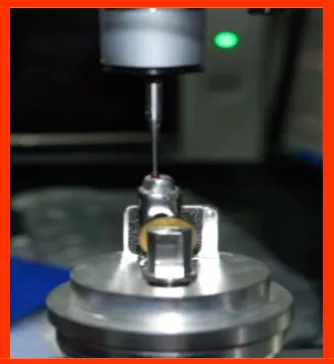
Fig 7:- Scanning of metal die for CAD/CAM coping of Cercon Fig 8:- Digital impression for CAD/CAM manufacturing Fig 9: - Cercon ingot for computer aided machining Fig 10:- Cercon coping and crown
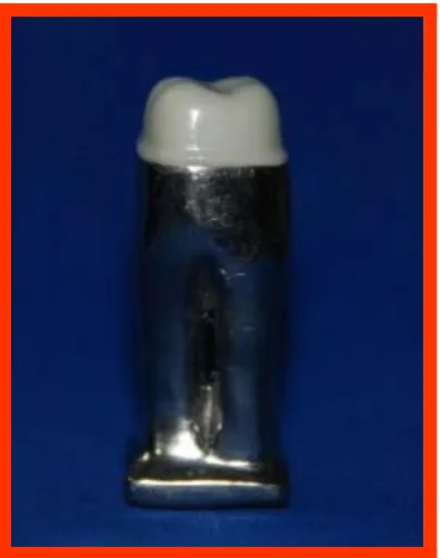
Fig 11:- Procera coping and crown
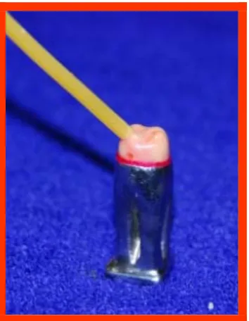
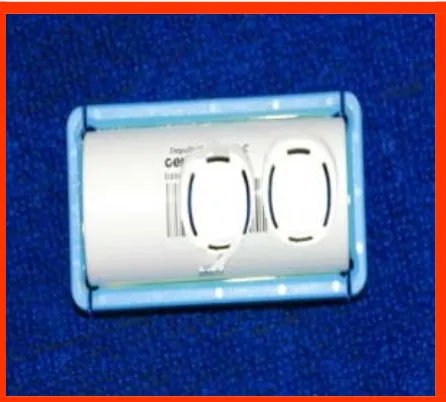
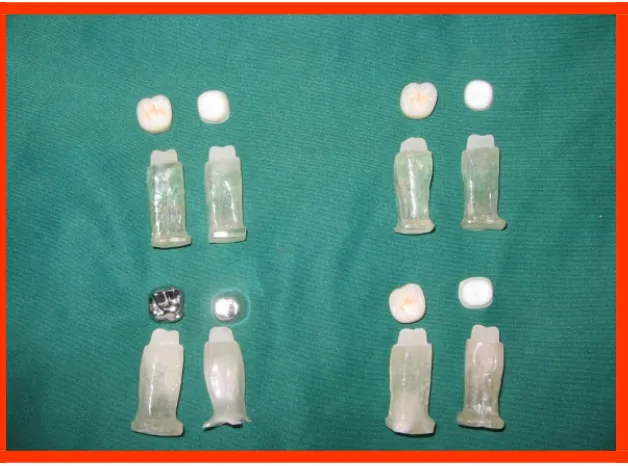
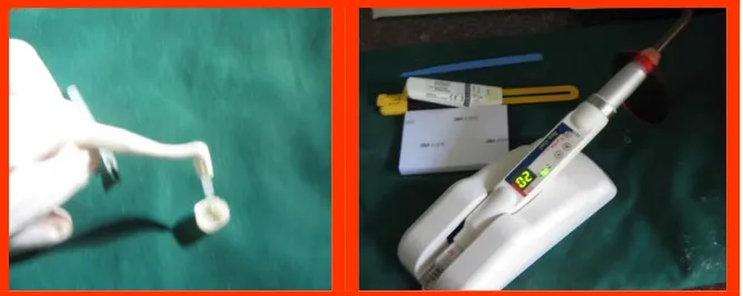
Fig 12:- Test samples of each system before luting Fig 13:- Luting procedure
Fig 14:- Instron Universal testing machine for assessing fracture resistance Fig 15:- Fractured Cercon crown
Fig 16:- Fractured Procera crowns Fig 17:- Fractured IPS e.max crowns
Fig 18:- Stereomicroscopic image depicting marginal gap in IPS e.max copings
Fig 19:- Stereomicroscopic image depicting marginal gap in Procera All Ceram
coping.
Fig 22:- Stereomicroscopic image depicting marginal gap in Procera All Ceram
crown
LIST OF GRAPHS
Graph 1:- Graph depicting the mean fracture resistance values of copings and
crowns of IPS-e.max group
Graph 2:- Graph depicting the mean fracture resistance values of copings and
crowns of Procera All Ceram
Graph 3:- Graph depicting the mean fracture resistance values of copings and
crowns of Cercon
Graph 4:- Graph depicting the mean fracture resistance values of copings and
crowns of metal Wirobond C.
Graph5:- Graph depicting comparison of fracture resistance between groups
(between materials) and within groups (coping and crown of each material)
Graph 6:- Graph depicting the mean marginal discrepancy values of copings and
crowns of IPS-e.max group
Graph 7:- Graph depicting the mean marginal discrepancy values of copings and
crowns of Procera All Ceram group.
Graph 8:- Graph depicting the mean marginal discrepancy values of copings and
crowns of Cercon group.
Graph 9:- Graph depicting the mean marginal discrepancy values of copings and
crowns of metal Wirobond C.
Graph 10:- Graph depicting comparison of marginal discrepancy between groups
A COMPARATIVE EVALUATION OF THE MARGINAL
ADAPTATION AND FRACTURE RESISTANCE OF THREE
DIFFERENT TYPES OF METAL FREE CERAMIC
SYSTEMS WITH METAL CROWN-AN
INVITRO STUDY
Abstract
Aims & objectives
The purpose of the present study is:
1. To compare the marginal adaptation of copings and crowns of three different metal free ceramic systems against a standard metal crown.
2. To compare the marginal adaptation between the copings of three different types of metal free ceramic systems.
3. To compare the marginal adaptation between the crowns of three different types of metal free ceramic systems.
4. To compare the marginal adaptation between the copings and crowns of three different types of metal free ceramic systems.
5. To compare the fracture resistance of three different types of metal free ceramic systems against a standard metal crown.
6. To compare the fracture resistance between the copings of three different types of metal free ceramic systems.
7. To compare the fracture resistance between the crowns of three different types of metal free ceramic systems.
Materials & Methods
Six copings and six crowns each of three commercial metal free ceramic systems, a) IPS e.max (Ivoclar Vivadent AG, Schan /Liechtenstein), b) Procera All ceram (Nobel biocare, Sweden), and c) Cercon (Dentsply, Degudent,) were fabricated in a standardized manner. Thus a total of 18 copings and 18 crowns were fabricated and compared against a Cobalt-chromium metal coping and crown (Wirobond C) for marginal adaptation and fracture resistance. Individual heat cure acrylic resin dies (Viade Products Inc. Camarillo, California) were fabricated for each coping and crown ( a total of 48) to check for marginal adaptation and fracture resistance. After copings were made, they were placed on individual dies to check for marginal adaptation using a stereomicroscope. Later crowns were fabricated and marginal adaptation checked . The copings and crowns were luted on to their definitive dies using dual cure resin luting agent ( Rely X, 3M ESPE, St Paul, Minn) and fracture resistance assessed with a Universal testing machine (Instron).
Results:
Analysis of variance ANOVA was the statistical tool employed to analyze the data. 1. The metal restoration showed better marginal adaptation for copings and crowns. 2. Procera All Ceram exhibited maximum marginal gap when the marginal adaptation of
copings were compared.
3. Cercon crown presented with maximum marginal gap.
4. Marginal adaptation of crowns of all ceramic systems was less than that of their respective copings. Metal crown and coping exhibited similar marginal adaptation. 5. Fracture resistance of IPS e.max crowns was considerably low when compared to the
6. Fracture resistance of crowns was higher than the coping, except for IPS e.max, which
had similar value.
Conclusion
Introduction
Ceramic materials are one of the oldest restorative materials used in dentistry. They are exquisite esthetic dental restorative materials owing to their extreme chemical stability and precise shade simulation with human teeth. They are also one of the most biocompatible materials in restorative dentistry. Ceramics are unique for their appearance, can be customized to simulate color translucency and fluorescence of human teeth.
The major limitation with use of ceramics as tooth replacement material is their very fragile fracture toughness and hence fracture occurs at a very low strain rate of 0.1%. The flexural strength of ceramics is relatively poor as compared to other esthetic resilient dental restorative materials.
The earliest successful porcelain systems used conventional feldspathic porcelain, derived from the natural mineral feldspar. This material was used for producing all ceramic jacket crowns, which were very esthetic, but extremely susceptible to fracture. To overcome this problem, Porcelain fused to metal systems were introduced by the incorporation of Leucite crystals into the feldspathic porcelain composition which were used to veneer the cast gold alloy substructure. The leucite crystals served to increase the thermal expansion of the porcelain to bring it closer to that of metal substructure, thus increasing bonding and strength.
components which are stronger than the traditional predominantly glassy, amorphous feldspathic porcelain. This type of core material can then be veneered with a more translucent ceramic material and an esthetically pleasing restoration could be accomplished.
Various all-ceramic systems have been discussed in the literature regarding their processing techniques, strength and wear characteristics. Although in-vitro studies have shown significant differences in the strength and hardness of some of these materials, the results of long term clinical studies are very less.
Newer ceramic materials and innovative ceramic processing techniques have been introduced in restorative dentistry since the early 1980s. Some of these ceramics still share roots with research that originated in Europe in the 18th century. Today most advances are derived from collaborations with the ceramics engineering community.
Notable recent progress includes the advent of newer ceramic materials and techniques for esthetic complete crowns, partial coverage and laminate veneer restorations, improved metal ceramic esthetics with the advent of opalascent porcelains and frame work modifications, introduction of CAD/CAM and machining as a route to fabrication of restorations, improved understanding of the clinical response of all ceramic prostheses and of the material factors that influence clinical longevity.
for single crown restorations. In addition to fracture resistance and esthetics, marginal fit is one of the most important criteria for the long term success of all ceramic crowns. Marginal discrepancies expose luting material to the oral environment thus leading to cement dissolution, caused by oral environment.
Ceramic materials can be classified based on material composition as silica based and non silica based. Silica based ceramic materials can be again divided into conventional feldspathic porcelain and reinforced pressed ceramics. The non silica based materials can be again classified into sintered, infiltrated (both are created according to slip cast technique), densely sintered, high putty aluminum, and zirconium oxide ceramic (fabricated through CAD/CAM technique .Due to their mechanical and aesthetic qualities and bio compatibility CAD/CAM generated restorations have gained acceptance in dentistry. They are called digital ceramic restorations. Among these materials Procera system has long term performance, mechanical properties and aesthetic capabilities. The versatility of Procera allows it to be used for veneers, crowns, abutments and fixed partial dentures.
Stereo microscope was used for evaluating the marginal discrepancies in copings and crowns of three different types of metal free ceramic systems used for the study. The studies conducted by Matty F. et al, (1989) and In –Sung Yeo et al, (2003) showed that IPS e.max has better marginal adaptability than the other types of all ceramic crowns. Ando et al, in their study concluded that marginal discrepancy increased after firing as a result of heat treatment for degassing. McLean et al, (1971) in their clinical study of 1000 restorations over a five year period, concluded that 120µm was the clinically acceptable marginal discrepancy (maximum).
Aims & Objectives
1. To compare the marginal adaptation of copings and crowns of three different types of metal free ceramic systems against a standard metal crown.
2. To compare the marginal adaptation between the copings of three different types of metal free ceramic systems.
3. To compare the marginal adaptation between the crowns of three different types of metal free ceramic systems.
4. To compare the marginal adaptation between the copings and crowns of three different types of metal free ceramic systems.
5. To compare the fracture resistance of three different types of metal free ceramic systems against a standard metal crown.
6. To compare the fracture resistance between the copings of three different types of metal free ceramic systems.
7. To compare the fracture resistance between the crowns of three different types of metal free ceramic systems.
Review of Literature
Gordon J. Christensen, (1971) has discussed some of the recent advances in
cast-gold dental treatment at that time. A study of the microscopic adaptation of the margins of crowns finished on various die materials showed superior results with silver-plated dies. He has suggested that margins be (1) burnished in the mouth with a blunt burnisher; (2) disked with extra-fine cuttle, coarse, medium, and fine 3/8 inch disks, respectively; and (3) cemented. The use of silver-plated dies eliminates the need for the use of all but the finest cuttle disks in the mouth, because the technician had previously finished the margins on the silver die. Castings finished on silver-plated dies will have closer marginal adaptation regardless of whether the margins are above or below the gingiva, since intraoral finishing of gingival margins is nearly impossible.1
Valderhaug (1977) studied oral hygiene, the gingival condition, pocket depth, and
recorded where the crown margins were located supra-gingivally. Caries lesions developed on 3.5% of the tooth surfaces which had received crowns.2
McLean J (1979) through his book titled the Science and Art of Dental Ceramics has
provided the dental profession with methods and materials for restoring teeth not only to function but to things of beauty. The mechanical properties of porcelain have been discussed and has reached the opinion that they are brittle materials with very low plastic deformation. However their strength values are much higher making them suitable for clinical usage in a variety of situations.3
Lang et al 1983 evaluated the changes occurring in the sub gingival microbiota in
Kwanchai A. Gomez, Arturo A. Gomez (1984), Statistical analysis – of variance
was done to assess the influence of various fixed partial dentures crown materials on marginal adaptation and fracture of copings and crowns.5
Belser et al (1985) carried out a scanning electron microscopy to estimate the fit of
three porcelain fused to metal marginal designs in vivo. Marginal gaps were measured with an SEM on replicas derived from elastomeric impressions. There was no significant difference among beveled metal margins, metal butt margins, or porcelain butt margins either before or after cementation at the 95% confidence level. This study has shown that it is possible under clinical conditions to consistently produce porcelain butt margins with less than 50 µ marginal opening in PFM restorations.6
Holmes (1989) has described in his work that the measurements of misfit at different
locations are geometrically related to each other and defined as internal gap, marginal gap, vertical marginal discrepancy, and horizontal marginal discrepancy, overextended margin, under extended margin, absolute marginal discrepancy, and seating discrepancy. The significance and difference in magnitude of different locations are presented. The best alternative is perhaps the absolute marginal discrepancy, which would always be the largest measurement of error at the margin and would reflect the total misfit at that point.7
Hopkins (1989) estimated the effect of specimen thickness on the strength of dental
load carrying capacity does not increase as much as might be expected with thicker sections. Possible reasons for this phenomenon and methods of limiting this effect are discussed.8
Jacobs MS, Windeler AS (1991) investigated the rate of type I zinc phosphate
cement solubility as it relates to the degree of marginal opening. Standardized test samples were constructed that would simulate clinically relevant marginal gaps of 25, 50, 75, and 150 microns and their subsequent cement lines. The study was divided into two phases, that is, cement solubility in a static environment and a dynamic environment. Both the phase 1 and phase 2 studies demonstrated no significant difference in the rate of cement dissolution for the 25-, 50-, and 75-micron test groups. The 150-micron test groups for both studies, however, demonstrated an increase in the rate of cement dissolution.9
Weaver et al (1991) evaluated the marginal adaptation of castable ceramic crowns,
Anusavice K J and Hojjatie B (1992) analyzed the relative effect of loading site,
occlusal thickness, ceramic flaws, elastic modulus of the cement, and voids in the cement layer on tensile stress that develops in molar glass-ceramic crowns under applied loads. Finite-element stress analyses were performed. For a ceramic thickness of 0.5 mm and a vertical distributed load applied at a distance of 1.3 mm from the vertical axis, the maximum tensile stresses were 100 MPa for a crown with flaws and a void, 87 MPa for a crown with no flaws and a void, and 75 MPa for a crown with flaws and no void. For a 1.5-mm-thick crown with flaws and a void, the tensile stress decreased to 22 MPa. When the load of 600 N was concentrated at the central point of the occlusal region, the peak tensile stress in a crown with flaws and no void was increased to 325 MPa. For the conditions analyzed in this study a large void in a flawed occlusal region of a thin molar crown (0.5 mm) is proposed as a mechanism of crown failure.11
Scherrer (1993) evaluated the fracture resistance of all-ceramic crowns as a function
Castellani et al (1994) evaluated the fracture resistance of three types of all-ceramic
crowns and compared these to the fracture values of metal ceramics. Uniform metal ceramic specimens; veneered, cast glass-ceramic; and porcelain fused to two different dispersion-strengthened ceramic cores (Hi-Ceram and In-Ceram) were investigated. The metal ceramic specimens demonstrated a significantly higher resistance to fracture than did the Hi-Ceram or veneered glass-ceramic units but did not significantly differ from the In-Ceram specimens. The metal ceramic crowns showed cracks only in the ceramic layer, whereas the all-ceramic specimens underwent global fracture.13
Sano et al (1994) studied the tensile properties of mineralised and demineralised
human and bovine dentin. Small slabs (4 x 0.5 x 0.5 mm) of bovine and human dentin were tested in a microtensile testing device in vitro. Human coronal mineralized dentin gave a mean ultimate tensile strength (UTS) of 104 MPa. Bovine incisor coronal dentin exhibited UTS of 91 MPa, and bovine root dentin failed at 129 MPa. The modulus of elasticity of mineralized bovine and human dentin varied from 13 to 15 MPa. When dentin specimens were demineralized in EDTA, the UTS and modulus of elasticity fell to 26-32 MPa and 0.25 GPa, respectively, depending on dentin specimens. The results indicate that collagen contributes about 30% of the UTS of mineralized dentin, which is higher than was expected.14
Yoshinari and Derand (1994) compared the fracture strengths of four types of
after a preload cycling in aqueous atmosphere. Preload cycling significantly decreased the strength of Vitadur crowns. Fracture strength of Vitadur crowns were improved when they were luted with either polyalkenoate or adhesive resin cement.
The In-Ceram crowns fractured in two modes: complete fractures at 1276 (207) N; and fractures with the core remaining intact at 808 (292) N.15
Burke FJ (1995) investigated the effect of dentinal bonding and ceramic etching
procedures on the fracture resistance of all-ceramic crowns. These results were compared with the fracture resistance of similar crowns placed with a nonadhesive conventional cement. All results indicated that superior fracture resistance was obtained when dentinal bonding was incorporated into the luting procedure together with etching of the ceramic fitting surface and the use of resin-based luting material. The fracture resistance of specimens luted with such a procedure was significantly greater than that of specimens in which conventional non-adhesive cement was used.16
Seghi RR and Sorenson JA (1995) studied the flexural strength of six recently
deflection appeared to be the principle strengthening mechanism in the highly crystalline materials.17
Tuntiprawon M, Wilson PR. (1995) evaluated the effect of cement thickness on the
fracture strength of all ceramic crowns. Thirty-three aluminous porcelain jacket crowns were divided into three groups. In Group 1, only platinum foil was used to provide cement space. In Group 2 two layers and Group 3 four layers of die spacer were painted onto the metal die before impression making. Each crown was cemented onto a metal die with zinc phosphate cement and loaded until fracture. It was concluded that increasing the cement thickness above 70 microns reduced the fracture strength of porcelain jacket crowns.18
Wagner and Chu (1996) compared the biaxial flexural strength and indentation
fracture toughness of three new dental core ceramics, Empress, In-Ceram, and Procera AllCeram ceramics. They were prepared according to their manufacturers' recommendations. The results revealed significant differences in flexural strength for the three materials (p ≤ 0.05). The average flexural strengths of AllCeram, In Ceram, and Empress Ceramics were 687 MPa, 352 MPa, and 134 MPa respectively. There was no statistically significant difference between the fracture toughness of Procera
( ) and In-Ceram ceramics ( ); however, both
ceramics had significantly higher fracture toughness (p < 0.005) than Empress
ceramic ( ).19
Sulaiman et al (1997) carried out an in vitro study to compare the marginal fit of
the greatest marginal discrepancy (161 microns), followed by Procera (83 microns), and IPS Empress (63 microns). There were no significant differences among the various stages of the crown fabrication: core fabrication, porcelain veneering, and glazing. The facial and lingual margins exhibited significantly larger marginal discrepancies than the mesial and distal margins.20
Andersson et al (1998) summarized from the data from the many studies on Procera
All Ceram crowns that have been conducted at clinical and laboratory centres around the world. The evidence reported in these studies clearly demonstrated that the Procera AllCeram crown represents a combination of computer technology and creativity for which a positive prognosis can be made. Today its application is restricted to single crowns; however, with continued development, multiple unit all-ceramic anterior and posterior fixed partial dentures will be avilable.21
May et al (1998) measured the precision of fit of the Procera AllCeram crown
fabricated with Procera CAD/CAM technology for the premolar and molar teeth fitted to a die. Laser videography was used to measure the gap dimension between the crowns and the dies at the marginal opening, the axial wall, the cusp tip, and the occlusal adaptation measurement locations. Mean gap dimensions and standard deviations (SDs) were calculated for marginal opening, internal adaptation, and precision of fit.22
Zeng et al (1998) describes the mechanical testing of dental ceramic core materials in
porcelains (Procera Porcelain AllCeram, Vitadur-N, and Vitadur Alpha); and densely sintered alumina-Procera Porcelain AllCeram two-layer composites, densely sintered Vitadur-N two-layer composites, and glass-infiltrated presintered alumina-Vitadur Alpha two-layer composites, with different thicknesses of densely sintered alumina or glass-infiltrated presintered alumina, respectively. The flexural tests were performed in ring-on-ring biaxial bending. Results indicated that the failure stress of densely sintered alumina is significantly higher than that of glass-infiltrated presintered alumina as a core dental material under the same testing conditions. The failure stresses of the three commercial dental porcelains are statistically the same. It was concluded that the densely sintered alumina-Procera Porcelain AllCeram two-layer composite has interesting dental applications.23
Strub JR, Beschnidt M. (1998) evaluated the fracture resistance of 5 different
all-ceramic crown systems (In-Ceram, Empress staining technique, Empress veneering technique, Celay feldspathic system, and Celay In-Ceram system) before and after cyclic preloading in an simulated mouth. It was found that the chewing simulation and the thermocycling significantly decreased the fracture strength of all tested crown systems (P < 0.01). There were no statistically significant differences between the all-ceramic crown groups and the PFM crowns.24
Sobrinho et al (1998) investigated the influence of fatigue on the fracture strength of
Empress. The fatigue values of the three ceramic systems decreased significantly in both dry and wet environments. This may be due to crack propagation from pre existing flaws. No difference was found between fatiguing in dry and wet environments25.
Brunton, Mc Cord and Wilson (1999) is discussing about a new ceramic material,
Procera AllCeram, with universal anterior and posterior applications. It was recently introduced to the UK by Nobel Biocare (Sweden). This type of restoration has a densely sintered, high purity, alumina core and was first described in 1993. These restorations are produced in a unique manner using technology initially developed to produce titanium copings for implant abutments by spark erosion. This article also offers suggestions for case selection, preparation design, and luting procedures.26
Kelly JR (1999) in the article, clinically relevant approach to failure testing of
Beschnidt and Strub (1999) evaluated the marginal fit of different all-ceramic crown
systems after simulation in the simulated mouth. The in vitro marginal fit of five different all-ceramic crown systems (In-Ceram®, Empress® staining technique, Empress® veneering technique, Celay® feldspathic system, Celay In-Ceram® system) was evaluated before and after cyclic preloading in an artificial mouth and were compared to those for porcelain-fused-to-metal (PFM) crowns with circular porcelain-butt margins which were cemented with zinc phosphate cement. It was observed that crown cementation increased the marginal gaps significantly (P<0·01). Empress® staining technique crowns showed the smallest marginal gaps (median 47
µm), followed by conventional In-Ceram® crowns (median 60 µm) and Empress® veneer technique crowns (median 62 µm). Celay In-Ceram® crowns displayed marginal openings with a median of 78 µm, followed by Celay® feldspathic crowns with a median of 99 µm. Ageing in the chewing simulator had no significant influence on the marginal fit of all specimens. The study indicated that all the tested all-ceramic crowns have clinically acceptable margins.28
Holand et al (2000) analyzed the microstructures of glass-ceramics of the IPS
Drummond et al (2000) has evaluated the flexure strength under static and cyclic
loading and the fracture toughness under static loading of six restorative ceramic materials. It was proved that the lithium disilicate containing ceramic had significantly higher flexure strength and fracture toughness when compared to the four pressable leucite strengthened ceramics and the low fusing conventional porcelain. All of the leucite containing pressable ceramics did provide an increase in mean flexural strength (17–19%) and mean fracture toughness (3–64%) over the conventional feldspathic porcelain. Further, the influence of testing environment and loading conditions implies that these ceramic materials in the oral cavity might be susceptible to cyclic fatigue, resulting in a significant decrease in the survival time of all-ceramic restorations.30
Stokes et al (2001) evaluated the dynamic fracture energies and patterns of fracture in
Guazzato et al (2002) compared the mechanical properties of In-Ceram Zirconia and
Ceram Alumina. Mean biaxial flexure strengths of Ceram Alumina and In-Ceram Zirconia were 600 MPa (SD 60) and 620 MPa (SD 61), respectively. Mean fracture toughness measured according to indentation strength was 3.2 MPa ·m 1/2 for
In-Ceram Alumina and 4.0 MPa ·m 1/2 for In-Ceram Zirconia. Mean fracture toughnesses of In-Ceram Alumina and In-Ceram Zirconia measured according to indentation fracture were 2.7 MPa m·1/2 and 3.0 MPa m 1/2 respectively. X-ray diffraction analysis showed that little phase transformation from tetragonal to monoclinic occurred when the specimens were fractured, supporting the existence of a modest difference of fracture toughness between the two ceramics. It was concluded that no statistically significant difference was found in strength. In-Ceram Zirconia was tougher (P < .01) than In-Ceram Alumina when tested according to indentation strength. However, no significant difference was found in the fracture toughness when tested with the indentation fracture technique.32
Blatz et al 2003 in the article resin ceramic bonding presents a literature review on
Webber et al (2003) investigated the effect of different thickness of veneer porcelain
on the compressive load at fracture of Procera AllCeram crowns. Sixty brass dies were fabricated with a crown-like preparation and a chamfer margin. Sixty crowns were fabricated with a 0.6-mm-thick core: Procera crowns with either a 0.4-mm- or 0.9-mm-thick veneer of AllCeram (Groups 1 and 2 respectively) or In-Ceram crowns with a 0.9-mm-thick veneer of Vitadur Alpha porcelain (Group 3). Each group consisted of 20 crowns. In-Ceram crowns were used as the control group. Panavia 21 TC Dental Adhesive served as the luting agent. After luting, fracture resistance was tested using Universal testing machine. It was proved that the axial thickness of veneer porcelain did not have a significant effect on the compressive load at fracture of Procera AllCeram crowns.34
Harrington et al (2003) investigated the load at fracture of Procera AllCeram
porcelain increased the load at fracture for Procera AllCeram crowns. There was no significant difference in load at fracture between the Procera and In-Ceram crowns.35
Olsson et al (2003) evaluated the long-term outcome of In-Ceram Alumina fixed
partial dentures (FPD) performed in a general dental practice from 1992 to1996. The study was conducted as a retrospective assessment of up to 9 years of patient records and a clinical follow-up examination of patients treated with In-Ceram Alumina FPDs. In 37 patients, 42 FPDs had been inserted during the selected period. The mean time in function for the 42 FPDs was 76 months, with 86% being followed for > 5 years. No adverse effects to either periodontal or pulpal tissues were recorded. The technical quality was very good, and patient satisfaction very high. Five FPDs fractured during the observation period, resulting in a total failure rate of 12%. Cumulative survival rate according to life table analysis was 93% after 5 years and 83% after 10 years. The results suggest that the In-Ceram Alumina short-span FPD is a viable prosthetic alternative.36
Anusavice (2003) has explained in detail about the mechanical properties of dental
materials in the textbook Phillip’s Art and Science of dental materials. The various factors affecting the ultimate strength as well as fracture toughness assessment of brittle materials has been explained. The importance of surface flaws on stress concentration is also dealt with in this chapter.37
Sundh and Sjogren (2004) evaluated the fracture strengths of stabilized all-ceramic
with different thickness in different parts of the restoration, called an 'adapted Denzir core'; or with a uniform core thickness of 0.5 mm. IPS Empress 2 all-ceramic crowns served as reference. There was no significant difference between the crowns with an 'adapted Denzir core' veneered with the two brands of glass-ceramics. No significant difference was seen between the crowns with a 0.5 mm Denzir core veneered with the two brands of glass-ceramics. The crowns with an 'adapted Denzir core' exhibited significantly higher values than those with a 0.5 mm Denzir core and than the IPS Empress 2 crowns used as reference.38
Kelly J R (2004) in the article Dental ceramics, Current thinking and trends has
presented all-ceramics within a simple framework allowing for easy understanding of their composition and development. He has also provided for a classification of ceramics based on composition and structure. The meaning of strength and details of the fracture process are explored, and recommendations are given regarding making structural comparisons among ceramics. Assessment of clinical survival data is dealt with, and literature is reviewed on the clinical behavior of metal-ceramic and all-ceramic systems. Practical aspects are presented regarding the choice and use of dental ceramics.39
Narong Potiket, (2004) evaluated the in vitro fracture strength of teeth restored with
minute with an angle of 30 degrees to the long axis of the tooth. It was found that there was no significant difference between groups. There was no significant difference in the fracture strength of teeth restored with all-ceramic and metal-ceramic restorations in this in vitro study. The all-ceramic crown may be considered to be an alternative restoration for highly esthetic areas.40
Quintas et al (2004) conducted an vitro study to evaluate the effect of different finish
lines, ceramic manufacturing techniques, and luting agents on the vertical discrepancy of ceramic copings. The two finish lines considered were heavy chamfer and rounded shoulder. Luting agents tested included zinc phosphate, resin-modified glass ionomer (Fuji Plus), and resin composite cements (Panavia F). Procera copings presented the lowest mean values of vertical marginal discrepancy before and after cementation (25/44 mm) when compared to Empress 2 (68/110 mm) and InCeram Alumina copings (57/117 mm), regardless of any combinations among all finish lines and luting agents tested. This study confirmed that the ceramic manufacturing technique influenced marginal discrepancy of all-ceramic copings.41
Komine et al (2004) evaluated the fracture resistance of aluminum oxide ceramic,
results of this in vitro study showed that the selection of the adhesive resin cement may influence the fracture strength of aluminum oxide ceramic posterior crowns.42
Pallis et al (2004) made an experimental observation of the failure loads of
all-ceramic crowns using a steel ball intender. The 95% confidence intervals for characteristic failure loads were 771 to 1115N for IPS Empress 2, 859 to 1086 for Procera All ceram and 998 to 1183 for In ceram Zirconia. There was no significant difference in fracture resistance, although there was a significant difference in failure origin. The origin of failure was most commonly found at the interface between the ceramic core and veneer porcelain for IPS Empress II and between the ceramic core and luting agent layer for the other systems. Two–way multivariant analysis of variance was used to analyze the thickness of the luting agent, ceramic core, and veneer porcelain layers.43
Reich S (2005) evaluated the clinical fit of all-ceramic three-unit fixed partial
micron for DIGI, 65 micron for LAVA and INLA and 54 micron for the conventional FPDs. Only the DIGI data differed significantly from those of the conventional FPDs. Within the limits of this study, the results suggested that the accuracy of CAD/CAM generated three-unit FPDs is satisfactory for clinical use.44
Bindal and Mormann (2005) made an experimental observation of the marginal
and internal fit of all ceramic CAD/CAM crown copings on chamfer preparation. Slip cast,(Inceram Zirconia) ,heat pressing (Empress II) and CAD/CAM (Cerec in Lab, DCS, Decim and Procera) crown copings were seated on 12 dies each . Marginal and internal gap width was measured in the SEM at 120X magnification. It was found that the marginal gap of Empress II was significantly larger than In-ceram zirconia. Procera and Decim had values similar to In-ceram. The internal mid orobuccal gap width was highest for Procera and lowest for Decim. In-ceram, Empress II, DCS and Cerec InLab had values in between. But the fit of conventional and CAD/CAM all-ceramic molar crown copings covered the range of assumed gap width.45
Luthy et al (2005) determined the strength and reliability of four-unit posterior
reported in the literature. Four-unit posterior frameworks require a connector size larger than7.3 sq.mm.46
Stappert (2005) evaluated the influence of preparation design on longevity and
failure load of ceramic veneers bonded to human maxillary central incisors. The control group remained unprepared (NP). For Group WP, a window preparation was made. Specimens in Group IOP were prepared with an incisal overlap of 2 mm without palatal chamfer. For Group CVP, specimens were prepared with a complete-veneer design of 3-mm incisal reduction and 2-mm palatal extension. All the specimens were subjected to cyclic loading and thermal cycling in a dual-axis masticatory simulator. Within the limits of this in vitro investigation, the use of adhesively luted IPS Empress 1 veneers prepared according to the three different preparation designs demonstrated adequate stabilization of residual tooth structure. Crack pattern analysis showed a higher risk of subcritical crack development when the indenter impact was applied on the palatal ceramic surface. They have concluded that the palatal contact point position of the antagonist should remain on the natural tooth structure after preparation. In particular, this is important for complete veneer preparation.47
Sundh et al (2005) evaluated the effect of heat treatment and veneering on the
Sadighpour et al (2006) reviewed the various mechanical test methods usually use to
evaluate dental ceramic materials. The various test methods for determining the flexural strength, fracture toughness and fracture resistance has been reviewed in detail. They have also discussed the clinical implication of these tests and their limitations.49
Steyern et al (2006) investigated the fracture resistance of zirconia crowns and
compared the results with crowns made of a material with known clinical performance (alumina). Sixty crowns were made, 30 identical crowns of alumina and 30 of zirconia and divided into three groups and subjected to (i) water storage only, (ii) pre-loading (10 000 cycles, 30–300 N, 1 Hz), (iii) thermocycling (5–55, 5000 cycles) + pre-loading (10 000 cycles, 30–300 N, 1 Hz). Subsequently, all 60 crowns were subjected to load until fracture occurred. It can be concluded that there is no difference in fracture strength between crowns made with zirconia cores compared with those made of alumina if they are subjected to load without any cyclic pre-load or thermocycling. There is, however, a significant difference in the fracture mode, suggesting that the zirconia core is stronger than the alumina core. Crowns made with zirconia cores have significantly higher fracture strengths after pre-loading.50
Studart et al (2007) in the article the in vitro lifetime of dental ceramics under cyclic
spite of its noticeable susceptibility to fatigue in water, the 3Y-TZP material was found to be particularly suitable for the preparation of posterior all-ceramic bridges due to its high initial mechanical strength. Guidelines are provided for the selection of materials and the design of all-ceramic posterior bridges exhibiting lifetime longer than 20 years under severe wet and cyclic loading conditions.51
Aboushelib (2007) evaluated both the fracture and impact strength of two core
veneered all-ceramic systems to reveal whether the speed of loading affects fracture mechanism. The absorbed energy by IPS Empress-Eris crowns and Cercon-Ceram S crowns in a fracture strength test was compared by the energy absorbed in an impact strength test. The principles of fractography were used to identify fracture origin and dimensions and to calculate the stress at failure. Finite element analysis (FEA) was used to rationalize the results. For the IPS Empress 2-Eris crowns, there was a significant difference in the energy absorbed for the fracture test and the impact test, where for the Cercon-Ceram S, there was no significant difference. Despite the high strength of the zirconia cores there was no significant difference in the energy absorbed between the two systems in the impact strength test. The dominant mode of failure of layered all-ceramic restorations under occlusal loading is cone cracking in the veneering ceramic.52
Di Lorio et al (2008) compared the resistance to fracture under a cyclic load applied
underwent a fracture test with a cyclic load for 24 hours. Fragments were retrieved for fracture characterization using scanning electron microscopy (SEM). The mean values of fracture resistance for the chamfer samples were 406.10, 67.271 N and 643.90 32.912 N for the shoulder samples. The results of this in vitro study indicate a relationship between the cervical thickness of the alumina cores and their fracture resistance. A shoulder margin could improve the biomechanical performance of posterior single crown alumina restorations.53
Shirakura et al. (2009) assessed the influence of veneering porcelain thickness of all
ceramic and metal ceramic crowns on failure resistance after cyclic loading. Two different frame work designs with 2 different incisal thickness of veneering porcelain were used (2mm,&4mm ) for all ceramic (Procera all ceram) and metal ceramic crown system. The all ceramic group showed significantly higher success and survival rates than the metal ceramic group. For the failure load ,the 2-way ANOVA showed significant effects for material and porcelain thickness. Procera All Ceram crowns may allow up to approximately 4mm of feldspathic porcelain on the incisal area without increasing the failure rate or decreasing the failure load.54
Tao J, Han D (2009) investigated the effect of finish line curvature on marginal fit of
all-ceramic crowns, but had a significant effect on the marginal fit of metal-ceramic crowns.55
Att et al. (2009) assessed the marginal adaptation of three different zirconium dioxide
three unit fixed dental prosthesis at different fabrication stages and after artificial aging. The purpose of this study was to evaluate the marginal adaptation of different zirconia 3-unit fixed dental prostheses at different fabrication stages and after artificial aging. Twenty-four zirconia 3-unit fixed dental prostheses (DCS, Procera, and VITA YZ-Cerec; n=8) were fabricated using different manufacturing systems and conventionally cemented with glass ionomer cement on human teeth. Each group was aged in a masticatory simulator with thermal cycling. The marginal gaps were examined on epoxy replicas for frameworks and for restorations before and after cementation, and after masticatory simulation, at 250x magnification. Group VITA YZ-Cerec showed significantly smaller marginal gap values than groups DCS and Procera at framework (P<.05) and before-cementation (P<.05) stages. The VITA YZ-Cerec group showed significantly smaller marginal gap values than the Procera group after cementation (P<.05). The marginal gap values between different stages were not significantly different for all groups (P>.05). They have come to a conclusion that the marginal accuracy of zirconia fixed dental prosthesis is influenced by manufacturing technique.56
Comlekoglu et al (2009) evaluated the effect of different cervical finish line designs
frameworks were manufactured by a copy-milling system (Zirconzahn) using prefabricated blanks and tried on the master models for initial adaptation of the framework; they were then sintered, followed by veneering (Zirconzahn). The finished crowns were cemented with a polycarboxylate cement (Poly F) under 300 g load and ultrasonically cleaned. The specimens were sliced and the marginal gap was measured, considering absolute marginal opening (AMO) and marginal opening (MO) for each coping under a stereomicroscope with image processing software (Lucia). It was found that the cervical finish line type had an influence on the marginal adaptation of the tested zirconia ceramic. For better marginal adaptation, both shoulder and mini-chamfer finish line types could be suggested for zirconia crowns.57
ElGuindy (2010) investigated the effect of different adhesive systems on the vertical
Hyun-Soon (2010) evaluated the influence of porcelain veneering on the marginal fit
of Digident and Lava CAD/CAM zirconia ceramic crowns before and after porcelain veneering. 20 crowns were made per each system and the marginal fit was evaluated through a light microscope with image processing (Accura 2000) at 50 points that were randomly selected. Each crown was measured twice: the first measurement was done after obtaining the means and standard deviations of the marginal fit were 61.52 ± 2.88 µm for the Digident CAD/CAM zirconia ceramic crowns before porcelain veneering and 83.15 ± 3.51 µm after porcelain veneering. Lava CAD/CAM zirconia ceramic crowns showed means and standard deviations of 62.22 ± 1.78 µm before porcelain veneering and 82.03 ± 1.85 µm after porcelain veneering. Both groups showed significant differences when analyzing the marginal gaps before and after porcelain veneering within each group. However, no significant differences were found when comparing the marginal gaps of each group before porcelain veneering and after porcelain veneering as well. It can be concluded that the 2 all-ceramic crown systems showed marginal gaps that were within a reported clinically acceptable range of marginal discrepancy.59
Abhishek Rastogi and Vikas Kamble (2011) studied the effect of design on
Materials & Methods
In the present study the following materials and methods were employed.
MATERIALS
1. Wax block for carving the die in the intended dimension (The Hindustan dental Products ,Chapel Road, Hydrabad)
2. Inlay wax (Bego Co, Bremen, Germany)
3. Phosphate bonded investment material (Bellavest T, Bego Co, Bremen, Germany)
4. Nickel-chromium alloy for the fabrication of the definitive die (Wiron 99, Bego Co,
Bremen, Germany).
5. Polyvinyl siloxane Impression material (Virtual VPS, Ivoclar Vivadent, Amherst,
NY)
6. Die stone Type IV (Gyprock, Rajkot, Gujarat, India)
7. Clear, heat cure acrylic for duplicating the metal die (Viade Products Inc. Camarillo,
California)
8. Die spacer (Siena, Cergo, Dentsply)
9. Cobalt-Chromium (WirobondC, Bego Co, Bremen, Germany )
10. IPS e.max ingot and veneering porcelain (Ivoclar Vivadent Amherst NY)
11. Procera Allceram ingot and veneering porcelain (Nobel Biocare, Sweden)
12. Cercon ingot and veneering porcelain (Dentsply, Degudent, Germany)
13. Investment material for fabrication of acrylic die (Cergo fit, Dentsply, DeguDent,
Germany)
14. Investment material for IPS e.max (IPS Press vest, Ivoclar Vivadent , Amherst, NY)
EQUIPMENTS
1. Stereomicroscope (Leica MZ 6, Germany )
2. Press Furnace for IPS e.max (EP600 Press, Ivoclar Vivadent, Amherst,NY)
3. Pressure casting machine (Shofu, Shofu Inc, Japan)
4. Microblaster (Penblaster,Shofu Inc, Japan
5. Ceramic furnace (Progrmat P500, Ivoclar Vivadent, Amherst, NY)
6. Universal testing machine (Model 3345; Instron Corp, Canton, Mass)
METHODOLOGY
A total of forty eight samples were fabricated for the study of which thirty six
die. The resin die material was selected because it has a modulus of elasticity (12.9 GPa) similar to that of human dentin (14.7 GPa).
Thirty six all-ceramic cores and crowns were fabricated from these three systems, IPS e.max, Procera AllCeram and Cercon. IPS E-max II is a recently introduced hot pressed ceramic.The major crystalline phase of the core material is a lithium disilicate. The material is pressed at 920º C (1690 ºF) For the fabrication of the core, a wax pattern is fabricated, it is invested using phosphate investment material .Then it is heated to 800 º C to burn out the wax pattern. A ceramic ingot of appropriate shade is placed in the special pressing furnace. After heating to 1150ºC, the softened ceramic is slowly pressed. Then it is allowed bench cool for 10 hours. The restoration is recovered from the investment by airborne particle abrasion with 55 µm glass beads, the sprue is removed and it is refitted to the die. Where as for Procera All Ceram and Cercon systems the fabrication procedure involves an industrial CAD/CAM process. The die is mechanically scanned by the technician. This data is send to the lab where an enlarged die is milled using a computer controlled milling machine. This enlargement is necessary to compensate for sintering shrinkage. Aluminum oxide powder is then compacted on to the die. The coping is milled before sintering at very high temperature. (1150 º C).The coping is further veneered with an aluminous ceramic with matched thermal expansion.
Inc, Melville, NY) The Procera AllCeram cores were presintered, milled, and sintered by the manufacturer (Nobel Biocare USA, Yorba Linda, Calif). Six Cercon cores were fabricated by Vident using a CAD-CAM system. All Ceramic cores were fabricated to a thickness of 0.5mm on all surfaces. The twenty four crowns were completed with the application of the appropriate dentin and veneer porcelains. Vitadur Alpha porcelain (Vident, Brea Calif) was used to complete Procera AllCeram and Cercon crowns, while Eris porcelain (Ivoclar Vivadent) was used to complete the IPS e. max crowns. After the desired thickness of porcelain was achieved and evaluated using micrometer, eighteen ceramic cores were further fired according to the manufacturers’ instructions. The restorations were ultrasonically cleaned in distilled water for 10 minutes.
The samples were grouped in to group I, group II. group III and group IV. IPS e.max, Procera All Ceram, Cercon and metal respectively. They were again subdivided in to A and B for convenience. Where A denotes coping and B crown.
MARGINAL ADAPTATION
measurement analysis to assess the marginal adaptation between crown groups, coping groups , crowns and copings, and all ceramic systems with metal samples. The average gap dimension at each measurement location within each crown group was determined by calculating the means and standard deviations (SD).
FRACTURE TESTING
A stainless steel ball bearing of 5.25 mm diameter was centered on the occlusal surface of each specimen. Each specimen was occlusally loaded along the long axis to fracture in a universal testing machine (Mode3345; Instron Corp, Canton, Mass) at a crosshead speed of 1mm/min, and the maximum load was recorded from the load-displacement trace.
DATA ANALYSIS:-
Analysis of variance was done to assess the influence of various fixed partial denture
crown materials on marginal adaptation and fracture resistance and of copings and crowns [5].
Source Degrees of freedom
Between groups (A) 3
Between copings and crowns (B) 1
Interaction (A x B) 3
Experimental error (E) 40
Total 47
Critical Difference (CD) = terror DF0.5 x √2 MSE/r, where MSE is the experimental
Results
Table – 1: Analysis of variance for fracture resistance of copings and crowns of 4 groups
Source Degrees of freedom Mean Square (MS) Fn1,n2
Table Fn1.n2
5% 1%
Between groups (A)
3 3.0699 41.37***** 2.84 4.31
Between copings and
crowns (B)
1 13.1461 177.17***** 4.08 7.31
Interaction
(AxB) 3 22.3557 301.29***** 2.84 4.31
Experimental error (E)
40 0.0742
Total
47
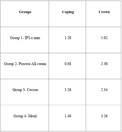
Table – 2 Mean fracture resistance (KN) of copings and crowns
Groups Coping Crown
Group 1- IPS e.max 1.20 1.02
Group 2- Procera All ceram 0.88 2.30
Group 3- Cercon 1.26 2.34
Group 4- Metal 1.40 3.26
SEm = 0.157
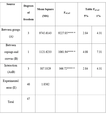
Table – 3: Analysis of variance for marginal discrepancy of copings and crowns
of 4 groups
[image:60.595.90.530.199.671.2]Source Degrees of freedom Mean Square (MS) Fn1,n2
Table Fn1.n2
5% 1%
Between groups (A)
3 9745.6143 9227.05***** 2.84 4.31
Between copings and
crowns (B)
1 1121.6233 1061.94***** 4.08 7.31
Interaction (AxB)
3 387.3329 366.72***** 2.84 4.31
Experimental
error (E) 40 1.0562
Total
47
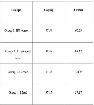
Table – 4 Mean marginal discrepancy (µm) of copings and crowns
Groups Coping Crown
Group 1- IPS e.max 57.54 60.23
Group 2- Procera All ceram
88.48 99.17
Group 3- Cercon 83.35 108.65
Group 4- Metal 37.17 37.17
SEm = 0.593
Table (1) shows Analysis of Variance ANOVA to compare fracture resistance of
copings and crowns of four groups of crown materials. ANOVA revealed that significant differences existed between the four groups compared.
Table (2) depicts the mean values of fracture resistance of copings and crowns of
four groups tested.
It was found that no significant difference in fracture resistance was observed between copings and crowns in Group 1, ie, IPS e,max. While in all other groups, i.e., Procera All Ceram, Cercon and metal (Wirobond C) fracture resistance of crowns were high in comparison with copings.
Fracture resistance of Procera All ceram coping (Group 2) was significantly low in comparison to copings of IPS e.max, Cercon and metal Wirobond C. There was no significant difference in fracture resistance of copings between group 1, group 3 and group 4. In other words the fracture resistance of IPS e.max and Cercon did not differ significantly from the control metal group.
Table (3) shows Analysis of Variance ANOVA to compare marginal
discrepancy of copings and crowns of four groups of crown materials. ANOVA revealed that significant differences existed between the four groups compared.
Table (4) depicts the mean values of marginal discrepancy of copings and
crowns of four groups tested.
It was found that there was a significant increase in marginal discrepancy of crowns of all three all-ceramic systems when compared to the copings. With metal Wirobond C the marginal discrepancy remained the same for both copings and crowns.
When the marginal discrepancy of copings was compared it was observed that there was a significant difference between all the four groups. Maximum discrepancy was registered for Procera All ceram followed by Cercon and IPS e. Max. Marginal discrepancies of three all-ceramic systems were significantly higher than the control metal group.
INFERENCES:-
1. There was no significant difference in fracture resistance values between copings and crowns of IPS e.max.
2. Fracture resistance values of crowns of Procera All Ceram, Cercon and metal Wirobond C were significantly higher than their corresponding coping.
3. Fracture resistance values of Procera Allceram coping was significantly low in comparison to copings of IPSe.max, Cercon and metal Wirobond C.
4. Fracture resistance values of IPS e.max, Cercon and metal Wirobond C coping were statistically similar.
5. Fracture resistance value of IPS e.max crown was significantly low in comparison to all other groups.
6. There was a statistically significant increase in marginal discrepancy of three all-ceramic system crowns when compared to their corresponding copings.
7. There was no difference in marginal gap between metal coping and crown.
8. Maximum marginal gap was exhibited by Procera All ceram coping when compared to copings of other three groups.
Figures
[image:66.595.196.340.469.573.2]
Fig 1:-Wax block carved for fabrication of metal die.
Fig 3 :- Definitive die fabricated in Nickel Chromium alloy Wiron 99
Fig 5 :- Completed IPS e.max core after heat pressing using lost wax technique
Fig 6 :- Completed IPS e. Max crown after veneering
Fig 7:- Scanning of metal die for CAD/CAM coping of Cercon
Fig 8:- Digital impression for CAD/CAM manufacturing
Fig 9 : - Cercon ingot for computer aided machining
[image:70.595.181.473.467.654.2]
Fig 11 :- Procera coping and crown
Fig 12 :- Test samples of each system before luting
Fig 13:- Luting procedure
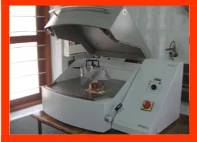
[image:72.595.146.480.170.303.2]Fig 14 :- Instron Universal testing machine for assessing fracture resistance
Fig 15 :-Fractured Cercon crown
[image:73.595.183.468.458.649.2]
Fig 17 :-Fractured IPS e.max crowns
[image:74.595.183.469.125.356.2]
Fig 19 :- Stereomicroscopic image depicting marginal gap in Procera Allceram coping.
[image:75.595.189.387.338.482.2]
Fig 20:- Stereomicroscopic image depicting marginal gap in Cercon
[image:75.595.168.388.519.688.2]
Fig 22 :- Stereomicroscopic image depicting marginal gap in Procera Allceram crown
[image:76.595.181.401.315.483.2]
Graph 1 :- Graph depicting the mean fracture resistance values of copings and crowns of IPS-e.max group
Graph 3 :- Graph depicting the mean fracture resistance values of copings and crowns of Cercon
Graph 6 :- Graph depicting the mean marginal discrepancy values of copings and crowns of IPS-e.max group
Graph 8 :- Graph depicting the mean marginal discrepancy values of copings and crowns of Cercon group.
In the search for the ultimate esthetic restorative material, many new all-ceramic systems have been introduced to the market. For many years development of dental ceramics has centered on creating materials with the same optical properties of natural teeth and the all- porcelain restoration closely matched the translucency and value of natural dentition. While the need for extremely esthetic and natural restorations has become predominant in recent years, a reasonably predictable long-term clinical life span is also paramount. One of the deficiencies of early all-ceramic crowns such as the feldspathic porcelain jacket crown was its lack of such predictability. Many of the newer systems have been marketed heavily on the basis that these crowns are stronger or are reinforced with a core material that will prevent clinical fracture.
Polycrystalline ceramics contain no glass; all of the atoms are packed into regular crystalline arrays through which it is more difficult to drive a crack than the irregular network found in glasses. Aluminia and zirconia are the only two polycrystalline ceramics suitable for use in dentistry as framework materials able to withstand large
stresses. Computer aided manufacturing has made possible the application of polycrystalline ceramics practical for well fitting restorations.
Metal free restorations have a strong ceramic core onto which layering ceramic is applied to achieve a natural appearance. High-strength ceramics for the cores of crowns and fixed partial dentures have been introduced and tested, both in vitro and in vivo [17, 36]. There are various factors affecting the failure loads of all-ceramic restorations including test methods, distribution of flaws, marginal gap [19] and veneering porcelain thickness [54]. In vitro studies do not account for the influence of prosthesis shape and dissimilar component material properties on the stress distribution. Hence it is preferable to test all components materials in a multilayered specimen to reproduce the flaw properties resulting from interfaces between the veneer materials, core materials, luting agent layer and dentin.
Although there are multiple studies analyzing the fracture resistance of all ceramic copings or veneered crowns separately, there are very few reports in literature dealing with fracture resistance of all-ceramic veneered crowns and copings in the same research project. Hence the present study is an attempt to compare the
fracture resistance of copings and veneered crowns individually of the same all ceramic material. A standard metal coping and crown served as the control.
Another variable that is being studied is the variability in marginal adaptation of the crown before and after veneering.
Three most commonly used posterior all-ceramic crown systems were compared for fracture resistance and marginal adaptation in the present study against a metal crown. The all-ceramic systems tested include IPS e.max (Ivoclar Vivadent), Procera All-ceram (Nobel Biocare, Sweden) and Cercon (Dentsply Degudent, Germany). A Cobalt chromium metal coping and crown (Wirobond C) of same dimensions, served as the standard.
crystals measure approximately 3 µm to 6 µm in length. A veneer porcelain consisting of fluorapatite crystals in an aluminosilicate glass may be layered on the core to create the final morphology and shade of the restoration. Fluorapitite is a fluoride-containing calcium phosphate, Ca5 (PO4)3F. The fluorapitite crystals contribute to the veneering
porcelain's optical properties and coefficient of thermal correction, so it matches the disilicate pressable or machinable material. Both the veneering and lithium-disilicate materials are etchable due to the glassy phase.
milling machine creates a duplicate of the preparation (enlarged by 20%) in the 'hub' laboratory onto which aluminum oxide is densely packed [26]. The aluminum oxide is machined to the proportions requested in the digital prescription and sintered to full density. Since the polycrystalline ceramics are opaque, they are veneered with glassy ceramics to achieve pleasing esthetics. Luting procedure can be carried out with zinc phosphate cement, glass ionomer cement or chemically cured resin cement such as Panavia 21 (Kuraray Co Ltd, Japan). As the fit surface of the aluminum oxide (Al2O3)
coping is microscopically rough there is little to be gained by acid etching; surface treatment of the fit surface is therefore usually restricted to sandblasting and the application of a silane-coupling agent. It is suggested that Panavia 21 is the cement of choice [26]. This is supplied with a priming agent and the use of this coupled with a total etch procedure is recommended. Glass-ionomer cement has been advocated for use when moisture control is not optimal.
that a precisely designed coping or framework is produced for an ideal fit. Because of the inherent opacity and high value of all original zirconia blocks, corresponding porcelain overlay materials were developed and introduced to improve the cosmetic look of the resulting restorations. In 2004, Dentsply Ceramco introduced Cercon Ceram S followed by Ceramco PFZ that offered impr