ANTICANCER AND ANTIVIRAL ACTIVITY OF MARINE ALGAE Thesis Submitted To
THE TAMILNADU Dr. M.G.R. MEDICAL UNIVERSITY, GUINDY, CHENNAI
As a partial fulfillment of the requirement for the award of the degree of
DOCTOR OF PHILOSOPHY
Submitted by
V. LAVAKUMAR, M.Pharm.,
Under the supervision of
Dr. V. RAVICHANDIRAN, M.Pharm., Ph.D.,
Principal
Vel’s College of Pharmacy Velan Nagar, Pallavaram,
CERTIFICATE
This is to certify that the thesis entitled “ANTICANCER AND
ANTIVIRAL ACTIVITY OF MARINE ALGAE” is submitted to The Tamilnadu
Dr. M.G.R. Medical University, Chennai in partial fulfillment of the requirements
for the award of degree of Doctor of Philosophy is the record of original research
work done by Mr. V.Lavakumar, M.Pharm., for the academic year 2008 –
2012 under my guidance, and the thesis has not formed the basis for the award of
any degree, diploma, associate ship, fellowship or other similar title.
Date:
DECLARATION
This is to certify that the thesis, entitled “ANTICANCER AND
ANTIVIRAL ACTIVITY OF MARINE ALGAE” is submitted to The Tamilnadu
Dr. M.G.R. Medical University, Chennai in partial fulfillment of the requirements
for the award of degree of Doctor of Philosophy is the record of original research
work done by me under the guidance of Dr. V. Ravichandiran, Principal, Vels
College of Pharmacy, Chennai-600117 for the academic year 2008 – 2012 and the
thesis has not formed the basis for the award of any degree, diploma, associateship,
fellowship or other similar title.
V.LAVAKUMAR
ACKNOWLEDGEMENT
During the processes of learning the principles of scientific work, imaging of subjects, preparing the articles and writing this thesis, I have received help and support from many people.
First and foremost I would like to thank my adorable Supervisor
Dr. V. Ravichandiran, M. Pharm., Ph.D., Principal, Vel’s College of Pharmacy for his valuable guidance, constructive ideas along with constant encouragement and intelligent decisions helped me in completing my work with ease.
I express my deepest thank to Dr. R. Govindh, Dr. Haja, Dr. Apo Mani and Dr. Balaji for their innovative aspects towards this project outcome which made me much confidence in completion of the thesis most successfully.
Words just cannot explain my pleasure in thanking my guru’s
Prof K. Chinnaswamy, Dr. B. Suresh and Dr. T. Ilango for their timely concerns, suggestions and moral support throughout the course of study without which the completion of work was achieved with no difficulties
I emphasize a special thanks to our Chairman, Dr. Isari K.Ganesh, Vel’s College of Pharmacy, (Registrar) Dr. P. Govindarajan and other office staff for providing the necessary facilities to carry out my project work successfully.
I express my deepest thank to Dr. SM. Sivakumar for his innovative aspects towards this project outcome and his knowledge has been a great source of inspiration.
I extend my sincere thanks to Dr. N. Murugesan, Professor, SAIF, IIT, Chennai and DVR Saigopal, Head, Department of Virology, S.V.University, Tirupathi, with his great ideas and timely help for the successful completion of the thesis work.
I owe my ultimate obedience and respect to my father Mr.V. Jayachandra naidu, my mother Mrs. M. Meera, Mrs. V. Kavitha, CL. Sagarika
and Mr.CL. Chandramouli who stood as pillars in entire my course work and made every thing possible.
It gives me immense pleasure and deep sense of gratitude in expressing my heartfelt thanks to my wife Mrs. H. Lavanya for her unmatched inspiration and constant support in the completion of the work most successfully.
I would like to express my gratitude towards my father-in-law Mr. SM. Haridass Naidu and Mother-in-law Mrs. H. Rukmani for their kind co-operation and rocking support for me in completion of this project with greater dimension
I code my special thanks to My uncle P. Govindhaswamy naidu and to my gradmother P. Saroja for their goodwill towards the development of my research.
My thanks and appreciations to H. Suresh, R. Saritha, R. Sathish Kumar, R. Nivetha, G. Ayeswaryaa, G. Sneka, V. Papitha and V. Nikitha for their contribution and goodwill in the completion of the work successfully.
Words are inadequate in offering my thanks to Dr. Srujana Chittibothu
I am grateful to K. Masilamani, S. Sundararajan, N. Deepa, K. Venkatrao, M. Suhasini, K. Swetha and Karnath for their valuable help rendered in shaping my project work.
It would be incomplete if I fail to express my heartfelt thanks to
Dr A. Nirmala, Dr Thilagavathi, Dr Sheela, Dr Mohana, Mr. R. Thangam, SM.
Gopal, Mrs .G. Vanaja, R. Vishnuvardan and R. Ajitha nayiac and Rehan Ahamed for their instant help in fulfilling the thesis.
My thanks and appreciations also go to J. Hemachandran, K. Sathis kumar, K. Narendiran, K. Chinna, N. Kishore babu and N. Sathish babu who have
willingly helped me out with their abilities.
I also thank V. Ragavendra, P. Ramados, S. Kannan, V. Mohanvel, all undergraduate and post graduate students of Vel’s college of Pharmacy and other teaching and non teaching staff for their help towards the completion of the thesis work.
Finally, my sincere thanks are extended to numerous other individuals and organizations whose contribution, directly or indirectly during this project work could never be ignored.
ABBRIVATIONS
WHO - World Health Organisation
DNA - Deoxyribo Nucleic Acid
E2F - E2 Transcription Factor
TNF - Tumor Necrosis Factor
FADD - Fas-Associated protein with Death Domain
P53 - Protein 53
BCl2 - B-cell lymphoma 2
HCV - Hepatitis C virus
HBV - Hepatitis B virus
NF-kB - Nuclear factor kappa
GIT - Gastro Intestinal Tract
PAHs - Polycyclic aromatic hydrocarbons
PE - Pet Ether
Bu-OH - Butanol
CMC - Carboxy Methyl Cellulose
OECD - Organisation for Economic Co-operation and Development
EAC - Earlich Ascites Carcinoma
KCl - Potassium chloride
TBARS - Thiobarbituric acid reactive substances
GSH - Glutathione
GPx - Glutathione peroxidase
GST - Glutathione S-transferase
CAT - Catalase
PCV - Packed cell volume
MSD - Mean Survival Day
5-FU - 5-fluorouracil
Hb - Haemoglobin
MDA - Malondialdehyde
ASE - Acanthophora spicifera Extract
BSLT - Brine Shrimp Lethality Test
DPPH - diphenyl-1-picrylhydrazyl
MTT - (3-(4,5-Dimethylthiazol-2-yl)-2,5-diphenyltetrazolium bromide
HPLC - High-performance liquid chromatography
HPTLC - High performance thin layer chromatography
DMSO - Dimethyl sulfoxide
DMEM - Dulbecco’s modified Eagle's medium
FBS - Fetal bovine serum
ELISA - Enzyme-linked immunosorbent assay
TLC - Thin layer chromatography
GLC - Gas chromatography
µg/ml - micro gram/milli liter
w/w - Weight/weight
v/v - Volume/volume
ng - nano gram
ADP - Adenosine diphosphate
AP-1 - Activator protein 1
FITC - Secondary dye in flow cytometry (Fluorescein isothiocyanate)
PI - Propidium Iodide (Primary Dye)
EDTA - Ethylene di-amine tetra acetic acid
NaOH - Sodium hydroxide
PBS - phosphate Buffer Saline
IC50 - Half maximal inhibitory concentration
HCC - Hepatocellular carcinoma
NADPH - Nicotinamide adenine dinucleotide phosphate
DEN - Diethylnitrosamine
LFT - Liver function test
AST - Aspartate transaminase
ALP - Alkaline phosphatase
ALT - Alanine transaminase
TBST - Tris-Buffered Saline and Tween 20
ISOQN - Isoquercetin
CHIKV - Chikungunya virus
HIV - Human immunodeficiency virus
CPE - Cytopathic effect
CONTENTS
S.NO CONTENT PAGE NO
1
Chapter 1: Introduction 1
2
Chapter 2: Seaweed Profile 21
3
Chapter 3: Aim and Objective 26
4
Chapter 4: review of Literature 26
5 Chapter 5: Antioxidant, Anticancer activity of
Alcoholic
Extracts of Acanthophora spicifera
6
5.1: Introduction 42
7
5.2: Materials and Methods 43
8
5.3: Results 47
9
5.4: Discussion 49
10
5.5: Figures 51
11 Chapter 6: Bioactive guided fractionation, Isolation
and
Characterization of Acanthophora spicifera
12
6.1: Introduction 54
13
6.2: Materials and Methods 54
14
6.3: Results 58
15
6.4: Column Chromatographic Separation 59
16
6.5: Characterization of Isolated compounds 60
20
7.1: Introduction 78
21
7.2: Material and Methods 81
22 7.3: Results 84
23
7.3: Discussion 84
24
7.4: Figures 85
25 Chapter 8 : Effect of Isoquercetin on Cell Cycle
Analysis of
HepG2 and HT29 Cell lines By Flow cytometry
26
8.1: Introduction 91
27
8.2: Materials and Methods 92
28
8.3: Results 95
29
8.4: Discussion 97
30
8.5: Figures 98
31 Chapter 9 : Effect of Isoquercetin from Acanthophora
spicifera in DEN induced Hepatocellular
carcinoma
32
9.1: Introduction 101
33
9.2: Materials and Methods 104
34
9.3: Result 107
35
9.4: Discussion 109
36
9.5: Figures 113
37 Chapter 10: Antiviral effect of Isoquercetin against
Chikungunya Virus
38
10.1: Introduction 118
39
10.2: Materials and Methods 121
10.3: Results
41
10.4: Discussion 123
42
10.5: Figures 125
43
10.6: Tables 129
44
Chapter 11: General Discussion and Conclusion
45
Discussion
130
46
Conclusion
132
47
Chapter 12 : Bibliography
I
48
Chapter 13 : Achievements
LIST OF FIGURES
S.NO CONTENT PAGE NO
CHAPTER :1
Fig 1 Represents the Cancer is caused by both Intrinsic
factors and Extrinsic factors 1
Fig 2
Represents the various cancers through genetic
mutation 2
Fig 3
Represents the tumour progression towards the normal cell to cancerous cell leads to apoptosis by cell
cycle arrest 4
Fig 4 Represents the normal cell cycle and its various phases 5
Fig 5 Represents the Apoptotic mechanism 9
Fig 6 Represents the Metastatic Cascade 10
Fig 7
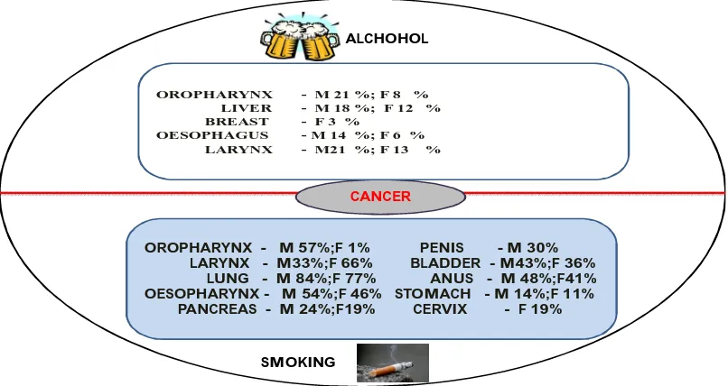
Represents Cancers that have been linked to alcohol
and
smoking 12
Fig 8 Represents the influence of diet on carcinogenesis 13
Fig 9 Represents the role of obesity on cancer 14
Fig 10 Represents the various neoplasm associated with
infection 15
Fig 11
Represents the effect of environmental factors
CHAPTER : 5
Fig 1
Effect of Acanthophora spicifera on tumour weight in
EAC treated mice 51
Fig 2
Effect of the Acanthophora spicifera on tumour
volume in EAC treated cancerous mice 51
Fig 3
Mean survival day of the tumour bearing mice treated with
5 FU and ASE (100 & 200 mg/kg)
52
Fig 4
The effect of Acanthophora spicifera on various blood haematological parameters treated with mice bearing with
EAC cell lines
52
Fig 5
The effect of Acanthophora spicifera on blood haematological parameters like Lymphocytes,
Nuetrophils and Monocytes 53
Fig 6
The effect of Acanthophora spicifera on biochemical
parameters 53
CHAPTER : 6
Fig 1 DPPH assay of ascorbicoaid and EAF 64
Fig 2 MTT assay of EAAS 64
Fig 3
MTT assay of Etoposide 64
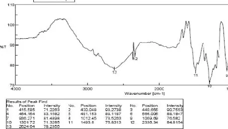
Fig 4 FTIR of compound -1
65
Fig 5 MASS Spectrum of Compound 1
66
Fig 6 NMR spectrum of Compound 1
67
Fig 10 FTIR of Compound-3
73
Fig 11 MASS spectrum of Compound-3
74
Fig 12 NMR spectrum of compound -3
75
CHAPTER :7 Fig
1 &2
HPLC profile of Ethyl Acetate
85
Fig 3-9 HPTLC document for Gallic acid
86
Fig
10-12 HPTLC document for Quercetin 89
CHAPTER: 8
Fig 1 MTT Assay ( Isoquercetin) using HT 29 and HepG2
cancer cell lines 98
Fig 2 Tryphan blue assay (Isoquercetin)
98
Fig 3-5. Effect of Isoquercetin on cell cycle progression in
HepG2 (Liver) cancer cells (DNA histogram) 99
Fig 6-8 Effect of Isoquercetin on cell cycle progression in
HT-29 (Colon) cancer cells (DNA histogram) 100
CHAPTER :9
Fig 1 Effect of ISQN on body weight change in DEN
induced Hepatacarcinoma in rats 113
Fig 2 Effect of ISQN on liver weight in DEN induced
Hepatocarcinoma in rats 114
Fig 3 Effect of ISQN on liver function test in rats treated
with DEN 114
Fig 4 Effect of ISQN on liver SOD, CAT and TBARS
115
Fig 5 Effect of ISQN on liver GPX, GR and GST
115
7 markers
Fig 9 Histopathology report
117
CHAPTER: 10
Fig 1 Represents the Host Control monolayer with
uni(BHK 21) 125
Fig 2 Represents the Virus control (CHIKV infected in to
BHK21) 125
Fig 3-5 Effect of Isoquercetin on CHIKV at different Time
[image:17.595.97.516.92.335.2]1.1 Introduction
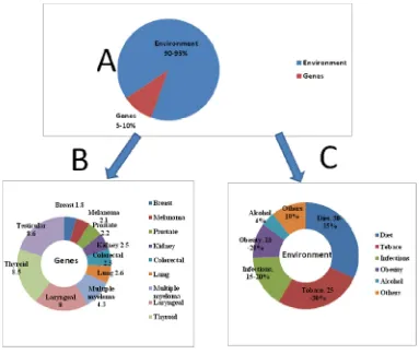
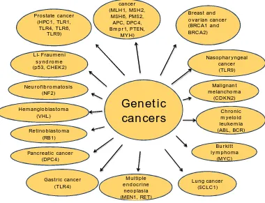
[image:19.595.116.499.352.676.2]Although all cancers are result of multiple mutations3 and these mutations are mainly due to the interaction with the environment4. Infact only 5–10% of all cancers are due to an inherited gene defects which is shown in the fig 2. These observations indicate that most cancers are not of hereditary origin and that lifestyle factors, such as dietary habits, smoking, alcohol consumption, and infections, have a profound influence on cancer development5.
Colorectal cancer (MLH1, MSH2, MSH6, PMS2, APC, DPC4, Bmpr1, PTEN, MYH) Breast and ovarian cancer (BRCA1 and BRCA2) Prostate cancer
(HPC1, TLR1, TLR4, TLR6, TLR9) Li- Fraumeni syndrome (p53, CHEK2) Nasopharyngeal cancer (TLR9) Neurofibromatosis (NF2) Malignant melanchoma (CDKN2) Hemangioblastoma
(VHL) myeloid Chronic
leukemia (ABL, BCR) Retinoblastoma
(RB1)
Burkitt lymphoma
(MYC) Pancreatic cancer
(DPC4) Lung cancer (SCLC1) Multiple endocrine neoplasia (MEN1, RET) Gastric cancer
(TLR4)
[image:20.595.118.498.240.531.2]Genetic
cancers
Fig:2 Represents the various cancers through genetic mutation
For millennia, these diseases processes remained the major area of detailed understanding to mankind. Current epidemiologic evidences links behavioural factors to a variety of malignancies which includes the most common cancers diagnosed in the developed world like lung, colorectal, prostate, and breast cancers. Owing to the tremendous impact of modifiable factors on risk, especially for the most prevalent cancers, it has been estimated that 50% of cancer is preventable7
However, to bring about dramatic reductions in cancer incidence there should be widespread of lifestyle changes are necessary. Cancer, in all forms, has been poorly understood, feared and usually fatal. This disease, thought to be a heterogeneous group of related disorders, was often diagnosed in the past on the basis of macroscopic features such as mass, relentless growth and metastatic spread. With better understanding of pathophysiology of the disease and advances in molecular biological techniques and the scenario is fast changing in the area of cancer biology.
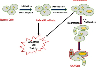
1.1.a. An Insight in to the Carcinogenesis8
first step in carcinogenesis. Body cells that divide rapidly are more susceptible to carcinogenesis because there is less opportunity for DNA repair before cell division. Mutagenic changes in the components of signalling pathways lead to cellular transformation (cancer).
Initiation
DNA Repair
Normal Cells Cells with adducts
Promotion
Cell Proliferation
Initiated cells
Progression Cell
Proliferation
Apoptosis Cell Toxicity
CANCER
Fig 3 Represents the tumour progression towards the normal cell to cancerous
cell leads to apoptosis by cell cycle arrest
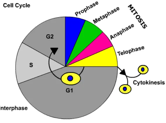
Normally, human cell life cycles consist of proliferation, differentiation, and cell death. The cell reproductive life cycle has four phases G1, S, G2, M. G0 is a
[image:22.595.110.494.219.489.2]next phase is G2 when the cell prepares other structures needed for mitosis. The M phase is mitosis itself , where two daughter cells are produced
[image:23.595.165.450.372.583.2]There are two distinct of hallmarks of cancer. The first family describes the conditions the cell of a tissue needs to exhibit uncontrolled proliferation. These are self sufficiency in growth signals, ignoring of anti-growth signals, evading apoptosis and immortalization. Acquiring all these four properties is a necessary condition for a cell to become malignant. However, in order to cause certain death of the organism, a tumor must also have the two hallmarks belonging to the second family: angiogenesis and metastasis. Angiogenesis, the process of generating new blood vessels which infiltrate the tumor, is essential for its growth beyond a (very small) critical size9
Fig 4 represents the normal cell cycle and its various phases
words imbalance between quantity of the signalling molecules (growth factors) and the number of receptors, i.e. the efficiency of receptions, is increased. The gene which encodes for the receptor can undergo a mutation. Over expression or mutation of a gene drive the cell towards cancer, the affected gene is called an oncogenes.
A healthy cell receives signals to proliferate as well as signals to not divide. Some of these signals are of extracellular, like e.g. anti-growth factors, other are internal. If such an anti-growth factor binds to its receptor, the complex initiates a cascade which blocks transcription. The main controller of these processes is the Rb-protein. It works as follows: When a transcription factor called E2F (elongation factor 2) is free and activated, the transcription of a large number of genes is initiated, the biological material needed for growth is synthesized and cell proliferation can proceed. When Rb is bound to E2F, it neutralizes E2F and makes it inactive10.
However, as soon as Rb is deactivated, it releases the E2F molecule which subsequently enters the nucleus and transcription starts. The transition from the active to the inactive state of Rb is induced by its phosphorylation, i.e. adding a phosphate group to the protein. Rb belongs to a family of proteins, or genes, which are called tumor suppressors. Under normal conditions wild type Rb arrests growth; if it undergoes a mutation such that freezes it in its inactive state, it loses its function. Finally, it is worth noticing that modification of one of the two copies of the oncogenes and tumor suppressors have different effects: while a mutation into an oncogene of one of the two copies may be sufficient for a physiological effect, generally both copies of the tumor suppressor have to be removed or inactivated in order to affect the cell.
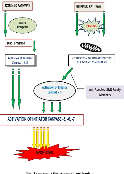
1.2 Apoptosis
pathway. Signal transduction may occur by two distinct pathways: extrinsic or receptor-linked apoptosis or intrinsic or mitochondria- mediated apoptosis12.
1.2.a. Extrinsic apoptotic pathway
This receptor-linked pathway requires the binding of a ligand to a death receptor on the cell surface. The cytokine, tumor necrosis factor (TNF ), binds to the death receptor, TNF receptor type 1, which recruits two signal transducing molecules: TNFR 1-associated death domain protein (TRADD) and Fas-associated protein with death domain (FADD). This complex then binds to procaspase 8 to activate caspase 8, which initiates the protease cascade leading to apoptosis13
1.2.b. Intrinsic apoptotic pathway
including Smac/DIABLO (caspase activator) and apoptosis-inducing factor17. Next, caspase 3 activation is detected following the formation of the apoptosome
1.3 Immortalization
A normal cell cannot divide an infinite number of times and there is a kind of clock which keeps count. This clock uses the fact that during cell division DNA is replicated. Replication of DNA is done by a protein called DNA polymerase. During cell division it attaches itself to a single strand of DNA of the mother cell, runs along it and synthesizes a matching strand of DNA. This way the number of copies of the entire DNA content of the dividing cell doubles, and subsequently, during mitosis, it is divided evenly between the two daughter cells. However, during each copying process the DNA polymerase cannot replicate the last stretch of the mother DNA strand to which it binds (these ends of each DNA are called telomeres). Consequently, the resulting DNA of the daughter cells is shortened compared to the original template. Each division reduces the length of DNA by about 100 nucleotides. In fully mature differentiated cells expression of telomerase is suppressed and therefore these cells cannot divide infinitely often. The division of cancer cells, on the other hand, is not restricted to a certain number of times. Cancer cells circumvent this limitation and acquire the ability to elongate their telomeres. This process is called immortalization18
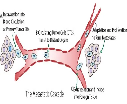
1.4. Metastasis and angiogenesis
Fig: 5 represents the Apoptotic mechanism
EXTRINSIC PATHWAY INTRINSIC PATHWAY
Disc Formation
Death
Receptors STRESS
ACTIVATION OF PRO-APOPTOTIC BCL2- FAMILY MEMBERS
Activation of Initiator Caspase – 8,10
Activation of Intiator Caspase – 9
ACTIVATION OF INTIATOR CASPASE -3, -6, -7
Anti Apoptotic Bcl2 Family Members
Fig 6 represents the Metastatic Cascade
But before being able to metastase, a tumor faces another problem, namely the supply of nutrients and oxygen. In normal tissues, those are delivered via the bloodstream but as a tumor mass expand (by division of its cells), only its edges can grow to the vicinity of existing blood vessels. Hence, during growth most of the tumor mass becomes increasingly distant from blood vessels and is consequently deprived of oxygen (the diffusion length of oxygen in human tissue is on the order of a few hundred microns). Thus, in order to grow beyond a critical size, a tumor induces vascularization: the in growth of blood vessels. This process is called angiogenesis19.
1.5. Risk Factors of Cancer
active carcinogen was said to be benzopyrenediol epoxide, where as a this particular carcinogen is having the direct etiological link with lung cancer20. Among all developed countries the prevalence of smoking has been slowly declining due to strong governamnt policies and awareness programmes. At the similar time the similar scenario exits in developing countries like India. The prevalence of smoking is increasing due to lack of governament policies and awareness towards smoking. The only way to minimise the sigar smoking is to accelerated tobacco control programs, will be the only way to reduce the rates of tobacco-related cancer mortality. How smoking contributes to cancer is not fully understood. It has been known that smoking can alter a large number of cell signalling pathways in lung tissue and it has been established that a direct link through the activation of NF-kB21.
1.5.a. Alcohol
CANCER
OROPHARYNX - M 21 %; F 8 % LIVER - M 18 %; F 12 %
BREAST - F 3 % OESOPHAGUS - M 14 %; F 6 %
LARYNX - M21 %; F 13 %
OROPHARYNX - M 57%;F 1% PENIS - M 30% LARYNX - M33%;F 66% BLADDER - M43%;F 36%
LUNG - M 84%;F 77% ANUS - M 48%;F41% OESOPHARYNX - M 54%;F 46% STOMACH - M 14%;F 11%
PANCREAS - M 24%;F19% CERVIX - F 19%
ALCHOHOL
[image:30.595.104.507.94.308.2]SMOKING
Fig 7 represents Cancers that have been linked to alcohol and smoking
1.5.b. Diet
Primly diet influences the lower GIT cancer deaths in world wide which is directly relay on diet (Fig. 7). The extent to which diet contributes to cancer deaths varies a great deal, but it purely depends on type of cancer25. Most carcinogens that are ingested, such as nitrates, nitrosamines, pesticides, and dioxins, come from food or food additives or from cooking. Another consecutive factor for many types of cancers is consumption of red meat. Especially, for those of the gastrointestinal tract, but also for colorectal, prostate26, bladder, breast, gastric, pancreatic, and oral cancers27.
Diet Vs cancer
35%
Endometrial 50%
Larynx, Bladder Mouth, Pharynx 20%
Breast 50 %
Gastric 35%
Others 10%
Gall Bladder
50% Prostate 75%
Pancreatic 50% Colorectal 70% Lung 20%
Fig 8 represents the influence of diet on carcinogenesis
1.5.c. Obesity
Obesity
14%
20%
Esophageal cancer
Colon cancer
Gastric cancer
Gall bladder cancer
Ovarian cancer
Breast cancer
Endometrial cancer Pancreatic cancer
Cervical cancer Non- Hodgkin’s
lymphoma Renal cancer
Fig 9 Inflence on obesity on cancer
1.5.d. Infectious Agents
Worldwide, an estimated 17.8% of neoplasms are associated with infections especially with viruses which accounts for most infection related cancers (Fig 9). The mechanisms by which infectious agents promote cancer are becoming increasingly evident. Infection-related inflammation is the major risk factor for cancer, and almost all viruses linked to cancer have been shown to activate the inflammatory markers31. Human papillomavirus, Epstein Barr virus, Kaposi’s sarcoma associated herpes virus, human T-lymphotropic virus 1, HIV,HBV, and HCV are associated with risks for anogenital cancer, cervical cancer, skin cancer, Burkitt’s lymphoma, nasopharyngeal cancer, Hodgkin’s lymphoma, Kaposi’s sarcoma, adult T-cell leukemia, B-cell lymphoma, and liver cancer. However, other microorganisms, including selected parasites such as Opisthorchis viverrini or
Infection
cancer
Hepatocellular Carcinoma (Hepatitis-B, Hepatitis- C Anogenital cancers (6, HPV-16, HPV-18) Kaposi sarcoma (HPV-8) Lymphoma (EBV) Lymphoma (HIV- 1) Mucosa- associated Lymphoid tissue Lymphoma (Helminthes: Schitosomes Clonorchis sinesis) Gastric cancers (Helicobacter pylori) Merkel cell carcinoma (Merkel cell polyvirus)
Fig 10 Represents the various neoplasm associated with infection
1.5.e. Environmental pollution
1.5.f. Radiation
Radiation like ionizing and nonionizing, typically from radioactive substances and ultraviolet (UV), pulsed electromagnetic fields increases the 10% of total cancer cases world wide. The cancers induced by radiations, includes some types of leukemia, lymphoma, thyroid cancers, skin cancers, sarcomas, lung and breast carcinomas32.
Fig 11. Represents the effect of environmental factors associated with
carcinogenesis
Natural products and their derivatives represent more than 50% of all the drugs in clinical use of the world. These natural products and their derivatives have historically been a major source of new pharmaceuticals and have made enormous contributions towards human health. Their role in the drug discovery process is especially renounced in the areas of anti cancer and infectious disease agents, where the fractions of the drugs derived from natural products amount to 60 and 75%, respectively. ENVIRONMENTAL CARCINOGENS Bladder cancer, colorectal cancer, leukemia (Chlorinated drinking water) Bladder cancer [Nitrate consumption]
Sarcoma & lymphoma [Dioxane, incinerators] Testicular cancer (Env. Organic pollutants)
[In utero]
Childhood leukemia & lymphoma, brain tumors,
Wilm’s tumors, Ewings sarcoma, germ cell tumors
[Pesticides] Lung cancer metastasis [Nitric oxide] Leukemia, lymphoma& colorectal cancer (Nitrates) [Drinking water] Childhood leukemia [Motor vehicle exhaust]
Childhood leukemia & lymphoma [Indoor air- pollutants]
1.6. Role of natural products on cancer
Over the past 50 years, natural products (NP, here the term is restricted to small molecules) have been the cornerstone of anti cancer pharmacology. The discovery of these antitumor NP opened the route to the ‘‘forty glory cytotoxic’’ four decades of cytotoxic agents, among which a significant number of molecules from plants used today for the treatment of cancer. Immediately, the anti tumor pharmacology community embraced the new concept and this vision of molecular targetbased drug discovery (or reverse pharmacology) became a standard anti cancer research.
Old good drugs from plants and microbes remain essential. Since the 1950s, a large number of novel antitumor drugs have been identified and validated clinically. There is no doubt that the treatment of cancers has profoundly changed, with the advances of targeted therapies. For example, the discovery of specific tyrosine kinase inhibitors like imatinib (Gleevec1), or nilotinib (Tasigna1) have opened the new vistas in pharmacotherapeutics in cancer33. There is a strong need for novel anticancer drugs effective against solid tumors, especially at advanced stages of the disease. This can be achieved by the innovation of small molecules from NP should continue to be consider as one of the most important sources of innovative products.
ointment as well as for the treatment of cough, wounds, gout, goiter, venereal disease, and so forth. But modern scientific world proves that pleothra of the biological potential of seaweeds as antioxidant, antimutagenic, anticoagulant, and antitumor. The seaweeds also play an important role in modification of lipid metabolism in the human body. Seaweed extract is interestingly similar to human blood plasma (Langseth, 1995). Although, the use of seaweeds in medicine is not as wide spread as once it was, the use of seaweed polymer extract in pharmacy, medicine, and biochemistry is well established. Clinical trials are also in progress to make diabetic patients free from injection by introducing insulin secreting “jelly capsule”made of seaweed-derived alginic acid36.
1.7. Anticancer Agents from Marine Floras
Marine microorganisms are a source of new genes, and exploitation his likely to the discovery of new drugs and targets. In this marine resources red algae are considered to be one of the potential organisms which can be the richest sources of known and novel bioactive compounds including toxins with potential for pharmaceutical applications37. Scytonemin is a protein serine/threonine kinase inhibitor, isolated from the cyanobacterium Stigonema sp. Scytonemin regulates mitotic spindle formation as well as enzyme kinases involved in cell cycle control and the compound also inhibits proliferation of human fibroblasts and endothelial cells. Thus scytonemin may provide an excellent drug as protein kinase inhibitors to have antiproliferative and anti-inflammatory activities38.
Edible seaweed like Palmaria palmate is shown to be effective antioxidant, capable of inhibiting cancer cell proliferation40. Algae have gained special interest owing to their biological properties. There are many reports on the immunomodulating and antitumor activities of algae41. An extract from the brown seaweed Sargassum thunbergii has shown antitumour activity and inhibition of tumour metastasis in the rat mammary adeno carcinoma cell.
Recent studies in the field of cancer research have revealed promising compounds, isolated from natural sources, with proven anticancer activity. Three examples are trabectedin, cytarabine and eribulin mesylate42,43, which represent the first three described marine anticancer drugs. Indeed, almost 50% of the anti tumor agents approved in the last 50 years of the 20th century were either compounds derived from natural sources or (semi-) synthetic analogs44. Natural compounds remain a high out put source of promising chemotherapeutic or chemo preventive agents in current cancer research45. Marine organisms are potentially prolific sources of highly bioactive secondary metabolites that might represent useful leads in the development of new pharmaceutical agents. During the last four decades, numerous novel compounds have been isolated from marine organisms and many of these substances have been demonstrated to possess interesting biological activities46.
Table: 1
Marine Flora Chemical
Composition Biological Activity
Acanthophora
spicifera Crude Extracts Antioxidants and inhibiting cancer
cell proliferation48
Acanthophora
spicifera Crude Extracts
Tumoricidal activity on Ehrlich’s ascites carcinoma developed in mice48
Ascophyllum
nodosum
Fucoidan Antiproliferative antitumour, anticancer, antimetastatic, and
fibrinolytic49
Caulerpa sp. Caulerpenyne
Cytotoxicity, anticancer, antitumour, and antiprolifer-
ating activity50
Chondria sp. Condriamide A Cytotoxicity51
Cyanobacteria Apratoxins Inhibit a variety of cancer cell lines52
Cyanobacteria Nostoc
linckia and Nostoc
spongiaeforme var.
Tenue
Borophycin
Cytotoxicity against human epidermoid carcinoma
(LoVo) and human colorectal adenocarcinoma activity53
Cystophora sp Meroterpenes and
Usneoidone Antitumour
A-(a pentamer), dieckol, and 8,8-bieckol (hexamers)
Leptolyngbya sp.
coibamide A Cytotoxicity against NCIH460 lung and mouse neuro-2a cells56
Nostoc linckia Cyptophycin 1 Cytotoxicity against human tumor cell lines and human solid tumors57
Nostoc
spongiaeforme Cryptophycin 8
Greater therapeutic efficiency and lower toxicity than
cryptophycin 14 in vivo58
Palmaria palmata
Phloroglucinol and its polymers, namely, Eisenia bicyclis
eckol (a trimer), phlorofucofuroeckol A-(a pentamer), dieckol, and 8,8-bieckol(hexamers)
Antioxidant activity of the phlorotannins59
Stigonema sp. Scytonemin Antiproliferative and
anti-inflammatory activities60
2.1. Classification
Kingdom : Plantae
Phylum/division : Rhodophyta
Class : Rhodophyceae
Order : Ceramiales
Family : Rhodomelaceae
Genus : Acanthophora
Species name : Acanthophora Spicifera(vahl) borgesen
Common name : Spiny Seaweed
Plate 1 : Habitat of Acanthophora spicifera
2.2. Species description
2.3. Morphology
Shape : Large irregularly shaped holdfast for attachment to hard bottoms.
Branches : Short, Determinate branchlets that are irregularly and Spinose. Branchlets are hook-like, brittle and fragment easily under heavy wave action.
Colour : Shades of red, purple, or brown62
Height : Upright to approximately 25 cm.
2.4. Regional Occurrence
2.5. Use
Aim & Objective
The cytotoxic and anticancer investigations of marine resources are increasing worldwide because these marine resources are economical, safe and effective, apart from this these marine resources posses enormous hidden phytoconstituents in the ocean with particular to the development of new chemical entity as anticancer drugs which has isolated from marine algae, seaweeds, tunicates etc..
The present aim of the investigation is to explore the anticancer and antiviral effect of marine algae Acanthophora spicifera belonging to the family: Rhodomelaceae which is widely distributed in Mandapam, Costal region of Tamilnadu, Gulf of Mannar, Bay of Bengal which is chosen its for in vivo and in vitro studies.
The objective of the present investigations is as follows.
Collection of sufficient quantity of marine alga, Acanthophora spicifera
for the present research investigation, identification and preservation of the marine algae
Extraction of marine algae using conventional extraction procedure followed by filtration and purification
Preliminary phytochemical studies of Acanthophora Spicifera extract followed by qualitative and quantitative phytochemical investigations.
Preliminary anticancer effect of crude extract in Earlich Ascites Carcinoma challenged mouse model
Bioactive guided fraction of crude extracts of Acanthophora Spicifera
against various human cancer cell lines and Free radical scavenging assay
Column chromatographic isolation of bioactive fraction High performance thin layer chromatography (HPTLC) finger print technique
Cell cycle analysis of anticancer isolates against HepG 2 and HT 29 cell lines
Anticancer efficacy of isolated compounds against Diethyl Nitrosamine (DEN) induced Hepato carcinoma in rats
CHAPTER 4
1. Mohamed et al., 2012 reported that edible seaweeds are rich in bioactive antioxidants, soluble dietary fibers, proteins, minerals, vitamins, phytochemicals, and polyunsaturated fatty acids. Although previously the seaweeds were only used as gelling and thickening agents in the food or pharmaceutical industries, recent researches have revealed their potential as complementary medicine. The red, brown and green seaweeds have been shown to have therapeutic properties for health and disease management, such as anticancer, antiobesity, antidiabetic, antihypertensive, antihyperlipidemic, antioxidant, anticoagulant, anti-inflammatory, immunomodu -latory, antiestrogenic, thyroid stimulating, neuro protective, antiviral, antifungal, antibacterial and tissue healing properties in in vivo. Active compounds include sulphated polysaccharides, phlorotannins, carotenoids (e.g. fucoxanthin), minerals, peptides and sulfolipids, with proven benefits against degenerative metabolic diseases.
2. Lavanya and Veerappan, 2011 has reported that in vitro antibacterial activity of six selected marine algae (seaweeds) which have been selected and their extracts have been tested as an alternative to commonly used antibiotics. Extracts of six seaweed samples namely Codium decorticatum, Caulerpa scalpelliformis, Gracilaria crassa, Acanthophora spicifera, Sargassum
wightii and Turbinaria conoides were collected from Gulf of Mannar, southeast cost region. The collected algae were subjected for antibacterial activity. Extraction was done by using the following solvents viz., Acetone, methanol, chloroform, diethyl ether, ethyl acetate, hexane and aqueous and screened against selected human pathogens like Vibrio parahaemolyticus, Salmonella sp, Shewanella sp, Escherichia coli, Klebsiella pneumoniae,
Streptococcus pyogenes, Staphylococcus aureus, Enterococcus faecalis,
3. Mayer et al., 2011 has clearly explained that preclinical pharmacology of structurally characterized marine compounds isolated from marine animals, algae, fungi and bacteria is discussed in a comprehensive manner. From 74 marine natural compounds various biological activities like antibacterial, anticoagulant, antifungal, antimalarial, antiprotozoal, antituberculosis and antiviral activities were reported. Additionally, 59 marine compounds were reported to affect the cardiovascular, immune and nervous systems as well as to possess antiinflammatory effects.
4. Schumacher et al., 2011 explained that the cancer is one of the most deadly diseases in the world. Although advances in the field of chemopreventive and therapeutic medicine have been made regularly over the last ten years, the search for novel anticancer treatments continues. In this field, the marine environment, with its rich variety of organisms, is a largely untapped source of novel compounds with potent antitumor activity.
5. Wijesekara et al., 2011 reported recently, a great deal of interest has been developed to isolate novel bioactive compounds from marine resources because of their numerous health beneficial effects. Among marine resources, marine algae are valuable sources of structurally diverse bioactive compounds. The cell walls of marine algae are rich in sulfated polysaccharides (SPs) such as fucoidans in brown algae, carrageenans in red algae and ulvans in green algae. These SPs exhibit many beneficial biological
activities such as anticoagulant, antiviral, antioxidative, anticancer and immunomodulating activities. Therefore, marine algae derived SPs have great potential for further development as products in nutraceutical, pharmaceutical and cosmoceutical areas. This contribution presents an overview of biological activities and potential health benefits of SPs derived from marine algae.
offering a great scope for discovery of new anticancer drugs. The marine floras are rich in medicinally potent chemicals predominantly belonging to polyphenols and sulphated polysaccharides. The chemicals have displayed an array of pharmacological properties especially antioxidant, immunostimulatory, and anti tumour activities. The phytochemicals possibly activate macrophages, induce apoptosis, and prevent oxidative damage of DNA, thereby controlling carcinogenesis. In spite of vast resources enriched with chemicals, the marine floras are largely unexplored for anticancer lead compounds. Hence, this paper reviews the works so far conducted on this aspect with a view to provide a baseline information for promoting the marine flora-based anticancer research in the present context of increasing cancer incidence, deprived of the cheaper, safer, and potent medicines to challenge the dreadful human disease.
7. Nurul et al., 2010 said that the antimicrobial activity of eight crude extracts of Acanthophora spicifera were screened against 18 bacteria, 3 yeast and 6 fungi by disc diffusion method. The results clearly reveals that methanol and ethyl acetate extracts exhibits broad spectrum activity against all tested bacterial strains like Bacillus Cereus, Pseudomonas aeruginosa, yersinia sp
and Citrobacter freuidii. The extract does not shown the any activity against tested strains of yeast and fungi.
The growing interest in marine-derived antiviral compounds, along with the development of new technology in marine cultures and extraction, will significantly expedite the current exploration of the marine environment for
compounds with significant pharmacological applications, which will
continue to be a promising strategy and new trend for modern medicine.
9. Liu et al., 2009 has stated that prevention and treatment of cancer require the continued development of novel and improved chemopreventive and chemotherapeutic agents. Throughout history, natural products have afforded a rich source of anticancer agents with diverse chemical structures and bioactivities. Recent technological and methodologic advances in structure elucidation, organic synthesis, and biological assay have resulted in the isolation and clinical evaluation of various novel anticancer agents. In this review, they present the anticancer activities, mechanism of action, structure and activity relationships of six important anticancer agents from natural products, that is, taxol, betulinic acid, camptothecin, resveratrol, podophyllotoxin and curcumin.
10. Moon et al., 2009 the invention of Laurinterol (LOEL) which was isolated from Laurencia okamurai is considered as invention for the prevention and inhibition of melanoma, LOEL can effectively inhibit the growth of melanoma cells by inducing apoptosis therein without adverse effect as in synthetic medicines. Thus, LOEL exhibited a dose dependent inhibitory effect on the growth of melanoma cells as it was observed that cells are treated with LOEL at 10 g/ml and the growth of melanoma cells by was inhibited 50%. Addition of 1 µg/ml of LEOL exerted 30% inhibition on the growth of melanoma cells in the presence of fetal bovine serum (FBS)
inhibits considerably the expression of Bcl-2 whilst increasing that of Bax; it also stimulates the release of mitochondrial cytochrome-c and activates caspase-9 and caspase-3. All these results point clearly to the activation of the mitochondrial apoptotic pathway in response to the treatment of HT29 colon-cancer cells with masilinic acid. Our results suggest that masilinic acid has the potential to provide significant natural defence against particular colon -cancer cell lines.
12. Bacac and Stamenkovic, 2008 said that metastasis is the result of cancer cell adaptation to a tissue microenvironment at a distance from the primary tumor. Metastatic cancer cells require properties that allow them not only to adapt to a foreign microenvironment but to subvert it in a way that is conducive to their continued proliferation and survival. Recent conceptual and technological advances have contributed to our understanding of the role of the host tissue stroma in promoting tumor cell growth and dissemination and have provided new insight into the genetic makeup of cancers with high metastatic proclivity.
and Acanthophora spicifera were evaluated. Total phenolic content and reducing power of crude methanol extract was determined. The antioxidant activities of total methanol extract and five different solvent fractions (viz.,
petroleum ether (PE), ethyl acetate (EA), dichloromethane (DCM), butanol (BuOH) and aqueous) were also evaluated. EA fraction of A. spicifera
exhibited higher total antioxidant activity (32.01 mg ascorbic acid equivalent/g extract) among all the fractions. Higher phenolic content (16.26 mg gallic acid equivalent/g extract) was noticed in PE fraction of G.edulis.
Reducing power of crude methanol extract increased with increasing concentration of the extract. Reducing power and hydroxyl radical scavenging activity of E. kappaphycus were higher compared to standard antioxidant (-tocopherol). The total phenol content of all the seaweeds was significantly different (P < 0.05). In vitro antioxidant activities of methanol extracts of all the three seaweeds exhibited dose dependency and increased with increasing concentration of the extract.
15. Lang et al., 2007 has done the phytochemical investigation on Acanthophora spicifera (Vahl) Borgesen (Ceramiales: Rhodophyta) rhodophycean alga and chemical substances was identified as 5-cholestane-3,6-dione, cholest-4-ene-3-one, 11-hydroxy-5-cholestane-3,6-dione, cholest-5-en-3-diol and cholest-4-ene-3,6-diol from Acanthophora spicifera were described. Flavonoids such as quercetin, (-)- catechin and tiliroside, acanthophorin A and B, acid derivates, dipeptides and anthraxanthin were also identified from the above species.
16. Ma et al., 2006 has stated that the red alga Rhodomela confervoides was the source of four bromophenols and they exhibited moderate cytotoxicity against several human cancer cell lines.
18. Vasanti et al., 2006 screened Acanthophora spicifera from the Gulf of Mannar for their biological activity. This particular species was screened for hepatoprotective effect and antioxidant effect in CCl4 intoxicated male albino
rats. Liver damage was induced in rats by injecting CCl4. The effect of the
seaweed extract (ethanolic) at different doses was determined by comparing with the controls. The algal extract at a dose of 200 mg/kg b.w orally was found to exhibit a significant decrease in the level of SGOT, SGPT and LDH as compared to those of the CCl4 induced liver damaged controls. The level
of lipid peroxidation was also decreased in the 200 mg/kg extract treated rats. The antioxidant status of the extract treated rats showed tremendous increase in the levels of the antioxidant enzymes SOD, catalase and glutathione peroxidase. The biological activity was related to the phyto-constituents such as flavonoids, vitamin – A, E, C, present in the algae.
19. Abatis et al., 2005 has screened brown alga Taonia atomaria, which was a source of meroditerpenes atomarianones A and B stated as cytotoxic agents which has screened against the NSCLC-N6 and A-549 cell lines.
20. Kubanek et al., 2005 explained that the red alga Callophyeus serratus was the source of three antibacterial and antifungal diterpene-benzoate compounds, bromophycolides A and B, and a non-halogenated compound. Bromophycolide A was cytotoxic against several human tumour cell lines by specific induction of apoptosis.
22. Bhaskar et al., 2004 has screened the three species of red marine macro algae (Rhodophyta) from the Indian Ocean and subjected to analysis for the occurrence of conjugated polyenes. The composition of different lipid classes in these seaweeds along with their fatty acid composition has also been reported. Analysis of lipid classes of these seaweeds revealed that both Acanthophora spicifera (Ceramiales, Rhodophyta) and two species of
Gracilaria, viz. G.edulis and G.folifera (Gracilariales, Rhodophyta) were rich in glycolipids. However, A. spicifera had significantly higher amounts of eicosapentaenoic acid (EPA) and arachidonic acid (AA) as compared to negligible amount of these fatty acids in both species of Gracilaria. The red seaweed Acanthophora spicifera contain conjugated eicosapentaenoic acid (CEPA) and conjugated arachidonic acid.
23. de Ines et al., 2004 has stated that furoplocamioid C, perfuroplocamioid, pirene and tetrachlorinated cyclohexane from the red alga Plocumium carttilagineum exhibited selective cytotoxicity against human tumour cell lines with pirene showing a specific and irreversible effect on SW480 cells.
24. Stein and Colditz , 2004 stated that over 6 million people around the world die from cancer each year. Modifiable risk factors have been linked to a wide range of malignancies, including cancers of the oropharynx, oesophagus, larynx, lung, kidney, bladder, pancreas, skin, stomach, ovary, breast, cervix, uterus, prostate, and colon. Research indicates that over half of all cancers in developed countries could be prevented if we implemented population-wide measures to promote the following behaviours: reduce tobacco use, increase physical activity, control weight, improve diet, limit alcohol, utilise safer sex practices, get routine cancer screening tests, and avoid excess sun expore.
25. Goncalves et al., 2002 has reported that the water-soluble acid agaran isolated from Acanthophora spicifera (Rhodophyta) was submitted to alkaline treatment for the complete cyclization of -L-Galp 6-sulfate to 3,6-An- -L-Galp units. The modified agaran was then partially depolymerized using
purified by gel-filtration chromatography and studied by ESIMS and NMR spectroscopy, including 1D, 1H,13C, DEPT and 2D 1H, 1H COSY, TOCSY and 1H,13C HMQC procedures
26. Dorta et al., 2002 showed the Stypolactone, a diterpenoid of mixed biogenesis has been isolated from the brown algae Stypopdium zonale and showed weak cytotoxic activity in vitro against the A-549 and H-116 cell lines.
Acanthophora spicifera. The structures were determined to be kaempferol 3-O--L-fucopyranoside(1) and quercetin 3-O--L-fucopyrinoside by spectroscopic methods and these compounds shown significant antioxidant activity.
29. Guardia et al.,1999 has stated that the enantioselective synthesis is of natural anti tumour(-) – Bifurcadio involving an alkylation key-step reaction is reported. In this paper, we report the first enantioselective synthesis of (-)-Bifurcadiol 6 involving an alkylation key-step reaction between a cyanohydrin and an allyl bromide (Scheme 1). trans trans Farnesol 1 was converted into the corresponding ether 2 by treatment with dihydropyran and subsequently oxidized using a mixture of pyridine, selenium dioxide and PDC to afford the expected aldehyde 3 in 34% yield. 4 Compound 3 was added to a solution of trimethylsilyl cyanide in the presence of a catalytic amount of sodium cyanide and 18-crown-6 ether complex to lead, after addition to a solution of LiN(SiMe3)2 in anhydrous THF (-78°C) and condensation with 1-bromo-3-methylbut-2-ene, to the silyl cyanide derivative 45 (52% yield). Subsequent treatment with triethylamine trihydrofluoride salt afforded the expected ketone 5 in 70% yield.
30. Iwamoto et al., 1998 explains the Penostatins F–I have been isolated from a strain of Penicillium sp. originally separated from the marine alga
Enteromorpha intestinalis, and their stereostructures have been established on the basis of spectral analyses. All the compounds exhibit significant
cytotoxicity against culture
31. Wahidulla et al., 1998 has isolated new steroid cholest-4-ene-3, 6-diol together with the known cholest-4-ene-3-one, lauric acid and o-phthalic acid bis-(2-ethyl nonyl)-ester were isolated from the red alga Acantophora spicifera. The structures of these compounds were established on the basis of their spectral data.
of Leptosphaeia sp. originally isolated from the marine alga Sargassum tortile. Their stereo structures, with a different configuration from that of leptosins A-C, have been elucidated by spectral and X-ray analyses and some chemical transformations. X-Ray and NMR and NOE spectral analyses of 4 revealed that it exists in a mixture of four conformers, of which two each closely resemble, in a single crystal, and in a single conformer in solution. NOE experiments of 5 and 6 demonstrated that they exist in a mixture of two conformers slowly exchanging in CDCI3.
33. Gerwick et al., 1994 taken an account on bioassay-guided fractionation of the organic extract of Lyngbya majuscula led to the isolation of a new lipid, curacin A, with exceptional brine shrimp toxic and antiproliferative activities. Its unique thiazoline-containing structure has been deduced from spectroscopic information. Pure curacin A is an antimitotic agent (IC60 values in three cell lines ranging from 7 to 200 nM )that inhibits microtubule assembly and the binding of colchicine to tubulin.
34. Numata et al., 1993 has stated that the isolated Communesins A and B, exhibiting cytotoxic activity against the cultured P-388 cells, were isolated from the mycelium of a strain of pencillium sp. stuck on the marine alga enteromorpha intestinalis and their structures was elucidated by spectroscopic analyses.
35. Fuller et al., 1992, has isolated polyhalogenated monoterpene from the red alga Portieria hornemanii is considered as a novel in vitro antitumor agent by National Cancer institute (NCl). The NCI Decision Network Committee selected halmon as a pre-clinical drug for development.
Aspergillus niger and Candida albicans. The content of agar in Acanthophora spicifera was found to be the highest (33.5%) of all the species studied.
37. Numata et al., 1991 shown that partition fraction of hexane, Chloroform, CHCL3 for the methanolic extracts of marine algae were examined for
cytotoxicity activities against cultured P-388 lymphocytic leukemia cells. Cytotoxic activities were found for partition fractions of 21 species of seaweed. Bioactivity guided fraction of the CCl4 partition fraction from sargassum tortile, exhibiting the most prominent activity, afforded dihydroxysargaquinone and sargatriol previously isolated from the alga. The former was evaluated as a cytotoxic principle and the latter showing the moderate activity, suggested to be an artefact from 1 during the isolation procedure.
38. Solambi et al., 1991 has done investigation on red algae, Acanthophora spicifera afforded the know peptide, aurantiamide acetate and new diastereoisomer of this dipeptide(dia- aurantiamide acetate). This is the first report of aurantiamide acetate form a marine source and of the natural occurrence of the diastereoisomer.
39. Cortes et al., 1990 reported that 2-acetoxy-15-bromo-6,17-dihydroxy3-palmitoyl-neoparguera- 4(19), 9(11)-diene, a novel seco-parguerane skeleton have been isolated from the red alga laurencia obtuse from Okinawa and showed a cytotoxic activity against various cancer cell lines.
2-aminoanthracene and ethyl methanesulfonate toward, respectively, the T-98 strain plus a metabolic activator and T- 100.
41. Kalaiperumal et al., 1986 has done experimental field cultivation of the red alga Acanthophora spicifera (Vahl.) Boergesen, by vegetative-propagation method, Vegetative fragments 5 cm in length were tied into clusters with polypropylene straw and were fastened to nylon fishing lines. The weight of seed material thus introduced was 4.85 kg. The algae grew rapidly and reached harvestable size of 15.9 cm mean length in 25 days. The weight of fresh harvested plants was 12.85 kg, having had a 2.6 fold increase over the weight of the seed material, indicating that the near shore area of Hare Island in Gulf of Mannar, where the experiment was conducted, is suitable for large-scale cultivation of this seaweed.
42. Higa, 1985 reported that Several cyclic monoterpenes have been isolated from the Japanese red alga Desmia hornemanni, and some chemical modification have been done on these compounds to get the most active moiety for cytotoxic activity and these compound exhibited relatively high activity against P-388 lymphoblastic cell line, A-549 lung carcinoma, and HCT-8 human colon adenocarcinoma.
43. Two new cyclic ethers consisting of squalene carbon skeleton, teurilene and thyrsiferyl 23-acetate, have been isolated from the red alga Laurencia obtuse
(Suzuki et al., 1985). Thysiferyl 23-acetate (bromo ether) showed remarkably cytotoxic property (EDso of 0.3 µg/ml) against P388 in vitro cell line.
CHAPTER 5
Anticancer, Antioxidant Effect
of Alcoholic Extracts of
5.1. Introduction
Free radical and reactive oxygen species (ROS) are by products produced in body due to various physiological and biochemical processes. Generally, most of these free radicals generated from cellular metabolism are scavenged by endogenous defence system such as superoxide dismutase, Catalase and Peroxidase–glutathione system110. However, in many cases, such as in unhealthy physical condition, ageing, or under stress environments, the endogenous antioxidants are either exhausted or insufficient to scavenge these free radicals.
Imbalance of these free radicals can cause oxidative damage to biomolecules like lipids, proteins and DNA111. These reactive oxygen species (ROS) has been implicated and having major role on human biological system which leads to diseases, like ageing, cancer, and neurodegenerative disorders etc112. Among this, cancer is a complex disease characterized by proliferation (uncontrolled cell division), cell transformation, and cape of apoptosis, invasion, angiogenesis and metastasis.
Cancerous cells are to produce reactive oxygen species (ROS) and their
inflammatory mediators. ROS may cause DNA mutation, which may be followed by
oncogenes activation and down regulation of tumour suppressor genes113. By the activity of ROS, scavenging system gets altered in tumour cells114. Over the past decades, seaweeds or their extracts have been shown to produce a variety of compounds and some of them have been reported to possess biological activity of potential medicinal values115.
and Acanthophora spicifera118. In this study Acanthophora spicifera (Vahl) Borgrsen (Ceramiales:Rhodophyta) are commonly known as spiny seaweed, is widely distributed throughout the tropic and subtropics throughout the Gulf of Mannar, Rameswaram63.
To the date, research on biologically active substances from this species is rather limited or not been clearly established119. Therefore the aim of the present study was to evaluate anticancer and antioxidant activity of Acanthophora spicifera, a red algae in suitable experimental conditions.
5.2. Materials and Methods
5.2.a. Marine algae collection
The red algae Acanthophora spicifera (Family: Rhodomelaceae, Ceramiales) was collected from Mandapam, during the month of March 2008 from Ramaeshwaram coast, Tamil Nadu, India. It is identified and authenticated by Dr. Krishnamurthy, Institute of algology, Anna nagar, Chennai. The voucher specimen (VCP/09-345) was deposited in the Department of Pharmacognosy, Vels College of Pharmacy, Chennai 117.
5.2.b. Extraction of Acanthophora spicifera
Dried, pulverized A. spicifera (1 kg) was extracted with 5 liters of ethanol using soxhlet apparatus for 24 hrs. The extract was filtered, and the filtrate was evaporated by rotary vacuum evaporator and sample was freeze dried for further use. The percentage yield of ethanol extract was found to be 20.22% w/w. The ethanolic extract was subjected to qualitative chemical test and thin layer chromatography studies121.
5.2.c. Chemicals and drugs
bi-tartrate (Sisco chemicals, Mumbai), Thiobarbituric acid (Sisco chemicals, Mumbai), Elman’s reagent reagent (SRL Chemicals, Mumbai) and all other chemicals and reagents used were purely of analytical grade.
5.2.d. Animals
Swiss male albino mice weighing around 20-22g were used for present investigation. Animals were obtained from the central animal house, Vels College of Pharmacy, Chennai. Mice were grouped and housed in poly acrylic cages (n=6) and maintained in standard laboratory conditions under the temperature 25 ± 20 C dark/light cycle. They were allowed free access to standard dry pellet diet (Hindustan Lever, Kolkata, India) and water ad libitum. The mice were acclimatized into a laboratory conditions for 7 days before the experiment. All procedures described were reviewed and approved by the Institutional Animal Ethical Committee.
5.2.e. Acute Toxicity Study
Toxicity study - up and down procedure was carried out as per the guidelines set by Organization for Economic Co-operation and Development (OECD). Oral toxicity study was done according to OECD guidelines 423. In this experiment two groups of wistar rats (n=3) were used. Animals were fasted over night with water ad libitum and foods were withheld for 3-4 hrs after oral administration of the e