0022-538X/96/$04.0010
Copyrightq1996, American Society for Microbiology
Second-Strand Synthesis Is a Rate-Limiting Step for Efficient
Transduction by Recombinant Adeno-Associated Virus Vectors
FORREST K. FERRARI,
1THADDEUS SAMULSKI,
2THOMAS SHENK,
3AND
RICHARD JUDE SAMULSKI
1,4*
Gene Therapy Center
1and Department of Pharmacology,
4University of North Carolina, Chapel Hill, North Carolina 27599;
Division of Radiation Oncology, Duke University Medical School, Durham, North Carolina 27352
2; and Howard Hughes Medical
Institute, Department of Molecular Biology, Princeton University, Princeton, New Jersey 08544-1014
3Received 2 November 1995/Accepted 24 January 1996
The ability of recombinant adeno-associated virus (AAV) to transduce cells with a marker gene in vitro was
found to be substantially increased by the presence of adenovirus. Transfection experiments with adenovirus
genomic DNA suggest that this increase is not facilitated by adenovirus-mediated viral uptake but is instead
dependent on adenovirus gene expression. Using various adenovirus mutants, we were able to map this
function to early-region E4 open reading frame 6. Plasmid expression of open reading frame 6 protein in cells
infected with recombinant AAV increased transduction between 100- and 1,000-fold. The increase in
trans-duction was not dependent on the recombinant AAV gene cassette but instead appeared to involve an
immediate early step of the AAV life cycle. Chemical and physical agents that have been shown to induce
helper-free replication of wild-type AAV were also able to stimulate recombinant AAV transduction, suggesting
that the phenomenon might affect AAV DNA replication. Further experiments showed that viral uncoating was
not affected and that the rate-limiting step involved synthesis of a second strand on the single-stranded
genomic AAV DNA. These data suggest that the adenovirus E4 region, as well as chemical and physical agents,
can play an essential role in an immediate-early step of the AAV life cycle, specifically in second-strand
synthesis, and have important implications for the use of AAV vectors in gene therapy protocols.
Adeno-associated virus (AAV) is a nonenveloped virus with
a single-stranded linear DNA genome of 4,680 bp (6, 28). AAV
relies on the presence of a helper virus, e.g., adenovirus (Ad)
or herpesvirus, for efficient lytic growth. When AAV infects a
cell in the absence of a helper virus, it generally does not
undergo lytic replication. Rather, it remains latent (46),
inte-grating into the host cell genome (11). This integration of the
AAV genome is preferentially targeted to the q terminus of
chromosome 19 (19, 20, 36). Ad superinfection of cells latently
infected with AAV results in the rescue and replication of the
AAV proviral genome, bridging the two components of the
biphasic life cycle of AAV (5).
The role of Ad in the early steps of an AAV infection is
complex, with at least five Ad genes contributing to the helper
effect: E1A, E1B, E2A, VA, and E4 open reading frame 6
(ORF6) (4, 7, 8, 16, 34). These effects of the Ad genes on AAV
include transcriptional activation (E1A), mRNA maturation
(E1B and E4 ORF6), and translation enhancement (VA and
E2A). Although these Ad genes are necessary for efficient
replication in a coinfection, limited replication of AAV can
occur in the absence of Ad helper virus if the cells are treated
with toxic agents such as UV light (45), hydroxyurea (HU)
(46), or heat shock (47). Taken together, these results suggest
that the specific cellular milieu, which is significantly
modu-lated by Ad infection, may provide all the rate-limiting factors
required for both the replicative and latent components of the
AAV life cycle.
The generation of infectious AAV plasmids has made it
possible to dissect the AAV life cycle and to test AAV as a
vector for gene therapy (21, 31, 32). Studies with such plasmids
have shown that the AAV 145-bp inverted terminal repeats are
the only cis-acting sequences necessary for rescue from the
infectious plasmid, replication, and integration of the AAV
genome (22, 31, 33, 35).
In this study, we have used an AAV recombinant, consisting
of the viral cis-acting termini and a marker gene, to further
explore the role of Ad in the early steps of an AAV infection.
By using an AAV
b
-galactosidase (lacZ) recombinant (rAAV),
we were able to determine that an immediate-early step,
namely, second-strand synthesis, was rate limiting in the
ab-sence of Ad. In coinfected cells, the E4 ORF6 region of Ad
facilitated this reaction. It appears that this function is
inde-pendent of previous AAV helper roles assigned to E4 ORF6
(16, 34). The demonstration that E4 ORF6 is required for an
immediate-early step in the AAV life cycle also suggests that it
may play an as yet unknown role in Ad infections. The Ad E4
ORF6 function can be supplied to various degrees in
rAAV-transduced cells by exposure to heat shock or genotoxic
re-agents. Our results suggest that all these factors are acting
through a similar pathway that results in the efficient
produc-tion of a duplex AAV molecule. These results have important
bearing on the AAV life cycle, as well as on the use of AAV
vectors for gene therapy. Second-strand synthesis is apparently
a rate-limiting step for transduction of therapeutic genes by
current AAV vectors.
MATERIALS AND METHODS
Cells, viruses, and DNA.Human 293 (15) and HeLa (13) cells were grown in Dulbecco’s modified Eagle’s medium (Gibco, Grand Island, N.Y.) supplemented with 10% heat-inactivated fetal bovine serum at 378C under 5% CO2. The Ad mutants dl309 (18), dl312 (12, 18), dl324 (39), dl327 (29, 38), and dl331 (39) have been described previously. The Ad mutants dl366*, E4dlORF1-4, E4inORF6, E4inORF3, E4inORF3/inORF6, dl366*1ORF1–2, dl366*1ORF3, dl366*1ORF4, and dl366*1ORF6/7 were generous gifts from Pat Hearing, State University of New York, Stony Brook, and are described elsewhere (16). When applicable, Ad virus titers were determined by a plaque assay (43). Ad particle numbers were determined by diluting 15ml of the virus preparation in 285ml of buffer con-taining 0.1% (wt/vol) sodium dodecyl sulfate, 10 mM Tris-HCl (pH 8.0), and 1 mM EDTA; agitating the mixture for 5 min; pelleting debris by centrifugation * Corresponding author. Mailing address: Gene Therapy Center,
CB 7352, 7119 Thurston Bowles, University of North Carolina-CH, Chapel Hill, NC 27599-7352. Phone: (919) 962-3285. Fax: (919) 966-0907. Electronic mail address: [email protected].
3227
on November 9, 2019 by guest
http://jvi.asm.org/
(13,0003g) for 5 min; and measuring the A260of the supernatant (1 A260unit
51012
particles per ml).
Plasmids pAB-11 and pAAV/Ad were described previously (14, 33). Briefly, pAB-11 contains theb-galactosidase gene under the control of the
cytomegalo-virus (CMV) immediate-early promoter and flanked by the AAV inverted ter-minal repeats. pAAV/Ad contains the AAV replication and capsid protein-coding regions flanked by the Ad terminal repeats. Plasmid pCMV/E4ORF6 was created by amplifying the Ad5 E4 ORF6 region by PCR and subsequently cloning it into the pBK/CMV (Stratagene, La Jolla, Calif.) eukaryotic expression plasmid. The sequence was confirmed by sequencing (27).
Viral infections and transient transfections.At 24 h before infection, 293 and HeLa cells were seeded at 8.53104and 4.33104cells per cm2, respectively. The cells were infected for 1 h at 378C with the indicated virus diluted in DMEM–2% heat-inactivated fetal bovine serum. For all infections involving rAAV, a multiplicity of infection of 1 was used. To stop the adsorption process, the virus-containing medium was removed and replaced with DMEM–10% heat-inactivated fetal bovine serum. The infections were allowed to continue for the indicated time.
Calcium phosphate (GIBCO-BRL, Grand Island, N.Y.)-mediated transfection was used for all transfections as specified by the manufacturer directions. All transfections were allowed to proceed for 12 h.
Generation of rAAV.rAAV was generated as described previously with minor modifications (14). Dishes (diameter, 10 cm) of subconfluent 293 cells were transfected with pAB-11 (1mg), pAAV/Ad (9mg), and dl309 DNA digested with
XbaI (10mg). The cells were collected 48 h after transfection, and the virus was
released from the cells by sonication. The rAAV particles were purified by isopycnic centrifugation in CsCl (r 51.4 g/ml) in an SW41 rotor (Beckman, Palo Alto, Calif.) at 40,000 rpm for 48 h at 108C. Fractions from the gradient were collected and assayed for lacZ transduction ability. Peak fractions were pooled, dialyzed against 15% glycerol–1 M NaCl–10 mM Tris-HCl (pH 7.5)–1 mM EDTA, and stored at2208C.
b-Galactosidase staining.Cells were fixed and stained for b-galactosidase activity by published procedures (37). We determined the number of blue nuclei 24 to 48 h after staining. Each plate or well was examined with an inverted microscope, and an optimum magnification that showed between 1 and 50 blue nuclei per field was chosen. The blue nuclei were counted in 10 randomly selected fields, the results were averaged, and the standard error of the mean was determined.
Treatment of cells.Cells were seeded as described above and subjected to X-ray or UV treatment the next day. For X-ray treatment, the cells were sub-jected to an X-ray beam from a linear accelerator, and for UV treatment, a Stratalinker (Stratagene, La Jolla, Calif.) was used. Immediately following the treatment, the cells were infected. For heat shock experiments, the medium was exchanged with medium heated to 42.58C and the cells were incubated at 42.58C for the indicated times. Immediately following the heat shock, cells were infected as described above. For HU treatment, the cells were seeded as described above and were treated 24 h later by supplementing the medium with HU to a final concentration of 10 mM.
Isolation of DNA from cells.Virion-protected DNA was isolated by resus-pending 53105cells in 500ml of 0.2% deoxycholate–10% ethanol–50 mM Tris (pH 8) for 1 h. The lysate was then centrifuged (13,0003g) for 10 min, the
supernatant was collected, RNase was added to 20mg/ml, CaCl2and MgCl2were added to 2 mM, and DNase was added to 200mg/ml. The mixture was incubated at 378C for 90 min. EDTA and ethylene glycol-bis(b-aminoethyl
ether)-N,N,N9,N9-tetraacetic acid (EGTA) were added to a final concentration of 10
mM, 1/20 volume of 10% sarcosine was added, and the mixture was heated to 708C for 10 min. The mixture was then cooled to 378C, proteinase K was added to a final concentration of 1 mg/ml, and the mixture was incubated for 2 h at 378C. The mixture was then subjected to phenol extraction followed by chloro-form-isoamyl alcohol (24:1) extraction, and DNA was precipitated with 1/10 volume of 3 M sodium acetate and 2.5 volumes of ethanol. The DNA was resuspended in Tris-EDTA (TE) and allowed to reanneal for 2 h at 378C before being subjected to fractionation on an 0.8% agarose gel.
[image:2.612.63.293.67.630.2]Low-molecular-weight genomic DNA was isolated by the method of Hirt as described by McMaster et al. (24). Agarose gel electrophoresis, alkaline agarose gel electrophoresis, and Southern blots were performed as described previously (23).
[image:2.612.319.555.84.158.2]FIG. 1. Effect of Ad on transduction by rAAV. (A and B) 293 cells infected with either rAAV alone (A) or rAAV plus Ad (B). (C) Effect of increasing amounts of Ad on rAAV transduction. Cells positive for transduction have dark blue nuclei. All cells were fixed and stained 24 h p.i. as described in Materials and Methods.
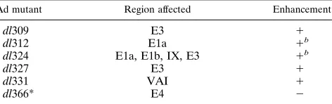
TABLE 1. Effect of Ad mutants on rAAV expressiona
Ad mutant Region affected Enhancement
dl309 E3 1
dl312 E1a 1b
dl324 E1a, E1b, IX, E3 1b
dl327 E3 1
dl331 VAI 1
dl366* E4 2
a
HeLa cells were coinfected with rAAV and the indicated Ad mutants. The cells were infected and stained forb-galactosidase activity as described in Ma-terials and Methods.
b
A high multiplicity of infection (.1,000 particles per cell) is needed to overcome the dependence of other genes on the E1 region (3, 17).
on November 9, 2019 by guest
http://jvi.asm.org/
RESULTS
rAAV vector transduction is enhanced by Ad coinfection.
We studied the effect of Ad on the efficiency of transduction by
an AAV vector by using an rAAV that carries the lacZ gene
controlled by the human CMV major immediate-early
pro-moter (CMV/lacZ). The number of cells expressing a
detect-able amount of
b
-galactosidase from the rAAV virus increased
by as much as a factor of 1,000 in the presence of Ad (Fig. 1A
and B). The maximal increase in rAAV transduction was
achieved at an input of about 100 Ad particles per cell (Fig.
1C). Since 100 Ad particles is equivalent to 1 to 2 PFU, this
result indicates that only about one functional Ad unit is
re-quired per cell. This effect is not unique to 293 cells; it is
equally dramatic in other cell lines such as HeLa (see Fig. 2B).
Ad could affect the efficiency of rAAV transduction by
di-rectly modulating the expression of the CMV/lacZ gene
car-ried on the rAAV genome. However, this mode of action
seems unlikely, since Ad did not influence expression of this
marker gene when it was introduced to cells on a plasmid by
transfection or when it was present integrated in a stable cell
line (data not shown). These results favor the hypothesis that
the enhancing effect of Ad is due to a unique aspect of AAV
virion-mediated gene delivery.
Ad induction of rAAV transduction maps to the Ad E4
transcription unit.
To determine which Ad gene product(s) is
necessary for the induction of rAAV gene expression, a panel
of Ad mutants was used in a coinfection assay. These viruses
carry specific mutations in the E1 (dl312 and dl324), VAI
(dl331), E3 (dl309, dl324, and dl327), or E4 (dl366) regions of
Ad (Table 1). Earlier studies demonstrated that the E1, VAI,
and E4 regions are necessary for efficient replication of
wild-type AAV (7, 8, 26, 34, 39). It is also possible that these regions
are involved in an immediate-early step of the AAV life cycle.
The above Ad mutants were individually coinfected into HeLa
cells with rAAV and scored for their ability to enhance
trans-duction by assaying lacZ expression 24 h postinfection (p.i.).
Analysis of these coinfections demonstrated that only dl366*,
an early-region E4
2mutant, was unable to enhance lacZ
trans-duction by rAAV (Table 1). This result focused the search for
the Ad gene responsible for the increased transduction to the
seven known ORFs of the E4 region.
Ad E4 ORF6 enhances rAAV transduction.
The Ad E4
re-gion has the potential to code for seven proteins, at least one
of which, ORF6, is necessary for a productive wild-type AAV
FIG. 2. Effect of various Ad E4 mutants on rAAV transduction. 293 or HeLacells were infected and stained forb-galactosidase activity as described in Materials and Methods. (A) Maps of various mutations (thick black lines indicate deletions; black arrows indicate insertions; gray boxes indicate ORFs). (B) Ability of various mutants to induce rAAV transduction. Lanes: 1, no virus; 2, dl309; 3, E4dlORF 1-4; 4, E4inORF3; 5, E4inORF6; 6, E4inORF3/
inORF6; 7, dl366*1ORF1–2; 8, dl366*1ORF3; 9, dl366*1ORF4; 10, dl366*
[image:3.612.344.522.73.246.2]1ORF6/7.
FIG. 3. Expression of E4 ORF6 from a plasmid is able to increase transduc-tion by rAAV. 293 cells were transfected with plasmid DNA as indicated. At 24 h later, the cells were infected with the appropriate virus. The cells were fixed and stained forb-galactosidase activity as described in Materials and Methods 24 h p.i. Lanes: 1, rAAV alone; 2, rAAV plus dl309; 3, rAAV plus pCMV/E4ORF6; 4, rAAV plus pCMV/E4ORF6 plus E4inORF6; 5, rAAV plus pUC119; 6, rAAV plus pUC119 plus E4inORF6.
on November 9, 2019 by guest
http://jvi.asm.org/
infection (9, 16, 26, 34). To determine which E4 gene(s) is
necessary for induction, a number of Ad mutants that express
one or more of the E4 proteins (Fig. 2A) were individually
coinfected into HeLa or 293 cells with rAAV and assayed for
transduction (Fig. 2B). In 293 cells, dl309 (wild-type Ad)
in-creased the transduction by rAAV approximately 40-fold
com-pared with rAAV alone (Fig. 2B, compare lanes 1 and 2). Ad
mutants that did not express ORF6 were unable to increase the
transduction by rAAV compared with rAAV alone (compare
lane 1 with lanes 5 to 10). Finally, Ad mutants that expressed
ORF6 were able to increase transduction to levels only twofold
lower than those achieved by use of dl309 (compare lane 2 with
lanes 3 and 4). Similar results were obtained with HeLa cells
(Fig. 2B). The identification of E4 ORF6 as a critical player in
the life cycle of rAAV is consistent with previous studies
indi-cating a role for this gene in the wild-type AAV life cycle (16,
34). While these data strongly suggest that the E4 ORF6 gene
is necessary for increased transduction by rAAV, the use of
mutant viruses precludes identification of this gene as being
sufficient. Since the Ad genome will provide a number of other
FIG. 4. Effect of various treatments on transduction by rAAV. 293 or HeLa cells were infected with rAAV, treated with the indicated agents, and stained forb-galactosidase activity as described in Materials and Methods. (A) Heat shock treatment at 42.58C. Lanes: 1, rAAV alone; 2, 1 h at 42.58C; 3, 2 h at 42.58C; 4, 3 h at 42.58C; 5, 4 h at 42.58C; 6, rAAV plus dl309. (B) HU treatment. Lanes: 1, rAAV alone; 2, rAAV plus dl309 plus HU; 3, rAAV plus HU until infection; 4, rAAV plus dl309. (C) UV treatment. Lanes: 1, rAAV alone; 2, 15 J/m2; 3, 20 J/m2; 4, 25 J/m2; 5, 30 J/m2; 6, rAAV plus dl309. (D) X-ray treatment. Lanes: 1, rAAV alone; 2, 200 rads; 3, 800 rads; 4, 1,400 rads; 5, 2,000 rads; 6, rAAV plus dl309.
on November 9, 2019 by guest
http://jvi.asm.org/
gene products that could be affecting rAAV transduction, we
attempted to determine if the E4 ORF6 product is active in the
absence of a viral background.
To determine if the expression of E4 ORF6 is necessary to
induce rAAV transduction, a plasmid that expresses only E4
ORF6, pCMV-E4ORF6, was transiently transfected into 293
cells and rAAV transduction was measured as described above
(Fig. 3). Both dl309 virus and the E4 ORF6 plasmid were able
to increase the transduction by rAAV approximately 40-fold
compared with that achieved by rAAV alone (Fig. 3, compare
lanes 1 through 3). When a nonspecific DNA (pUC119) was
used, no increase in transduction was observed (lane 5). We
assayed for the effect of other Ad genes on the E4 plasmid
induction by transfecting pCMV-E4ORF6 and infecting 293
cells with a viral mutant that does not express E4 ORF6 (Fig.
3, E4 in ORF6). This combination did not augment the amount
of rAAV transduction provided by E4 ORF6 alone (compare
lanes 3 and 4), suggesting that the E4 ORF6 gene product is
necessary and sufficient to enhance rAAV transduction in cells.
Induction of rAAV gene expression in the absence of Ad
genes.
The results thus far have suggested that transduction of
cells by rAAV follows an infectious path similar to that used by
wild-type AAV and that E4 ORF6 plays a critical role in an
immediate-early step of the AAV life cycle. Previous studies
have shown that cells subjected to genotoxic agents or stress
can support limited helper-independent replication of
wild-type AAV (29, 38, 44–47). These observations would suggest
that at least for wild-type AAV, the dependence on Ad helper
functions can be substituted by treatment of cells with these
agents. If the increase in transduction seen with rAAV and E4
ORF6 is mimicking the wild-type AAV life cycle, we would
predict that agents which stimulate productive wild-type AAV
infections should also affect transduction of cells by rAAV. To
test this hypothesis, HeLa and/or 293 cells were subjected to
conditions that induce helper-independent replication of
wild-type AAV, such as heat shock, HU, and UV irradiation. These
cells were then infected with rAAV and assayed for lacZ
trans-duction 24 h p.i. (Fig. 4A to C). All these treatments increased
the number of lacZ-transduced cells by at least 2 orders of
magnitude. In addition, X-ray treatment, which has not been
reported to induce helper-independent replication of wild-type
AAV, was able to increase transduction by rAAV by slightly
more than 1 order of magnitude (Fig. 4D). Similar effects of
DNA-damaging agents on AAV transduction have recently
been reported by others (1, 30). These data, as measured by
rAAV transduction, suggest that the E4 ORF6, genotoxic, and
physical stresses all may be acting through a common pathway
which is essential for the AAV life cycle.
Ad E4 ORF6 and UV treatment enhance second-strand
DNA synthesis by rAAV.
To identify the rate-limiting step in
rAAV transduction, we initially assayed the effect of E4 ORF6
on rAAV uncoating. 293 cells were transfected with the E4
ORF6 plasmid or control DNA and then with rAAV. At 24 h
p.i., virion-protected DNA was isolated by treating the cell
lysate with DNase to remove all uncoated rAAV genomes.
This was followed by inactivation of the DNase, protease
treat-ment of the residual virions, and detection of the protected
virion DNA by Southern hybridization. As illustrated in Fig. 5
(lane 1), rAAV DNA added to control samples was completely
degraded after the addition of DNase. No difference was
ob-served between the number of protected genomes in E4
ORF6-transfected cells and a plasmid control (lanes 2 and 3).
Comparison of the initial input rAAV dose (lane 4) with the
remaining uncoated virions (lanes 2 and 3) suggests that most
of the rAAV genomes are uncoated efficiently regardless of the
presence of E4 ORF6.
[image:5.612.81.266.69.218.2]The previous experiment suggests that an immediate-early
step in rAAV transduction, which occurs after uncoating, is
being facilitated by E4 ORF6. A unique aspect of the AAV life
cycle is that the viral template is packaged as a single-stranded
DNA molecule and that second-strand synthesis is required
before gene expression can take place. Second-strand synthesis
is facilitated by a self-priming mechanism involving the AAV
terminal repeat and results in the specific production of a
double-stranded AAV molecule nicked at one end (see Fig. 8).
To test the possibility that second-strand synthesis from rAAV
was rate limiting for transduction, UV-irradiated or E4
ORF6-transfected cells were infected with rAAV.
Low-molecular-weight DNA was isolated by Hirt extraction 24 h p.i. and
fractionated by electrophoresis on alkaline agarose gels. Southern
hybridization (Fig. 6) detected input rAAV DNA migrating as
single-stranded DNA in all samples. However, rAAV molecules
migrating at the expected molecular weight for the duplex
replicative intermediate were detected at significantly greater
levels only in the UV-irradiated cells (Fig. 6A, lane 2), and E4
ORF6-transfected cells (Fig. 6B, lane 1). Figure 6B, lane 2,
shows the mobility of the AAV genome in the absence of
transfected ORF6. This result indicates that the conversion of
FIG. 5. Effect of E4 ORF6 on rAAV uncoating. 293 cells were transfectedand then infected with rAAV as described in Materials and Methods. At 24 h p.i., nuclease-resistant virion DNA was isolated and electrophoresed through 0.8% agarose, transferred to a nylon membrane, and probed with a32
P-labeledb -ga-lactosidase sequence. Lanes: 1, 293 cells plus 1.6 ng of rAAV DNA; 2, 293 cells plus pCMV/E4ORF6 plus rAAV; 3, 293 cells plus rAAV plus pUC119; 4, 106 rAAV virions. M, monomer size; SS, single stranded.
FIG. 6. Effect of E4 ORF6 and UV treatment on rAAV second-strand syn-thesis. 293 cells transfected with a plasmid expressing E4 ORF6 and 293 cells treated with UV (20 J/m2
) were infected as described in Materials and Methods. At 24 h p.i., the low-molecular-weight DNA was isolated by Hirt extraction, subjected to electrophoresis in a 0.8% agarose gel, transferred to a nylon mem-brane, and probed with a [32
P]DNA corresponding to theb-galactosidase coding region. (A) Lanes: 1, 293 cells plus rAAV; 2, 293 cells plus rAAV plus UV. (B) Lanes: 1, 293 cells plus rAAV plus E4 plasmid; 2, 293 cells plus rAAV.
on November 9, 2019 by guest
http://jvi.asm.org/
[image:5.612.361.508.564.661.2]rAAV single-stranded DNA to a double-stranded state is
en-hanced by UV light and E4 ORF6.
While the indication was that second-strand synthesis
corre-lated with increased transduction from rAAV in the presence
of E4 ORF6 and UV irradiation, we used our ability to dose
escalate transduction by UV as a means of determining the
direct relationship between second-strand synthesis and rAAV
transduction. 293 cells were irradiated with various doses of
UV and then infected with rAAV. The cells were assayed for
lacZ staining at 24 h p.i., and a duplicate plate was
character-ized for second-strand synthesis as described above. Increased
lacZ transduction by rAAV at different doses of UV (Fig. 7A)
directly correlated with increased accumulation of duplex
rAAV replicative intermediates (Fig. 7B), supporting the view
that the rate-limiting step of rAAV transduction is the
conver-sion of the single-stranded rAAV genome into a
double-stranded intermediate.
DISCUSSION
By
using
AAV
b
-galactosidase
(lacZ)
recombinants
(rAAV), we demonstrated up to a 1,000-fold increase in
trans-duction frequency when cells were coinfected with Ad (Fig. 1
and 2) or exposed to genotoxic and physical stress (Fig. 4).
These analyses may help resolve some of the discrepancies
observed when characterizing rAAV generated with crude
ly-sate compared with gradient-purified virus. For Ad
coinfec-tion, enhanced transduction occurred when the Ad genome
was introduced into rAAV-infected cells via infection or
trans-fection (data not shown). These data suggest that the enhanced
transduction is not mediated by cointernalization of AAV with
Ad virions but support the premise that expression of an Ad
gene product is necessary for this effect. By using various Ad
mutants, we have mapped this function to the E4 ORF6 region
of Ad (Table 1; Fig. 2). Transfection experiments with plasmid
DNA (Fig. 3) suggest that ORF6 of the E4 region is necessary
to supply the Ad helper function needed for an early step in the
AAV life cycle, specifically, second-strand synthesis (Fig. 6 and
7).
Effect of Ad E4 on AAV.
The E4 region of Ad has been
implicated in AAV helper function in various assays (9, 16, 26,
34). Early studies demonstrated the need for this region by
assaying AAV replication or yield. Extended characterization
of E4 mutants in Ad infection revealed the role of E4 ORF6 in
the efficient accumulation of Ad late mRNAs (16, 42), a
func-tion also used by wild-type AAV for accumulafunc-tion of its
mRNA (34). All these previous studies were complicated by
the fact that measurements of wild-type AAV DNA replication
or gene expression were influenced by the AAV rep gene. AAV
rep expression, in turn, is dependent on numerous Ad
[image:6.612.99.258.70.347.2]early-gene functions (E1A [transcriptional activation], E1B and E4
[mRNA maturation], and VA and E2a [translation
enhance-ment]) (7, 9). As a result, it has been difficult to distinguish
effects of rep from direct effects of Ad helper functions on
other aspects of the AAV growth cycle. By using rAAV vectors
to assay the immediate-early steps in AAV infection, we have
avoided this complication in interpretation. This assay is
func-tionally different from previous studies because reporter gene
expression is independent of rep and its impact on the AAV life
cycle. Our data show that AAV uncoating was not altered in
the presence of E4 ORF6. However, it should be noted that
although we saw similar amounts of DNase-protected DNA
from E4 ORF6-positive and -negative cells, this assay could not
distinguish between potential uncoating intermediates.
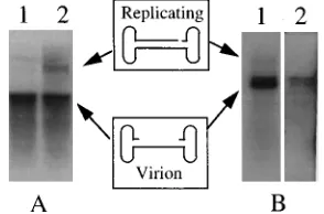
FIG. 7. Increased transduction corresponds to increased second-strandsyn-thesis. Duplicate plates of 293 cells were irradiated with the indicated amounts of UV as described in Materials and Methods. The cells were then infected with rAAV. At 24 h p.i., the cells were either stained and the number of transduced cells was calculated (A) or subjected to the Hirt procedure to isolate low-molecular-weight DNA (B). The DNA samples were electrophoresed through a 0.8% alkaline agarose gel, transferred to a nylon membrane, and probed with a 32
P-labeledb-galactosidase sequence. R, replicating form; V, virion form.
FIG. 8. Early events in the AAV life cycle cascade. When AAV encounters a host cell, it must first adsorb to the cell, uncoat its viral genome to release the single-stranded genome (solid line), and finally convert the genome to a double-stranded form (dashed line) before transcription and translation can take place. Second-strand synthesis appears to be the rate-limiting step of this cascade and can be facilitated by a number of agents including expression of the Ad E4 ORF6.
on November 9, 2019 by guest
http://jvi.asm.org/
[image:6.612.332.528.449.663.2]Our experiments clearly demonstrate that Ad E4 ORF6
facilitates second-strand synthesis of AAV DNA delivered to
cells as a single-stranded molecule in a virus particle (Fig. 6
and 8). The mechanism by which the ORF6 protein enhances
this reaction is unclear. It is interesting that E4 ORF6 prevents
the generation of Ad DNA concatemers (41). We might
spec-ulate that the roles of E4 ORF6 in Ad DNA concatemer
formation and AAV second-strand synthesis are directly
re-lated. Future studies are required to elucidate this
relation-ship.
The phenomenon elicited by the Ad E4 ORF6 can be
re-produced to different degrees in rAAV-infected cells by
expo-sure of cells to heat shock or genotoxic reagents (Fig. 4, 6, and
7). Our studies support the hypothesis that increased rAAV
transduction is due to enhanced AAV second-strand synthesis
and that E4 ORF6, genotoxic, and physical stresses may all be
acting through a common, as yet undefined, mechanism.
Impact on AAV vectors.
AAV is being tested as a vector for
gene delivery in vivo (2, 25, 40). Recently, a clinical protocol
involving AAV-mediated gene delivery for cystic fibrosis was
approved by both the Recombinant DNA Advisory Committee
and the Food and Drug Administration. It is clear that rAAV
vector transduction can be improved dramatically in cultured
cells by the physical and chemical manipulations described
above (Fig. 4, 6, and 7) and elsewhere (1, 30), suggesting that
such reagents, which manipulate the intracellular milieu, could
be coupled with current rAAV vector strategies to enhance the
delivery of therapeutic genes. For example, rAAV
transduc-tion of bone marrow stem cells may be enhanced through the
use of HU, a reagent which is currently being used in the
treatment of sickle cell anemia (10). On the basis of our
stud-ies, current AAV vectors may be best suited for cancer gene
delivery when the target cell is usually treated with agents such
as X rays or chemotherapy that also enhance transduction by
AAV. The use of AAV vectors aimed at transducing the bone
marrow stem cells of patients with Fanconi’s anemia (an
au-tosomal recessive disorder characterized by eventual
suscepti-bility to malignancy) in the presence of mitomycin has
achieved promising results (40). Alternatively, systematic
iden-tification of cells in vivo which are competent for efficient AAV
transduction may simultaneously obviate the need for
addi-tional steps required for enhanced transduction and direct the
use of vector to these appropriate targets. The direct role of E4
ORF6 in increasing rAAV vector transduction could also be
exploited by dual vector systems involving both rAd and rAAV
carrying the therapeutic gene. Such a combination could
com-bine high-efficiency gene transduction (rAd and rAAV) along
with long-term persistence (rAAV). Alternatively,
understand-ing the mechanism by which E4 functions to enhance rAAV
transduction may suggest the possibility of identifying small
molecules which could replace the role of Ad and enhance the
efficiency of gene delivery by AAV vectors.
ACKNOWLEDGMENTS
We thank P. Hearing for kindly providing E4 mutants, T. Van Dyke for giving advice and for critical reading of this manuscript, and Can-dace Summerford for expert technical assistance.
This work was supported by NIH grants DK42701, HL48347, and HL51818. T. Shenk is an American Cancer Society Professor and an Investigator of the Howard Hughes Medical Institute.
REFERENCES
1. Alexander, I. E., D. W. Russell, and A. D. Miller. 1994. DNA-damaging agents greatly increase the transduction of nondividing cells by adeno-asso-ciated virus vectors. J. Virol. 68:8282–8287.
2. Bartlett, J. S., K. B. Quattrocchi, and R. J. Samulski. 1995. The development of adeno-associated virus as a vector for cancer gene therapy, p. 27–40. In
R. E. Sobol and K. J. Scanlon (ed.), Gene therapy of cancer. Appleton & Lange, East Norwalk, Conn.
3. Berk, A. J., F. Lee, T. Harrison, J. Williams, and P. A. Sharp. 1980. Pheno-types of adenovirus-5 host-range mutants for early-mRNA synthesis. Cold Spring Harbor Symp. Quant. Biol. 44:429–436.
4. Berns, K. I. 1984. Adeno-associated virus, p. 563–592. In H. S. Ginsberg (ed.), The adenoviruses. Plenum Press, New York.
5. Berns, K. I. 1984. The parvoviruses. Plenum Press, New York.
6. Berns, K. I., and J. A. Rose. 1970. Evidence for a single-stranded adenovirus-associated virus genome: isolation and separation of complementary single strands. J. Virol. 5:693–699.
7. Carter, B. J. 1990. Adeno-associated virus helper functions, p. 255–282. In P. Tijssen (ed.), CRC handbook of parvoviruses, vol. 1. CRC Press, Inc., Boca Raton, Fla.
8. Carter, B. J., and C. A. Laughlin. 1983. Adeno-associated virus defectiveness and the nature of the helper function, p. 67–127. In K. I. Berns (ed.), The parvoviruses. Plenum Press, New York.
9. Carter, B. J., C. J. Marcus-Sekura, C. A. Laughlin, and G. Ketner. 1983. Properties of an adenovirus type 2 mutant, Ad2dl807, having a deletion near the right-hand genome terminus: failure to help AAV replication. Virology 126:505–516.
10. Charache, S., M. L. Terrin, R. D. Moore, G. J. Dover, F. B. Barton, S. V. Eckert, R. P. McMahon, and D. R. Bonds.1995. Effect of hydroxyurea on the frequency of painful crises in sickle cell anemia. N. Engl. J. Med. 332:1317– 1322.
11. Cheung, A. K., M. D. Hoggan, W. W. Hauswirth, and K. I. Berns. 1980. Integration of the adeno-associated virus genome into cellular DNA in latently infected human Detroit 6 cells. J. Virol. 33:739–748.
12. Esche, H., M. B. Mathews, and J. B. Lewis. 1980. Proteins and messenger RNAs of the transforming region of wild-type and mutant adenoviruses. J. Mol. Biol. 142:399–417.
13. Gey, G. O., W. D. Coffman, and M. T. Kubicek. 1952. Tissue culture studies of the proliferative capacity of cervical carcinoma and normal epithelium. Cancer Res. 12L:264.
14. Goodman, S., X. Xiao, R. E. Donahue, A. Moulton, J. Miller, C. Walsh, N. S. Young, R. J. Samulski, and A. W. Nienhuis.1994. Recombinant adeno-associated virus-mediated gene transfer into hematopoietic progenitor cells. Blood 84:1492–500. (Erratum, 85:862, 1995.)
15. Graham, F. L., J. Smiley, W. C. Russell, and R. Nairn. 1977. Characteristics of a human cell line transformed by DNA from human adenovirus type 5. J. Gen. Virol. 36:59–74.
16. Huang, M. M., and P. Hearing. 1989. Adenovirus early region 4 encodes two gene products with redundant effects in lytic infection. J. Virol. 63:2605– 2615.
17. Jones, N., and T. Shenk. 1979. An adenovirus type 5 early gene function regulates expression of other early viral genes. Proc. Natl. Acad. Sci. USA 76:3665–3669.
18. Jones, N., and T. Shenk. 1979. Isolation of adenovirus type 5 host range deletion mutants defective for transformation of rat embryo cells. Cell 17: 683–689.
19. Kotin, R. M., R. M. Linden, and K. I. Berns. 1992. Characterization of a preferred site on human chromosome 19q for integration of adeno-associ-ated virus DNA by non-homologous recombination. EMBO J. 11:5071–5078. 20. Kotin, R. M., M. Siniscalco, R. J. Samulski, X. D. Zhu, L. Hunter, C. A. Laughlin, S. McLaughlin, N. Muzyczka, M. Rocchi, and K. I. Berns.1990. Site-specific integration by adeno-associated virus. Proc. Natl. Acad. Sci. USA 87:2211–2215.
21. Laughlin, C. A., J. D. Tratschin, H. Coon, and B. J. Carter. 1983. Cloning of infectious adeno-associated virus genomes in bacterial plasmids. Gene 23: 65–73.
22. Lusby, E., K. H. Fife, and K. I. Berns. 1980. Nucleotide sequence of the inverted terminal repetition in adeno-associated virus DNA. J. Virol. 34: 402–409.
23. Maniatis, T., E. F. Fritsch, and J. Sambrook. 1982. Molecular cloning: a laboratory manual. Cold Spring Harbor Laboratory, Cold Spring Harbor, N.Y.
24. McMaster, G. K., P. Beard, H. D. Engers, and B. Hirt. 1981. Characteriza-tion of an immunosuppressive parvovirus related to the minute virus of mice. J. Virol. 38:317–326.
25. Miller, J. L., R. E. Donahue, S. E. Sellers, R. J. Samulski, N. S. Young, and A. W. Nienhuis.1994. Recombinant adeno-associated virus (rAAV)-medi-ated expression of a human gamma-globin gene in human progenitor-de-rived erythroid cells. Proc. Natl. Acad. Sci. USA 91:10183–10187. (Erratum, 92:646, 1995.)
26. Richardson, W. D., and H. Westphal. 1981. A cascade of adenovirus early functions is required for expression of adeno-associated virus. Cell 27:133– 141.
27. Roberts, R. J., G. Akusjarvi, P. Alestrom, R. E. Gelinas, T. R. Gingeras, D. Sciaky, and U. Pettersson.1986. A consensus sequence for the adenovirus-2 genome, p. 1–51. In W. Doerfler (ed.), Adenovirus DNA. Martinus Nijhoff Publishers, Boston.
28. Rose, J. A., K. I. Berns, M. D. Hoggan, and F. J. Koczot. 1969. Evidence for
on November 9, 2019 by guest
http://jvi.asm.org/
a single-stranded adenovirus-associated virus genome: formation of a DNA density hybrid on release of viral DNA. Proc. Natl. Acad. Sci. USA 64:863– 869.
29. Routes, J. M. 1992. IFN increases class I MHC antigen expression on ade-novirus-infected human cells without inducing resistance to natural killer cell killing. J. Immunol. 149:2372–2377.
30. Russell, D. W., I. E. Alexander, and A. D. Miller. 1995. DNA synthesis and topoisomerase inhibitors increase transduction by adeno-associated virus vectors. Proc. Natl. Acad. Sci. USA 92:5719–5723.
31. Samulski, R. J., K. I. Berns, M. Tan, and N. Muzyczka. 1982. Cloning of adeno-associated virus into pBR322: rescue of intact virus from the recom-binant plasmid in human cells. Proc. Natl. Acad. Sci. USA 79:2077–2081. 32. Samulski, R. J., L. S. Chang, and T. Shenk. 1987. A recombinant plasmid
from which an infectious adeno-associated virus genome can be excised in vitro and its use to study viral replication. J. Virol. 61:3096–3101. 33. Samulski, R. J., L. S. Chang, and T. Shenk. 1989. Helper-free stocks of
recombinant adeno-associated viruses: normal integration does not require viral gene expression. J. Virol. 63:3822–3828.
34. Samulski, R. J., and T. Shenk. 1988. Adenovirus E1B 55-Mr polypeptide facilitates timely cytoplasmic accumulation of adeno-associated virus mRNAs. J. Virol. 62:206–210.
35. Samulski, R. J., A. Srivastava, K. I. Berns, and N. Muzyczka. 1983. Rescue of adeno-associated virus from recombinant plasmids: gene correction within the terminal repeats of AAV. Cell 33:135–143.
36. Samulski, R. J., X. Zhu, X. Xiao, J. D. Brook, D. E. Housman, N. Epstein, and L. A. Hunter. 1991. Targeted integration of adeno-associated virus (AAV) into human chromosome 19. EMBO J. 10:3941–3950. (Erratum, 11:1228, 1992.)
37. Sanes, J. R., J. L. Rubenstein, and J. F. Nicolas. 1986. Use of a recombinant retrovirus to study post-implantation cell lineage in mouse embryos. EMBO J. 5:3133–3142.
38. Stein, R., and E. B. Ziff. 1984. HeLa cell beta-tubulin gene transcription is stimulated by adenovirus 5 in parallel with viral early genes by an E1a-dependent mechanism. Mol. Cell. Biol. 4:2792–2801.
39. Thimmappaya, B., C. Weinberger, R. J. Schneider, and T. Shenk. 1982. Adenovirus VAI RNA is required for efficient translation of viral mRNAs at late times after infection. Cell 31:543–551.
40. Walsh, C. E., A. W. Nienhuis, R. J. Samulski, M. G. Brown, J. L. Miller, N. S. Young, and J. M. Liu.1994. Phenotypic correction of Fanconi anemia in human hematopoietic cells with a recombinant adeno-associated virus vec-tor. J. Clin. Invest. 94:1440–1448.
41. Weiden, M. D., and H. S. Ginsberg. 1994. Deletion of the E4 region of the genome produces adenovirus DNA concatemers. Proc. Natl. Acad. Sci. USA 91:153–157.
42. Weinberg, D. H., and G. Ketner. 1986. Adenoviral early region 4 is required for efficient viral DNA replication and for late-gene expression. J. Virol. 57:833–838.
43. Williams, J. F. 1970. Enhancement of adenovirus plaque formation on HeLa cells by magnesium chloride. J. Gen. Virol. 9:251–255.
44. Winocour, E., L. Puzis, S. Etkin, T. Koch, B. Danovitch, E. Mendelson, E. Shaulian, S. Karby, and S. Lavi.1992. Modulation of the cellular phenotype by integrated adeno-associated virus. Virology 190:316–329.
45. Yakobson, B., T. A. Hrynko, M. J. Peak, and E. Winocour. 1989. Replication of adeno-associated virus in cells irradiated with UV light at 254 nm. J. Virol. 63:1023–1030.
46. Yakobson, B., T. Koch, and E. Winocour. 1987. Replication of adeno-asso-ciated virus in synchronized cells without the addition of a helper virus. J. Virol. 61:972–981.
47. Yalkinoglu, A. O., R. Heilbronn, A. Burkle, J. R. Schlehofer, and H. zur Hausen.1988. DNA amplification of adeno-associated virus as a response to cellular genotoxic stress. Cancer Res. 48:3123–3129.