ETIOLOGICAL PATTERN OF ANTERIOR UVEITIS
IN A REFERRAL HOSPITAL
DISSERTATION SUBMITTED FOR
MASTER OF SURGERY DEGREE
BRANCH – III - OPHTHALMOLOGY
MARCH 2009
THE TAMILNADU
DR.M.G.R. MEDICAL UNIVERSITY
ACKNOWLEDGEMENT
I am deeply indebted to
Dr. P. THIYAGARAJAN. MS., D.O,
Professor and Head of the department of Ophthalmology,
Madurai Medical college, Madurai
for the able guidance,
inspiration and encouragement he rendered at every stage of this
study.
I acknowledge with gratitude the dynamic guidance and
persistent encouragement given to me by my guide
Dr. N.
PARVATHA SUNDARI, M.S., DO, Assistant Professor in
Ophthalmology, Department of Ophthalmology, Madurai
Medical College, Madurai
for having taken keen interest in sharing
her ideas throughout the study period. Her valuable suggestions and
patronage has been a driving force to make this endeavor possible.
My Sincere thanks to
Dr. Dr.M. SIVAKUMAR, M.D
.,
Dean,
Madurai Medical
College, & Govt Rajaji Hospital,
,
Madurai
for
permitting me to utilize the clinical materials of the hospital.
Last, but not the least, my profound gratitude to all the
‘patients’, to whom I owe everything because, this venture would not
DECLARATION
I,
Dr. JEEVAKALA.C.
solemnly declare that the
dissertation titled
“
ETIOLOGICAL PATTERN OF ANTERIOR
UVEITIS IN A REFERRAL HOSPITAL
”
has been prepared by
me.
This is submitted to The Tamil Nadu Dr. M.G.R. Medical
University, Chennai, in partial fulfillment of the requirement for
the award of M.S.,(Ophthalmology) Branch - III degree
Examination to be held in MARCH 2009.
Place : Madurai
Dept. of Ophthalmology,
Govt. Rajaji Hospital,
Madurai.
CERTIFICATE
This is to certify that this dissertation entitled
“
ETIOLOGICAL PATTERN OF ANTERIOR UVEITIS IN A
REFERRAL HOSPITAL
”
has been done by
DR.JEEVAKALA.
C.
under my guidance in Department of OPHTHALMOLOGY,
Madurai Medical College, Madurai.
I certify regarding the authenticity of the work done to
prepare this dissertation.
DR. P.THIYAGARAJAN. M.S., D.O.,
PROFESSOR & H.O.D.
DEPARTMENT OF OPHTHALMOLOGY GOVT. RAJAJI HOSPITAL &
CONTENTS
S.NO.
TOPIC
PAGE NO.
1
.
INTRODUCTION
1
2.
HISTORICAL
BACKGROUND
3
3.
CLASSIFICATION
OF
UVEITIS
4
4.
REVIEW
OF
LITERATURE
14
5.
AIM OF THE STUDY
45
6.
MATERIALS AND METHOD
46
7.
RESULTS & COMPARATIVE ANALYSIS
50
8.
SUMMARY 65
9.
DISCUSSION
67
10.
CONCLUSION
69
ANNEXURE
INTRODUCTION TO UVEITIS
Uveitis is such a small word and yet in common usage in most
medical circles it encomposes the entire spectrum of intraocular
inflammation:-
iritis, iridocyclitis, parsplanitis, posterior uveitis, choroiditis,
retinitis and retinal vasculitis
The Uvea of the eye consist of iris, ciliary body and choroid
which is the eyes major blood supply.
Uveitis is broadly defined as inflammation of [i.e:-itis]of uvea
[from the latina uva meaning grape]
The study of uveitis is complicated by the fact that the cause of
inflammatory reaction of the inner layer can be infection, traumatic,
neoplastic or autoimmune.
Inflammation may be acute, subacute or chronic. With
inflammation and repair that as a protective response may not only
damage inflamed target tissue but also may participate in collateral
Pathological classification of uveitis is into granulomatous and
non granulomatous with distinct etiologies, features, sequelae and
treatment for each category.
Some uveitis entities occur bilaterally eg;-APMPPE and others
occur unilaterally-ARPE ( acute retinal pigment epithelitis)
Age sex and race may also help the clinician to narrow the
diagnostic possibilities
HISTORICAL BACKGROUND
The problem of inflammation of the eye including uveitis was
known to the Egyptians. The Edwin Smith surgical papyrus, the
oldest known existing ophthalmic document, contains references to
inflammatory condition of the eye.
The name iritis was introduced in 1801 by Johnn adam
schmid-anatomist, surgeon and ophthalmologist of Vienna.
Inflammation of the ciliary body was originally described as an
inflammation of orbicularis ciliaris by Friedrich Von Ammon. It was
called cyclitis by August Bererd, which was later fully elucidated by
Albricht Von Graefe. Ernst fuchs madde it clear that acute
inflammation was best designated as iridocyclitis and pure cyclitis
existed only in the chronic form characterised by keratic precipitates
and vitreous opacities .
In the later part of 19
thcentury and during present century the
amount of work done on all forms of uveitis has been immense and
among the many Alan Churchill Woods laboured both in the clinic
Etiological classification
Uveitis may also be classified and organised etiologically and
pathophysiologically according to the fallowing mechanism-traumatic
infectious immunologic masquerade
Epidemiology - uveitis may affect individuals from any age
from infancy on ,affects people from all parts of the world and is a
significant cause of blindness.
Classification of Uveitis :
Duke Elder’s Classification
1.
Uveitis wherein the infective element is dominant
a. Exogenous
- Wound infection
- Parasitic entry
b. From neighbouring structures, from direct continuity
- Extraocular
- Ocular
c. Endogenous-metastatic or ocurring in course of a infection -
bacterial, rickettsial, viral, mycotic or parasitic
b. Uveitis due to bacterial [delayed] allergy
c. Autoimmune uveitis focal infection
3.Toxic uveitis
a. Endogenous toxins
-
Autotoxication
-
Organismal toxin
b. Endocular toxin -atrophic,haemorrhagic,neoplastic
c. Exogenous Chemical irritants
4. Traumatic Uveitis
5. Uveitis associated with noninfective systemic diseases
a. Sarcoidosis
b. The collagen and related diseases
c. Diseases of central nervous system
d. Diseases of the skin
6.Uveitis of unknown etiology
a. Sympathetic
ophthalmitis
Nussenblatt’s Classification
IUSG *
TESSLER
--- Sclerouveitis--- Keratouveitis Anterior Uveitis Iritis Anterior cyclitis Iridocyclitis Anterior Uveitis Iritis Iridocyclitis Intermediate Uveitis Posterior cyclitis Hyalitis Iridocyclitis Intermediate Uveitis Cyclitis Vitritis Parsplanitis Posterior uveitis
Focal, Multifocal or diffuse choroiditis,chorioretinitis
Retinitis
Panuveitis Choroiditis
IUSG*
From Bloch Michael E, Nussenblatt RB. International Uveitis
Study Group recommendation for the evaluation of intraocuJlar
inflammatory disease. Am J Ophthalmology 103
Tesslar +
Causes of Acute and Chronic Uveitis
1. Acute Uveitis
-
most cases of anterior uveitis
Idiopathic
ankylosing spondylitis
reiters syndrome
fuchs heterochromic iridocyclitis
-
VKH syndrome
-
toxoplasmosis
-
AMPPE
-
MEWS
-
Acute retinal necrosis
-
Post surgical bacterial infection
-
Trauma
2. Chronic Uveitis
-
Juvenile rheumatoid arthritis
-
Birdshot choroidopathy
-
Serpiginous chroidopathy
-
Postsurgical uveitis [ propionibacterium acnes,Fungal]
-
Intraocular lymphoma
-
Sympathetic ophthalmia
-
Multifocal choroiditis
-
Sarcoidosis
-
Intermediate uveitis/parsplanitis
3.
Causes of Granulomatous Inflammation in Eye
- Sarcoidosis
- Sympathetic
ophthalmia
- Uveitis
associated
with multiple sclerosis
- Lens
induced
uveitis
- Intraocular
foreign
body
- VKHsyndrome
- Syphilis
- Tuberculosis
-
Other infectious agents
4.
Causes of Unilateral Uveitis
- Sarcoidosis
- Intraocular
foreign
body
- Parasitic
diseses
- Acute
retinal
necrosis
- Behcet’s
disease
5.
Causes of Anterior Uveitis
- Idiopathic
- Ankylosing
spondylitis
- Reiter’s
syndrome
- Inflammatory
bowel
disease
- Psoriatic
arthritis
- Behcet’s
disease
- HLA-B27
associated
disease
-
Juvenile rheumatoid arthritis
-
Fuchs heterochromic iridocyclitis
- Sarcoidosis
- Syphilis
- Glaucomatocyclitic
crisis
6. Causes of Intermediate Uveitis
- Sarcoidosis
- Inflammatory
bowel
disease
- Multiple
sclerosis
- Lyme
disease
- Parsplanitis*
*
not an etiological diagnosis but patients with intermediate
uveitis of parsplanitis subtype tend to have worst prognosis
7. Causes of Posterior Uveitis
Focal Retinitis
-
Toxoplasmosis
-
Onchocerciasis
-
Cysticercosis
-
Masquerade syndrome
Multifocal Retinitis
-
Syphilis
-
Herpes simplex virus
-
Sarcoidosis infection
-
Masquerade syndrome
-
Candidiasis
-
Meningococcus
Focal Choroiditis
-
Toxocariasis
-
Toxoplasmosis
-
Nocardiosis
-
Masquerade syndromes
Multifocal Choroiditis
-
Histoplasmosis
-
Sympathetic ophthalmia
-
Vogt- Koyanagi- Harada syndrome
-
Sarcoidosis
-
Serpiginous choroidopathy
-
Birdshot choroidopathy
8. Causes of Panuveitis
- Syphilis
- Sarcoidosis
- VKH
syndrome
- Infectious
endophthalmitis
DEMOGRAPHIC CONSIDERATION IN UVEITIS
FACTOR DISEASE RISKS
Female Pauciarticular JRA,chronic anterior uveitis Male Ankylosing spondylitis,sympathetic ophthalmia American Black Sarcoidosis
Native American VKH Syndrome
Japanese VKH syndrome, Behcet’s syndrome Mid Western American POHS
Mediterranean ancestory Behcet’s syndrome
Central American Cysticercosis ,Onchocerciasis South American Cysticercosis,Toxoplasmosis West African Onchocersiasis
IV Drug abuser Fungal endophthalmitis,AIDS
Promiscuous AIDS, Syphilis
Frequent hiking in wooded area Lyme disease
Uveitis associated with medications
Anticholinesterases and direct acting agonists
Anterior uveitis
Hydralazine Lupus like syndrome
Nitrogen mustard Necrotising uveitis with retinal vasculitis
Rifabutin anterior uveitis
Procainamide lupus like syndrome with episcleritis Intraocular gases Air Perflurocarbon Silicone oil A chymotrypsin Anterior uveitis
Fibrinous anterior uveitis Anterior uveitis
REVIEW OF LITERATURE
Anterior uveitis is the most common form of uveitis and
accounts for approximately ¾ of the cases with annual incidence rate
of about 8 cases/100,000 population. Although anterior uveitis is the
most easily managed form of uveitis, associated complications like
glaucoma may result in severe visual loss. In addition many disease
that can cause panuveitis such as sarcoidosis, behcet’s syndrome and
endophthalmitis start as anterior uveitis.
Clinical description
Anterior uveitis includes disease previously categorised as both
iritis-inflammation of iris and iridocyclitis-inflammation of iris and
ciliary body.
Patients with anterior uveitis often complain of redness, pain,
photophobia and blurred vision. Ciliary flush, conjunctival injection
in the perilimbal area are characteristic form of the disease.
Pupillary miosis, posterior synechiae and dilated iris vessels are
Conjunctival hyperaemia is a common sign of acute anterior
inflammation but is rare in chronic posterior segment disease.
Peripheral anterior synechiae should be looked for on
gonioscopy because patients with severe synechiae are at risk for
secondary glaucoma.
Major indicators of anterior uveitis are presence of cells and
flare in the anterior chamber. Anterior chamber inflammation is
assessed on slit lamp biomicroscopy
Gnex-crosier and colleagues recently used laser flare cell
photometry to demonstrated that blood aqueous barrier disruption was
very pronounced in idiopathic anterior uveitis, acute retinal necrosis
but minimal in patients with toxoplasmosis or Fuchs’ heterochromic
cyclitis
Fibrin may accumulate in the anterior chamber and may cause
the once mobile cells that circulate in the aqueous to become frozen.
The plasmoid aqueous is a sign of severe anterior uveitis that
requires aggressive therapy. Another sign of severe anterior uveitis is
with behcet’s disease or infectious endophthalmitis and rifabutin
toxicity in patients with aids
Recently anterior uveitis including some cases with hypopyon
has been described in patients with AIDS who are receiving the drug
rifabutin as treatment or prophylaxis for mycobacterial infection.
A pseudohypopyon, composed of tumour cells or haemorrhagic
debris, can occur in some of the masquerade syndromes after vitreous
haemorrhage
Inflammatory cells collect and adhere to corneal endothelium
and form keratic precipitates. Mechanism appears to involve the
expression of cell adhesion molecules that are upregulated in the
presence of inflammatory cytokines such as IL-1.in most
inflammatory reactions, the neutrophil is the first cell present and the
transformed macrophages [epithelioid cells] and lymphocytes
accumulates the inflammation becomes chronic. KPs therefore mimic
the course of the inflammation in the tissue.
Large greasy appearing keratic precipitates are suggestive of
Other corneal findings ; corneal dendrities in uveitis as a result
of herpes simplex virus infection, interstitial keratitis may be
associated with syphilis, with the presence of ghost vessels, similar
findings seen in patients with sarcoidosis.
During this episode the iop is often decreased due to ciliary
body shutdown.
Posterior synechiae and peripheral anterior synechiae are
responsible for pupillary block glaucoma and obstruction of aqueous
outflow respectively.
Iris nodules are accumulations of inflammatory cells in the iris
or on its surface.Koeppe nodule develops on the pupillary border,
busacca’s nodules occur on the iris surface.
Many patients with uveitis develop cataracts because of
underlying inflammation and the use of corticosteroids to treat the
disease
Inflammation of the Vitreous is characterised by increased cells
and protein. In patients with pars planitis who have cells in the
inflammation is more severe. Vitreous cells aggregate into clumps
called snow balls. These settle in the inferior periphery near the
retinal surface and are seen best with indirect ophthalmoscope.
Vitreous traction by inflammatory fibrin membranes causean
incomplete posterior vitreous detachment may be associated with the
development of cystoid macular oedema.
Cystoid macular oedema is a common retinal finding in patients
with uveitis. Vascular sheathing of the arteries or veins caused by in
infiltration of inflamatory cells around the vessels is easily seen in the
posterior pole. Retinal haemorrhages and cotton wool spots frequently
accompany retinal vasculitis.
Choroidal lesions with or without retinal involvement are
common in posterior inflammatory disease.
Uveitis may affect the optic nerve in several ways. Disc
Signs of uveitis Eyelid and skin
– vitiligo, nodules Conjunctiva
– perilimbal or diffuse injection - nodules
Corneal endothelium
- keratic precipitates[diffuse or gravitational] - fibrin
- pigment[non specific] Anterior/posterior chamber - inflammatory cells
- flare[protinaceous influx] - pigment[non specific] Iris
- nodules
- posterior synechiae - atrophy
- heterochromia
Angle-peripheral anterior synechiae - nodules
- vascularization Intraocular pressure - hypotony
Vitreous
- inflammatory cells[single/clumped] - traction bands
Parsplana
- snowbanking Retina
- Inflammatory cells
- Inflammatory cuffing of blood vessels - Oedema
- CMO
- RPE hypertrophy/clumping/loss - Epiretinal membrane
Choroid
- Inflammatory infiltrate - Atrophy
- Neovascularisation Optic nerve
The SUN working group grading scheme for anterior chamber cells No.of Cells Grade
0 <1
0.5 1-5
1+ 6-15 2+ 16-25 3+ 26-50
4+ >50
The SUN working group grading system for anterior chamber flare
Grade Description
0- none 1+ faint 2+ Moderate
[iris &lens details clear
3+ Marked [iris &lens details hazy]
4+ Intense [fibrin or plasmoid aqueous]
The standardization of uveitis nomenclature [SUN] working group.
Standardization of nomenclature for reporting clinical data. Results of the
IDIOPATHIC ANTERIOR UVEITIS
After a through medical history and an ocular and general
physical examination almost 50% of patients are found to have an
anterior segment inflammation that is not associated with other
defined clinical syndromes.
This form of anterior uveitis is referred to as idiopathic anterior
uveitis. The diagnosis of idiopathic anterior uveitis depends greatly on
the extend of evaluation for an underlying condition
Diagnostic workup:
A complete medical history and through examination can
help target the workup. When a through history and examination fail
to suggest specific diagnosis patients have limited workup with
anterior uveitis even if it is their first episode.
If uveitis is nongranulomatous FTA-Abs test is done to rule out
syphilis
A complete blood count and urine analysis to rule out
because the associated renal disease or anaemia may be asymptomatic
but may warrent therapy.
In case of granulomatous anterior uveitis - a mantoux test,
chest x-ray and serum and urine calcium test and serum ACE levels
HLA-B27 ASSOCIATED ANTERIOR UVEITIS
HLA-B27 associated anterior uveitis appears to be a distinct
clinical disorder. This form of disease has frequent association with
Ankylosing spondylitis
Psoriatic arthritis
Reiter syndrome
Reactive inflammation
Inflammatory bowel disease
Nevertheless patients withHLB-B27 haplotype and anterior
uveitis have no associated systemic illness
ANKYLOSING SPONDYLITIS
Ocular involvement occurs in 25% of patients with ankylosing
spondylitis. Both eyes are involved in 80% of patients but they are
rarely inflammed. Ocular findings are conjunctivitis and iritis. The
disease course is variable. Recurrance of inflammation can occur as
frequently as every 2-3 weeks. Anterior uveitis with ankylosing
spondylitis usually has a presentation similar to idiopathic anterior
PSORIATIC ARTHROPATHY
Psoriasis is a skin disease caused by hyperproliferation of the
epidermis with resultant scaling. Uveitis occurs predominantly in
patients who develop arthropathy.20% of patients with psoriasis
develop psoriatic arthropathy and about 20%of these patients develop
uveitis, sacroilitis and ascending spine disease. The arthropathy
usually involves the distal joints of hands and feet as well as
sacroiliac joint. Uveitis predominently involves the anterior segment
of eye and is similar to HLA-B27 associated disease.
REITER’S SYNDROME
It is a systemic disorder characterised by arthritis, conjunctivitis
and urethritis.
First described in 1818 but named after Reiter who described
the entity in1916.it is the most common cause of inflammatory
oligoarthropathy in young males and similar to ankylosing spondylitis
and it is related to both HLA-B27 and to a specific infection that may
trigger the disease. The disease develops in atleast 1% of patients with
nonspecific urethritis and occurs in about 2% of patients with shigella
called keratoderma blennorrhagicam,balanitis and aphthous
stomatitis. Rheumatologic features of disease include arthralgias,
plantar fasciitis and tenosynovitis. About 20% of patients with reiter
syndrome develope sacroilitis and ascending spinal disease similar to
ankylosing spondylitis. Hyperkeratotic skin lesions occur and may be
indistinguishable from psoriasis. Conjunctivitis is the most common
ocular finding in patients with Reiter’s syndrome and occur in 30%
-60% of patients. Iritis and keratitis are less common. Iritis occur in
3%-12% of patients and is nongranulomatous and mild. The keratitis
is characterised by multifocal punctuate subepithelial and anterior
stromal infiltrates. A small pannus may also develope. Reiter’s
syndrome may occur after gram negative dysentery or after
nongonoccocol urethritis as a result of chlamyidia trachomatis and
ureaplasma urealyticum.
INFLAMMATORY BOWEL DISEASE
Patients with ulcerative colitis and Crohn’s disease can develop
uveitis.5% of patients with ulcerative colitis will develop ocular
disease. Conjunctivitis, episcleritis and anterior uveitis are most
Similar ocular inflammatory disease has been associated with
Crohn’s disease.
WHIPPLE DISEASE
Systemic disorder characterized by malabsorption causing
chronic diarrhoea. Anterior uveitis and vitritis have been rarely
reported. Because antibiotic therapy can effectively treat this disease
it is important to diagnose whipple’s disease in uveitis patients with
gastrointestinal symptoms.
JUVENILE RHEUMATOID ARTHRITIS
The diagnosis of JRA is based on the presence of arthritis in
children under the age of 16 years and is usually found by negative
Rh factor test result and there is no other cause for the joint disease.
Joint inflammation maybe polyarticular or pauciarticular and majority
of the patients are Rh factor negative. Systemic form of JRA is
characterized by systemic polyarthritis with an associated fever, rash,
hepatosplenomegaly and leucocytosis
Uveitis is rare in children with systemic JRA. Patients with
with pauciarticular form of JRA are at much higher risk for the
development of ocular inflammation. Girls with pauciarticular
arthritis and positive result of an ANA test are at higher risk for
developing chronic iridocyclitis. High risk of uveitis is associated
with arthritis sparing the wrist but involving a lower extremity
joint.75% of boys with pauciarticular arthritis are HLA-B27 positive
and the ocular inflammation they develop is similar to ankylosing
spondylitis.
Uveitis is characterised by an acute episodic non granulomatous
anterior uveitis and some of the boys go on to develop ankylosing
DISEASE ASSOCIATIONS
FUCHS’ HETEROCHROMIC IRIDOCYCLITIS
In 1906 Fuchs described 7 patients with heterochromic
iridocyclitis and later described 38 patients with the disease and
reported on the pathologic features of 6 eyes. Fuchs observed that
most patients with this disorder develop cataract. The typical patient
is young and presents with iris heterochromia and a mild disturbance
of vision. Anterior chamber inflammation is mild and low grade
vitritis may be seen. Many patients are unaware of their disease until
their vision decreases because of cataract or glaucoma.
KAWASAKI DISEASE
It is also called mucocutaneous lymphnode syndrome is a
disease of children and young adults, characterised by an
erythematous and desquamative exanthema, oral mucosal erythema,
conjunctivitis, fever, and an asymmetric cervical adenopathy. Anterio
uveitis has also been noted in many patients with this disorder. The
uveitis does not appear to be severe or chronic and usually resolves
without therapy. Optic disc odema and dilated retinal veins may also
endothelial cells from the umbilical vein that express major
histocompatability complex class 2 antigens. In one study 66% of
patients had evidence of anterior uveitis that occurred most
commonly during the first week of illness.
ANTERIOR UVEITIS ASSOCIATED WITH RENAL DISEASE
Anterior uveitis has been associated with interstitial nephritis.
Patients usually present with an acute uveitis and later develop
manifestations of acute interstitial nephritis with cellular casts in the
urine. Anterior uveitis has been reported in patients with IgA
Nephropathy. This disease often occurs in children and is associated
with an upper respiratory tract infection. The uveitis appears to occur
with the pulmonary disease and responds to standard therapy.
GLAUCOMATOCYCLITIC CRISIS
Also known as Possner Schlossman syndrome is inflammatory
glaucoma manifested by fine KPs mydriasis and elevated intraocular
SCHWARZ SYNDROME
Rhegmatogenous retinal detachment is frequently associated
with a mild reduction of IOP. In some patients IOP is elevated and
associated with anterior uveitis .
ANTERIOR SEGMENT ISCHAEMIA
Anterior segment ischemia caused by carotid artery
insufficiency may simulate an anterior uveitis in older patients with
the presence of cells and flare.
LENS INDUCED ANTERIOR UVEITIS
Anterior uveitis occurs in association with several forms of lens
induced glaucoma. Phacolytic glaucoma occurs when a hypermature
cataract leaks liquefied cortical material into the anterior chamber.
Phaco antigenic glaucoma occurs after rupture of the lens capsule is
characterised by conjunctival chemosis and anterior segment
inflammation that develops within days to weeks after the capsule
disruption. Phacotoxic glaucoma may also occur after cataract
ANTERIOR UVEITIS ASSOCIATED WITH AIDS
Anterior uveitis is found in a number of patients AIDS. The
anterior uveitis is often associated with other infectious disease such
as syphilis. Cytomegalovirus retinitis, toxoplasmosis, herpes simplex
or herpes zoster. A mild anterior uveitis may be associated with HIV
virus infection in the absence of coinfection. In addition anterior
uveitis may result as an adverse reaction to one of the many
INFECTIOUS CAUSE OF ANTERIOR UVEITIS
OCULAR TUBERCULOSIS
Ocular involvement, particularly intraocular by the tubercle bacillus
[mycobacterium species] remains a presumptive one because these bacilli
are only rarely isolated for want of adequate tissue specimen. This should
be suspected in any patient of uveitis [anterior/posterior] wherein
¾ the disease is chronic and non responsive to conventional treatment
¾ history of systemicTB is positive or radiographic signs
¾ mantoux reaction is strongly positive
¾ patient responds to a therapeutic trial
¾ PCR for mycobacteria is positive
Clinically, tubercular uveitis does not have any distinct features
excepting that it is usually granulomatous in nature. Reaction in the
anterior chamber and vitreous is usually variable. Choroidal involvement
can be unifocal or multifocal choroiditis. Miliary tubercles and giant
granuloma may sometimes be present. The mainstay of treatment of
LEPROSY
Uveal involvement may occur in both the tuberculoid and
lepromatous forms of systemic disease caused by mycobacterium
leprae.due to decrease in endemicity and appropriate drug regimens. Cases
of leprosy with ocular involvement are less frequently encountered now. In
leprosy uveal involvement is confined to the anterior uvea and the usual
manifestation is that of an acute or chronic iridocyclitis and associated
complications such as cataract and glaucoma. Clinically in chronic
iridocyclitis there may be present white granulomas [lepra pearls] on the
iris stroma. With time the pupil becomes miosed and the iris may have a
moth eaten appearance. Most eyes with uveal involvement, other obvious
systemic and ocular signs are present [eg; madarosis, lagophthalmos, loss
of corneal sensation, neuroparalytic keratitis]. In the management such
uveitis it is important to remember that both initiation and withdrawal of
VIRAL
HERPES ZOSTER UVEITIS
Anterior uveitis is usually nongranulomatous iritis with mild to
moderate flare and cells. 40% of patients with herpes zoster ophthalmicus
eventually develop iritis. It is particularly more likely in those with
involvement of the nasociliary branch. [Hutchinson’s rule]. The uveitis has
certain features such as fine scattered keratic precipitates, decreased
corneal sensation, sectoral iris atrophy and a hypopyon tinged with blood.
HERPES SIMPLEX
Granulomatous CAU which may be associated with trabeculitis and
high IOP [hypertensive uveitis], may occur with or without active corneal
disease. Iris atrophy is often patchy and occasionally sectoral is common.
Spontaneous hyphema may also occur but is uncommon.
OTHER DISEASE ASSOCIATION
¾ Infectious causes of uveitis such as syphilis. Herpes simplex,
herpes zoster and toxoplasmosis are frequently accompanied by
an anterior uveitis.
¾ Non infectious causes of uveitis such as Behcet’s syndrome.
Sarcoidosis multiple sclerosis are also associated with anterior
¾ Anterior uveitis may also be caused by postoperative infection.
¾ Acute bacterial endophthalmitis may present with a hypopyon.
¾ Propionibacterium acnes infection has been associated with an
indolent smoldering anterior uveitis after cataract surgery.
¾ Anterior uveitis may also be sign of corneal allograft rejection,
especially when seen in conjunction with corneal inflammation such
as a Koudadoust line.
¾ Anterior uveitis can occur after immunisation or vaccination and
may develope as a toxic effect of certain medications.
¾ Finally a number of malignancies such as Leukaemia,
Retinoblastoma and Intraocular lymphoma may masquerade as an
TREATMENT OF ANTERIOR UVEITIS :
Treatment of uveitis is undertaken in a rational manner only by
first categorizing the nature of uveitis and then defining the treatment
objectives.
Categorization is based on several attributes such as
-
anatomical (anterior, intermediate, posterior)
-
etiological (infectious, non infectious)
-
clinical (acute, chronic, recurrent)
-
with / without systemic association
-
vision threatening
-
peripheral / dormant
Objectives of treating any patient with anterior uveitis is
a)
To rapidly curtail the inflammatory process so as to prevent
or decrease risk of vision threatening complications.
Eg. Cyclitic membrane formation
Secondary glaucoma
Scarring
b)
To restore vision when possible
Eg : by cataract surgery
c)
Symptomatic relief
Eg. During acute iridocyclitis
d)
To manage associated systemic disease
Eg. Tuberculosis
Syphilis
MEDICAL TREATMENT
I - Mydriatics Preparations :
Topical Medications : a) Short acting
i) Tropicamide (0.5% & 1%) has duration of 6 hours
ii) Cyclopentolate (0.5% & 1%) has duration of 24 hours
iii) Phenylephrine (2.5% & 10%) has duration of 3 hours but no cycloplegic effects
b) Long acting
ii) Atropine 1% is the most powerful cycloplegic and mydriate with duration of up to 2 weeks
Sub conjunctival mydricaine INDICATIONS :
a) To promote comfort by relieving spasm of ciliary musle and papillary sphincter
b) To prevent formation of posterior synechiae
c) To bread down recently formed posterior synechiae
II - TOPICAL STEROIDS
Topical steroids are useful only for anterior uveitis because therapeutic levels are not reached behind the lens.
Solution penetrates the cornea better than a suspension or ointment
The frequency of instillation of drops depends on the severity of the inflammation.
PREPARATIONS
A) SUSPENSION
i) Prednisolone acetate (0.125% - 1%)
ii) Dexamethasone acetate (0.1%)
B) SOLUTIONS
i) Prednisolone sodium phosphate ( 1%)
ii) Dexamethasone phosphate (0.1%)
iii) Betamethasone phosphate
INDICATIONS
a) Treatment of AAU : is relatively straight forward and depends on the severity of inflammation
* Initial intensive therapy involves instillation either hourly or every minute for the first 5 minutes of every hour
* Once the inflammation is well controlled the frequency should be carefully tapered to 2 hourly and followed by 3 hourly and gradually tapered.
b) Treatment of CAU :
It is more difficult because the inflammation may last for months and even years so that long term steroids are often required with the risk of complications such as cataract and steroid induced elevation of intraocular pressure.
ii) It may be difficult to discontinue therapy since exacerbation are common. Exacerbations are initially treated in the same way as AAU. If the inflammation is controlled with no more than +1 aqueous cells, the rate of instillation can be gradually reduced to one drop / month.
iii) The measurement of the flare gives a direct quantification of the integrity of the blood aqueous barrier. The intensity of the flare can also indicate an active process which may respond to therapy.
Complications :
a) Elevation of IOP is common in susceptible individuals (steroid – reactors) but long term exposure to topical steroids may eventually result in ocular hypertension in many patients.
b) Cataract is induced by both systemic and less frequently, topical steroid administration. The risk increases with dose and duration of therapy.
c) Corneal complications which are uncommon include secondary infection with bacteria and fungi, recrudescence of herpes simplex keratitis and corneal melting, which may be enhanced by inhibition of collagen synthesis.
III – SYSTEMIC STEROIDS
Occasionally depending upon the severity of the presentation and bilateral involvement systemic corticosteroids can be used
Dosage and preparation oral prednisone 1-2 mg / kg / day
The high dose is maintained until one sees clinical effect. Slow tapering plan permits the treating physician to see if the reduction will cause a reactivation.
IV – SPECIFIC TREATMENT
ATT for ocular tuberculosis associated anterior uveitis
SURGICAL MANAGMENT
Despite even optimal medical therapy to control ocular
inflammation, structural damage will develop in many eyes with chronic
inflammation which can be repaired only by surgical intervention.
Considerations :
Corticosteroids are used in the preoperative period for control of
inflammation
It is critical to examine patients with uveitis daily during the first 5-7
days after surgery.
1. Band Keratopathy :
Calcium hydroxyl apatite accumulates in Bowman’s membrane of
the cornea in some patients JRA. Chelation with 1% to 2% ethylene
diamino tetraacetic acid (EDTA) can be performed under LA, the age of
most patients with this condition makes general anaesthesia necessary.
2. Cataract Surgery :
¾ Cataract is extremely common in Fuch’s heterochromic cyclitis and
is often the presenting feature. Results of surgery of PCIOL
¾ The treatment for phacolytic glaucoma is lens removal, done after
IOP is controlled medically. Care should be taken not to rupture the
zonules when performing anterior capsulotomy.
3. Glaucoma Surgery :
It has been estimated that about 18% of uveitis eyes and almost 20%
of patients with uveitis will develop secondary glaucoma. Secondary
glaucoma that cannot be controlled by medical therapy is sometimes a
consequence of severe anterior segment inflammation. It is important to
differentiate between papillary block glaucoma and secondary angle
closure by PAS.
It may be difficult to distinguish papillary block glaucoma in some
patients with chronic inflammation and posterior synechiae that involve
the entire lens surface. Laser iridectomy will confirm the diagnosis and
also be therapeutic.
Filtering surgery such as trabeculectomy in performed when the eye
is quiet. Drugs such as 5-Fluorouracil and Mitomycin C are used to
improve the success of filtering surgery. Another approach in most cases
of uveitis in the use of aqueous draining device.
AIM OF THE STUDY
Etiological pattern of anterior uveitis in a referral hospital
1. To identify the different causes of anterior uveitis and
syndromes causing anterior uveitis in our hospital population.
2.
To compare the pattern of anterior uveitis with that of the other
MATERIALS AND METHOD
STUDY DESIGN AND METHODOLOGY
A prospective study of 83 cases of anterior uveitis was conducted at
uvea clinic, Department of Ophthalmology, GRH during the period from
January 2008 to June 2008. Any case of anterior uveitis that was presented for
the first time to our hospital was included in the study. Lens induced uveitis
like phacolytic glaucoma was also included in the study. Cases of anterior
uveitis secondary to trauma was also included. Cases with corneal pathology
like infection were excluded from the study. Cases were followed up for a
period of 4-5 months from the period of onset and were documented thereby
avoiding repetition. After clinical examination the defined cases were
subjected to a battery of questions regarding the time of onset progression and
regarding various etiologies associated. The patients were subjected to
relevant laboratory investigations, a clinical diagnosis made and appropriate
treatment started.
AGE :
¾ HLA-B27 associated uveitis and Behcet syndrome usually affect young
adults
¾ It is less common for primary uveitis to first manifest in old age, suspect
a masquerade syndrome.
SEX :
¾ JRA common in girls than boys
¾ Ankylosing spondylitis common in males
¾ Behcets and trauma more common in males
ADDRESS :
Geographic location may be of importance because infectious uveitis
may be endemic in certain locations laterality. Often unilateral in Fuch’s
heterochromic cyclitis ankylosing spondylitis often bilateral in Behcet’s
disease
¾ Socioeconomic status
¾ Presenting complaint in detail
¾ Leading questions regarding various etiological factors
¾ Any systemic disease associated : diabetes / cancer / syphilis / tuberculosis / leprosy
¾ Treatment history : corticosteroids – mode of administration,dose and duration
EXAMINATION
Local Examination : Lids and surrounding skin
Lacrimal gland enlargement
Ocular examination
1. Visual acuity [Best corrected]
2. IOP at presentation
3. Slit lamp examination, specifically for type of keratic precipitates,
anterior chamber reaction and consistency of hypopyon, iris changes,
syenechiae, lens changes and examination of anterior vitreous face.
4. Fundus examination after dilatation by IDO and +90 D slitlamp
biomicroscopy.
SYSTEMIC EXAMINATION
Skin / hair /nails / mouth ulceration /arthritis
Gut involvement / lungs / urethritis /CNS involvement
According to the need and specification patients were subjected to
laboratory investigation.
Differential count
Erythrocyte sedimentation rate
Mantoux test
VDRL
ELISA for HIV I & II
Rheumatoid factor
Ultra sound B scan for posterior segment
X ray chest and sacroiliac joint
The final aetiological diagnosis was made based on the clinical
features, relevant investigations and systemic evaluation by medical
specialities.
RESULTS AND COMPARATIVE ANALYSIS
[image:55.612.178.447.200.532.2]DEMOGRAPHY BY LOCATION
Table 1:
Age distribution
Cases Age group
No. %
Upto 20 years 6 7.2
21-30 11 13.3
31-40 20 24.1
41-50 15 18
51-60 17 20.5
Above 60 years 14 16.9
Total 83 100
Table -1 shows the age distribution. Most number of cases of
anterior uveitis occurred in the third to fourth decade of life with
Table 2:
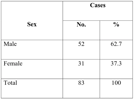
Sex distribution
Cases
Sex No. %
Male 52 62.7
Female 31 37.3
Total 83 100
Table -2 shows the sex distribution wherein around 52 [62.7% ] of
patients were males and the rest of the patients with anterior uveitis about
Table 3:
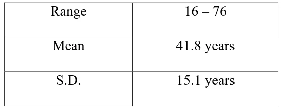
Range of Occurrence
Range 16
–
76
Mean 41.8
years
S.D. 15.1
years
Table 3 shows, Age of the patient who were included in the study ranged from 16 to 76 years with mean age of 44.8 with S.D of 15.1 years.
Table 4:
Laterality
Cases
Laterality
No. %
Right eye
45
54.2
Left eye
34
41
Both eyes
4
4.8
Total 83
100
[image:58.612.162.466.433.630.2]Table 4 shows the laterality, where out of 83 cases 45 [ 54.2 % ] had right eye involvement while 34 patients 41 % ] had their left eye affected. 4 cases [4.8 % ] had bilateral involvement.
Table 5:
Chronology
Cases
Acute / Chronic
No. %
Chronic 6
7.2
Acute 77
92.8
Total 83
100
Table 6 : Type of Inflammation
Cases
Type of Inflammation
No. %
Granulomatous 6
7.2
Non granulomatous
77
92.8
Total 83
100
Table 7 : Iris Nodules
Cases
Iris Nodules
No. %
Present 7 8.4
Absent 76
91.6
Total 90
100
The type of inflammation is showed in table 6 where the non granulomatous type was present in 7.2% and the rest 92.8% had non granulomatous type of inflammation.
[image:59.612.165.462.111.288.2]In contrast to the study by Rodriquez et al we have more of acute uveitis than chronic uveitis and recurrent uveitis. This is probably because of early referral by the physician / ophthalmologist from rural areas to the tertiary hospital because of lack of facilities like slit lamp and indirect ophthalmoscopy for diagnosing and early intervention , whereas in developed countries where there are facilities they refer cases only after a proper initial treatment in the peripheral centre. This might be the cause of a higher percentage of chronic uveitis in tertiary centre than acute cases.
The percentage of granulomatous and nongranulomatous uveitis detected in our study is based only on the clinical picture and not by histopathological examination which is difficult in our setup and also that the patients cannot afford. With the clinical identification in our study the percentages are in par with major studies as that of Rodriquez et al which was 89.8% and 10.2% respectively.
Laterality of the ocular involvement cannot be predicted properly as it depends on the presentation of uveitis. As in our study 95.2 % had unilateral involvement and 4.8 5% had bilateral involvement.
Table 8:
Final Diagnosis
Cases
Final Diagnosis
No. %
1. Idiopathic
36
43.9
2. HLA – B27 related
4
4.9
3. Viral Kerato uveitis
2
2.4
4. Traumatic anterior
uveitis16
19.5
5. Tuberculosis
3
3.7
6. Fuch’s heterochromic
uveitis1 1.2
7. Hansen’s
uveitis2
2.4
8. Lens induced
uveitis5
6.1
9. RD induced
uveitis1
1.2
10. Sclerokerato
uveitis1
1.2
11. Inflammatory bowel disease
1
1.2
12. Post operative uveitis
10
12.2
Table 8 shows the etiological association in patients with anterior
uveitis. The arrival to the final diagnosis was based on only clinical
findings in certain cases, while others were confirmed with the help of
laboratory investigations that are mentioned and a few cases were arrived
wherein the etiology was strongly suspected based on the clinical
presentation an negative laboratory results. Our study showed the
commonest cause of anterior uveitis to be idiopathic where 36 [43.9 %]out
of 83 cases had no identifiable aetiology. Next came trauma and post
operative uveitis that were 16 [19.5%] and 10 [12.2]respectively.
HLA-B27 associated anterior uveitis was suspected in about 4 [4.9%] of the
patients.3 [3.7%] cases of anterior uveitis were of tuberculosis etiology.2
[2.4%] cases were due to leprosy .one case each of fuch’s heterochromic
iidocyclitis, inflammatory bowel disease, sclerokeratitis and retinal
detachment associated anterior uveitis. Herpetic anterior uveitis was the
Table – 9
COMPARATIVE ANALYSIS OF ASSOCIATED CONDITIONS IN ANTERIOR UVEITIS England study 1916 USA study 1986 Israel study 1988 Present study 2008
Idiopathic 271 /44.1 72 /43.1 94 /51.4 36 /43.9 Trauma / surgery --/-- 10 /5.9 32 /17.5 26 /31.7 Herpes simplex ---/-- 10 /5.9 14 /7.7 ---/- JRA 1 /0.2 17 /10.2 7 /3.8 ---/-- Fuch’s hetero
chromic iridocyclitis 30 /4.9 11 /6.6 6 /3.3 1 /1.2
Leukaemia ---/-- ---/-- 4 /2.2 ---/--- Reiters disease ---/--- 6 /3.6 3 /1.6 ---/---
TB 158 /25.7 ---/-- 3 /1.6 3 / 3.7 Leprosy 2 /0.3 ---/--- 3 /1.6 2 /2.4 Syphilis ----/--- 5 / 3.0 1 10.5 ---/-- Herpes zoster ---/-- 5 /3.0 1 /0.5 2 /2.4 IBD --/-- 2 /1.2 1 /1.05 1 /1.2 Glaucomatocyclitic
crisis ---/--- 2 /1.2 1 /0.5 ---/-- Others 55 /8.9 --/-- 5 /2.7 12 /14.4 Total 614 /100 167 /100 183 /100 83 /100
[1961] showing 44.1% ,USA study [1986] showing 43 .1%, Israel study [1988] showing 51.4 % and our study showing 43.9 %.trauma and surgery formed 5.9% in the USA study and 17.5% and 31.7 % in Israel and our study respectively.
Herpes simplex was the causative factor of 5.9 % and7.7 % in the USA and Israel study respectively, the England study and ours had none. JRA formed 0.2 % in the England study ,10.2 5 in the USA study and 3.8 % in the Israel study with our study having no such case. Fuch’s was detected in 4.9 % in England study, 6.6% in USA study ,3.3% in Israel study and 1.2 % in our study. Leukaemia was the cause in2.2% of cases in the Israel study while there were no such cases in the England,USA and our study.
Table – 10
COMPARISON OF AETILOGICAL DIAGNOSIS OF ANTERIOR UVEITIC ENTITIES.
Diagnosis
Henderly et al
1987(I) %
Latanza et al
1991(II) %
Das et al
1995(III) %
Present Study
2008(IV) %
Idiopathic 12.1 36.8 17.8 43.9 Fuch’s
hetrochromic cyclitis
1.8 1.4 1.5 1.2
Herpetic uveitis
2.5 3.00 0.2 0.2
Traumatic 0.7 0 1.1 19.5
Collagen diseases
8.3 6.8 10.7 0
Iol Induced 1 0 3.9 0
Others 1.4 4.00 1.3 24.6 Total 27.8 52 36.5 89.4
uveitis was 0.7% in henderly study, 1.1% in Das study and 19.5% in our study. collagen disease as the etiological factor was a significant finding in Henderly study with 8.3%, Latanza study showed 6.8% and Das study showed10.7% and our study did not show any cases with this aetiology. IOLinduced anterior uveitis as the cause was seen with 1% and 3.9% in Henderly study and Das study respectively, while our study and Latanza study showed no cases of anterior uveitis which were IOL induced.
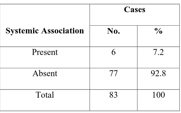
Table 11: Systemic Association
Cases
Systemic Association
No. %
Present 6
7.2
Absent 77
92.8
Total 83
100
[image:66.612.169.459.311.492.2]Table 12: Complications
Cases
Complications
No. %
None 67
80.5
Corneal Opacity
11
13.4
Glaucoma 1
1.2
Complicated cataract
3
3.6
Others 1
1.2
Total 83
100
Table 13: Intra Ocular Pressure
Cases
Intra Ocular Pressure
No. %
Normal 75
90.4
Raised 8
9.6
We cannot compare our study with that of hatzeistefanou et al where
it was 36.2% , because the study was characteristic of uveitis in the elderly
[image:67.612.169.456.115.373.2] [image:67.612.140.488.422.565.2]Table : 14 :
Final Visual Acuity
Presenting
Visual acuity
Final visual
acutity
Vision
No. % No. %
6/6
–
6/18
30 38.1 64 77.1
<
6/18
–
6/60
29 36.8 15 18.1
< 6/60 – 3/60
14
16.8
4
4.8
< 3/60
10
8.3
0
0
Total
83 100 83 100
These final tables show the complications that had resulted from the
episode of both acute and chronic anterior uveitis in our study.67 cases
[80.5%] had no complications and these were mostly idiopathic acute
anterior uveits.1 case [1.2%] had secondary glaucoma which was the
resultant of chronic topical medication of steroids. complicated cataract
was seen in Fuch’s heterochromic iridocyclitis and secondary to
trauma.corneal opacity resultd secondary to corneal ulcer and trauma in 11
Table 13 shows the raise of IOP during the episode of anterior
uveitis both acute and chronic form.75 patients [90.4%] a normal IOP
whereas 8 cases [9.6%] showed a raise of IOP which was transient that
was due to lens induced uveitis that normalised on lens extraction among
which only one resulted in secondary glaucoma as complication due to
topical steroid use in IBD.
Visual acuity was between 6/6 – 6/18 in 38.1% and improved
77.1% after the episode of anterior uveitis. It was 36.8% initially and
improved 18.1% were visual acuity < 6/18 – 6/60 and preexisting
SUMMARY
¾
That the mean age of presentation of the 83 cases of anterior
uveitis was found to be 41.8 years with S.D of 15.1 years.
¾
Sex wise the number of male patients were 62.7% and female
patients were 37.3%
¾
Acute anterior uveitis was seen in 92% of the cases and the rest
7.2% were chronic anterior uveitis.
¾
Majority of acute anterior uveitis was unilateral with 79 case
(95.2%). CAU involved in both eyes seen in 4 cases (4.8%).
¾
Koeppe’s nodule were seen in CAU in 8.4%.
¾
That 43.9% the majority of the cases that had anterior uveitis
were idiopathic
¾
Trauma and postoperative anterior uveitis were the next major
cause with 31.7%
¾
One case of uveitis was seen in Fuch’s heterochromic cyclitis,
¾
Lens induced uveitis was seen in 5 cases (6.1%), Phacolytic
glaucoma and treatment consisted of surgical removal of lens.
¾
Tuberculosis as cause of uveitis was seen in 3 (3.7%) patients.
¾
Systemic association was absent in 92.8% of the patients while
7.2% had anterior uveitis associated with a systemic condition
¾
Complication included complicated cataract 3.6%, secondary
glaucoma in 1.2% of patients and corneal opacity following
corneal trauma in 13.4%.
¾
Final visual acuity following treatment of an episode of acute
anterior uveitis was excellent in 77.1% of cases. 18.1% had
acuity between <6/18 – 6/60 and preexisting lenticular changes
DISCUSSION
Anterior uveitis is the most common form of uveitis.
The mean age of the presentation of anterior uveitis in our study
was found to be 41.8% with S.D. of + 15.1 years. This finding is
more or less correlated with one study by Rodrigurez et al where it
showed a mean of 39.8% and S.D of + 18.3.
In our study males and females contributed 62.7% and 37.3%
respectively whereas study by Rodrigurez et al showed as slightly
higher percentage of men. Such low percentage of female population
in our study is probably due to poor socio economic status of our
study population where they had to work with disease of the eye than
to attend hospital.
The same study showed high number of chronic and recurrent
uveitis whereas our study shows acute uveitis because our patients
had early referral from rural areas to the tertiary hospital because of
The percentage of granulomatous and non granulomtous uveitis
based on clinical identification was in par with major studies as that of
Rodriguerez et al.
Commonest cause of anterior uveitis was idiopathic in our study
and was 43.9% which was more or less similar to that of other
international studies like England study (1916) 44.1%, the USA study
1986 which showed 43.1% and the Israel study 1988 which had
51.4%. Similar findings were seen with respect to Herpes Zoster
ophthalmicus, IBD, Leprosy.
The idiopathic anterior uveitis as the etiological factor was also
found in the Latanza et al study. Fuch’s heterochromic cyclitis was
1.2% in our study and was comparable to Henderly et al study which
showed 1.8%, Latanza et al study 1.4%, Das et al study 1.5%.
Herpetic uveitis was 0.2% in our study showing similarity with
Das et al study which was also 0.2%
Our study was not a population based study but we still find that
CONCLUSION
The cause of uveitis vary greatly by geographical region
throughout the world. Such variation is due largely to complex
ecological, racial, nutritional and socioeconomic differences. Many
developing countries have a tropical climate allowing unique disease
pathogens, vectors and host reservoir to flourish. Poverty,
overcrowding, limited formal and public education, poor hygiene and
finite medical resources also play a role. The perineal migration of
people from city to city and around the world requires that all
phycisians be aware of global variations in disease pattern to provide
optimal medical care.
There are an estimate 45 million blind people in the world today
of whom approximately 75% live in developing nations. Although
data on the prevalence and incidence of uveitis as a cause of vision
loss in developing region are scarce, it is probably safe to say that
ocular complications of well recognised and endemic infection
constitute a major cause of blindness. we review those infectious and
non infectious cause of anterior uveitis encountered most often in the
BIBLIOGRAPHY
1. Wakefield, Denis; Montanaro, Anthony and Cluskey, Peter M C "Current Research: Acute Anterior Uveitis and HLA-B27"
SURV OPHTHALMOL VOL: 36 (3) 1991 NOV.-DEC. P.223-232
2. Saunders, Richard A Bluestein, Ettaleah C; Wilson, M Edward; Berland, Jerry E "DIAGNOSTIC AND SURGICAL TECHNIQUES: Anterior Segment Ischemia After Strabismus Surgery"
SURV OPHTHALMOL VOL: 38 (5) 1994 MAR.-APR. P.456-466
3. Lim, Jennifer I; Tessler, Howard H and Goodwin, James A
"Anterior Granulomatous Uveitis in Patients with Multiple Sclerosis"
OPHTHALMOLOGY VOL: 98 (2) 1991 FEB. P.142-145
4. Wakefield, Denis Stahlberg, Tom H; Toivanen, Auli; Granfors, Kaisa; Tennant, Carolyn "Serologic Evidence of Yersinia Infection in Patients with Anterior Uveitis" ARCH OPHTHAL VOL: 108 (2) 1990 FEB. P.219-221
6. Ophir, Avinoam; Ticho, Uriel "Remission of Anterior Uveitis by Subconjunctival Fluorouracil" ARCH OPHTHAL VOL: 109 (1) 1991 JAN. P.12-13 Type: Letter
7. Kolker, Richard J "Medication-induced Bilateral Anterior Uveitis" ARCH OPHTHAL VOL: 109 (10) 1991 OCT. P.1343 Type: Letter
8. Goldey, Stacia H Stern, George A; Oblon, David J; Mendenhall, Nancy P; Smith, Linda J; Duque, Ricardo E
9. "Immunophenotypic Characterization of an Unusual T-Cell Lymphoma Presenting as Anterior Uveitis: a clinicopathologic case report" ARCH OPHTHAL VOL: 107 (9) 1989 SEP. P.1349-1353 Type: Report
10.Fearnley, Ian R; Spalton, David J and Smith, Stephen E "Anterior Segment Fluorophotometry in Acute Anterior Uveitis" ARCH OPHTHAL VOL: 105 (11) 1987 NOV. P.1550-1555
11.Kanski, Jack J "Anterior Uveitis in Juvenile Rheumatoid Arthritis" ARCH OPHTHAL VOL: 95 (10) 1977 OCT. P.1794
13.Cherubini, Thomas D; George L Spaeth "Anterior Nongranulomatous Uveitis Associated with Rocky Mountain Spotted Fever" ARCH OPHTHAL VOL: 81 (3) 1969 MAR. P.363
14.Giles, CL "Anterior Uveitis in Children" ARCH OPHTHAL VOL: 70 (6) 1963 DEC. P.779
15.Coles, Robert S; Arthur Nathaniel "The Role of the Streptococcus in the Pathogenesis of Anterior Uveitis" ARCH OPHTHAL VOL: 61 (1) 1959 JAN. P.45
16.Biswas, Jyotirmay; Neelakantan, Arvind and Rao, Sridhar B "Adenoma of Nonpigmented Epithelium of the Ciliary Body Presenting as Anterior Uveitis and glaucoma: a case report" INDIAN J OPHTHALMOL VOL: 43 (3) 1995 SEP. P.137-140 Type: Letter
17.Rosenbaum, James T "Systemic Associations of Anterior Uveitis" INT OPHTHALMOL CLIN VOL: 31 (3) 1991 SUMMER P.131-142
18.Pavesio, Carlos E; Nozik, Robert A "Anterior and Intermediate Uveitis" INT OPHTHALMOL CLIN VOL: 30 (4) 1990 FALL P.244-251
20.Linssen, Annelise; Meenken, Christina "Outcomes of HLA-B27-Positive and HLA-B27 Negative Acute Anterior Uveitis" AM. J. OPHTHALMOL VOL: 120 (3) 1995 SEP. P.351-361
21.Ishimoto, Sei-ichi Wu, Guey-Shuang; Hayashi, Seiji; Zhang, Jie; Rao, Narsing A "Free Radical Tissue Damages in the Anterior Segment of the Eye in Experimental Autoimmune Uveitis" INVEST. OPHTHALMOL. VIS. SCI. VOL: 37 (4) 1996 MAR. P.630-636
22.Dunn, James P "New Treatments for Anterior Uveitis" OPHTHALMOLOGY VOL: 103 (3) 1996 MAR. P.355-356 Type: Editorial
23.Watanabe, Todd M; Hodes, Barton L "Bilateral Anterior Uveitis Associated with a Brand of Metipranolol" ARCH OPHTHAL VOL: 115 (3) 1997 MAR. P.421-422
24.Pavan-Langston, Deborah "Mycoplasmal anterior and posterior uveitis: I. Clinical manifestations of the experimental disease" ARCH OPHTHAL VOL: 82 (2) 1969 AUG. P.245-252
26.Patel, Niraj P Patel, Ketan H; Moster, Marlene R; Spaeth, George L "Metipranolol-Associated Nongranulomatous Anterior Uveitis" AM. J. OPHTHALMOL VOL: 123 (6) 1997 JUN. P.843-844 Type: Report
27.Van Der Lelij, Allegonda; Rothova, Aniki "Diagnostic Anterior Chamber Paracentesis in Uveitis: a safe procedure" BR J OPHTHALMOL VOL: 81 (11) 1997 NOV. P.976-979
28.Khan, Yasser A Pavlin, Charles J; Cykiert, Robert; Rootman, David
S "Uveitis-Glaucoma-Hyphema Syndrome after Handmade, Anterior Chamber Lens Implantation" J CATARACT REFRACT SURG
VOL: 23 (9) 1997 NOV. P.1414-1417
29.HOGAN Michael J "Signs and symptoms of uveitis- I. Anterior uveitis"AM. J. OPHTHALMOL VOL: 47 (5- PT.2) 1959 MAY P.155
31.WOLTER J Reimer "Acquired autosensitivity to degenerating descemet's membrane in a case with anterior uveitis in the other eye"
AM. J. OPHTHALMOL VOL: 72 (4) 1971 OCT. P.782
32.BELL Randall; FONT Ramon L "Granulomatous anterior uveitis caused by coccidioides immits" AM. J. OPHTHALMOL VOL: 74 (1) 1972 JUL. P.93
33.NOZIK Robert A; DORSCH William "A new chorioretinopathy associated with anterior uveitis" AM. J. OPHTHALMOL VOL: 76 (5) 1973 NOV. P.758
34.MASI Robert J "Anterior uveitis in geographic or serpiginous choroiditis" AM. J. OPHTHALMOL VOL: 86 (2) 1978 AUG. P.228
35.PUIG-LLANO Manuel "Pupillary membrane excision and anterior vitrectomy in eyes after uveitis" AM. J. OPHTHALMOL VOL: 87 (4) 1979 APR. P.533
36.SAARI K M "Genetic background of acute anterior uveitis" AM. J. OPHTHALMOL VOL: 91 (6) 1981 JUN. P.711
38.Rothova, A Van Veenendal, W G; Linssen, A; Glasius, E; Kijlstra, A; De Jong, P T V M "Clinical Features of Acute Anterior Uveitis" AM. J. OPHTHALMOL VOL: 103 (2) 1987 FEB. P.137-145
39.Secchi, A G Magni, G; Tognon, M S; Rupolo, G; Angi, M R; Arsie, D; Turrini, B "A Psychosomatic Approach to Idiopathic Recurrences of Anterior Uveitis" AM. J. OPHTHALMOL VOL: 104 (2) 1987 AUG. P.174-178
40.Rosenbaum, James T; Bennett, Robert M "Chronic Anterior and Posterior Uveitis and Primary Sjogren's Syndrome" AM. J. OPHTHALMOL VOL: 104 (4) 1987 OCT. P.346-352
41.D'Alessandro, Leonardo P; Forster, David J and Rao, Narsing A "Anterior Uveitis and Hypopyon" AM. J. OPHTHALMOL VOL: 112 (3) 1991 SEP. P.317-321
42.MAIER Gunthilt "HLA antigens in acute anterior uveitis in South African blacks" BR J OPHTHALMOL VOL: 64 (5) 1980 MAY P.329-331
43.EILON L A "Clinical evaluation