Received 26 August 1996/Accepted 11 December 1996
RNA tertiary structures, such as pseudoknots, are known to be biologically significant in a number of virus
systems. The 3* untranslated regions of the RNA genomes of all members of theEnterovirusgenus of
Picor-naviridaeexhibit a potential, pseudoknot-like, tertiary structure interaction of an unusual type. This is formed by base pairing between loop regions of two secondary structure domains. It is distinct from a potential, conventional pseudoknot, studied previously in poliovirus, which is less conserved phylogenetically. We have analyzed the tertiary structure feature in one enterovirus, coxsackievirus A9, using specific mutagenesis. A double mutant in which the potential interaction was destroyed was nonviable, and viability was restored by introducing compensating mutations, predicted to allow the interaction to reform. Phenotypic pseudorever-tants of virus mupseudorever-tants, having mutations designed to disrupt the interaction, were all found to have acquired nucleotide changes which restored the potential interaction. Analysis of one mutant containing a single-base mutation indicated a greatly increased temperature sensitivity due to a step early in replication. The results show that, in addition to secondary structures, tertiary RNA structural interactions can play an important role in the biology of picornaviruses.
Enteroviruses make up one genus of thePicornaviridae, a
diverse family of small viruses including several of clinical or economic importance such as polioviruses, hepatitis A virus, rhinoviruses (the major cause of the common cold), and foot-and-mouth disease virus of cattle (22, 27). They have a posi-tive-sense, single-stranded RNA genome, 7,100 to 8,500
nucle-otides long, which is 39polyadenylated. The genome encodes
one polyprotein which is cleaved by virus-encoded proteases to yield several proteins required for replication and assembly of new virus particles. RNA replication proceeds via the synthesis of negative-sense copies of the virus genome, which then serve as templates for the synthesis of new positive strands. The single open reading frame is preceeded by a long (ca. 750
nucleotides in enteroviruses) 59 untranslated region (59UTR)
which is followed by a much shorter (70 to 100 nucleotides in
enteroviruses) 39UTR and the poly(A) tract (22). The 59UTR
has two major structural and functional domains (20). The
59-most 90 nucleotides fold into a cloverleaf configuration
es-sential for positive-strand synthesis, while much of the rest of
the 59UTR comprises the internal ribosome entry site (IRES),
which is involved in cap-independent translation initiation (1, 14). IRES function depends on specific sequences and its com-plex secondary structure (14).
Despite the fact that the 39UTR probably plays a role in
RNA replication, since it must be in close proximity to the site of initiation of negative-strand synthesis, it has been
compar-atively little studied. The 39UTR of all enteroviruses appears to
fold into the same core structure consisting of two stem-loop domains, X and Y (10, 16). An additional domain, Z, is seen in two of the 5 enterovirus genetic subgroups which have been described previously (18, 19). Covariance between enterovirus sequences, maintaining domains X and Y, is strong evidence that these are significant, and there is direct support from structure-probing experiments (16). It has been pointed out
that the loop regions of domains X and Y have the potential to participate in a phylogenetically conserved tertiary structure interaction, involving Watson-Crick base pairing, which is sim-ilar to a pseudoknot (16). This is a less common structural motif than a typical pseudoknot, and its further study is thus of general importance to an understanding of RNA tertiary struc-tures. In addition, an alternative, but less phylogenetically con-served, typical pseudoknot has been proposed to occur in the
39UTR of one enterovirus, poliovirus (11). An understanding
of the structure and function of the enterovirus 39UTR
re-quires the determination of which, if either, of these alterna-tive structures is correct. We describe here experiments on the enterovirus coxsackievirus A9 (CAV-9) which provide genetic evidence that the phylogenetically conserved tertiary structure plays a critical role in the replication of enteroviruses.
MATERIALS AND METHODS
Virus cDNA.All mutants were produced from pCAV-9, a full-length cDNA
representing the complete CAV-9 genome, contained within the vector pBS (3, 9).
Oligonucleotides.A PCR mutagenesis procedure was used to make most of
the mutants. Two overlapping regions of pCAV-9 were amplified with one universal, outer primer (OL256, based on the genomic sense sequence at posi-tions 7099 to 7118 or OL419, complementary to the CAV-9 poly(A) tract and containing aMluI site and anXhoI site) and one of a pair of specific, inner primers. The latter were complementary and contained the specific mutation to be introduced. The oligonucleotides used were:
General OL256 59GAGCAGATGAACAATACCCC 39
OL419 59GAGAGACTCGAGACGCG(T)16 39
dm OL365 59CTGTACTAA(A,C)(A,C)GAACTAGATAACG 39
OL366 59CGTTATCTAGTTC(A,C)(A,C)TTAGTACAG 39
y4C OL581 59CTGTACTAACC(C,A,T)AACTAGATAACG 39
OL582 59CGTTATCTAGTT(G,T,A)GGTTAGTACAG 39 y3C and OL534 59CTGTACTAAC(A,G,C)GAACTAGATAACG 39
y3A OL535 59CGTTATCTAGTTC(A,C,T)GTTAGTACAG 39
XA and OL623 59GCAGTAGGGGTAAATTCT(C,A)AGCATTCGGTGCGG 39
XAA OL624 59CCGCACCGAATGCT(T,G)AGAATTTACCCC 39
In each case, the altered nucleotide is underlined and mixed nucleotides intro-duced at certain positions are indicated in parenthesis. The overlapping ampli-cons were then subjected to a second PCR amplification to produce a continuous
fragment of DNA. This fragment was cleaved withBamHI (at a site within
* Corresponding author. Phone: 44 1206 873308. Fax: 44 1206 873416. E-mail: [email protected].
2363
on November 9, 2019 by guest
http://jvi.asm.org/
CAV-9 cDNA at position 7178) andXhoI (introduced into the PCR product) and cloned into these sites in the vector pUBS. The mutated fragment was completely sequenced to ensure the presence of only the required mutation and then removed from the vector usingBamHI andXhoI and inserted into pCAV-9,
which had been digested withXhoI and partially digested withBamHI. The
sequence of this region in the mutated pCAV-9 was then reconfirmed. Some of the mutants were produced by an alternative strategy which made use
of aBsmI site close to the 39terminus of CAV-9 cDNA (position 7435). A
BamHI-XhoI subclone was cleaved with eitherBsmI andXhoI orBsmI andClaI, and pairs of annealed, complementary oligonucleotides were introduced by li-gation. The manipulated subclone was then introduced into pCAV-9. The oli-gonucleotides used in this strategy were:
dmrep OL471 59CATTC(A,G)(C,G)TGCGGAGG(A)15CGCG 39
OL472 59TCGACGCG(T)15CCTCCGCA(C,G)(C,T)GAATGCG 39
x4A and OL455 59CATT(A,G,T)GGTGCGGAGG(A)15T 39
x4G OL456 59CGA(T)15CCTCCGCACC(A,C,T)AATGCG 39
Virus generation and propagation.All transfections and experiments based on virus propagation were performed in 25 cm2tissue culture flasks, containing
monolayers of GMK cells. These were maintained in 7 ml of minimal Eagle’s medium (MEM) containing 10% heat inactivated fetal bovine serum, 1% MEM amino acids, and gentamycin (100 mg/liter). RNA transcription from the mu-tated derivatives of pCAV-9, using T7 polymerase followed by transfection into GMK cells was as described previously (9). Well-separated plaques were picked and passaged once in GMK cells. Viruses were liberated from infected cells by
freeze-thawing three times, and the resultant virus-containing suspension was used directly for analysis.
Sequence analysis of mutant and revertant viruses.RNA isolation (from 100
ml of the virus suspension), reverse transcription, and PCR were as described previously (6). The PCR product was purified by agarose gel electrophoresis and sequenced by means of a commercial cycle sequencing kit (Gibco BRL), using
the primer OL441 (59CAGGAGCGTCCCAGTTGG 39), complementary to
positions 7271 to 7292 of CAV-9 cDNA.
Growth curve and temperature shift experiment.To derive growth curves,
equal quantities (105PFU) of C9wt or C9y3G in 0.5 ml of growth medium were
adsorbed to several GMK monolayers at 208C for 60 min. Unbound viruses were removed by washing with medium, and the monolayers were incubated at 378C to allow virus replication, which was monitored by freeze-thawing three times, followed by plaque assay. A temperature shift experiment was performed in the same way except that after adsorption and washing, the infected monolayers were incubated at 378C before being shifted to 39.58C. Incubation was carried out for a total of 9 h (C9wt) or 12 h (C9y3G), and the level of replication was assessed by plaque assay.
RESULTS
3*UTR structure in enterovirus genetic groups. The
pre-dicted RNA secondary structures of the 39UTR of
represen-tative enteroviruses and of a typical human rhinovirus
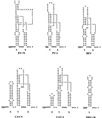
(HRV-FIG. 1. Schematic representation of the proposed secondary and tertiary structures of the 39UTR of viruses exemplifying the five genetic groups identified among enteroviruses, together with HRV-1B (15, 17, 18). Abbreviations: PV-3, poliovirus type 3; EV-70, enterovirus 70; BEV, bovine enterovirus. The asterisk in the PV-3 structure marks a nucleotide difference seen between the Leon (A) and Sabin (G) strains of this virus serotype (25).
on November 9, 2019 by guest
http://jvi.asm.org/
[image:2.612.139.474.76.463.2]viruses, enterovirus 70, and bovine enteroviruses, exhibit only these features and differ in the size of stem-loop Y (enterovirus 70) or the primary sequence between the stem-loops (bovine enterovirus). Two of the genetic groups, exemplified by CAV-9 and CAV-2, have an additional stem-loop (Z), which differs in size between the two groups and is located upstream of domain Y. Rhinoviruses have a single stem-loop, possibly equivalent to Y (16, 30, 31). Members of all the enterovirus genetic groups exhibit the potential for a tertiary structure interaction, involv-ing either 5 or 6 bp, between nucleotides in the loops of stem-loops X and Y (shown diagrammatically in Fig. 1). It is interesting that in each genetic group the sequences involved are quite different, yet the potential interaction is evident, which is strong phylogenetic evidence that it is significant. Within a genetic group, the sequences making up the interac-tion are conserved (data not shown).
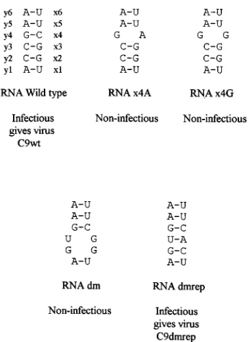
Mutagenesis and repair of the tertiary structure interaction. In order to study the significance of the tertiary structure interaction, we have exploited the infectivity of RNA tran-scribed from a complete cDNA copy of the RNA genome of picornaviruses. Mutations were introduced into CAV-9 cDNA, and their effect was monitored by determining whether the corresponding RNA gave rise to infectious virus and, if so, analyzing the properties of the mutant viruses. Mutant cDNAs were produced by PCR mutagenesis, using synthetic primers to introduce specific changes. Manipulated fragments were com-pletely sequenced to ensure the absence of undesired muta-tions and were reintroduced into the full-length cDNA. RNA was transcribed and transfected into the green monkey kidney cell line GMK, in which CAV-9 replicates efficiently. The mu-tant RNAs initially produced, together with the effect of the mutations on virus viability, are summarized in Fig. 2. For convenience, the nucleotides involved in the CAV-9 interac-tion are denoted y1 to y6 for those in loop Y and x1 to x6 for their complements in loop X. It can be seen that mutations which disrupt the potential interaction have a profound affect on the viability of CAV-9. In both cases tested, single
muta-tions in the x3 position (C3A or C3G) gave rise to RNA
which, despite several transfections, yielded no infectious virus. This indicates that these single nucleotide substitutions are lethal. A double mutant RNA, dm (positions y2 and y3,
CC3GU), was also produced and shown to be noninfectious,
again following several transfections. However, when this con-struct was modified by the introduction of two additional,
com-plementary mutations (positions x2 and x3 GG3CA), the
transcribed RNA, dmrep, proved to have an infectivity
com-parable to that of wild-type CAV-9 RNA (3 3 103 virus
plaques formed per mg of RNA) and produced viruses of a
wild-type plaque phenotype (Fig. 2). Two virus plaques were picked, passaged once in GMK cells, and analyzed, following reverse transcription-PCR, by cycle sequencing. In the region
sequenced (position 7300 to the 39 terminus), the virus
ge-nomes exhibited the four mutations specifically introduced
(positions y2 and y3, CC3GU, and x2 and x3, GG3CA) and
no other differences from the original CAV-9. The results indicate that the introduction of the two additional mutations,
designed to repair the interaction between domains X and Y, restores the viability of the virus. This suggests that the tertiary structure interaction plays an important role in CAV-9 repli-cation.
Analysis of CAV-9 pseudorevertants. Pseudorevertants, in
which a wild-type phenotype is restored or partially restored by second-site mutations, have proved useful for the demonstra-tion of specific RNA-RNA and RNA-protein interacdemonstra-tions in picornaviruses. Since no pseudorevertants were obtained from the noninfectious RNAs described above, two other mutant cDNA constructs were produced as described before, and
RNA was transcribed. These were y3G (position y3, C3G)
and y3A (position y3, C3A), and both proved to be highly
infectious. Three plaques were picked from each transfection, and the viruses were passaged once in GMK cells. Upon anal-ysis of passaged virus, all three y3C viruses and two y3G viruses had a wild-type plaque size phenotype, while one virus derived from y3G (denoted C9y3G) had a small plaque phenotype. All the passaged viruses were sequenced from position 7300 to the
39terminus, and mutations were observed only in the region of
the proposed tertiary structure interaction as shown in Fig. 3. The genomes of all viruses derived from y3A had an identical sequence, which differed from that of the transfected RNA at
the mutated position (y3, C3A, was introduced into y3A and
an A3U change was observed at this position). This mutation
restores the proposed tertiary structure interaction through the
formation of a UzG base pair (Fig. 3). The small plaque virus
from y3G, C9y3G, had maintained the mutation introduced
(position y3, C3G), and had no other mutations in the region
sequenced. Both large plaque viruses from y3G were identical, unusual revertants, which had retained the mutation
intro-duced (position y3, C3G) but exhibited an additional
muta-tion two bases upstream (posimuta-tion y1, A3C). One of these,
[image:3.612.345.526.66.315.2]denoted C9y3Gy1C, was chosen for further study. Curiously,
FIG. 2. Simplified version of the tertiary structure interaction between the loops of domains X and Y in CAV-9. x1 to x6 and y1 to y6 denote the nucleotides in loops X and Y, respectively, which take part in the interaction. The locations of several mutations in mutated constructs and their effects on virus viability are also indicated.
on November 9, 2019 by guest
http://jvi.asm.org/
analysis of the sequences involved indicates that they now have the potential to be involved in an alternative tertiary structure interaction, using the same loop X nucleotides but effectively shifted one nucleotide upstream with respect to loop Y (Fig. 3). The structure predicted is therefore between y0 to y5 and x1 to x6, where y0 is the nucleotide immediately preceeding the loop Y region involved in the wild-type interaction.
To extend the analysis, a further mutant cDNA clone (y4C)
was produced which had a mutation at the y4 position (G3C).
Repeated transfection of y4C gave only one plaque, the virus from which was passaged in GMK cells. Sequence analysis
from position 7300 to the 39terminus indicated that the virus
had retained the mutation introduced (position y4, G3C) but
exhibited a further, complementary change at the
correspond-ing position in the X domain (position x4, C3G), presumably
allowing the tertiary structure interaction to reform (Fig. 4).
This virus also had one further change (C3U at genomic
position 7433), predicted to be in the stem of domain X but
maintaining this structure through a UzG base pair (data not
shown). Thus, although different patterns of reversion were observed, there is a correlation between efficient virus replica-tion, in terms of plaque size, and the potential to form the tertiary structure interaction.
Growth properties of a mutant with a disrupted tertiary
structure.The properties of the mutant and revertant viruses
obtained are summarized in Table 1. It can be seen that, in terms of plaque size, the revertants are comparable to the unmanipulated virus, C9wt, while C9y3G has a smaller plaque size. The viruses were also analyzed with respect to
tempera-ture sensitivity by plaque assay at 34, 37, and 39.58C. All,
including C9wt, demonstrated some temperature sensitivity, since there was a significant reduction in plaque number at
39.58C compared to 348C and 378C (43-fold for C9wt at 39.58C
compared to that at 378C). However, temperature sensitivity
was extremely marked in the small plaque mutant C9y3G,
which gave a 104-fold reduction in plaque numbers at 39.58C
compared to that at 34 and 378C. The virus from a plaque
arising during the assay of C9y3G at 39.58C was propagated
and found to have a wild-type plaque phenotype. Upon
se-quencing, a position y3 (G3C) change, restoring the wild-type
sequence, was observed. Although giving a normal plaque size
at 378C, the revertant which had a shifted tertiary structure
(C9y3Gy1C) was much more temperature sensitive than C9wt (Table 1).
To further study the effect of the single mutation in the small plaque mutant, C9y3G, a single-cycle growth experiment was performed (Fig. 5). The results show that the growth of C9y3G is greatly retarded, since infectious virus particles are not pro-duced until 8 h postinfection compared to 4 h for C9wt. In
addition, the yield of C9y3G is reduced (by a factor of 102),
further indicating the radical effect of the single mutation in C9y3G.
The temperature-sensitive nature of the y3, C3G, mutation
in C9y3G was exploited in temperature shift experiments, in order to determine the timing of the replication step affected by this mutation (Fig. 6). C9y3G or C9wt were adsorbed at
228C to several GMK monolayers, and these were incubated at
378C to initiate infection. At various time intervals, the
in-fected monolayers were shifted to 39.58C, a temperature
non-permissive for C9y3G. After a total time of 9 h for C9wt and 12 h for C9y3G, the cells were lysed and the level of virus replication was established by plaque titration. It can be seen that, although C9wt exhibits a degree of temperature
sensitiv-ity, since incubation at 39.58C results in a reduced yield
com-pared to that at 378C, replication occurred in all flasks. In
contrast, no replication of C9y3G occurred unless it was
[image:4.612.59.297.67.348.2]incu-bated at 378C for at least 1 h before the temperature shift.
[image:4.612.368.509.68.183.2]FIG. 3. Summary of the point mutations introduced into the y3 position of the tertiary structure interaction seen in CAV-9 RNA, together with the se-quences of the viable viruses which resulted. The revertant C9y3C, which has the wild-type sequence, was derived from the mutant C9y3G by growth at 39.58C.
FIG. 4. Predicted structure of the noninfectious RNA produced by mutation of the y4 position of the tertiary structure interaction and of the viable pseudor-evertant, C9y4Cx4G, which was recovered.
TABLE 1. Comparison of the infectivity ratio of wild-type and mutant CAV-9 strains at different temperatures, together
with plaque sizes at 378C
Virus
Titer at indicated temp switch
Plaque size (mm)
34 to 39.58C 37 to 39.58C
C9wt 3.53102 4.33101 1.460.1
C9y3G 5.03103 1.03104 0.660.2
C9y3Gy1C 1.53103 1.33103 1.060.5
C9y3U 1.13102 1.63102 1.460.4
C9dmrep 5.33102 5.63101 1.060.5
on November 9, 2019 by guest
http://jvi.asm.org/
[image:4.612.318.556.645.727.2]Incubation at 378C for periods greater than 2 h had little further effect on the yield of C9y3G. This experiment demon-strates that the temperature-sensitive lesion affects a step early in replication, rather than a later event such as particle assem-bly.
Evidence for the existence of stem X. Although there is
strong phylogenetic evidence for the existence of both stems X and Y, stem X is relatively weak in many enteroviruses (Fig. 1). It is therefore possible that stem X does not form and that nucleotides interacting with stem-loop Y are not themselves in
a loop region, i.e., that the interaction is a typical pseudoknot. To determine whether stem X is functionally significant, two other constructs were produced. These had either one or two mutations designed to disrupt base pairs in stem X (Fig. 7). RNA transcribed from the single mutant (XA) was highly infectious and gave a virus which phenotypically approximated the wild type (data not shown). Several plaques were picked, and the virus was passaged once in GMK cells. All were found,
by sequence analysis from position 7300 to the 39terminus, to
have only the mutation introduced. In contrast, RNA tran-scribed from the double mutant (XAA) was completely non-infectious. The results imply that stem X can still form in the single mutant, despite the presence of a single mismatch, but that the structure is so weakened in the double mutant that it cannot form. The data suggest that stem X is indeed function-ally significant and that the tertiary structure proposed is of a complex form, involving the loop regions of two stem-loops (16).
DISCUSSION
In many cases, RNA tertiary structure is vital to biological function (17). Frequently, non-Watson-Crick interactions sta-bilize these structures, a prime example being tRNA in which several such interactions maintain the compact structure (8). However, critical tertiary structures are frequently stabilized by Watson-Crick base pairs between nucleotides of a loop and residues outside that loop, giving a pseudoknot (17). This motif is important in the structure of rRNA and the control of
translational regulation of ribosomal proteins in Escherichia
coli, while a pseudoknot downstream of a slippery sequence is
involved in21 ribosomal frameshifting in several virus families
(2, 17, 25, 32). 39UTRs of plant viruses frequently harbor
pseudoknots, which are required for the folding of a tRNA-like
structure seen in some viruses (28). Other plant virus 39UTRs
have sequential arrays of pseudoknots which may play a role in translation or replication (17).
The secondary structure of the picornavirus 59UTR plays an
essential role in replication (24). Although conserved tertiary structural features have also been reported, as yet there is little experimental evidence that they are functionally significant
(13). The 39UTR has been less studied, but phylogenetic
[image:5.612.321.551.69.214.2]con-servation of secondary and tertiary structures between all en-teroviruses suggests that these structures are important to the life cycle of these viruses (16). The conserved features are two
[image:5.612.63.291.69.289.2]FIG. 5. Growth curves of C9wt (F) and C9y3G (■) on GMK monolayers.
[image:5.612.67.291.454.683.2]FIG. 6. Temperature shift experiment. C9wt (F) and C9y3G (■) were ad-sorbed to monolayers of GMK cells and then incubated at 378C for the time indicated before being shifted to 39.5C. After a total incubation time of 9 h for C9wt and 12 h for C9y3G, the level of virus produced was monitored by plaque titration.
FIG. 7. Predicted structure of the stem-loop X region of wild-type CAV-9 RNA and that of single (XA) and double (XAA) mutants, designed to probe the significance of this potential secondary structure. Nucleotides which take part in the interaction with loop Y are shown in bold type.
on November 9, 2019 by guest
http://jvi.asm.org/
stem-loops (X and Y) and a tertiary structure interaction, similar to a pseudoknot, between the loops of these domains. We have shown here that in CAV-9, mutations in sequences involved in the tertiary structure are lethal or have a radical effect on replication ability, particularly at elevated tempera-tures. However, the effects of a double mutation in loop Y can be suppressed by directed mutagenesis of loop X, designed to restore the tertiary structural interaction (Fig. 2). Further-more, in some cases, pseudorevertants of loop Y mutants could be isolated and were analyzed by sequencing. In each, the reverting mutation was consistent with the recreation of the tertiary structure interaction (Fig. 3 and 4). The data therefore provide compelling evidence that the proposed tertiary struc-ture interaction is critical for the ability of CAV-9 to replicate. Phylogenetic conservation makes it likely that this interaction plays an important role in all enteroviruses. Indeed, the intro-duction of four nucleotide changes into the loop Y domain has been shown to be lethal for poliovirus (15). Although no at-tempts were made to repair the mutant and no pseudorever-tants were obtained, this effect is likely to be due to disruption of the tertiary structure interaction.
In the present work four different classes of revertant were identified: a direct back mutation (observed upon growth of
C9y3G at 39.58C), a second-site mutation in loop X
compen-sating a mutation in loop Y, an A3U change which restores
the interaction through a UzG base pair, an additional
mu-tation which allows a new interaction shifted one base up-stream (Fig. 2 to 4). In the latter two groups of mutants, the interaction is weaker than that of the wild-type virus, since they
contain UzG base pairs (Table 1). This probably accounts for
their increased temperature sensitivity, relative to C9wt, which is particularly evident for C9y3Gy1C. Both result from trans-versions, and it is not clear why these revertants appear fre-quently. One possibility is that a weaker loop-loop interaction may be advantageous in the experimental system employed. The lack of any viable mutants or pseudorevertants from RNA mutated at the x4 position and the generation of only a single pseudorevertant from RNA mutated at the y4 position is strik-ing, since y3-mutated RNAs were highly infectious, producing several revertants and one viable mutant C9y3G. This suggests that disruption of the y4-x4 base pair completely prevents RNA replication and so does not allow the possibility of re-vertants or pseudorere-vertants arising (Fig. 2 and 4). In contrast, replication of RNA with a disrupted x3-y3 interaction can take place, possibly indicating that the interaction can still occur (albeit inefficiently since C9y3G grows poorly and is highly temperature sensitive) by virtue of the other base pairs in this structure (Fig. 3). Both y3-x3 and y4-x4 base pairs are near the center of the interaction, but y4-x4 may be more critical as it is
adjacent to two AzU base pairs, which may not form without
the strong CzG y4-x4 base pairing (Fig. 2). The quadruple
mutant, C9dmrep, and the slipped interaction pseudorever-tant, C9y3Gy1C, both have different loop Y sequences inter-acting with the loop X residues than the wild type, suggesting that tertiary structure, not primary sequence, is the critical feature involved. This is consistent with the variation in the sequence making up this structure in the five enterovirus ge-netic subgroups (Fig. 1).
There is strong phylogenetic support for the proposed
en-terovirus 39UTR structure (Fig. 1). However, the relative
weakness of stem X makes it possible that this structure does not occur and, therefore, that the region interacting with loop Y is not itself part of a loop. The mutation of nucleotides predicted to form stem X suggests that this structure is impor-tant, since a double mutant (XAA) in which two potential base pairs are removed is nonviable (Fig. 7). Using the program
FOLD, the stability of stem X in the wild type, single mutant
XA, and double mutant XAA is210.0,24.3, and 20.2 kcal/
mol, respectively, showing the marked effect of the mutations introduced (33). Further evidence for the occurrence of stem
X in enteroviruses, is the sequence variability seen at the 39
terminus of some poliovirus strains. For instance, the Sabin
vaccine strain of poliovirus type 3 has an A3G change relative
to its parental strain Leon immediately preceding the poly(A) tract (26). In both these strains, base pairing can occur at this
position through U-A or UzG interactions (Fig. 1). Other
direct experimental evidence for the importance of stem-loops X and Y is the abrogation of RNA replication in poliovirus replicons when either stem-loop is precisely deleted (21). Mu-tations introduced into the base of stem-loop Y also had a lethal effect on poliovirus replication, indicating, in agreement with the strong phylogenetic conservation, that this stem-loop
is also an important feature of the 39UTR (15).
Evidence has been presented that in poliovirus, some of the nucleotides corresponding to both the stem and loop of Y interact with an upstream sequence located within the coding region, forming a conventional pseudoknot (11). This is based on a temperature-sensitive mutant with an 8-nucleotide inser-tion which reverts to a wild-type phenotype by deleinser-tion of all or part of the insertion (11, 23). Coincidentally, the original in-sertion is located immediately between the nucleotides making up the left hand side of the Y stem and the loop Y nucleotides which we suggest are involved in the inter-domain interaction (see the poliovirus structure shown in Fig. 1). This insertion disrupts completely the proposed conventional pseudoknot but affects only one extremity of the loop X-loop Y tertiary struc-ture interaction and both potential strucstruc-tures are restored in the revertants (data not shown). The poliovirus revertant data presented do not therefore argue against the conserved inter-domain tertiary structure. The conventional pseudoknot de-scribed in poliovirus has less phylogenetic support, because no analogous structure is observed in some of the enterovirus genetic groups, although a similar structure was proposed in coxsackievirus B1 (11). Furthermore, the analysis of the CAV-9 pseudorevertants described here gives no support to the conventional pseudoknot, since no second-site mutations
were observed in other regions proximal to the 39UTR and all
the results can be explained in terms of the loop X-loop Y tertiary structure. The observed effect on RNA amplification in the poliovirus mutants may therefore be due to the disturbance of the interloop tertiary structure rather than disruption of the conventional pseudoknot (11).
The 39UTR tertiary structure resembles a pseudoknot,
al-though there is discussion whether interactions between two loop regions should be so described (17, 28). It has been
suggested that the interaction allows the 39UTR to fold into an
L-shaped, tRNA-like structure (16). This would occur through stacking of the pseudoknot-like unit with either stem X or stem Y, giving a long quasicontinuous helix. The involvement of two loop regions means that the interdomain interaction is of an unusual form. The extra topological constraints may require an unstructured region near the tertiary structure and this may explain why in loop Y, downstream of y1 to y6, there are a number of unpaired nucleotides in all enteroviruses (Fig. 1).
In view of the relatively close overall structural relationship between enteroviruses and human rhinoviruses (HRVs), it is
surprising that the latter have a much shorter, A/U-rich 39UTR
which is predicted to have a single stem-loop, apparently equivalent to Y (16, 31). The absence of domain X precludes a tertiary interaction. The difference may be related to distinct cell tropism and temperature optima between enteroviruses and HRVs, but its final explanation must await a fuller
on November 9, 2019 by guest
http://jvi.asm.org/
ruses have diverged into distinct structural entities, while main-taining functional identity. They can thus be considered as modules, constrained in terms of individual sequences and structures, but functionally interchangeable. This implies that
the specificity of the interaction between 39UTRs and virus
proteins is low, since the poliovirus polymerase must be able to
recognize the HRV-14 39UTR. Although superficially distinct
structurally, one possible resolution of the apparent paradox raised by the CAV-9 and poliovirus-HRV work, may be stack-ing of stem Y and the duplex produced by interaction between loops X and Y. This may give a long helical structure, which is effectively similar to that achieved in HRVs by having a con-tinuous stem.
Work on HRV-14 itself has indicated that the requirements for RNA structural determinants may be less stringent than are suggested by the high degree of structural conservation
be-tween HRV 39UTRs (30, 31). It was reported that viable virus
mutants with large deletions could be constructed, including
one which lacks 37 of the 44 39UTR nucleotides. All the
de-letion mutants had defective growth phenotypes, suggesting
that even if the rhinovirus 39UTR stem-loop is not essential, it
is necessary for efficient replication. Furthermore, it should be
noted that a large 39UTR deletion abolished RNA replication
in the poliovirus-HRV-14 replicon and two point mutations, which disrupted the predicted stem-loop, had a similar effect (21). These observations strongly support a functional role for the HRV stem-loop.
Our results indicate that the tertiary structure interaction plays a central role in the CAV-9 life cycle. The temperature shift experiment, using the viable but temperature-sensitive mutant C9y3G, shows that the step involving the interaction occurs early in the replication process (Fig. 6). Roles in both translation and RNA synthesis have been suggested for con-ventional pseudoknots identified in plant viruses and the present experiments do not formally exclude the latter possi-bility (5, 12). However, translation proceeds throughout most of the replication cycle in picornaviruses and hence is unlikely to be the temperature-sensitive step in the mutant C9y3G,
since this can be overcome by incubation at 378C for only 1 to
2 h prior to the 39.58C shift. Wild-type CAV-9 cDNA and
C9y3G were transcribed/translated with apparently similar ef-ficiency in a coupled in vitro system, suggesting that the mu-tation has little effect on translation (data not shown). The
proximity of the 39UTR to the likely origin of negative-strand
synthesis makes it probable that the tertiary structure has a role in this process. The low efficiency of growth of CAV-9, however, makes it difficult to study RNA synthesis, and this will require the generation of suitable replicons, containing effi-cient reporter genes, such as have been produced for poliovirus (1, 20, 21).
The present experiments do not indicate what the role of the pseudoknot might be. Binding of both cellular and virus
pro-teins to the 39UTR of enteroviruses, HRVs, and another
pi-cornavirus, encephalomyocarditis virus, has been demon-strated, and since pseudoknots are frequently recognized by proteins, the tertiary structure may be involved in an obligatory
strand RNA in poliovirus by an apparently autocatalytic mech-anism, and it is conceivable that the loop-loop tertiary structure interaction could be involved in such a process (29). In view of the evident importance of the tertiary structure in enterovirus replication and the unusual loop-loop interaction involved, these aspects are worthy of further study.
ACKNOWLEDGMENTS
This work was supported by the Medical Research Council, grant number G9225250PB, and by a studentship from the Iranian Ministry for Culture and Higher Education awarded to M.H.M.
REFERENCES
1.Andino, R., G. E. Rieckhof, P. L. Achacoso, and D. Baltimore.1993. Polio-virus RNA synthesis utilizes an RNP complex formed around the 59-end of
viral RNA. EMBO J.12:3587–3598.
2.Brierley, I., P. Digard, and S. C. Inglis.1989. Characterization of an efficient coronavirus ribosomal frameshifting signal: requirement for an RNA pseudoknot. Cell57:537–547.
3.Chang, K. H., P. Auvinen, T. Hyypia¨, and G. Stanway.1989. The sequence
of coxsackievirus A9: implications for receptor binding and classification. J. Gen. Virol.70:3269–3280.
4.Cui, T., and A. G. Porter.1995. Localization of binding site for encephalo-myocarditis virus RNA polymerase in the 39-noncoding region of the viral RNA. Nucleic Acids Res.23:377–382.
5.Gallie, D. R., and V. Walbot.1990. RNA pseudoknot domain of tobacco
mosaic-virus can functionally substitute for a poly(A) tail in plant and ani-mal-cells. Genes Dev.4:1149–1157.
6.Gama, R. E., P. R. Horsnell, P. J. Hughes, C. North, C. B. Bruce, W.
Al-Nakib, and G. Stanway.1989. Polymerase chain reaction amplification of
rhinovirus RNA in clinical samples—a novel detection method. J. Med. Virol.28:73–77.
7.Harris, K. S., W. Xiang, L. Alexander, W. S. Lane, A. V. Paul, and E.
Wimmer.1994. Interaction of poliovirus polypeptide 3CDprowith the 59and
39 termini of the poliovirus genome. Identification of viral and cellular cofactors needed for efficient binding. J. Biol. Chem.269:27004–27014.
8.Hou, Y. M., E. Westhof, and R. Giege´.1993. An unusual RNA tertiary
interaction has a role for the specific aminoacylation of a transfer RNA. Proc. Natl. Acad. Sci. USA90:6776–6780.
9.Hughes, P. J., C. Horsnell, T. Hyypia¨, and G. Stanway.1995. The coxsack-ievirus A9 RGD motif is not essential for virus viability. J. Virol.69:8035– 8040.
10. Hyypia¨, T., and G. Stanway.1992. Biology of coxsackie A viruses. Adv. Virus Res.42:343–373.
11. Jacobson, S. J., D. A. M. Konings, and P. Sarnow.1993. Biochemical and
genetic evidence for a pseudoknot structure at the 39terminus of the polio-virus RNA genome and its role in viral RNA amplification. J. Virol.67:2961– 2971.
12. Lahser, F. C., L. E. Marsh, and T. C. Hall.1993. Contributions of the brome mosaic virus RNA-3 39nontranslated region to replication and translation. J. Virol.67:3295–3303.
13. Le, S. Y., J. H. Chen, N. Sonenberg, and J. V. Maizel.1992. Conserved
tertiary structure elements in the 59untranslated region of human enterovi-ruses and rhinovienterovi-ruses. Virology191:858–866.
14. Meerovitch, K., and N. Sonenberg.1993. Internal initiation of picornavirus RNA translation. Semin. Virol.4:217–227.
15. Pierangeli, A., M. Bucci, P. Pagnotti, A. M. Degener, and R. Perez Bercoff. 1995. Mutational analysis of the 39-terminal extra-cistronic region of polio-virus RNA: secondary structure is not the only requirement for minus strand RNA replication. FEBS Lett.374:327–332.
16. Pilipenko, E. V., S. V. Maslova, A. N. Sinyakov, and V. I. Agol.1992. Towards identification of cis-acting elements involved in the replication of enterovirus and rhinovirus RNAs: a proposal for the existence of tRNA-like terminal structures. Nucleic Acids Res.20:1739–1745.
17. Pleij, C. W. A.1994. RNA pseudoknots. Curr. Opin. Struct. Biol.4:337–344. 18. Po¨yry, T., T. Hyypia¨, C. Horsnell, L. Kinnunen, T. Hovi, and G. Stanway.
on November 9, 2019 by guest
http://jvi.asm.org/
1994. Molecular analysis of coxsackievirus A16 reveals a new genetic group of enteroviruses. Virology202:982–987.
19. Po¨yry, T., L. Kinnunen, T. Hyypia¨, B. Brown, C. Horsnell, T. Hovi, and G. Stanway.1996. Genetic and phylogenetic clustering of enteroviruses. J. Gen. Virol.77:1699–1717.
20. Rohll, J. B., N. Percy, R. Ley, D. J. Evans, J. W. Almond, and W. S. Barclay. 1994. The 59-untranslated regions of picornavirus RNAs contain indepen-dent functional domains essential for RNA replication and translation. J. Vi-rol.68:4384–4391.
21. Rohll, J. B., D. H. Moon, D. J. Evans, and J. W. Almond.1995. The 39
untranslated region of picornavirus RNA: features required for efficient genome replication. J. Virol.69:7835–7844.
22. Rueckert, R. R.1990. Picornaviridae and their replication, p. 507–548.In
B. N. Fields and D. M. Knipe (ed.), Virology. Raven Press Ltd., New York, N.Y.
23. Sarnow, P., H. D. Bernstein, and D. Baltimore.1986. A poliovirus
temper-ature-sensitive RNA synthesis mutant located in a noncoding region of the genome. Proc. Natl. Acad. Sci. USA83:571–575.
24. Skinner, M. A., V. R. Racaniello, G. Dunn, J. Cooper, P. D. Minor, and J. W.
Almond.1989. New model for the secondary structure of the 59non-coding
RNA of poliovirus is supported by biochemical and genetic data that also show that RNA secondary structure is important in neurovirulence. J. Mol. Biol.207:379–392.
25. Spedding, G., and D. E. Draper.1993. Allosteric mechanism for translational
repression in the Escherichia-coli alpha-operon. Proc. Natl. Acad. Sci. USA 90:213–216.
26. Stanway, G., P. J. Hughes, R. C. Mountford, P. Reeve, P. D. Minor, G. C.
Schild, and J. W. Almond.1984. Comparison of the complete nucleotide
sequences of the genomes of the neurovirulent poliovirus P3/Leon/37 and its attenuated Sabin vaccine derivative P3/Leon 12a1b. Proc. Natl. Acad. Sci.
USA81:1539–1543.
27. Stanway, G. 1990. Structure, function and evolution of picornaviruses.
J. Gen. Virol.71:2483–2501.
28. ten Dam, E., K. Pleij, and D. Draper.1992. Structure and functional aspects of RNA pseudoknots. Biochemistry31:11666–11676.
29. Tobin, G. J., D. C. Young, and J. B. Flanegan.1989. Self-catalyzed linkage of poliovirus terminal protein VPg to poliovirus RNA. Cell59:511–519. 30. Todd, S., J. H. C. Nguyen, and B. L. Semler.1995. RNA-protein interactions
directed by the 39end of human rhinovirus genomic RNA. J. Virol.69:3605– 3614.
31. Todd, S., and B. L. Semler.1996. Structure infectivity analysis of the human rhinovirus genomic RNA 39non-coding region. Nucleic Acids Res.24:2133– 2142.
32. Vila, A., J. Viril-Farley, and W. E. Tapprich.1994. Pseudoknot in the central domain of small-subunit ribosomal-RNA is essential for translation. Proc. Natl. Acad. Sci. USA91:11148–11152.
33. Zuker, M.1989. Computer prediction of RNA structure. Methods Enzymol.
180:262–288.