PREVALENCE OF ESBL PRODUCING GRAM
NEGATIVE BACILLI IN POST OPERATIVE
WOUND INFECTIONS
Dissertation Submitted to
Coimbatore medical College for
M.D. DEGREE in Microbiology
Branch IV
The Tamil Nadu
Dr. M.G.R. Medical University
Chennai
CERTIFICATE
This is to certify that this dissertation work entitled
“PREVALENCE OF ESBL PRODUCING GRAM NEGATIVE
BACILLI IN POST OPERATIVE WOUND INFECTIONS” is a
bonafide record of work done by Dr. J. VASUDEVAN in the department
of Microbiology, Coimbatore Medical College and Hospital, Coimbatore
- 641 004 under the effective guidance of Dr. R.K. GEETHA M.D.,
D.C.P., Professor and HOD of Microbiology during the period of study
(2003-2006).
DECLARATION
I solemnly declare that the dissertation titled “PREVALENCE OF
ESBL PRODUCING GRAM NEGATIVE BACILLI IN POST
OPERATIVE WOUND INFECTIONS” was done by me at
Coimbatore Medical College Hospital during the period from October
2004 – September 2005 under the guidance and supervision of Professor
Dr. R.K. GEETHA M.D., D.C.P., Professor and HOD of Microbiology,
Coimbatore Medical College, Coimbatore.
This dissertation is submitted to the Tamilnadu Dr. M.G.R.
Medical University towards the partial fulfillment of the requirement for
the award of M.D. Degree (Branch – IV) in Micro Biology.
Place:
ACKNOWLEDGEMENT
I extremely thankful Dr. T.P. KALANITI, M.D., our beloved
Dean, for allowing me to utilize the Hospital facilities for doing this
study.
I express my heartfelt thanks and deep sense of gratitude to Prof.
Dr. R.K. GEETHA, M.D. D.C.P., Professor and H.O.D. of
Microbiology for their whole hearted guidance and immense help given
to me.
I take this opportunity to express my sincere thanks to
Prof. Dr. ANBU. N. ARAVAZHI M.D., for his constant encouragement
and continuous support throughout the period of this work.
I am ever grateful to Prof. Dr. K. RAJENDRAN M.D., for his
valuable guidance and suggestions given to me.
I wish to extended my special thanks to Dr. M. BASKAR M.D.,
Asst. Prof. Department of Microbiology, Coimbatore Medical College for
guiding me throughout the period of study with constant encouragement
My sincere thanks to Mrs. SELVANAYAKI and
Mr. MURUGANANTHAM, Laboratory Technicians for their help in
doing all investigations.
Last but not the least I would like to thank my COLLEAGUES
and all other in the department for their kind co-operation.
I bestow my worship to Almighty and my Parents for their
CONTENTS
S. No. Page No.
1. INTRODUCTION 1
2. AIMS AND OBJECTIVE 3
3. REVIEW OF LITERATURE 4
4. MATERIALS AND METHODS 35
5. RESULTS 41
6. DISCUSSION 56
7. CONCLUSION 62
8. SUMMARY 65
PREVALANCE OF ESBL PRODUCING GRAM
NEGATIVE BACILLI IN POST OPERATIVE WOUND
INFECTION
INTRODUCTION
Resistant bacteria are emerging world wide as a threat to the
favorable out come of common infections in community and hospital
settings. Even before the development of penicillin the first beta lactam
anti biotic there was an emergence of resistance to betalactam antibiotics
(Ref.30). The most important single mechanism of resistance to these
antibiotics is due to the production of beta lactamases by gram positive
and gram-negative organisms. The enzymes are thought to have evolved
from penicillin binding proteins with which they show some sequence
homology. This development was likely due to the selective pressure by
beta lactam producing soil organisms found in the environment. Because
of their increased spectrum of activity against the
oxyimino-cephalosporins, these enzymes are called extended spectrum beta
lactamases which are capable of hydrolyzing and inactivating a wide
variety of beta lactam antibiotics like third generation cephalosporin’s
increasing importance on a worldwide basis and these pathogens are
beginning to pose a serious threat. There is some evidence to suggest that
over use of β lactams has imposed a selective pressure on pathogens to
acquire resistance genes and mutate these to confers a broader range of
activities (Ref. 34). ESBLs have been reported from all parts of the
world. However there is wide variation of the prevalence even in closely
related regions. ESBLs have been found in a wide range of gram-negative
bacteria and the majority of the strains belong to the family
Enterobacteriacae. Enterobacteriacae producing ESBLs enzymes are a
clinical threat and have been associated with increased mortality in severe
infection.
This study was undertaken to determine the prevalence of ESBL
producing gram negative bacilli in post operative wound infections and
AIM AND OBJECTIVES
1. Collection of specimens from post operative wound infections.
2. Inoculation of specimens onto suitable culture media for isolation of
organisms.
3. Selection of gram negative bacilli that are resistant to third
generation cephalosporins.
4. Detection of ESBL from these strains using screening (Double Disk
Synergy Test - DDST) and confirmatory test (Phenotypic
Confirmatory Disk Diffusion Test)
5. Analysis and comparison of different methods for ESBL detection.
6. To study the prevalence of ESBL producing Gram negative bacilli
REVIEW OF LITERATURE
Penicilins and cephalosporins are called β lactam antibiotics as
they posses β lactam ring in their structure. Bacteria acquired resistance
to these antibiotics by production of enzymes called β lactamases which
hydrolyse the β lactam ring. Extended spectrum β lactams are those
antibiotics which are not affected by common β lactamases. Enzymes
which confer resistance to these oxy imino β lactams and mono bactams
are called Extended spectrum β lactamases (ESBL). These are derivatives
of common β lactamases that have under gone one or more amino acid
substitution(s) near the active site of the enzyme thus increasing their
affinity and the hydrolytic activity against third generation
cephalosporins and monobactams.
The ESBL enzymes are plasmid mediated enzymes capable of
hydrolyzing and inactivating a wide variety of β lactams – third
generation cephalosoporins, penicillins and aztreonam. These enzymes
are the result of mutations of TEM 1 and TEM 2 and SHV1. All of these
β lactamase enzymes are commonly found in the family
Enterobacteriacei TEM 1, TEM 2 and SHV1 enzymes confer high level
generation cephalosoporins and aztreonam may be the major cause of
mutations in these enzymes that has led to the emergence of ESBLs. The
first ESBL isolates were discovered in Western Europe in mid 1980's and
subsequently in the US in the late 1980's. The resistant organisms are
now a worldwide problem. The majority of ESBL producing strains are
Klebsiella pneumoniae, K oxytoca, and Escherichia coli. Other enzymes
in which the frequency of ESBL production is low were;
Enterobacter.spp, salmonella.spp, morganella.morgani, protus mirabillis,
Serratia.marcescen and Pseudomonas.aeruginosa.
Structure of β lactamase and mechanism of action
All ESBLs have serine at their active sites except for a small but
rapidly growing group of metallo β lactamases belonging to class B.
They share several highly conserved aminoacid sequences with Pencillin
Binding Proteins (PBPs). β lactamases attack the amide bond in the β
lactam ring of penicillin and cephalosoporins with subsequent production
of pencillinoic acid and cephalosoporic acid, ultimately rendering
Classification Schemes
Various classification schemes have been proposed by many
researchers. Classification of Sawai et al in 1986 was based on response
to anti sera. Amblers molecular class A – majority of ESBLs contain a
serine at the active site and belong to these class characterized by a
molecular mass of approximately 29,000 Da. Richmond and Sykes
scheme in 1973 was on the basis of substrate profile. Extension of the
Richmond and Sykes scheme by Sykes and Mathew in 1976 was based
on differentiation by Isoelectric Focusing. In the scheme proposed by
Mitsuhashi and Inoue in 1981 the category "cefuroxime hydrolyzing β
lactamases" was added to "pencillinase and cephalosoporinase"
classification. Recently any clasxsificaiton scheme has been developed to
integrate functional and molecular characteristic. The Bush Jacoby,
Medeiros Scheme puts 178 β lactamases from naturally occurring
bacterial isolates into four groups based on substrate and inhibited
profiles.
β lactam Antibiotics
Of all the antibiotics, β lactam antibiotics are among the most
useful and frequently prescribed. They are so called due to the presence
will come under this group. Mechanism of action : Inhibition of synthesis
of bacterial peptidoglycan cell wall. Cephalosporins : First generation –
having G +ve and modest G -ve activity. Second Generation : Having
some what better activity against G –ve bacteria and some with anti
anaerobe activity. Third Generation : Less activity against G +ve
organisms but much more activity against the Enterobacteriaceae. Fourth
Generation : Spectrum similar to the third but having increased stability
to hydrolysis by β lactamases. Combination of β lactam antibiotics with β
lactamase inhibitors – sulbactam/ cefoperezone combination is an
effective and safe alternative to Aminoglycsoide / clindamycin
combination for intra abdominal infections. SHINAGAWA et al –
studied the efficacy and safety of sulbactam/ cefoperezone in the
treatment of billary tract infections. It had an efficacy of 79.9% with the
efficacy in patients with cholecystits, cholangits and liver abscess at 89%,
77.3% and 21.4%.
IAKOLEV et al – studied the efficacy of sulbactam /
cefoperezone in wound infections. The clinical efficacy was 92% and the
aData from Ambler's classification and the classification of Bush
et al.(Ambler, 1980; Bush et al., 1995) This table includes some simplifications. In particular,
Group 2d includes
(i) Molecular class A oxacillinases from Actinomadura and Streptomyces spp., as well as class D enzymes from gram-negative rods; (ii) hydrolytic activity varies within each group, and (iii) sequences remain to be determined for many enzymes included in Bush's scheme.
b +++, preferred substrate (highest Vmax); ++, good substrate; +,
hydrolyzed; ±, barely hydrolyzed; -, stable; V, variable within group; ?, uncertain.
c None of Bush's group 4 enzymes has yet been sequenced; they are
The enzyme first associates noncovalently with the antibiotic to
yield the noncovalent Michaelis complex. The β-lactam ring is then attacked by the free hydroxyl on the side chain of a serine residue at the active site of the enzyme, yielding a covalent acyl ester. Hydrolysis of the ester finally liberates active enzyme and the hydrolyzed, inactive drug.
This mechanism is followed by β-lactamases of molecular classes A, C,
Types Of ESBLs
Most are derivatives of TEM or SHV enzymes . there are now >
90 TEM type N beta lactamases and > 25 SHV type Beta lactamases.
They are most often found in E.coli and K.pneumoniae also found in
proteus spp. Providencia spp. With both of these groups of enzymes a
few point mutations at selected loci with in the gene give rise to the
extended spectrum phenotype. TEM-1 : First plasmid mediated B
lactamases in gram negatives described in the 1960’s found in a single
strain of E.coli isolated from a blood culture from a patient Temoniera in
Greece which is able to hydrolyze penicillin and early cephalosporins
such as cephalothin and cephaloridine. TEM-2: First derivative of
TEM-1 had a single amino acid substitution from the original Blactmases,
caused a shift in the isoelectric point from a PI of 5.4 to 5.6 but did not
change the substrate profile. TEM-3: Reported in 1989 first TEM type B
lactam ase that displayed the ESBL phenotype. SHV type Blactmase –
another common plasmid mediated b lactam ase found in K.penumnoiae,
E.coli is SHV-(sulphhydryl) variable. Inhibitor Resistant TEM
B.Lactamases (IRT) – B.Lactamases : In early 1990’s B.Lactmases that
were resistant to inhibition by clavulanic acid were discovered which
were variants of TEM-1 or TEM-2 revealed by nucleotide sequencing.
Inhibitor Resistant TEM B.Lactamases (IRT) found mainly in clinical
P.mirabilis, citrobater.freundii. Although Inhibitor Resistant TEM
B.Lactamases (IRT) variants are resistant to inhibition by clavulanic acid
and sulbactan, there by showing resistance to the B.Lactamase –inhibitor
combination , they remain susceptibile to inhibition by tazobactam and
subsequently the combination of piperacillin-tazobactam combination.
CTXM : In recent years a new family of plasmid mediated ESBLS called
CTXM that preferentially hydrolyze cefotaxime mainlyfound in strains of
slamonella enterica serovar.Typhimurium & E.coli. they included :
CTXM1 (Germany, Italy)M2 (Argentina) M3(Poland),
M4(Russia),M6,M7 Greece), M5(Latvia), M8
(Brazil),M9,10(Spain).Tohoenzymes 1,2 prevalence in Japan. OXA: type
enzymes are another growing family of ESNBLS belong to molecular
class D and functional 2d, confer resistance to ampicillin and cephalothin,
has high hydrolytic activity against oxacillin and cloxacillin, poorly
inhibited by clavulanic acid. OXA type ESBLS are found mainly in
P.aeruginosa. OXA 11 14 15 16 17 (Turkey) OXA 13 (France) 18 19
28(France).
Pathogenesis of wound infection
Wound infection has probably always been a major complication
of surgery and Trauma. Bacterial Contamination is inevitable but
Surgical incision itself – an initial act – which by breaching the
skin disrupts the primary barrier to infection. Then microorganisms may
gain access to the blood stream and deep tissues through the incision.
Dead space may result with increased risk of infection. Areas of tissue
ischaemia, necrosis and inadequate blood flow are created – predisposing
to the formation of exudates and haematomas.
Types Of Post Surgical Wounds
South ampton wound grading system
GRADE
0 Normal Healing
I
Normal Healing with mild bruising or erythema
Ia Some bruising
Ib Considerable bruising
Ic Mild erythema
II Erythema + other signs of
inflamation
IIa At one point
IIb Around sutures
IIc Along wound
IId Around wound
III Clear or haemoserous discharge
IIIa At one point only ≤ 2 cms
IIIb Along wound > 2 cms
IIIc Large volume
IIId Prolonged > 3 days
Major complications IV pus
IVb Along wound > 2 cms
V Deep severe wound infections with
or without tissue breakdown - Hematoma requiring aspiration
Trends of Surgical wound infection
In a study (Ref. 19) that investigated surgical wound infection rates
in 47 hospitals from 1986 – 1990 –Surgical wound infection rates were :
After colon surgery = 6.8 % highest. Coronary artery bypass
graft = 4.6%. Gastric surgery = 4.5%. In a comparative study of medical
intensive care unit (ICU) patients and surgical ICU patients ----
Nosocomial infection rates were higher in the SICU patients (31% vs
24%). Incidence of post operative wound infection has fallen over the
years ---From over 40% - for dirty wounds 1970’s to less than 30% by
the end of the 1980’s.From 20 to 11-12% for contaminated wounds and
from 9 to 7-8% for clean contaminated wounds.
Common Surgical Pathogens
In one study : 33% of all surgical infections caused by staph
aureus. most common wound pathogens after intrabdominal surgery
were : Enterococci, Entrobacter, Spp, E.coli.
Type of operation Anticipated Pathogen
Open heart Surgery, Vascular
surgery
S.aureus, Coagulase positive
staphylococcus GNB
Lower Extremity amputations S.aureus, Coagulase positive
staphylococcus GNB
Gastroduodenal Streptococci, Bacteroides,
Spp.colifoms
Billary Tract high risk Coliforms, enterococcus Vaginal/Abdominal
hysterectomy/Cessarian Section
Group B, Strptococi, Enterococcis,
anaerobes Enerobacteriaceae Colorectal, Appendicetomy Enerobacteriaceae, Anaerobes
Cranitomy / Lamiinectomy S. aurues coagulase positive
staphylococcus.
SSI – SURGICAL SITE INFECTION RATES BY WOULD
CLASSIFICATION AND NNIS SYSTEM RISK INDEX
Incision classification Risk index category(% infection)
0 1 2 3
Clean 1.0 2.3 5.4
-Clean contaminated 2.1 4.0 9.5
-Contaminated - 3.4 6.8 13.2
Dirty infected - 3.1 8.1 12.8
Over all 1.5 2.9 6.8 13.0
NNIS – National Nosocomial infection surveillance. Post operative
wound infections constitutes to major source of morbidity and mortality
for surgical patients. Surgical wound infections divided into:
1. Incision involving tissues above facia – 60%
2. Deep (invaliding tissue at or below facia)
3. Nosocomial surgical wound infections – 40%
Variables increasing risk of post Operatives infection
Patients variables - Age over 60yrs Malnutitirtions, obesity, Diabetes
mellitus, Radiation therapy, steroid therapy, malignancy, Immuno
Suppressive therapy.
Preoperative variables - Duration of preoperative hospitalization,
Duration of Surgery, present of hematoma, use of drains, repeat operation
(during same operation), insertion of foreign body or implant.
ESBL Detection Techniques
ESBL Detection Techniques : (Ref. 30)
Clinical Micro biology Techniques
4. Three Dimensional Test 5. E-Test, ESBL Strips 6. Viteke ESBL Test
Molecular Detection Techniques
1. DNA Probes 2. PCR
3. Oligotyping 4. PCR-RFLP 5. PCR-SSCP 6. LCR
7. Nucleotide Sequencing
Several ESBL detection test are based on the KIRBY-BAUER DISK
DIFFUSION Methodology
Routine antibiotic susceptibility testing methods are not capable of
detecting ESBL resistance without modification. There are chances of
ESBL producers likely to be reported falsely as susceptible to the
cephalosporins,unless specific ESBL screening and confirmation tests
ESBL Detection is a Two-Step Process
A screening step with an indicator cephalosporin to indicate
possible ESBL production followed by a phenotypic confirmatory test.
(Ref.34).
To establish the prevalence of ESBL strains in clinical isolates we
have to examine the available phenotypic ESBL detection methods and to
develop an optimal protocol for routine ESBL testing. Two different
protocols were used : (Ref. 34)
Protocol-1 : Three test panels were used for ESBL detection using the
NCCLS method. (K.pneumoniae ATCC 700603 was used as positive
control for ESBL).
1. ESBL –E-TEST PANEL : Cefepime,ceftazidime,cefotaxime, each + clavulanate.
2. CONVENTIONAL PANEL OF ESBL COMBINATION DISCS – Cefpodoxime, ceftazidime,cefotaxime each + clavulanate.
3. EXTENDED PANEL OF ESBL COMBINATION DISCS :
-Cefpirome (oxoid) and cefepime (MAST),each + calvulanate.
Protocol : 2 : Single plate disc approximation method. Four substrates
cefpodoxime, ceftazidime, cefotaxime, cefepime disks were placed using
a template approximately 2.5 cm from a co-amoxiclav disk in the center
on a single Muller Hinton Agar Plate inoculated with a 0.5 McFarland
suspension of isolate to be tested. Use of more than one substrate for
screening is recommended by NCCLS. The use of Cefepime permits
ESBL detection in isolates simultaneously Producing Ampc.
Double Disk Approximation Test (Ref. 30)
1. Described by Jarlier et al In this test the organism is swabbed onto a Mueller-Hinton Agar Plate. A susceptibility Disk Containing
amoxicillin Clavulanate is placed in the center of the plate and Disk
Containing one of the Oxyimino - β lactam antibiotics are placed
30 mm (center to center) from the amoxicillin - Clavulanate Disk,
Enhancement of the Zone of Inhibition of the Oxyimino- β lactam
caused by the Synergy of the Clavulalate in the
amoxicillin-Clavulalate disk is a positive test. The Sensitivity of the test was
increased by the reducing the distance between the disks to 20 mm
in this study.( Ref : Bradford volume 14 no.4,2001)
2. Jacoboy and Han test : in which 20 micro grams of sulbactam was
added to susceptibility disks containing one of the oxyimino B
disk containing sulbactam- compared to the drug alone was
considered a positive test.
3. Three Dimensional test by Thomson and sanders : Following
inoculation of the test organism onto the surface of a Mueller
Hinton agar plate, slit is cut into the agar into which a broth
suspension of the test organism is introduced. Subsequently
antibiotic disks are placed on the surface of the plate 3mm from the
slit.Distortion or discontinuity in the expected circular zone of
inhibition is considered a positive test.
4. Phenotypic Confirmatory Double Disk Diffusion test : The NCCLS
recommended the use of ceftazidime in combination with clavulanic
acid. Perform the antibiogran using Muller-Hinton agar and
Mcfarland 0.5 inoculum. Test both ceftazidime + clavulanic acid
and ceftazidime alone. An increase in zone diameter for the
combination of ceftazidime + clavulanicacid compared to
ceftazidime.alone of more then >5 mm is confirmatory of the
presence of ESBL.
NCCLS Screening and Confirmatory tests for ESBLS :
(K.pneumoniae, K oxytoca, E.coli) (Ref .34)
Disc R.
(BP.mm) S.(BP.mm)
ESBL Screening
BP(mm)
Cefpodoxime 10 mg 17 27 17
Ceftazidime 30mg 14 18 22
Aztreonam 30mg 15 22 27
Cefotaxime 30mg 14 232 27
Ceftriaxone 30mg 13 21 25
Zone diameter less than or equal to ESBL Screening BP for any
one of the above antibiotics MAY indicate ESBL production. Go to
Step 2.
Step 2 : ESBL Confirmatory Test
Discs Interpretation
Ceftazidime 30 mg
Ceftazidime + Clavulanic acid 30/10
mg AND
Cefotaxime 30 mg
Cefotaxime + Clavulanic acid 30/10
mg
A 5 mm increase in zone
diameter
For EITHER antibiotic tested
in combination with
Clavulanic acid . Versus it
zone when tested alone
January 2001 NCCLS Vo1.21 No.1
Table 2 A. (Continued) Screening and Confinnatory Tests for ESBLs in Klebsiella pneumoniae, K. oxytoca, and Escherichia coli.
Method Initial Screen Test Phenotypic Confinnatory Test
Medium Mueller-Hinton Agar Mueller-Hinton Agar
Antimicrobial Disk
Concentration Cefpodoxime 10 micro g or ceftazidime 30 micro g or aztreonam 30 micro g or cefotaxime 30 micro g or ceftriaxone 30 micro g or
(The use of more than one antimicrobial agent for screening improves the sensitivity of detection).
ceftazidime 30 micro g
ceftazidime/clavulanic acida 30/1 0
micro g I and
cefotaxime 30 micro g
cefotaxime /clavulanic acida 30/1 0
micro g
(Confinnatory testing requires use of both cefotaxime and ceftazidime, along and in combination with clavulanic acid.
Inoculum Standard disk diffusion
recommendations
Standard disk diffusion recommendations
Results Cefpodoxime zone < 22 mm
Ceftaz~dime zone < 22 mm Aztreonam zone < 27 mm· Cefotaxime zone < 27 mm Ceftriaxone zone < 25 mm = suspicious for ESBL
production
A > 5 mm increase in a zone diameter for either antimicrobial agent tested in combination with clavu1anic acid versus its zone when tested alone = ESBL (e.g., ceftazidime zone = ESBL (e.g., ceftazidime zone = 16; ceftazidime/clavulanic acid zone= 21)
QC Recommendations E. coli ATCC 25922 (see control limits in Table 3)
Kleb'siella pneumomae ATCC 700603 :
Cefpodoxime zone 9-16 mm Ceftazidime zone 10-18 mm Aztreonam zone 9-17 mm Cefotaxime zone 17-25 mm Ceftriaxone zone 16-24 mm
E. coli ATCC 25922 : < 2 mm increase in zone diameter for antimicrobial agent tested alone versus its zone when tested in combination with clavulanic acid.
Kebsiella pneumoniae ATCC 700603 : > 5 mm increase in ceftazidime / clavulanic acid zone diameter;
[image:32.595.67.561.160.688.2]Controls :
E.coli ATCC 25922(-ve QC)
K.pneumoniae ATCC 700603 (+ve QC)
Quality Control : (Ref.35) The NCCLS recommends Klebsiella
pneumoniae ATCC 700603 as an ESBL producing QC control(+ve
control) & Escherichia.coli 25922-as a negative control in ESBL
confirmation tests. zones of the cephalosporins and
cephalosporins+Clavulanate disks for ESBL negative E.coli should be
equal or at worst within + 2 mm.Any greater difference implies
malfunction or deterioration. This screen is than followed by a
phenotypic confirmatory test that consist of determining MICs of either
cefotaxime or ceftazidime with and without the presence of clavulanic
acid 4 µ gm/ml.
ETEST : Etest ESBL strip carries two Gradients on the one end
Ceftazidime on the opposite end Ceftazidime + Clavulanic acid. MIC is
interpreted as the point of intersection of the inhibition ellipse with the E
test Strip edge. Ratio of Ceftazidime MIC and Ceftazidime + Clavulanic
MIC Reduction Test : An 8 fold reduction in the MIC of Cephalosporin
in the presence of clavulanic acid indicates – production of ESBL.
(Ref .11). For MIC testing - A decrease of >3 doubling dilutions in an
MIC for either Cefotaxime or Ceftazidime tested in combination with
4 µ gm /ml micro gram clavulanic acid vesus the MIC when tested alone
confirms an ESBL producing Organism.(Ref.37). In one study (Ref.24)
Hansotia et al where 2 micro gram per ml of clavulanic acid as been
recommended. The minimum inhibitory concentrations of (MIC) of
Ceftazidime, Cefotaxime and Ceftriaxone have to be determined for each
of the resistant Isolates by AGAR Dilution method. The inoculum has to
be standardized which is essential to provide reproducible MICs. A
properly prepared Mc Farland 0.5 turbidity standard is helpful to achieve
the correct working inoculum of 107 cfu / ml. After inoculation with a
multi point inoculator, delivering 1-2 microl. The final inoculum on the
agar surface will be approximately 104 cfu / spot. For broth dilution MICs
A method of preparing antibiotic dilutions for MICs Ref : Mackie
& Mccartney practical medical microbiology 14th edition page162.
Three stock solutions were prepared in step 1 and used to prepare
the working dilutions in steps 2 -4 before addition of agar or broth in
step5.
Step 1 : I II III
Initial Solution 9 ml + 1ml 9.9 ml + 0.1 ml
10ml Distilled Water
(10,000 mg/ltr) (1,000 mg/ltr) (100mg/ltr)
STEP 2
(10,000 mg/ltr)256 µ ltr 128 µltr 64 µltr 32 µltr
128mg/ltr 64mg/ltr 32mg/ltr 16 mg/ltr
STEP 3
(1,000 mg/ltr) 160 µltr 80 µltr 40 µltr
8mg/ltr 4mg/ltr 2mg/ltr
STEP 4
(100 mg/ltr) 200 µltr 100 µltr 50 µltr
STEP 5
20ml agar was added to all dilution bottles including control bottle
to give 10 different concentrations of each antibiotic in MHA and mixed
before pouring plates or dispensing broth.
MIC Determination : (Ref.24) (Hansotia.et.al.,) The minimum
inhibitory concentrations (MICs)of cefotaxime, ceftazidime has to be
determined for each of the resistant isolates by Agar Dilution method
using an inoculums size of 105 CFU/ML. Serial two fold dilutions of each
test antibiotic were incorporated in Mueller Hinton Agar to give 13 or 10
different concentrations of each antibiotic in MHA. (0.03125-128 4 µ
gm / ml. In one study (Ref. 8) - eight two fold dilutions of test antibiotic
has been recommended.
MIC Reduction Test (Hansotia. et.al.,)
The MICs of Third Generation Cephalosporins test antibiotics
determined earlier for each of the 45 resistant isolates has to be noted.
Similarly the MICS of each 3 generation cephalosporins, test antibiotic
combined with 4 µgm/Ml of clavulanic Acid has to be determined for
test antibiotic in MHA. The reduction in MIC was calculated by the
fractional. Inbitory concentrations (FIC) index. (Ref. 24)
FIC Index = MICof 3GC MICtest antibioticof 3GCTestcombinedAntibioticwithaloneclavulanic. Acid
An index of less than 0.5 was considered as evidence of synergism
II Molecular Techiques
1. DNA Probes : Early detection of B. Lactamase genes was performed
using DNA probes, that were specific for TEM & SHV. enzymes. DNA
sequencing has become the gold standard for analyzing novel beta
lactamase genes 2. Obligo Typing - Developed by the Queltelle etal.
Used to discriminate between TEM 1 and TEM 2 3. PCR – PCR with
oligonucleotide primers that are specific for a betalactamase gene.
Obligonucleotide primers can be chosen from sequences available in
public databases such as Genbank. 4. PCR – RFLP add Restriction
Fragment length prolymophism analysis to PCR. 5. PCR – SSCP – Single
strand conformational polymorphism. 6. LCR : Ligase chain reaction.
7. ISO electric Forcussing Analysis. 8. PFGE – Pulse field gel
electrophoresis – An epidemiological tool is used to study clonal spread
Epidemiology
ESBLs are now a problem in hospitalized patients worldwide. The
frequency of their occurrence in clinical infection is increasing globally.
It Ranges from 5% In Canadian Hospitals to > 25 % in Western Pacific
and > 50 % in Indian Hospitals.
The source of ESBL : Hospital or community : (Ref. 31).
Community acquired outbreaks of ESBL infections are emerging due to
increasing use of broad spectrum antibiotics in the community and due to
large number of ESBL positive patients who carry the organisms from
hospitals to community. Out breaks may be associated with inadequately
disinfected equipments or contaminated surface levels – Hand Touch
Sites – Hands are chief route whereby patients acquire hospital strains
and hand washing programmes for staff are likely to impact on hospital
acquired infection rates.
Molecular epidemiology (Ref. 36) : The mechanism of spread may
be clonal strain dissemination, clonal plasmid dissemination and
selection among polyclonal strains or both. The methods of transmission
includes – clonal dissemination of an ESBL producer strain or the
dissemination of a plasmid carrying an ESBL gene. Selective antibiotic
of subsequent infection. Thus fecal colonization may play a critical role.
Out breaks may be associated with procedures – catheterization and
contamination of medical devices. Through health care personal hands
spread then appears to occur. Because of patient colonization,
environmental contamination and hand transmission endemic strains may
persists in health care settings for years. To prevent the spread and
outbreak of ESBL producing microorganisms, proper infection
prevention and control practices are essential.
Surveillance : Systematic method of collecting, consolidating, analyzing
and distributing data with critical information on the distribution and
determination of a given disease or event.
Best practices : The following practices are organized into five
categories. Antibiotic stewardship – Infection control professionals play a
role, Surveillance and screening, Precautions, Hand hygiene and
antisepsis, Disinfection / environment.
Surveillance and screening : Preventing and controlling the spread of
ESBLs is having an effective and consistent approach to surveillance.
The activity of surveillance starts with micro laboratory reporting and
high risk target population, Neutropenic elderly patients, transplant
recipients, premature neonates, post gastrointestinal surgery, prolonged
extensive antibiotic use (Cephalosporins) and high risk unit admissions,
ICUS, Hematology, oncology units, transplantation units, are all
necessary.
The frequency of occurrence of an outbreak is dependent whether
the successful clone begins to survive within hospitals and causes an
outbreak. These outbreaks are often fueled by the large number of patient
transfers between units & between hospitals.
Specific Risk Factors which will predispose to an outbreak were : Length
of hospitals stay, time in the ICU, intubation and mechanical ventilation,
urinary or arterial catheterization, prior hospitalisation, old age 60 years,
Prior use of Antibiotic therapy within previous 3 months, Males,
debilitated patients confined to bed, many of the patients infected with
ESBLs are found in ICUs, surgical and neonatal wards.
There is a need to study the role of environment, animal sources,
hospitals sludge play in the transmission of ESBLS between the species
for containment of outbreaks – will require intervention strategies such as
restriction of the use of Oxyimino- cephosporins and Antibiotic cycling.
A successful approach to the control of the spread of ESBL
producing organisms involved switching to different classes of broad
spectrum antibiotics. Two most successful replacement antibiotics have
been imipenem and piperacillin – tazobactam (Ref. 30)
Molecular methods for studying the epidemiology of the strains
involved in outbreaks are
Plasmid Profiles
Pulse field gel electro phoresis
Ribotyping
RAPD – Random Ampliphied Polimorphic DNA
AP – PCR – Arbitrarily Primed - PCR
Prevention of spread of ESBL positive organisms in the ward
Infection control precautions – barrier nursing, cohorting of patients
and nurses. Contact precautions – use of disposable gloves, gowns and
strict attention to handwashing. Infection control policy.Formulation of
Co resistance to quinolones and amino glycosides are common.
Usage of quinolones should be restricted as far as possible as they are
strong selectors of ESBL producers.
Treatment : Treatment of ESBL producing strains of entrobactriaceae
has emerged as a major challenge in hospitalized as well as in community
based patients. These organisms are responsible for a variety of
infections.
Urinary tract infection
Septicemia
Hospital acquired Pneumonia
Intra abdominal abscess
Brain abscess
Device related infections
Management decisions in the treatment of ESBL producers
Non antibiotic approach, Removal of the source of the infection –
Removal or replacement of the foreign body or prosthetic device
becomes necessary because infections of surgical implant and devices are
associated with bio-film formation. Removal of a ESBL colonized
intravascular line (Central venous catheter – Peripheral venous catheter)
abdominal abscess removal of an infected prosthetic device, heart, valve
prosthetic joint. Combination of imipenem + Amikacine was great in the
treatment of life threatening infections like septicemia hospital acquired
peneumonia intra visceral abscess due to faster killing rates of amikacin.
A few Beta lactams, 7 Alfa methoxy cephalosphorins – Cefoxitin,
Cefotetan are effective. Beta Lactam, β lactamase inhibitor combination
is very useful.Organisms such as Klebsiella pneumoniae - may become
deficient in the crucial outer membrane proteins thereby rendering the
Beta lactam – Beta lactamase inhibitor combination clinically in
effective.
OPAT – Outpatient Parentral Antibiotic Therapy :- Intra venous
administration of ertapenem and aminoglycosides (Gentamicin) once
MATERIALS AND METHODS
The study was conducted in the Department Microbiology,
Coimbatore Medical College Hospital, Coimbatore from October 2004 to
September 2005.
The subjects considered for our study were 120 patients admitted
in the post operative surgical wards with surgical wound infections and
those who had inclusive criteria of Grade IV (pus) of south ampton
wound grading system. The study material consist of wound swabs from
post operative surgical wound site. Samples were collected from them
using the following protocol – sample collection technique - The wound
area was wiped with sterile saline (or 70% alcohol) swabbed along the
leading edge of the wound. Wound swabs were collected from 120
patients with clinical infected wounds and 15 from out patients attending
the Review OPD (After discharge surveillance).
In each case two swabs were taken from the clinically infected
wound site, on the third post operative day i.e. at the time of first change
of dressing, used for culture. From the other swab smears were made
gram stained and examined microscopically. After discharge surveillance
preparation. When Gram stained smears were examined under the
microscope, gram negative rods were seen (GNB). All the specimens
were inoculated onto the following media. Nutrient Agar, Blood Agar,
Macconkey Agar. Incubated at 37oC for 24 hours. After incubation for
24 hours the plates were examined for growth and the morphology of the
colonies has been observed. The gram negative bacilli colonies were
tested for a panel of biochemical tests.
Based on colony morphology and a panel of biochemical tests – the
organisms were identified upto the species level. The antimicrobial
susceptibility of all the isolated aerobic bacterial organisms was done by
KIRBY BAUER Disk diffusion method using Mueller-Hinton –Agar and
the following anti microbial disks.
Amikacin 30 micro gram Cefotaxime – 30 micro gram
Gentamicine 10 micro gram Ceftazidime – 30 micro gram
Ciprofloxacine 5 micro gram Ceftriaxone – 30 micro gram
Oflaxacine 5 micro gram Co-trinaxazole 25 micro gram
Inoculum standardization : (According to NCCLS ( KIRBY-BAUER))
When using the technique of KIRBY-BAUER, inoculum is
standardized according to the method described by the NCCLS, which
Culture Plates (MacConkey Agar) showing Escherichia coli (LF), Klebsiella pneumoniae (MLF), Proteus mirabilis (NLF) and Pseudomonas aeruginosa in nutrient Agar (bluish green pigment production).
BIOCHEMICAL TESTS
Escherichia coli
1. Indole = +ve 2. Gulcose = +ve 3. Lactose = +ve 4. Sucrose = +ve 5. Manitol = +ve
6. Citrate utilization = - ve 7. Urease production = -ve
8. TSI = A/A = Acid slant / Acid butt and gas production
9. Methyl red test = +ve
10. Voges – proskauer test = -ve
Proteus mirabilis
Klebsiella pneumoniae
1. Indole = -ve
2. Gulcose = +ve (Acid and Gas) 3. Lactose = +ve
4. Sucrose = +ve 5. Manitol = +ve
6. Citrate utilization = + ve 7. Urease production = +ve
8. TSI = A/A = Acid slant / Acid butt and gas production
9. Methyl red test = -ve
10. Voges – proskauer test = +ve
Pseudomonas aeruginose
Escherichia coli
11. Indole = -ve
12. Gulcose = +ve (Acid and Gas) 13. Lactose = -ve
14. Sucrose = -ve 15. Manitol = -ve
16. Citrate utilization = - ve 17. Urease production = +ve
18.TSI = K/A = Alklaline slant / Acid butt, gas and H2S production +ve
Proteus mirabilis
Klebsiella pneumoniae
11. Indole = -ve
12. Gulcose = Only Acid 13. Lactose = - ve
14. Sucrose = - ve 15. Manitol = - ve
16. Citrate utilization = + ve 17. Urease production = +ve
18.TSI = K/K = Alklaline slant and butt negative for H2S and gas production.
zones than for semi confiuent growth. Two to three colonies have been
suspended into 10 ml physiological saline and thereafter diluted 0.1 ml /
10 ml. For Enterobacteriaceae and other gram negative rods 1 to 2 drops
(for 9cm plate 3 to 4 drops for 14cm plate) of the final suspension have
been taken and applied on to the Agar surface and distributed with a glass
rod. The open plate is then dried at 35-37oC for 10-15 minutes before the
disks are placed onto the Agar surface.
The plates were incubated at 37oc for 24 hours and the Diameter of
the zone of inhibition for each antimicrobial disk was measured and
recorded as resistant, intermediate and susceptible according to the
standard NCCLS interpretative criteria. Those isolates which were gram
–ve and showed resistance to Cefotaxime, Ceftazidime, Ceftriaxone were
included in this study. 45 isolates which were gram –ve and were found
to be resistant to all 3 GC antibiotics were selected for further study. All
of them were screened and confirmed for ESBL production. The
following methods were adopted for ESBL detection.
1. Screening by Double Disk approximation test
2. Confirmation by phenotypic confirmatory Double Disk
Diffusion test.
1. Double Disk approximation test : In this test the organism is swabbed
DDAT shows enhancement in the zone of cefotaxime Disk on the side facing the Augmentin indicating the production of ESBL by the isolate.
Amoxycillin 20µg and 10µg Clavulanic acid (Augmentin) is placed in the
center of the plate. Disk containing one of the (30µg) oxyimino
betalactum antibiotics are placed 30mm center to center from the
Amoxycillin – Clavulanate Disk incubated at 37oC for 24 hours. It was
observed that the zone size around the test antibiotic was increased
towards the Augmentin Disk.
2. Confirmation by phenotypic confirmatory Double Disk Diffusion
test :
This was done as per the NCCLs recommendations. On Muller
Hinton Agar, Four Disks containing cefotaxime (30µg), cefotaxime /
clavulanic acid (30µg / 10µg), ceftazidime (30µg), and ceftazidime /
clavulanic acid (30µg / 10µg) were used. There was a > 5mm increase in
zone diameter for either antimicrobial tested in combination with
clavulanic acid versus its zone when tested alone, which confirmed ESBL
production. ESBL was deducted in 20 isolates of Escherichia.coli and 25
isolates of Klebsiella pneumoniae (Table IV) and in none of the isolates
MIC Determination : Agar Dilution Technique – All antimicrobial agents
were prepared freshly before each use to avoid any loss of potency.
MHA was prepared and sterilized at 121oC for 15 minutes. The pH of
each batch of MHA was checked when the medium was prepared – The
medium is allowed to cool to 45-50oC in a water bath. Appropriate
dilutions of antimicrobial agents were added to molten test agars. The
agar and anti microbial solutions were mixed thoroughly and the mixture
was poured into petri dishes to result in a agar depth of 3-4 mm.
Control plates – Drug free plates prepared from the base medium was
used as growth controls.
Antimicrobial concentration and inoculum preparation – serial two fold
dilutions of test antibiotic were incorporated in Muller – Hinton Agar to
give 13 different concentrations of each antibiotic in MHA (0.03125-128
µgm/ml). The test inoculum was prepared with an overnight growth of
each isolate, which was adjusted to a turbidity equivalent to 0.5 Mc
Farland standard. Test organism was inoculated in each concentration
Agar dilution plate with one µl of this suspension (105 CFU/ml) and the
each of the resistant isolates were determined and noted. Similarly the
MICs of cefotaxime, ceftazidime combined with 4µgm/ml of clavulanic
MIC of cefotaxime by Agar dilution method for ESBL producers.
1. Photograph showing cefotaxime (cef) antibiotic dilution.
2. MIC determination by Agar dilution method.
acid, were also determined for each resistant isolate at different
concentrations (0.0625 -4µgm/ml). It was found that there was a decrease
of >3 doubling dilutions in MICs of cefotaxime, ceftazidime with
RESULTS
Table -I
Sex distribution among post surgical patients in this study
Sl.No Sex Total Number of
patients
Percentage of Post surgical patients
1 Male 76 63.33%
2 Female 44 36.67%
The total number of patients screened were 120 of which Males =
Chart -I
Sex distribution among post surgical patients in this study
36.67%
63.33%
Table -II
Age distribution among post surgical patients in this study
Sl.No Age Group Percentage of Post surgical
patients
1 25-35 years 25.4%
2 36-55 years 54.1%
3 Above 60 years 20.5%
Patients included in this study were of 25-30 years (25.4%), 36-55
years (54.1%), above 60 years (20.5%) (Table – II)
Socio Economic Status : Patients included in this study were belong to
Chart -II
Age distribution among post surgical patients in this study
20.5% 54.1%
25.4%
0.0 10.0 20.0 30.0 40.0 50.0 60.0
25-35years 36-55 years Above 60 years
Table - III
Distribution of the various organisms isolated form from post
operative surgical wound infections
Organism Percentage
Escherichia.coli 35.83
Klebsiella.pneumoniae 43.33
Proteus spp 14.17
Pseudomonas.aeruginosa 6.67
A total of one hundred and twenty organisms were isolated. They
were Escherichia.coli = 43, Klebsiella.pneumoniea = 52, Proteus.spp =
Chart-III
Distribution of the various organisms isolated form from post operative surgical wound infections
6.67%
14.17%
43.33%
35.83%
Escherichia.coli Klebsiella.pneumoniae Proteus spp
Table -IV
Distribution of ESBL +ve Strains among the different organisms
isolated
Organism
Number of Organisms
isolated
Number of ESBL +ve
strains
Percentage of ESBL
Strains
Escherichia.coli 43 20 46.50%
Klebsiella.pneumoniae 52 25 48.07%
Proteus.mirabilis 17 -
-Pseudomonas.aeruginosa 8 -
-Acinetobacter - -
-Citrobacter - -
-Enterobacter - -
-Morganella morgani - -
-TOTAL 120 45 37.50
Out one hundred and twenty isolates 45 were found to be ESBL
producers (37.50% of the total isolates) Escherichia.coli = 20 (46.50%),
Char - IV
Distribution of the Various Organisms Isolated from Post Operative Surgical Wound Infections
43
52
17
8
0 0 0 0
0 10 20 30 40 50 60 Esch erich ia.co li Kleb siella .pne umon iae Prot eus. mira bilis Pseu dom onas .aer ugino sa Acine toba cter Citro bact er Ente roba cter Mor gane
lla m
orga
ni
Nam e of the Organism
Chart - V
Number of ESBL positive isolates detected among isolates from post operative wound infections
43
52
17
8
0 0 0 0
20
25
0 0 0 0 0 0
0 10 20 30 40 50 60 Esch erich ia.co li Kleb siella .pne umon iae Prot eus. mira bilis Pseu dom onas .aer ugino sa Acine toba cter Citro bact er Ente roba cter Mor gane
lla m orga
ni
Nam e of the Organism
Chart - VI
Percentage of ESBL positive isolates detected among isolates from post operative wound infections
43
52
17
8
0 0 0 0
0 0 0 0 0 0
48.07% 46.50% 0 10 20 30 40 50 60 Esch erich ia.co li Kleb siella .pne umon iae Prot eus. mira bilis Pseu dom onas .aer ugino sa Acine toba cter Citro bact er Ente roba cter Mor gane
lla m
orga
ni
Name of the Organisms
ESBL producing gram negative bacilli - screening versus
confirmatory test
When the results of the initial screening tests for the two major gram
negative isolates were compared with the results of the confirmatory test
for ESBL test it was found that the majority of the isolates that were
positive in the screening in the screening test were also positive by the
confirmatory testing, although some of the isolates that were positive in
the screening test were negative for ESBL productions when tested by the
[image:69.595.82.553.475.594.2]confirmatory method. (Table-V)
Table -V
ESBL producing gram negative bacilli - screening versus
confirmatory test
Confirmatory Test Result
Isolates Positive on Screening Test
Isolates Negative on Screening Test
E-coli Klebsiella Spp E-coli Klebsiella Spp
Positive 20 (46.50%) 25 (48.07%) 0 0
Table VI
Antimicrobial resistance of ESBL producing isolates to Third
Generation Cephalosporins
Organism CTAX CTRX CTAZ
Escherichis .coli (20) 42.17% 40.01% 41.97% Klebsiella
pneumoniae (25) 43.45% 38.83% 43.41%
Antimicrobial Resistance Pattern of ESBL producing isolates (45)
to CTAX, CTRX, CTAZ, showed resistance percentage of 85.62%
(CTAX), 78.84% (CTRX), 85.38% (CTAZ). In this study among the
betalactum antibiotics the maximum degree of resistance was seen to
Table VII
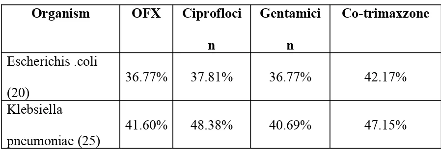
Antimicrobial resistance of ESBL producing isolates to
Non beta lactum antibiotics
Organism OFX Ciprofloci
n
Gentamici
n
Co-trimaxzone
Escherichis .coli
(20)
36.77% 37.81% 36.77% 42.17%
Klebsiella
pneumoniae (25)
41.60% 48.38% 40.69% 47.15%
Antimicrobial resistance of ESBL producing isolates to non beta
lactum antibiotics were also studied and noted that among ESBL
producing isolates (45) there was associated high level co-resistance to
Co-trimaxzone (89.32%), ciprofloxcine (86.19%), oflaxacin (78.37%),
Table VIII
Antimicrbial Susceptibility Pattern of ESBL Positive Isolates
to Non Beta lactam antibiotics
Organism R/All S/G S/Cip S/AK S/OFX S/COTX
Escherichia.coli (20) - 10% 5% 100% 10% 5%
Klebsiella
pneumoniae (25)
-8% 8% 88% 16%
-Antimicrbial Susceptibility Pattern of ESBL Positive Isolates to
Non Beta lactam antibiotics were also studied and found that most of
them were found to be highly susceptible to amikacin, but the level of
susceptibility to co-trimaxazole (5%) quinolones (cipfloxacin 13%,
Table IX
Susceptibility Results for ESBL Positive isolates by
Agar Dilution method
Method MIC in microgram/ml
Agar dilution method
CTAX
CTAX +
CLA
CTAZ CTAZ + CLA
Resistant isolates ESBL
producing (45) 4(8) 8(9) 16(9) 32(7) 64(7) <128(5) 0.25 0.5 >0.25 0.5 >0.5 1 2(9) 16(7) 8(10) 4(7) 32(8) 64(4) 0.125 >0.5 0.5 0.25 0.5 >0.5
A fixed concentration of 4 micro gram of clavulanic acid per ml
was used in conjunction with each antimicrobial agent. Figures in
parentheses = ( ) Number of isolates, CTAX = Cefotaxime,
CTAZ = Ceftazidime, CLA = Clavulanic acid
The susceptibility Results for ESBL Positive isolates by Agar
Dilution method (MIC determination) were studied and analysed. It was
found that there was a decrease of >3 doubling dilutions in MICs of
cefotaxime, ceftazidine with clavulanic acid compared to the MIC of
DISCUSSION
Of one hundred and twenty isolates – 45 were found to be ESBL
producers. Which were screened and confirmed by NCCL methods. This
study, showed a prevalence rate of 37.5% of the total isolates of ESBLS,
among Enterobactericeae in post operative wound infection (world wide
prevalence <1 to 74%). In India the prevalence rate varies from 28 to
84% whereas is the U.S it was 0 to 25% and the national average is 3%
(CDCNNIS) The high percentage of ESBL producing isolates (37.5%)
deducted in this study may be due to selective pressure imposed by
extensive use of antimicrobials in the ICU ward and Post operative
wards. The prevalence rate of ESBL detected in this study was 37.5%.
But the prevalence rates deducted by both Priya Datta in a territory care
hospital in India and Kanungo et al Department of Micorbiology
JIPMER, were 12.6% and 20.5% respectively which were found to be
lower than the value reported in the study (32,18). But the prevalence rate
reported by C. Rodrigues et al was 53% which was found to be higher
than the value detected in this study (28). The prevalence rate of ESBL
producing Klebsiella pneumoniae detected in this study was 48.07%. But
as per the studies of Hansotia et al in 1997, the prevalence of ESBL
producing Klebsiella isolates was detected as 76.5% (24) in central India
studies stated that the prevalence of ESBL producing Klebsiella isolates
was 43.75% (18) which was lower than the value reported in this study.
The results of foreign studies have been studied by Subha et al in 2001.
The incidence of ESBL producing Klebsiella isolates in the United States
has been reported to the 5%. In France and England – 14 to 16% ESBL
producing Klebsiella isolates have been reported. In a report from France,
24.8% ESBL positive Klebsiella pnemoniae were found in patients of
more than 16 year of age. In another report from France – 3.5% of ESBL
producing Klebsiella pnemoniae was obtained in patients with a mean
age of 66 year. This shows that there is a wide disparity between Indian
studies and Foreign studies. In this study a prevalence of ESBL
producing Escherichia coli was identified as 46.50%. But Kanungo et al
(18) and Anandhakrishna of JIPMER (18) in 2000 had reported incidence
of 58.06% which was higher than the value of this study. Further they had
reported 57.14% for ESBL producing Enetrobacter species (18).
Sensitivity and Specificity of ESBL detection test – while referring the
studies of Priya Datta it was found that a comparison of three test DDST,
3 Dimensional Test (3D) and IPT was made for the screening of ESBL
strains (32). In their study IPT was found to be best screening method
when combined with use of a ceftriaxone disk. IPT detected the
maximum number of ESBL producing strains. Ceftriaxone detected the
ceftazidime (32). Courdron et al found the sensitivity of ceftriaxone to be
88% ceftazidime 79% for these ESBL screening methods. Further it was
confirmed by Hoe et al studies which has reported IPT was 100%
sensitive and DDS was 96% sensitive (32). According to C.S. Anders –
ESBL detection was made, in their study, by the comparison of 3
Dimensional Test (3D) and double disk Test. In which the 3D test
provided more sensitivity of 93% of the ESBL producing strains than
Double Disk Test (26). By DDST, 76.5% of Klebsiellaisolates resistant to
3G cephalosporins antibiotics was found to produce ESBLs, which was
studied by Suba et al in 2002 in Central India (13). When analyzing the
sensitivity of DDST it was found that Thomson Sandars has reported
DDS to be 79% sensitive, Vercauteren et al found the sensitivity to be
93% and Shukla et al reported 90.6% sensitive, which were co-related
with this study. Some studies questioned the sensitivity of Double Disk
Test. Several modifications have been recommended including changing
the distance between the Disks. A distance of 20mm center to center has
been recommended by Priya Datta, Archana Thakkur in their study (32).
A distance of 30 mm from center to center has been recommended by U.
Chaudhry, R. Agarwal (11). The MICs of most cephalosporins rose
dramatically when the inoculum of susceptibility test was raised from 105
to 107 CFU/ml. which was found by Medeiros and Crellin. It was
SHV–1 in a strain of K.Pnemoniae that was also lacking an Outer
membrane porin protein caused a false positive in ESBL detection tests
that looked at the differential between the MICs of oxyimino – B lactum
antibiotics with and without clavulanate. The presence of an ESBL can
also be masked by the expression of an Amp C. type enzyme in the same
strain. (Ref: above).
The prevalence of ESBLs in post operative wound infection may
be reduced in hospitals by instituting a hospital infection control cell,
which will advise periodic Antibiotic rotation. (Every 6 months).
Apart from this there was associated resistance to other antibiotics
(Table VIII). The enzymes were disseminated by the spread of either
plasmids or strains, as suggested by epidemiological studies. The location
of the ESBL genes on plasmids may also explain a problem of
co-resistance. Such plasmid responsible for the development of Multi Drug
Resistant Strains asa they may also carry co-resistance genes to
aminoglycosides, quinolones, co-timaxazome. In this study prevalence of
ESBL was associated with a high degree of co-resistance to the above
drugs, hence there is a necessasity for using alternate antibiotics to which
these organisms may be susceptible. Formulation of an appropriate
Because of selective pressure due to heavy use of expanded spectrum
cephalosporins and also due to lapses in effective infection control
measures, there are chances of increase of institutional out breaks. From
one ESBL enzyme to another the optimal substrate profile may varies.
For this reason susceptibility panels with one extended spectrum
cephalosporins cannot predict resistance to other extended spectrum
cephalosporins. NCCLS has recommended use of more than one
substrate for screening (Ref. 32). The use of cefepime allows ESBL
detection in isolates simultaneously producing Amp.c.
The findings detected in this study were co-related with risk factors
like prolonged hospital stay, period of stay in SICU, urinary
catheterization for post operative patients, all of which will predispose to
an out break. There was empirical prescription of Ampicilli, gentamicine
and metronidazole for post operative patients. Ciprofloxacin, ceftriaxone
or ceftazidime or cefotaxine was added later on which will lead to
prolonged hospital stay. The carbapenam antibiotics were not used at all.
Further these ESBL strains will acquire co-resistance genes on plasmids.
To prevent the acquisition of these infections, precautionary
measures should be taken – control of their spread by having an effective
specificity of ESBL deduction test. Even though there are merits and
shortcomings of each of the detection test Double disk approximation test
of Jarlier et al and the Broth Dilution MIC reduction method (NCCLS
confirmatory Test) are the easiest and cost effective methods for use in
clinical laboratories. However none of the detection test are 100%
sensitive and specific (30).
The techniques required for the identification of the exact
ESBL sub type – DNA Probing – Polymerase chain reaction, RFLP,
Isoelectric focusing – are available only at research centers. Perhaps with
the advent of gene chip technology in the near future – the sub type
CONCLUSION
The prevalence of ESBL producing gram negative bacilli in
operative wound infection identified in this study was 37.50% which was
with in the range of 28 to 84% reported in India
ESBL production was found to be coexsisted with resistance to
several other antibiotics. ESBLs are encoded by plasmids which also
carry co- resistant genes for other antibiotics. We found such associated
co-resistance with cotrimaxazole, 44.66% gentamicin, 38.73%
ciprofloxacin, 41.09% oflaxacin 39.18%. Since co-resistance to non β
lactum antibiotics, cotrimaxazole, gentamicin and quinolones was
observed, combination of Imipenem+amikacin were found to be
alternatives for the treatment of life threatening infections, like
septicemia, hospital acquired pneumonia, intravisceral abscess due to
faster killing rates of Amikacin. Our study highlights the emergence of
ESBL producing Klebsiella pneumoniae (48.01%) isolates endowed with
extremely wide spectrum of antibiotic resistance including resistance to
cotrimaxazole, gentamicin and quinolones suggest that ESBL producing
Klebsiella pneumoniae isolates have emerged as one of the major