Copyright © 2000, American Society for Microbiology. All Rights Reserved.
NOTES
Role of Simian Virus 40 Vp1 Cysteines in Virion Infectivity
PEGGY P. LI,1AKIRA NAKANISHI,1MARY A. TRAN,1ADLER M. SALAZAR,1ROBERT C. LIDDINGTON,2ANDHARUMI KASAMATSU1*
Department of Molecular, Cell and Developmental Biology and Molecular Biology Institute, University of California,
Los Angeles, California 90095,1and the Burnham Institute, La Jolla, California 920372
Received 11 May 2000/Accepted 7 July 2000
We have developed a new nonoverlapping infectious viral genome (NO-SV40) in order to facilitate structure-based analysis of the simian virus 40 (SV40) life cycle. We first tested the role of cysteine residues in the for-mation of infectious virions by individually mutating the seven cysteines in the major capsid protein, Vp1. All seven cysteine mutants—C9A, C49A, C87A, C104A, C207S, C254A, and C267L—retained viability. In the crystal structure of SV40, disulfide bridges are formed between certain Cys104 residues on neighboring pen-tamers. However, our results show that none of these disulfide bonds are required for virion infectivity in culture. We also introduced five different mutations into Cys254, the most strictly conserved cysteine across the polyomavirus family. We found that C254L, C254S, C254G, C254Q, and C254R mutants all showed greatly reduced (around 100,000-fold) plaque-forming ability. These mutants had no apparent defect in viral DNA rep-lication. Mutant Vp1’s, as well as wild-type Vp2/3, were mostly localized in the nucleus. Further analysis of the C254L mutant revealed that the mutant Vp1 was able to form pentamers in vitro. DNase I-resistant virion-like particles were present in NO-SV40-C254L-transfected cell lysate, but at about 1/18 the amount in wild-type-transfected lysate. An examination of the three-dimensional structure reveals that Cys254 is buried near the surface of Vp1, so that it cannot form disulfide bonds, and is not involved in intrapentamer interactions, consistent with the normal pentamer formation by the C254L mutant. It is, however, located at a critical
junc-tion between three pentamers, on a conserved loop (G2H) that packs against the dual interpentamer Ca2ⴙ
-binding sites and the invading C-terminal helix of an adjacent pentamer. The substitution by the larger side
chains is predicted to cause a localized shift in the G2H loop, which may disrupt Ca2ⴙion coordination and
the packing of the invading helix, consistent with the defect in virion assembly. Our experimental system thus allows dissection of structure-function relationships during the distinct steps of the SV40 life cycle.
Proper virion assembly is important for the spread of a DNA tumor virus such as simian virus 40 (SV40), a member of the papovavirus family. The icosahedral capsid of SV40, which houses the viral minichromosome along with the minor struc-tural proteins Vp2 and Vp3, is made from 72 pentamers of the major structural protein Vp1. These Vp1 pentamers form the building blocks which interact via the five long carboxy-termi-nal arms extending from each pentamer into neighboring pen-tamers (13). The crystal structure of SV40 shows bound Gd3⫹
(which acts as a Ca2⫹mimetic) at two neighboring sites per
Vp1 subunit, forming bridges between the pentamer core and the invading C-terminal arms of another pentamer (13, 19). Structural refinement (19) indicates the presence of inter-pentamer, but not intrapentamer or intramonomer, disulfide linkages among some of the Vp1 cysteine (Cys104) residues. For mouse polyomavirus, intrapentamer disulfide bridges have been observed between Vp1 cysteines 19 and 114 (18). In ad-dition, evidence indicates that calcium ion chelation and disul-fide linkage may further stabilize the interaction between Vp1 pentamers on the capsid. Disruption of both SV40 and mouse polyomavirus capsids requires the reducing agent dithiothrei-tol (DTT) (3, 20) and can be accomplished with the
combina-tion of DTT and the calcium chelator EGTA (2). Bacterially produced polyomavirus Vp1 can be induced to self-assemble in vitro into a capsid-like structure by Ca2⫹addition at
physio-logic salt concentration (15). Recently reported in vitro studies have hinted that the ways in which Vp1 cysteines contribute to assembly and/or capsid disassembly may be different for SV40 and polyomavirus. Cysteine-free mutant SV40 Vp1 synthesized in vitro forms pentamers but not postpentameric complexes, and the conversion into these complexes has been suggested to involve Vp1 disulfide bonding (10). In contrast, bacterially made cysteine-free mouse polyomavirus Vp1 forms virus-like parti-cles, though at 50% of the wild-type level (17). The greater stability of the wild-type capsids is apparently conferred by the intrapentamer C19–C114 disulfides (17). These results collec-tively point to calcium ions and disulfide bonds as integral parts of the virion structure (13, 19). However, it is not known whether the observed Vp1 disulfide formation, such as that through cysteines 104 in SV40, is essential for infectious virion formation. We wished to determine which, if any, of the seven SV40 Vp1 cysteines are essential for infectivity, and to define the roles of the essential residues, using a mutagenesis ap-proach. We report here that neither the C104 disulfide nor any other single disulfide bridge is required for virion infectivity. We also provide evidence for the importance of Cys254, a cysteine that is conserved across the polyomavirus family (see references within reference 14), for virion infectivity.
First, seven single-cysteine Vp1 mutants were constructed. Except for the previously created C9A mutation in pSV-Vp1
* Corresponding author. Mailing address: Molecular Biology Insti-tute, 456 Boyer Hall, University of California at Los Angeles, 611 East Charles E. Young Dr., Box 951570, Los Angeles, CA 90095-1570. Phone: (310) 825-3048. Fax: (310) 206-7286. E-mail: harumi_K@mbi .ucla.edu.
11388
on November 9, 2019 by guest
http://jvi.asm.org/
(9), the systematic mutation of the remaining cysteines began with the construction of plasmid pBS-Vp1 (Fig. 1), which not only serves as an intermediate for cloning the Vp1 mutant viral genomes but also allows for the in vitro synthesis of Vp1. Within the Vp1 coding sequence of pBS-Vp1, several silent base pair substitutions have been made to introduce three additional unique restriction sites—BstBI at SV40 nucleotide 2143,SpeI at nucleotide 2324, andMluI at nucleotide 2449— which facilitated the creation of individual cysteine mutations and of combination mutations for future studies. Individual mutations at cysteines 49, 87, 104, 207, 254, or 267 were intro-duced into pBS-Vp1 by oligonucleotide-directed mutagenesis via PCR or by ligation of short mutant coding segments (Fig. 1); then suitable fragments of the resulting plasmids were transferred to the nonoverlapping, infectious viral genome (NO-SV40) (8). In this manner, a series of single-cysteine NO-SV40 mutants were created: the alanine-substituted C9A, C49A, C87A, C104A, and C254A mutants; the serine-substi-tuted C207S and C254S mutants; the leucine-substiserine-substi-tuted C254L and C267L mutants; the glycine-substituted C254G mutant; the glutamine-substituted C254Q mutant; and the arginine-substituted C254R mutant.
All subcloning techniques were performed as described pre-viously (16). Oligonucleotides for PCR, for linkers, and for sequencing were synthesized by Genosys (The Woodlands, Texas) or by the Oligonucleotide Preparation Laboratory of the University of California—Los Angeles (UCLA) Molecular Biology Institute. All mutations were confirmed by double-stranded DNA sequencing using the ABI PRISM Dye Termi-nator Cycle Sequencing Ready Reaction Kit (Perkin-Elmer). All DNA sequences below are given in uppercase letters except for mutated SV40 Vp1 nucleotides, which are lowercased. Rel-evant restriction sites are underlined.
To create pBS-Vp1, pBluescript II KS(⫹) (Stratagene, La Jolla, Calif.) was first inserted with a linker between theSacI and ApaI sites to introduce unique XbaI, PstI, BstBI, SpeI,
MluI, SacI, and XhoI sites and to knock out the plasmid’s original SacI and ApaI sites via this insertion. The resulting pBS plasmid was sequentially inserted with five Vp1-encoding
fragments: the 495-bpXbaI-to-PstI (Vp1 amino acids 1 to 162) fragment from pSV-Vp1 (8); the 155-bp PstI-to-BstBI (Vp1 amino acids 162 to 214) PCR fragment with template pSV-Vp1 and the 5⬘-TTTGGAGCTGCAGGGTGTG sense and
5⬘-GTTTTCATTTTTcgaaGGATCAGGAACCCAGCACTC
CACTGGATAAGC antisense primers; the 181-bpBst
BI-to-SpeI (Vp1 amino acids 214 to 274) PCR fragment using the
5⬘-GTTCCTGATCCttcgAAAAATGAAAACACTAGATAT
TTTGGAACCTACACAGGTGG sense and 5⬘-CTGTAAAC
ACCCGACAAATGGTTGTGAtcACCTTGTGTC antisense primers; the 125-bpSpeI-to-MluI (Vp1 amino acids 274 to 316) PCR fragment using the 5⬘-TTACCAACACTagTGGAACAC
AGCAGTGGAAGGGACTT sense and 5⬘-GACCAATACgC
gtTGTGTCCTCCTGTTAATTAGGTCAC antisense prim-ers; and the 172-bpMluI-to-XhoI (Vp1 amino acids 316 to 362)
PCR fragment using the 5⬘-GAGGACACAacGcGTGGATG
GGCAGCCTATGATTGGA sense and 5⬘-ATTCCAAACTC
GAGGCGCGCTGAGCTCTAAGCACCGCGGCCGCTCT
GCATTCTAGTTGTGGTTTGTCC-3⬘antisense primers.
Single-cysteine mutant pBS-Vp1’s were then derived by placing Vp1-coding fragments of wild-type pBS-Vp1 with re-spective mutant fragments as follows. pBS-Vp1-C49A was made by substituting the 630-bpAccI-to-ApaI PCR fragment derived using template pSV-Vp1 and the 5⬘-GGAGTAGACA GCTTCACTGAGGTGGAGgcCTTTTTAAATCCTCAA
ATG sense and 5⬘-CAAGGGCCCAACACCCTGCTC
anti-sense primers. pBS-Vp1-C87A was made by substituting the 289-bpXbaI-to-EcoRI PCR fragment derived with the 5⬘-TG
GTCTAGATGAAGATGGCCC sense and 5⬘-AAGGAATTC
TAGCCACACTGTAGgcAGGCAGTTGTTCTTTGTCTGG antisense primers. pBS-Vp1-C104A was made by substituting the 849-bp EcoRI-to-EcoRI PCR fragment derived with the
5⬘-CTAGAATTCCTTTGCCTAATTTAAATGAGGACTTA
ACCgcTGGAAATATTTTGATGTGGG sense and 5⬘-CTTC
AAGAATTCGAGCTCGCC antisense primers. pBS-Vp1-C207S was constructed by substituting the 155-bpPstI-to-BstBI PCR fragment derived with the 5⬘-TTTGGAGCTGCAGGG
TGTGTTAG sense and 5⬘-TTCATTTTTCGAAGGATCAG
GAACCCAagACTCCACTGGATAAGC antisense primers. pBS-Vp1-C254A, -C254S, -C254L, -C254G, -C254Q, and -C254R were made by substituting the 66-bp ApaI-to-SpeI linker in which the Cys254 codon was converted into GCT, TCT, CTT, GGC, CAA, and CGC, respectively. pBS-Vp1-C267L was made via a similar linker in which the Cys267 codon was converted into CTT.
pBS-Vp1-⌬C58 and its C207S and C254L mutants were cre-ated by replacing theSpeI-to-XhoI region of pBS-Vp1 and its C207S and C254L mutants with a linker that begins with the same sequence as pBS-Vp1 from theSpeI site to the 304th Vp1 codon (TTT) and ends with a stop codon and theXhoI site.
To create viral genomes containing single-cysteine Vp1 mu-tations, the 1,179-bpXbaI-to-SacI region of the wild-type NO-pSV40 (8) was replaced with the corresponding fragment from pSV-Vp1-C10A (containing the C9A mutation) (9) to yield NO-pSV40-C9A or was replaced with the corresponding frag-ment from pBS-Vp1-C49A, -C87A, -C104A, -C207S, -C254A, -C254S, -C254L, -C254G, -C254Q, -C254R, or C267L to pro-duce the respective NO-pSV40 mutant. Mutant NO-SV40 viral genomes were prepared from their respective NO-pSV40 plas-mids by digestion with BamHI and recircularizing with T4 DNA ligase as described elsewhere (8).
Individual cysteine mutations and viability. Each mutant
[image:2.612.88.260.73.206.2]NO-SV40 was tested for viability by one or both of two types of plaque-forming assays. In the first assay, cells were microin-jected with each NO-SV40 DNA by a previously described method (21). In the second assay, cells were infected with serial FIG. 1. Construction of Vp1 cysteine mutant plasmid DNAs. Individual Vp1
cysteine residues except cysteine 9 were mutagenized within the plasmid pBS-Vp1, which encodes wild-type Vp1 amino acid sequence and whose Vp1-encod-ing and immediately flankVp1-encod-ing regions are shown as a bold horizontal line. Loca-tions of unique restriction sitesXbaI (Xb),AccI (Ac),AflII (Af),EcoRI (E),PstI (P),ApaI (Ap),BamHI (Ba),SacI (Sa), andXhoI (Xh) are indicated by vertical lines. Three other unique sites indicated by diamonds and stars—BstBI (Bs),SpeI (Sp), andMluI (M)—are not present in the natural Vp1 coding sequence [Vp1 (natural)] but were introduced into pBS-Vp1 (Vp1-BSM) at the SV40 nucleotide positions noted above the respective sites via silent base pair substitutions. Dots indicate the locations of the seven cysteines, with their respective amino acid numbers noted underneath. Truncated pBS-Vp1 derivatives with the carboxy-terminal 58 amino acids deleted, the pBS-Vp1-⌬C58 (Vp1-⌬C58) series, were also created.
on November 9, 2019 by guest
http://jvi.asm.org/
dilutions of the NO-SV40 DNA-transfected cell lysate, which was prepared by transfecting CV-1 cells on a 60-mm dish with 1g of NO-SV40 DNA using the Effectene transfection kit (Qiagen), harvesting the cells at 72 h posttransfection in 800l of serum-free culture medium, and freeze-thawing the cell suspension three times to release potential virions or virion-like particles. For either assay, infected cells or microinjected cells were incubated for 21 days under an agar medium and the plaques formed were visualized as before (21). The viability results are summarized in Table 1. The microinjection type assays gave a plaquing or nonplaquing phenotype, and the ensuing infection type assays permitted measurement of the number and sizes of the plaques. NO-SV40-BSM, which con-tains the three additional restriction sites of pBS-Vp1 but no amino acid mutations, formed plaques at nearly the wild-type NO-SV40 level (1.8⫻108PFU/ml), or 1.6⫻108PFU per ml
of transfected cell lysate. Seven of the NO-SV40 single-cys-teine mutants, the C9A, C49A, C87A, C104A, C207S, C254A, and C267L mutants, had PFUs that were either similar to or no less than one-fifth that of the wild type. The average plaque diameters of the mutants ranged from 5.2 to 1.6 mm, which are equivalent to, or somewhat smaller than, that of the wild type, 5.0 mm. In contrast, five different C254 substitution mutants, the C254S, C254L, C254G, C254Q, and C254R mutants, showed dramatically reduced viability, either producing no plaques at all (C254R) or producing 20,000- to 400,000-fold fewer plaques than the wild type. Mutant C254L plaques were also exceptionally small (about 0.5 mm). Nonetheless, these results indicate that individual Vp1 cysteines are not required for SV40 infectivity. In particular, the highly viable C104A mutant suggests that the crystallographically observed inter-pentamer disulfide linkages among some of the Cys104 resi-dues on the CD loops (19) are not essential for infectious virion formation, at least in culture. They presumably formed after particle release in an extracellular oxidizing environment.
Capsid protein subcellular localization and DNA
replica-tion of mutants.The mutant viral genomes were tested for the
structural proteins’ subcellular localization as described previ-ously (4). Vp1-BSM localized to the nucleus as well as wild-type Vp1. The Vp1’s of all Vp1 single-cysteine mutants more or less localized to the nucleus, indicating conservation of the nuclear targeting function of each mutant Vp1. We noted that
though nuclearly localized, C254R mutant Vp1 frequently ac-cumulated in several large aggregates in the nucleus (data not shown). The amount and stability of the mutant proteins syn-thesized are not addressed at this time. As expected from our previously reported result (8), the wild-type Vp2/3 encoded in all the NO-SV40 constructs was similarly found in the nucleus (data not shown).
The mutants were also tested for viral DNA replication. CV-1 cells on a 150-mm dish were transfected with 4g of wild-type or cysteine mutant NO-SV40 DNA (prepared by restriction enzyme digestion and ligation of bacterially propa-gated NO-pSV40 [8]) and harvested at 72 h posttransfection in TD buffer (25 mM Tris-Cl [pH 7.4], 140 mM NaCl, 5 mM KCl, 1 mM NaPi). The cells were collected by centrifugation and
resuspended and sonicated in 0.5 ml of hypotonic buffer (25 mM Tris-Cl [pH 7.6], 1 mM MgCl2, 0.4 mM CaCl2, 0.5 mM
DTT). It is known that transfected wild-type viral DNA has replicated to a readily detectable amount by this time point and that some of the progeny DNA is already packaged into virion particles. To quantitate the replicated DNA, total DNA in 12.5l of the cell lysate was purified by digestion with 0.75 g of proteinase K/l in 25 mM Tris-Cl (pH 7.6)–10 mM EDTA at 50°C for 3 h, extraction with phenol-chloroform, and precipitation with ethanol. The resulting DNA was then di-gested with eitherKpnI alone or the combination ofKpnI and
DpnI and was detected by Southern blotting with a32P-labeled
SV40 DNA probe (Fig. 2). For both the C207S and C254L mutants as well as the wild type, a vast majority of the total viral DNA was resistant toDpnI digestion (Fig. 2) and hence represented host cell-replicated viral DNAs rather than bacte-rially propagated input DNAs. For all other single-cysteine mutants, intracellular episomal DNA was extracted by the Hirt method (7) from 72-h-transfected cells, similarly analyzed with
[image:3.612.52.293.83.239.2]DpnI, and detected by Southern blotting or ethidium bromide staining following agarose gel separation. We found similar extents of replication (data not shown). Thus, there is no ap-parent defect in viral genome replication, and no substantial alteration in the mutant proteins’ nuclear localization, for all of
TABLE 1. Viability of Vp1 single-cysteine mutants
NO-SV40
mutant Formation ofplaquesa cell lysate (PFU/ml)Titer in transfectedb Plaque diam(mm)b
Wild type Yes 1.8⫻108 5.0⫾1.2
BSM Yes 1.6⫻108 4.0⫾1.0
C9A Yes 1.2⫻108 3.3⫾1.5
C49A Yes 1.4⫻108 4.1⫾1.3
C87A Yes 3.5⫻107 3.2⫾1.2
C104A Yes 8.0⫻107 5.2⫾0.3
C207S Yes 3.9⫻107 1.6⫾0.5
C254A ND 2.2⫻108 4.0⫾0.9
C254S ND 1.6⫻103 2.4⫾0.2
C254L No 4.0⫻103 0.5c
C254G ND 8.0⫻103 2.6⫾0.8
C254Q ND 4.4⫻102 2.0⫾0.6
C254R ND 0d
C267L Yes 1.5⫻108 2.7⫾0.6
aIn microinjection assay. ND, not done.
bDetermined from an infection type plaque assay. Cells were infected with
serially diluted, freeze-thawed cell lysates obtained from transfection with re-spective viral DNAs, as described in the text.
cTiny, barely visible plaques.
dNo plaques detected in 0.1 ml of transfected cell lysate.
FIG. 2. DNA replication and packaging by C207S and C254L mutants. To analyze viral DNA replication, total DNA was prepared from the sonicated lysate of cells transfected with wild-type or mutant (C207S or C254L) NO-SV40 as described in the text, and identical aliquots thereof were digested withKpnI alone or with bothKpnI andDpnI, followed by separation on a 0.9% agarose gel, Southern transfer, and hybridization with a nick-translated 32P-labeled SV40
DNA probe (0.1g, or 107cpm, in 10 ml). The radioactivity of the linearized
5.2-kbp viral DNA band for theKpnI-DpnI double reaction, quantitated by a phosphorimager, was expressed as a percentage of the radioactivity of the same band for theKpnI-only reaction. To analyze viral DNA packaging, identical aliquots of each transfected cell lysate were either treated or not treated with DNase I as described in the text, and the remaining DNA in the lysate was pu-rified and digested withKpnI and analyzed by Southern blotting as above. The radioactivity of the 5.2-kbp DNase I-resistant viral DNA band was expressed as a percentage of the radioactivity of the same band for the non-DNase I-treated sample.
on November 9, 2019 by guest
http://jvi.asm.org/
[image:3.612.347.515.483.596.2]the single-cysteine mutants. The observation that five of the Cys254 mutants have greatly reduced viability is intriguing. The following experiments were thus carried out with the C254L mutant to examine the stages of the life cycle that are affected by the cysteine mutations.
C254L mutant Vp1 forms pentamers in vitro.We looked at
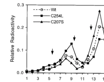
the ability of C254L mutant Vp1 to form pentamers. The carboxy-terminal-truncated wild-type, C207S mutant, and C254L mutant Vp1 proteins were in vitro synthesized from the T7 promoter-based pBS-Vp1-⌬C58 plasmid series (Fig. 1), and the oligomerization states of the resulting [35
S]methionine-labeled Vp1’s were analyzed by velocity sedimentation in su-crose gradients (Fig. 3).
Four micrograms of pBS-Vp1-⌬C58, pBS-Vp1-C207S-⌬C58, or pBS-Vp1-C254L-⌬C58 DNA was transcribed and translated in a 125-l reaction mixture containing 50Ci of [35S]methionine (⬎1,000 Ci/mmol; Amersham) and 100l of
TNT Coupled Reticulocyte Lysate Quick Master Mix (Pro-mega) at 30°C for 90 min. The resulting reaction was treated with 78 Worthington units of RNase A and 24 Kunitz units of DNase I in the presence of protease inhibitors (1,000 U of aprotinin/ml, 1g of pepstatin A/ml, 1g of leupeptin/ml, 10 nM phenylmethylsulfonyl fluoride) at 37°C for 1 h, diluted twofold with distilled water, layered onto a 10.5-ml, 5 to 20% continuous sucrose gradient in 50 mM HEPES (pH 7.5)–140 mM NaCl, and centrifuged at 35,000 rpm for 23 h at 4°C in an SW41 rotor. Seventeen fractions were collected from the bot-tom of the gradient, and each entire fraction was reacted over-night at 4°C with 10l of a 50% (vol/vol) slurry of protein A-Sepharose (Pharmacia) that had been freshly complexed with the immunoglobulin G (IgG) fraction of rabbit anti-Vp1 serum. The Vp1 immunoprecipitates were collected by centrif-ugation, washed twice with 10 mM Tris-Cl (pH 8.0)–140 mM NaCl–0.1% Triton X-100–0.25% gelatin–protease inhibitors and twice more with the same buffer without the Triton and gelatin, and then analyzed by sodium dodecyl sulfate–10% polyacrylamide gel electrophoresis (SDS–10% PAGE) (12) and fluorography using the Amplify reagent (Amersham).
The 58-amino-acid deletion at the carboxy-terminal end of SV40 Vp1 is analogous to the 57-amino-acid deletion at the carboxy-terminal end of polyomavirus Vp1, which preserves the protein’s ability to form pentamers but abolishes its inter-pentameric interaction (6). If the C254L mutation affected pentamer formation, we would expect to observe a reduced presence of Vp1 in the expected pentamer peak. The results in Fig. 3 showed no obvious effect of C254L or C207S mutations on Vp1 pentamer formation. About 30, 40, and 20% of the Vp1 radiolabel was found in the 7S-to-8S pentamer region for Vp1-⌬C58, Vp1-C254L-⌬C58, and Vp1-C207S-⌬C58, respec-tively, with the remaining Vp1 label being in the monomeric form. Thus, the ability of C254L mutant Vp1 to form penta-mers was not compromised.
The C254L mutant forms virion-like particles but at a
re-duced amount.Since the C254L mutant replicated its DNA,
and its mutant Vp1 could form pentamers and localize to the nucleus along with Vp2/3, this mutant may be able to form virion particles. So we next examined the amount of virion or virion-like particles formed by the C254L mutant. Sonicated, transfected cell lysates, prepared as described above for the replication analysis, were digested with 500 U of DNase I/ml in 20 mM Tris-Cl (pH 7.6)–2 mM MgCl2to degrade unpackaged
DNA. Under such conditions, protein-free SV40 DNA, but not DNA in intact virions, was effectively digested and not de-tected by Southern blotting (data not shown). It must be noted, however, that the nuclease could penetrate the particles and cleave the viral chromosome if the integrity of the particles was
[image:4.612.342.518.72.211.2] [image:4.612.339.519.514.603.2]somehow compromised by mutations. After the nuclease di-gestion, the remaining DNA was purified as described earlier and linearized withKpnI before being analyzed by Southern blotting. As shown in Fig. 2, about 70 and 50% of intracellular wild-type and C207S mutant viral DNAs, respectively, were resistant to DNase I, whereas only a small proportion, about 4%, of the C254L mutant viral DNA was protected from DNase I digestion. Either the mutant particles had a structure different from that of wild-type particles or the mutant formed much fewer particles than the wild type, or both. To confirm that the C254L mutant did package a fraction of the viral DNA into virion particles, an aliquot of wild-type- or C254L mutant-transfected cell lysate that was DNase I treated and contained the same amount of remaining viral DNAs was analyzed by velocity sedimentation in sucrose gradients, followed by South-ern blotting. Although the viral DNA peak was found in frac-tion 4 of both wild-type and C254L mutant samples, as ex-pected for the sedimentation of mature virions (Fig. 4), the FIG. 3. C207S and C254L mutant Vp1’s form pentamers in vitro. In vitro-transcribed and -translated wild-type (Wt) or mutant (C207S or C254L) Vp1-⌬C58 proteins were analyzed by sucrose gradient sedimentation as described in the text.For each fraction, the radioactivity of the Vp1 band in the gel lane was quantitated using a phosphorimager. Each quantitated value was expressed as a decimal fraction of the summed Vp1 radioactivity values for all 17 fractions (taken to be 1.0), and was plotted against the fraction number. Arrows indicate the peak positions for three sedimentation markers, from the bottom to the top of the gradient: catalase B (11.3S), IgG (7S), and bovine serum albumin (4.5S).
FIG. 4. The C254L mutant forms virion-like particles. Sonicated lysate alignots of wild-type (Wt) or C254L mutant NO-SV40-transfected cells, esti-mated to contain similar amounts of DNase I-resistant viral DNA (200l for the wild type and 1 ml for the C254L mutant), were centrifuged at 350⫻gfor 5 min at 4°C to pellet cellular debris. The supernatant was treated with DNase I as described in the text and was sedimented through a 5 to 32% sucrose gradient in 50 mM HEPES (pH 7.5) at 37,000 rpm at 4°C for 80 min in an SW41 rotor. Eighteen fractions were collected from the bottom of the gradient, and the DNA in each fraction was extracted following proteinase K treatment. One-half of each DNA sample was separated on a 0.9% agarose gel, Southern transferred, and hybridized with a nick-translated32P-labeled SV40 DNA probe. To the left of each panel, upper and lower lines indicate the positions of 7,554- and 5,243-bp marker DNA fragments, respectively, and an arrow points to the covalently closed circular DNA band. An arrowhead above fraction 4 indicates the location of control wild-type virions sedimented in a parallel gradient.
on November 9, 2019 by guest
http://jvi.asm.org/
C254L mutant distribution curve trailed on the righthand (lighter) side of the peak. It is possible that a small portion of mutant particles had become structurally altered during the centrifugation. We noted in the gradients three viral DNA species: a singly nicked DNA appearing near the 7.3-kbp marker, broad bands of linear DNA centering at the expected 5.2-kbp position, and a covalently closed circular DNA. The last species was present in a reduced amount in the mutant lysate relative to that in the wild-type lysate. These particle-derived DNA species were not investigated further at this time, but they may have arisen from the heterogeneous populations of particles that were in various stages of particle maturation in the nucleus (1, 5, 11). Some of these particles may be accessible to the nuclease, leading to the DNA band patterns observed. Also, the variation could in part reflect artifacts of sonication or fractionation that helped release immature, cell-associated particles. Nonetheless, these results indicate that NO-SV40-C254L is capable of forming virion-like particles in the trans-fected cells, though at an approximately 18-fold reduced level compared to wild-type NO-SV40. Since the reduction in the physical particles does not fully account for the 45,000-fold reduction in plaque number for this mutant (Table 1), we conclude that the low viability of the C254L mutant is due to the sum of the reduced production of mutant virion particles in the nucleus and the apparently poor infectivity of the mutant particles that are produced.
Structural environment of C254.In this report, we examined
effects on the viral life cycle of mutating each of the seven
SV40 Vp1 cysteines and showed that (i) each cysteine could be mutated without significant loss of viability and (ii) certain mutations of Cys254 resulted in a dramatic reduction in virus viability. Cys254, along with the Vp1 G2H loop on which it lies, is highly conserved across the polyomavirus family. Inspection of the three-dimensional crystal structure suggests that most mutations cause subtle effects that interfere with the ability to form capsids. As illustrated in Fig. 5, Cys254 lies on a short loop connecting strands G2 and H, and it is not involved in Vp1–Vp1 interactions that stabilize the pentamer. Its side chain points into the hydrophobic core of Vp1, so that it cannot participate in disulfide bonding. Substitution by the longer side chains, leucine, glutamine, and arginine, would be expected to create a local perturbation of the structure of this loop to accommodate the longer side chain (e.g., the loop might move as a rigid body by 1 to 2 Å away from the core), while substi-tution by the smaller alanine would not. We cannot readily explain why the C254S and C254G mutants lost viability. The G2H loop lies at a critical junction between three pentamers within the capsid, such that subtle alterations in structure might be expected to affect capsid assembly significantly. On one side of the G2H loop are the twin Ca2⫹-binding sites (Fig.
5) (13, 19), in which the Ca2⫹ ions are coordinated each by
three to four acidic residues from different pentamers that are brought into close apposition as a result of capsid assembly. Residues from the G2H loop form direct hydrogen bonds and salt bridges to the Ca2⫹-coordinating residues, including a salt
[image:5.612.55.547.75.360.2]bridge between the side chains of Lys255 and Glu157 and the FIG. 5. Structural environment of the Vp1 cysteines. (Left) Ribbon structure of an SV40 Vp1 pentamer, with one of the Vp1 monomers shown in white. An invading arm from a neighboring pentamer is pinkish red. The locations of six Vp1 cysteines mutated in this study are shown by yellow or red (Cys254) circles. Cys9 lies within an amino-terminal arm that is disordered in the crystal structure, and is not shown. Two blue circles represent the twin Ca2⫹sites. (right) An enlarged view of the Ca2⫹-binding sites shows the location of C254 in the G2H loop and its proximity to the Ca2⫹ions, which are coordinated by glutamate and aspartate side chains from three Vp1 monomers on two different pentamers. Glu48 (E48, marked with a star) is from a neighboring monomer in the same pentamer. Glu330 and Asp345 (E330 and D345, marked with double stars) are from a second pentamer, and the helix␣C** is from a third pentamer. Thus, Cys254 lies close to the junction of three pentamers, and mutation to the longer leucine residue is expected to disrupt these interactions and hence capsid assembly. The image on the left is adapted from
Structure(19) with permission of the publisher.
on November 9, 2019 by guest
http://jvi.asm.org/
interaction of the main-chain C¢O of Leu253 with the side chain of Asn217; thus, subtle changes in the structure or po-sition of the G2H loop might well disrupt Ca2⫹binding. The
G2H loop also makes close contact with a third pentamer, making hydrophobic packing contacts with an invading C-helix (the “␣C**” depicted in Fig. 5). Additionally, a shift of the G2H loop could alter the angle of C-helix packing. The C-helix interaction is likely to be important in defining the curvature of the assembling capsid, and alterations in the angle of packing could lead to some of the aberrant assembly products, such as T⫽1 particles and tubes, observed in in vitro assembly exper-iments (15).
In conclusion, we have developed an experimental system that allows dissection of structure-function relationships dur-ing distinct steps of the SV40 life cycle. For all polyomavirus family Vp1’s, amino acids within the G2H loop region are well conserved and may hold a key to capsid formation in the nucleus. By assessing the overall viability and the cell entry capability of alternative Cys254 mutants, as well as of mutants in which the metal binding residues are altered, we may shed light on the importance of the G2H loop in the virus life cycle.
This work was supported by Public Health Service grant CA50574 from the National Institutes of Health (NIH) and by a grant from the UCLA Academic Senate. A.M.S. was supported in part by an under-graduate fellowship from the NIH Minority Scientist Development program (GM55052).
REFERENCES
1.Baumgartner, I., C. Kuhn, and E. Fanning.1979. Identification and charac-terization of fast-sedimenting SV40 nucleoprotein complexes. Virology96:
54–63.
2.Brady, J. N., V. D. Winston, and R. A. Consigli.1977. Dissociation of polyoma virus by the chelation of calcium ions found associated with purified virions. J. Virol.23:717–724.
3.Christiansen, G. T., T. Landers, J. D. Griffith, and P. Berg.1977. Charac-terization of components released by alkali disruption of simian virus 40. J. Virol.21:1079–1084.
4.Clever, J., and H. Kasamatsu.1991. Simian virus 40 Vp2/3 small structural proteins harbor their own nuclear transport signal. Virology181:78–90.
5.Coca-Prados, M., and M.-T. Hsu.1979. Intracellular forms of simian virus 40 nucleoprotein complexes. II. Biochemical and electron microscopic analysis of simian virus 40 virion assembly. J. Virol.31:199–208.
6.Garcea, R. L., D. M. Salunke, and D. L. D. Caspar.1987. Site-directed mutation affecting polyomavirus capsid self-assembly in vitro. Nature (Lon-don)329:86–87.
7.Hirt, B.1967. Selective extraction of polyoma DNA from infected mouse cell cultures. J. Mol. Biol.26:365–369.
8.Ishii, N., A. Nakanishi, M. Yamada, M. H. Macalalad, and H. Kasamatsu.
1994. Functional complementation of nuclear targeting-defective mutants of simian virus 40 structural proteins. J. Virol.68:8209–8216.
9.Ishii, N., N. Minami, E. Y. Chen, A. L. Medina, M. M. Chico, and H. Kasamatsu.1996. Analysis of a nuclear localization signal of simian virus 40 major capsid protein Vp1. J. Virol.70:1317–1322.
10. Jao, C. C., M. K. Weidman, A. R. Perez, and E. Gharakhanian.1999. Cys9, Cys104, and Cys207 of simian virus 40 Vp1 are essential for interpentamer disulfide linkage and stabilization in cell-free lysates. J. Gen. Virol.80:2481– 2489.
11. La Bella, F., and C. Vesco.1980. Late modifications of simian virus 40 chromatin during the lytic cycle occur in an immature form of virion. J. Virol.
33:1138–1150.
12. Laemmli, U. K.1970. Cleavage of structural proteins during the assembly of the head of bacteriophage T4. Nature (London)227:680–685.
13. Liddington, R. C., Y. Yan, J. Moulai, R. Sahli, T. L. Benjamin, and S. C. Harrison.1991. Structure of simian virus 40 at 3.8-Å resolution. Nature (London)354:278–284.
14. Pipas, J. M.1992. Common and unique features of T antigens encoded by the polyomavirus group. J. Virol.66:3979–3985.
15. Salunke, D. M., L. D. Caspar, and R. L. Garcea.1986. Self-assembly of purified polyomavirus capsid protein Vp1. Cell46:895–904.
16. Sambrook, J., E. F. Fritsch, and T. Maniatis.1989. Molecular cloning: a laboratory manual, 2nd ed. Cold Spring Harbor Laboratory, Cold Spring Harbor, N.Y.
17. Schmidt, U., R. Rainer, and G. Bo¨hm.2000. Mechanism of assembly of recombinant murine polyomavirus-like particles. J. Virol.74:1658–1662. 18. Stehle, T., and S. C. Harrison.1996. Crystal structures of murine
polyoma-virus in complex with straight-chain and branched-chain sialyloligosaccha-ride receptor fragments. Structure4:183–194.
19. Stehle, T., S. J. Gamblin, Y. Yan, and S. C. Harrison.1995. The structure of simian virus 40 refined at 3.1 Å resolution. Structure4:165–182. 20. Walter, G., and W. Deppert.1975. Intermolecular disulfide bonds: an
im-portant structural feature of the polyomavirus capsid. Cold Spring Harbor Symp. Quant. Biol.39:255–257.
21. Yamada, M., and H. Kasamatsu.1993. Role of nuclear pore complex in simian virus 40 nuclear targeting. J. Virol.67:119–130.