0022-538X/94/$04.00+0
Copyright © 1994, American Society for Microbiology
Localization
of a
34-Amino-Acid Segment Implicated in
Dimerization
of
the
Herpes
Simplex
Virus
Type
1
ICP4
Polypeptide by a Dimerization Trap
PAOLA GALLINARI, KARIN WIEBAUER, MARIA CHIARANARDI, AND JOSEF JIRICNY* Istitutodi Richerche di Biologia MolecolareP.Angeletti 00040 Pomezia,
Italy
Received 20 December 1993/Accepted 5 March 1994
The herpes simplex virustype1 immediate-early protein ICP4 plays an essential role in the regulation of the expression of all viral genes. It is the major trans activator of early and late genesand also has a negative
regulatoryeffect on immediate-early gene transcription. ICP4 is a sequence-specific DNA-binding protein and has always been purified in a dimeric form. The part of the protein that consists of the entire highly conserved region2and of the distal portion of region 1 retains theabilityto specifically associate with DNA and to form homodimers insolution. In an attempt to map the dimerization domain of ICP4, we used a dimerization trap assay,inwhichwe screened deletionfragmentsofthis 217-amino-acid stretch for sequencesthatcouldconfer dimerizationpropertieson aheterologous cellular transcription factor (LFB1), which binds to its cognateDNA sequence only as adimer. The analysis of these chimeric proteins expressed invitro ultimately identified a stretch of 34 aminoacids (343 to 376) that could still confer DNA-bindingactivityon theLFB1reporter protein and thus apparently contained the ICP4 dimerization motif. Consistent with this result, a truncated ICP4 protein containing amino acids 343 to 490,inspite of the complete loss of DNA-binding
activity,
appeared to retain the capacity to form a heterodimer with a longer ICP4 peptide after coexpression in an in vitro translation system.The o(4 open reading frame of herpes simplex virus type 1 (HSV-1) encodes a protein of 1,298 amino acids known as ICP4 orVmwl75,with a predicted molecular mass of 132,835 Da.ICP4 is generally accepted to be the major trans activator ofHSV-1 genes, and for this reason, it has been thesubject of intensive study.
During the very early phase of infection, the ao4 gene is transcribed and the ICP4 protein is synthesized in high amounts (reviewed in reference 33). This newly synthesized polypeptide is thought to mediate the transition from the immediate-early(IE)phase tothelater phases of the viral life
cycle by shutting off thetranscription of the IE genes andby
activating the expression of the early (E) and late (L) ones (4, 7,30,31). The protein has been shown to bind with nanomolar
affinity to a bipartite consensus sequence, RTCGTCNNY NYSG (whereRisA orG, Nis any nucleotide, Yis C or T, and S is CorG) (6, 8), which is found in the promoter regions of the ICP4 andICPOgenes.ThebindingofICP4tothesesites
is generally thought to be involved in the transcriptional
downregulation of these genes (11, 22, 24,32). Although the molecular mechanism of this process issubjectto speculation atpresent, elegantinvitro studies carriedoutbyDeLuca and
colleagues (2) demonstrated that the binding of ICP4 to the
high-affinity consensus sequence at the startsite of (x4 tran-scriptionisstrictlyrequired for the repression of Spl-induced transcription.
Numerousstudiescarried out in recent years led to extensive biochemical characterization ofICP4. The protein has been partially purified from infected-cellextractsand showntohave unimpaired binding properties with respect tothehigh-affinity consensus site (10, 11, 14-17, 23, 24, 29, 41). It is largely
*Correspondingauthor.Mailingaddress:Department of
Biochem-istry, Istituto di Richerche di Biologia Molecolare P. Angeletti, Via Pontina Km30,600, 00040 Pomezia, Italy. Phone: 39-6-91093203. Fax: 39-6-91093654. Electronic mail address:[email protected].
believedtobind DNAas a dimer(21, 35), mainly because it hasalways been found to exist as such in solution (20). The protein was arbitrarily subdivided into five regions (Fig. 1), two ofwhich, 2 and 4, are conserved among the functional ho-mologs of ICP4 from other members of theAlphaherpesviridae subfamily (19). Recently, a combination of mutational and functional studies (3, 5, 27, 28, 37) showed that, like many other regulatory proteins involved in transcription, ICP4 is modular in composition, consisting of multiple discrete do-mains that collectively contribute to its function during viral infection. In particular, the integrity of region 2 is strictly necessaryfor theproteintobindtoDNAandtoregulate viral gene transcription. Everett et al. (9) showed that a domain
encompassingthedistal end ofregion 1 andasubstantial part ofregion 2 could be liberated byproteinase Kdigestionas a functional unit that retains theabilitytobindDNA.Thesame region could be expressed in Escherichia coli as a stable
polypeptide, capableofsequence-specificDNAbinding(8, 29, 44). Thisfinding was further extendedby the demonstration thatasetofN-andC-terminal deletionmutantsof this domain couldbindtotheconsensussitesolongasamino acids 275to 490 were present (8, 44). Further deletions both at the N terminus (uptoamino acid306)andatthe C terminus(upto amino acid464) completelyabolishedbinding toDNA,
dem-onstrating that both the distal end ofregion 1 and the distal part of region 2 are required for binding and sequence recognition.
The minimal functional ICP4DNA-bindingdomain (amino
acids276 to523)expressed in bacteriawasshowntoexistasa
dimer in solution(8),butnoinformation isavailable about the multimerization state of the shorter inactive forms. It is thus unclearatpresentwhether theirinabilitytobindDNAis due to a lack of dimerization or to a loss of aminoacid residues critical for sequence recognition. Studies aimed at
resolving
this dilemma have been frustrated
by
the fact that theDNA-binding anddimerization regions appearto eitheroverlapor 3809
on November 9, 2019 by guest
http://jvi.asm.org/
3810 GALLINARI ET AL.
1 2
ICP4
3
315 488
*
274 490
rAA
274 376
X 103aa
,
I_AUG 248 aa
313 376
AUG
343 376
AUJG
274 355
AUJG
248aa
4 5
797 1224 1298
1 M
201
202/LFB1 UAA
5'AIILFB1
UAA
B C F
248aa UAA
5'A2/LFB1
B C e 3'AIJLFB1
248aa
343 410
AUG
UAA
B C IF 5'A2(410)/LFB1
248aa UAA
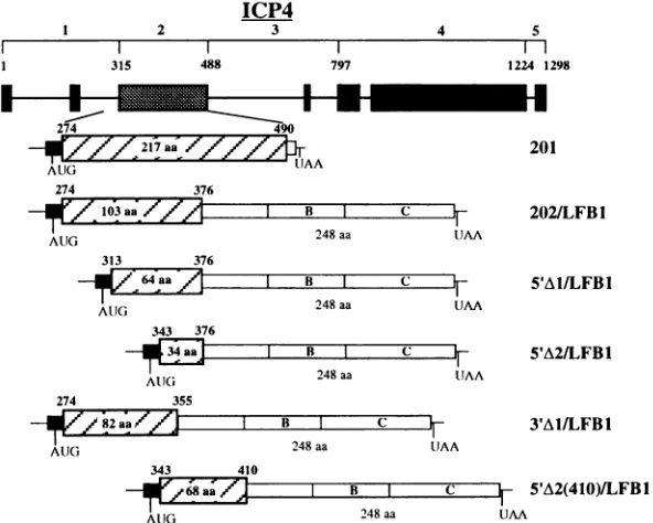
FIG. 1. StructuresofICP4-LFB1 chimericconstructs. (Top) Diagram of the five ICP4 polypeptide domains proposed by McGheochetal.(19)
isshown (modified from reference 5). Shadedboxes,regions ofaminoacid similarity with the ICP4 homologs from otheralphaherpesviruses. (Bottom) Schematic representation of the201 constructandof the ICP4-LFB1chimericconstructs.Hatched boxes, ICP4sequences.Boundaries
areindicatedbytheoriginalICP4amino acid(aa)numbers.Open boxes, LFBIsequences;B,t~POU domain;C,homeodomain; solid boxes, pT7.7
sequences.
be interspersed. It has thus not been possible to map the minimaldimerization domain of ICP4 within thecontextof the wild-type amino acid sequence and thus to demonstrate whether dimerization of ICP4per seis required for bindingto
DNAorforoneof itsother associated activities.
Inanattemptto mapthedimerization domain ofICP4,we
decided to adopt a strategy different from the deletion
mu-tagenesis studies carriedout to dateinotherlaboratories. We substituted the dimerization domain of the cellular transcrip-tion factor LFB1 (for which high-affinity binding to DNA is strictly dependent on dimerization) (42) with progressively smaller portions of a region of ICP4 spanning amino acid
residues274 to490 and examined these chimericproteins for LFBI-specific DNA binding in the form ofadimer.Inthisway, we could assess the dimerization capacity of ICP4 indepen-dentlyofits DNA-binding properties. To determinewhether dimerization isnecessaryforbindingtoDNA,weexpressed in an invitro translation system several polypeptides containing ICP4 DNA-binding domains of various lengths and analyzed theircapacitytobindtothe DNA high-affinity siteas dimers. It wasanticipated that the functional definition ofa minimal ICP4 dimerization domain would allow us to search for compounds that could specifically impair dimerization and
thus interfere withthefunctionof ICP4 inthereplicative cycle
ofHSV-1.
MATERIALS AND METHODS
Synthetic oligonucleotides and plasmid constructs. The following oligonucleotides, corresponding to the indicated binding sites and their respective complementary strands,were
synthesised: LFB1 Pal, GCTTGGTTAATGATTAACC
AAGC; LFB1 C, CACTGCCCAGTCAAGTGTFCTTGA
(42); IE3 cap site ICP4, GTGAATTCCCGGGATCCGCC
CGAGGACGCCCCGATCGTCCACACGGAGCGCGGCT
GCGTCGACTGCAGGTACGC; and BCU, GTGAATTCCC GGGATCCCGCTFTTCGAGTGTAATCCCCAGATATAGC TATGGAGCCAGGTCGACTGCAGGTACGC. To obtain the ICP4-LFBI chimeric constructs 201/LFB1 (not shown), 202/LFB1,5'A1/LFB1, 5'A2/LFB1, 3"AI/LFB1, and 5'A2(410)/ LFBI (Fig. 1), the corresponding DNA fragments coding for amino acids 274to490, 274to376, 313to376, 343to376, 274
to 355, and 343 to 410, respectively, were amplified by PCR
withsuitableprimers containing aSac! site(5' primer) andan
Sphlsite (3' primer).
Plasmid pcICP4, which contains the Sall-Ddel restriction fragment encompassing bp -122 to +4363 from the
transla-tion start site of ICP4, was used as a template in PCR.
Amplified DNA fragments obtained afterdigestion with Sacl andSphIenzymes wereinserted into theunique Sacl and SphI
sites of the LFB1 mutant vector, renamed LFB1/A8 in this article,generously donated by R. De Francesco and L.Tomei.
This backbonevector isthe modified version of LFBI mutant
Al (25) described in reference 42. Briefly, it contains the
N-terminalportion of LFB1, in which the dimerization domain
of the protein (region A) has been replaced with a shorter
sequence containing suitable restriction sites for cloning
het-erologous dimerization domainsinframe with the distalpartof the LFBI DNA-binding domain (4POU/homeodomain,
re-gions B and C, respectively). The chimeric ICP4/LFBI
frag-ments were cloned into the EcoRI (filled in with Klenow
enzyme) and BamHI restriction sites of a pT7.7 expression
vectorcontaininganartificialstopcodon. The LFB1 sequence
wasdeleted fromthe 201/LFBI chimericconstruct,generating
ICP4 mutant 201, in which the sequence coding for amino
acids 274to490wasincluded between theSacl and SphI sites.
The 5'A1BD200 and 5'A2BD200 mutants (see Fig. 4A) were
generated by PCR amplification of the DNA fragments coding
for amino acids 313 to 490 and 343 to 490 with the same
I-*
-J. VIROL.
on November 9, 2019 by guest
http://jvi.asm.org/
[image:2.612.156.452.75.312.2]suitable primers and the same DNA template as for the chimeric constructs. After Sacl and SphI digestion, the frag-ments were inserted into the unique SacI-SphI sites of the modified pT7.7 vector, isolated after SacI-SphI digestion of construct 201. All constructs contained an additional sequence encoding M A R I N S S S in front of the ICP4 N terminus.
Construct pT7I1OX was kindly provided by R. Everett and has been described in detail before (8, 29).
Invitrotranscription and translation. In vitro transcriptions were performed with 2 ,ug of linearized template DNA in a 40-plI reaction volume containing 40 mM Tris-HCl (pH 8.0), 50 mMNaCl, 8 mMMgCl2, 10 mM dithiothreitol, 2 mM spermi-dine, 0.5 mM each of the four nucleoside triphosphates (NTPs), 0.56 U of m7GpppG (Boehringer Mannheim), 80 U of RNasin, and 40 U of T7 RNA polymerase (Promega). The mixture was incubated for 1 h at 37°C, diluted to 100
[lI
with H20, and extracted with phenol-chloroform. The in vitro-transcribed complementary RNA (cRNA) was purified through a 1-ml Sephadex G50 prespun column, ethanol pre-cipitated, washed with 70% ethanol, dried, and resuspended in 40[lI
of H20.Translation of the cRNA (2.5,lI)was carried out for 90 min at 30°C in a30-,lI reaction mix containing 3,uMamino acid mixlacking methionine, 30,uCi of[35S]methionine (1,000 Ci/mmol, 10
mCi/ml;
Amersham SJ115), 1 mM dithio-threitol, and 12ptl
of rabbit reticulocyte lysate (Promega).35S-labeled
translation products were analyzed by sodium dodecylsulfate-polyacrylamide gel-electrophoresis (SDS-PAGE), and the gel was treated with Amplify (Amersham), dried, and autoradio-graphed.
Expression and purification of protein 201 from bacteria. The 201 protein was expressed in E. coli BL21(DE3) as described before (40). After a 3-h induction with 0.4 mM IPTG
(isopropyl-3-D-thiogalactopyranoside), cells were harvested
andlysed withaFrench pressure cell in a buffer containing 20 mMHEPES (N-2-hydroxyethylpiperazine-N'-2-ethanesulfonic
acid, pH 8.0), 2 mM EDTA, 20% (vol/vol) glycerol, 1 mM
phenylmethylsulfonylfluoride, and 1 mM dithiothreitol (buffer
A).The extract was treated with 20 ,ug of DNase I and 100,ug of RNase per ml for 10min on ice. NaCl and polyethylenimine were then added to a final concentration of 0.5 M and 0.1% (vol/vol), respectively, and insoluble material was pelleted by centrifugation at 27,000xgfor 30min in a Sorvall SS34 rotor. The clarified supernatant was dialyzed against buffer A con-taining 0.1 M NaCl andsubsequently purified by fast protein liquidchromatography (FPLC; Pharmacia) as follows. The 201 protein collected in the flowthrough of a DEAE column equilibrated in the same buffer as the final crude extract was loaded on a Mono S column and eluted with a 0.1 to 0.5 M NaCl gradient in buffer A. The 201 protein eluted as a sharp peak at 0.250 M NaCl. This process yielded about 0.5 mg of purified 201 from 1 liter of culture.
Gel retardation assays. Gel retardation experiments were carried out by mixing the appropriate amount of protein with the specific radiolabeled double-stranded oligonucleotide (40 fmol)ina
20-pA
reaction mix containing 10 mM Tris-HCl (pH8.0), 100 mM NaCl, 1 mM EDTA, 10% (vol/vol) glycerol, and either 0.1% (vol/vol) Nonidet P-40, when binding of ICP4 mutants wastested, or 5 mM MgCl2, whenbinding of LFB1 chimeras was tested. Either 0.1 or 2 ,ug of poly(dI: dC) *poly(dI:dC) was used as nonspecific competitor DNA when proteins expressed either in vitro or in bacteria were analyzed.Binding reaction mixes were incubated for 30min on ice. The samples were then loaded onto a 6% native polyacryl-amide gel(30:1,acrylamide-bisacrylamide;0.25x Tris-borate-EDTAelectrophoresis buffer) and analyzed by autoradiogra-phy.
Gel filtration chromatography. The native molecular weight of the 201 protein purified from bacteria was determined on a Pharmacia Superdex 75 HR 10/30 prepacked column in a buffer containing 50 mM HEPES (pH 8), 0.2 M NaCl, 10% (vol/vol) glycerol, and 0.1 mM dithiothreitol. The flow rate was 0.5 ml/min, and 0.5-ml fractions were collected and analyzed by both SDS-PAGE and gel retardation assays. Bovine serum albumin (67 kDa) and chymotrypsinogen (25 kDa) were obtained from Sigma and used as standards for calibration of the column.
RESULTS
Construction of ICP4-LFB1 chimeric expression plasmids. We aimed to devise a dimerization trap which would allow us to separate the analysis of two distinct functions of ICP4, namely, binding to DNA and dimerization, a task that would otherwise be very difficult to accomplish because of the high stability of ICP4 dimers in solution in the absence of DNA (20) and the probable partial colocalization of the DNA-binding and dimerization functions within the same domain of the protein. Indeed, the minimal portion of ICP4 identified so far which retains DNA-binding activity was demonstrated to be a dimer in solution (8). As a reporter for dimerization, we used the 4POU/homeodomain of LFB1, a liver-specific transcrip-tion factor that binds with high affinity to its cognate DNA palindromic sequence in the form of a homodimer. Previous studies showed that deletion of the dimerization domain of this protein resulted in the loss of DNA-binding activity but that this activity was restored in chimeric proteins containing the LFB1 DNA-binding domain linked to any one of a number of different heterologous dimerization motifs (42). On the basis of these findings, it was anticipated that the LFB1
qPOU/
homeodomain would also tolerate the ICP4 dimerization motif.To make the chimeric constructs shown in Fig. 1, we used the backbone vector LFB1/A8, which contains the N-terminal portion of LFB1 in which the dimerization domain of the protein (32 N-terminal amino acids [region A]) was replaced with a shorter sequence containing restriction sites for conve-nient in-frame cloning with the LFB1 DNA-binding domain (region B,
*POU;
region C, homeodomain). We initially assembled the construct 201/LFB1 (not shown), in which the LFB1 dimerization domain was replaced with the sequence corresponding to amino acids 274 to 490 of ICP4. From previous mutational studies (8, 9, 29, 44), this region was predicted to represent the minimal functional DNA-binding domain of the protein and to maintain the capacity to dimer-ize. The polypeptide product of 201/LFB1, expressed in vitro, could bind neither the LFB1 nor the ICP4 cognate DNA consensus sequence (data not shown). We took this negative result to indicate a functional interference between the two adjacent DNA-binding domains contained in this polypeptide. Inorder to avoid a recurrence of this phenomenon, we decided to exclude from the other constructs amino acids 377 to 490, which correspond to the most highly conserved part of region 2 and have been demonstrated to be strictly necessary for binding to DNA (5, 27, 28, 37, 44). Insertion of ancx4fragment encoding amino acid residues 274 to 376 into the LFB1/A8 polylinker gave construct 202/LFB1, into which were intro-duced two 5' deletions(5'A1, amino acids 313 to 376, and5'A2, amino acids 334 to 376) and a 3' deletion(3'A1, amino acids 274 to355). In addition, we also cloned into a pT7.7 vector, modified as described in Materials and Methods, an ICP4 domaincomprising amino acids 274 to 490 (region 2 and distal part ofregion 1), which encoded polypeptide 201 (Fig. 1).on November 9, 2019 by guest
http://jvi.asm.org/
A | Protein
Input
Oligoprobe ICP4 LFBI
c
j Oligocomp./s NS /
/S
5NS /
e-I~~
__N,
SN-97.4kODa
- -69k0a
~
- -46k0a
4..
-30k1Da
-2lkDa
1 2 3 4 5
1 2 3 4 5 6 7 8
B
-r4 Zn Zn in 22.J
-2001Da
97.4kDa
- 69kDa
_~
4_
__e_
--
46kDat~~~~~~~~. ... ..
- S
1 2 3 4 5 6 7
'43
~~~~~~~~~~~~~~~~~376
_
_
....~~~~~~~~...
,... ,_ ...,HiSV-l ICP4 PE:AEE-ARiRR EVA1SCGAPAAVWAP EL G AAQQY _,
EHV-IIEI D) VI--AAwSRYRA E AQAPVPVVF V PmErGDSTKQY NAL
PRV IE180 DDVPQV A RYlAA CPVPVPI G KQ HEAL:
V7V ORF621.AVVPRAA R ,Y.E A fDTTPPVPL't VP-LG-DPAP
R2Q
YP..A;
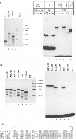
FIG. 2. Analysis ofICP4-LFB1 chimericproteins synthesized in vitro.Invitro transcription-translationreactionswerecarriedout asdescribed
in Materials and Methods. (A andB,left panels) A1.5-k1amountof the reactionmix containing the35S-labeledprotein productswasanalyzed
by SDS-PAGE (12% polyacrylamide) and autoradiography. R.R.L., invitro translation reaction with no RNAincluded; M, 14C-labeled size
markers(Amersham).(A andB,rightpanels)Approximatelyequalamountsof proteinweretested inagelshiftassay, asdescribed in Materials
and Methods. See the text for the oligonucleotide probes. Oligonucleotide competitors: S (specific), ICP4 oligonucleotide for 201 and Pal
oligonucleotideforICP4-LFBI; NS(nonspecific), BCUoligonucleotide for 201 and Coligonucleotide forICP4-LFBI. (C) Sequence alignment
of HSV-1 ICP4 amino acids 343 to376with the corresponding regions ofICP4 homologs fromdifferent alphaherpesviruses. EHV-1, equine
herpesvirus 1; PRV,pseudorabiesvirus; VZV,varicella-zostervirus.
3812
on November 9, 2019 by guest
http://jvi.asm.org/
[image:4.612.142.466.52.645.2]Analysis of ICP4-LFB1 chimeric proteins synthesised in vitro. The constructs depicted in Fig. 1 were used as linear
templates in an in vitro transcription-translation system. We chose this system because it permits rapid analysis of the recombinant products because the proteins of interest need
not be purified to homogeneity prior to use. In addition, no
complications from nonspecific aggregation of the protein arise,asthe concentrationof the polypeptide in the translation
mixture is relatively low.
The 35S-labeled protein products were analyzed by
SDS-PAGE (Fig. 2Aand B, left panels) andwere tested for DNA
binding either to a 23-mer duplex oligonucleotide probe
containingthe LFB1palindromicconsensus sequence
(chime-ra) or to a 74-mer oligonucleotide probe bearing the ICP4
high-affinity binding site (201) (Fig.2AandB,rightpanels).As shownin Fig. 2A, the 217-amino-acid region of ICP4 (polypep-tide 201) synthesized in vitro (left panel, lane 1) behaved as a
sequence-specific DNA-binding protein, giving a retarded
band that couldbe specifically competedwith byan excess of
unlabeledICP4oligonucleotide (right panel, lanes 1 to3). To
ourknowledge, this protein is the smallest version ofanactive
nonfusion DNA-binding domain observed to date. However, themost significant result in Fig. 2A shows that the chimera 202/LFB1 (left panel, lane 2) interacted specifically with the LFB1 palindromicconsensus sequence(right panel, lanes4to
6) with an affinity and specificity comparable to those of the
intactLFB1 DNA-bindingdomain either purified from
bacte-ria (Fig. 2A, right panel, lane 8) orsynthesized in vitro (Fig.
2B, right panel, lane 5). In contrast, the in vitro-translated
LFB1/A8 mutant (Fig. 2A, left panel, lane 3), lacking the
N-terminal dimerization domain, produced only avery faint
band (Fig. 2A, right panel, lane 7). Furthermore, the 201
polypeptide and the 202/LFB1 chimeric protein did notshow any appreciable binding to the LFB1 probe or to the ICP4
probe, respectively(datanotshown). Itthereforeappearsthat the ICP4 sequence between amino acids 274 and 376 can
confer onLFB1 the capacitytobind toDNAspecifically as a
dimer,implyingthat the ICP4 dimerization domain is located
in thisregion.
Figure2Bshows the results ofafurther deletionanalysis of
the putative dimerization domain from both the 5' and 3' directions. Approximately equal amounts of the in
vitro-translatedproductsshown in the left panelweretested inagel
shiftassayfor bindingto the LFB1 oligonucleotide probe. As can be seenin lanes 2 and 3,mutants with 5' deletions up to
amino acid343 retained the capacity tobind DNAwith high
affinity. The specificity of binding was confirmed both by
competition with an excess of LFB1-specific oligonucleotide
and bythe interactionwith anLFB1-specific antiserum,which
supershiftedthe retardedcomplexes(datanotshown).Onthe
otherhand,the 3' deletionmutanttruncatedatamino acid 355
possessed very low DNA-binding activity (Fig. 2B, lane 4),
comparabletothe backgroundactivity of the negative control
LFB1/A8 (lane 6). The stretch of 34 amino acids containing
ICP4 residues 343 to 376 thus appeared to be sufficient to
functionally replace the dimerization domain of the LFB1
reporterprotein,andwethereforeproposethat itcontainsthe
ICP4 protein dimerization motif. Our data also demonstrate
that no dimerization was apparent in the absence of the
C-terminal 22 amino acid residues within this stretch (355 to
376).
Protein sequence analysis of this ICP4 peptide revealed a
high degreeof conservationwith thecorresponding regions of
theICP4homologs fromthe other members of the
Alphaher-pesviridae subfamily, with a block of homology of 11 amino
acids inthe C-terminal part (Fig. 2C). On the other hand, it
showed no homology with other known dimerization motifs, and no prediction of secondary structure could be made.
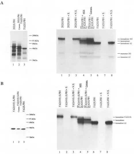
Exchange of monomers between chimeric proteins in vitro. In order to study the stability of the ICP4-LFB1 dimers, we decided to find out whether the individual monomers were free to exchange. To this end, the 202/LFB1 and 5'A2/LFB1 proteins were synthesized in vitro either separately or in cotranslation (Fig. 3A, left panel). Since the two polypeptides differ in size by about 7.6 kDa, a heterodimeric protein bound to DNA was expected to migrate through a polyacrylamide gel with a mobility intermediate between those of the two respec-tive homodimeric protein-DNA complexes. The right panel of Fig. 3A shows the result of such an experiment. In lane 4, the 202/LFB1 and 5'A2/LFB1 proteins were mixed in approxi-mately equal amounts and incubated for 30
min
at37°C
before the addition of the LFB1 oligonucleotide probe. Both cotrans-lation and premixing of the two different chimeras resulted in diminution of the relative amounts of the two homodimer-containing complexes. In contrast, a novel complex (het-erodimer) was formed, which had an intermediate mobility and an intensity about twofold higher than that of the single homodimeric complexes. We concluded that the ICP4-medi-ated dimerization of the LFB1 reporter occurred via the association of free monomers in solution, rather than during the translation-associated process of polypeptide folding.In a similar experiment, the dimerization partner for the
5'A2/LFB1 protein was chimeric polypeptide5'A2(410)/LFB1, which contains amino acids 343 to 410 of ICP4 fused to the LFB1 DNA-binding domain (Fig. 1). This protein differs from
5'A2/LFB1 only by the additional 35 amino acid residues at the C terminus of the ICP4 sequence. As shown in Fig. 3B, the two in vitro-synthesized proteins were able to exchange monomers both after mixing of the individually translated proteins in solution and during cotranslation (lanes 4 and 5), demonstrat-ing that amino acids 274 to 343 are not required for dimeriza-tion. Furthermore, as free monomer exchange was still ob-served, the region of ICP4 containing amino acids 376 to 410 did not appear to contain residues that are involved in the stabilization of ICP4 dimerization.
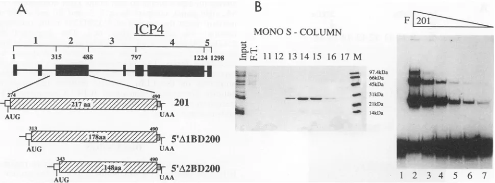
Analysis of the ICP4 DNA-binding region expressed in bacteria. In addition to the truncated ICP4 protein 201 (amino acids 274 to 490; see Fig. 1), we inserted in the modified pT7.7 expression vector two further ICP4 segments in which addi-tional amino acids were removed from the N terminus to yield polypeptides containing residues 313 to 490
(5'AlBD200)
and 343 to 490 (5'A2BD200) (Fig. 4A). Our aim was to express and purify these proteins in large amounts to allow future biochem-ical and structural studies. Protein 201 was expressed in bacteria and purified by FPLC ion-exchange chromatography. It was shown to bind to the ICP4 consensus site with high affinity (Fig. 4B) and with the same specificity as the full-length protein (data not shown). In gel shift experiments, the protein gave rise to at least two protein-DNA complexes; the ratio between the slower- and the faster-migrating species increased with increasing protein concentration (Fig. 4B, right panel). This phenomenon was not observed with 201 expressed in vitro, which reproducibly formed only a single complex with the target DNA (Fig. 2A) migrating with a mobility similar to that of the faster-moving band seen in Fig. 4B. This result confirmed previous observations by Everett et al. (8), who proposed that the slower-migrating complexes represent mul-tiples of the basic dimeric form. Multimerization could derive either from nonspecific aggregation of two or more dimeric units at high protein concentrations or from the interaction of the additional dimers with the nonspecific DNA tails in the probe molecule.on November 9, 2019 by guest
http://jvi.asm.org/
3814 GALLINARI ET AL. J. VIROL.
A
+ + + + + +
51 _
E51s
N vNini kin]
---200kDa
_ _ _ ~~~~~~~~~~~~~-homodimer 202
- 97.4kD:a - hewerodlimer
-69k:a
_sS|8|@-
~~~~~~~~~~~~~~~~~~~-
UIP -homodlimer A2monomer A2(
-30kDa
1 2 3
1 2 3 4 5 6 7 8
BE
+ + +
- 1 tt+
i_-
_W
9
C]
) <iCD <i a <<<<i
<C1 U m
-200kDa
-*_-homodimer5A2(410)
_97.4kDa _heterodimer
-69kDa homodimer A2
-46kDa
-3OkDa
12 3
[image:6.612.76.545.73.607.2]1 2 3 4 5 6 7 8
FIG. 3. Monomer exchange between different ICP4-LFB1 chimeric proteins synthesized in vitro. For protein synthesis, SDS-PAGE analysis (A and B, left panels) and gel retardation assays (A and B, rightpanels), the experimental conditions and the abbreviations were the same as in Fig. 2 and in Materials and Methods. Incotranslation reactions, 2.5 ,u1of each cRNAspecies was included as for single translation reactions, and 3 ,ulof the protein sample wasanalyzed by gel shift assay. In the premixing experiments, 1.5p.1of each of thetwo proteins wasmixedand incubated for 30minat37°CunderDNA-binding conditions before the LFB1 Pal probe was added. The identity of the retarded complexes is indicated.
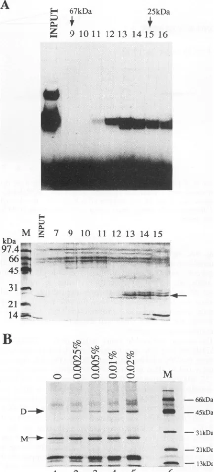
Physical analysis of 201 protein purified from bacteria. To shift assays (Fig. 5A). The 201 protein eluted between the investigate the multimerization state of 201, a purified prepa- bovine serum albumin (67 kDa) and chymotrypsinogen A (25 ration was applied to a Pharmacia Superdex 75 gel filtration kDa) markers, and its molecular mass was estimated to be column. The elution profile was monitored by UV absorbance, about 45 kDa, in agreement with the predicted molecular mass and the eluted fractions were analyzed by SDS-PAGE and gel of the dimeric form of 47.4 kDa (the faster-migrating band in
on November 9, 2019 by guest
http://jvi.asm.org/
A
1 2 3
ICP4
4 5
1 315 48 797 12241;
274 490
<///E////////217
;;i/;u////,
201AIrV UAA
B
MONOS-COLUMN
298 r ; 1112 1314 15 16 17 M
_ 97.4kDa _ 66kDa
_ 45kDa _ 3lkDa _ 2IkDa
- 14kDa
l
AUlJ
313 490
1i//LYY/,P78aaA7////I///i
5'AlBD200
AUG UAA
Ly////X///
A48aa
Jl/
5'A2BD200 [image:7.612.73.558.77.257.2]AUG UAA 1 2 3 4 5 6 7
FIG. 4. Analysisof truncated ICP4 proteins expressed in E.coli. (A)Diagram of ICP4 mutant constructs.SeethelegendtoFig. 1for details. (B) In the left panel, the 201 protein-containing fractions of a Mono S column eluted with a linear NaCl gradient as described in Materials and Methods were analyzed on a 12% polyacrylamide-SDS gel stained with Coomassie blue. Lane M, size markers (Bio-Rad). F.T., flowthrough. In the right panel, fractions containing the 201 protein were pooled, and serial 1:2 dilutions of the sample were tested for binding to the ICP4 probe in a gelshift assay (lanes 2 to 7) as described in Materials and Methods. F, free probe.
the gel shift was most likely the result of proteolysis). To confirm this result, an aliquot of the 201 preparation was treated with increasing concentrations of the cross-linking agent glutaraldehyde, and the products were analyzed by SDS-PAGE. The result(Fig.5B) shows clear formation of 201 dimers, supporting the finding that this protein retains the ability of the full-length ICP4 protein to exist in a dimeric form insolution. The fact that the cross-linking was not quantitative isprobably the consequence of poor efficiency of the reagent usedrather than of anexistingequilibrium between dimers and monomers.Thisinterpretation is also supported by our results with the invitro-synthesized ICP4 proteins (see below).
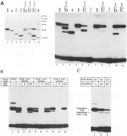
Analysis of ICP4 deletion mutants in vitro.The two trun-catedpolypeptides 5'A1BD200 and 5'A2BD200 expressed in bacteriawere moredifficulttoisolate inlargeamountsbecause of their lowsolubility. We therefore hadto resort tostudying the dimerization and DNA-binding properties of these polypeptides in the in vitro transcription-translation system described above. In addition to the polypeptides already described, we also expressed in vitro the polypeptide I1OX
(from the pT7I1OX vector, kindly provided by R. Everett) that extends 34amino acids further towards the C terminus of ICP4 than our constructs (amino acids 276 to 523) and has been showntoexist inadimeric form insolution(8, 28).
Inorder toanalyze the dimerization capacity of each of the truncated proteins, we carried out a monomer exchange ex-perimentsimilartothat shown inFig.3, this time with I1OXas a partner for heterodimerization with either the 201, 5'A1BD200, or 5'A2BD200 polypeptide (Fig. 6A). Approxi-mately equal amounts of the proteins synthesized in vitro
(eitherseparatelyorincotranslation) (Fig.6A, leftpanel)were tested in a gel shift assay for binding to the ICP4 DNA oligonucleotide probe(Fig.6A,rightpanel).Incubationof the ICP4 probe with the cotranslationmixofI1OXand 201 (Fig. 6A, left panel, lane 5) resulted in the appearance of anovel complex withamobility intermediate between andanintensity
about twofold higher than those of the respective bound homodimercomplexes (Fig. 6A, right panel, lane2). Interest-ingly, thisnewcomplex didnotform aftermixingand incuba-tion of the two individual polypeptidesbefore DNAbinding
(Fig. 6A, right panel, lane 10). This demonstrated that a heterodimercanformsolely during the translation of the ICP4 fragments and that, once formed, the individual monomer subunits are no longer able to exchange in solution. This observation is consistent with previous results (38), which showed thatheterodimers between thewild-typeICP4 and the mutanttruncatedproteinX25formed inHSV-1-infected X25 cells but not in vitro when cell extracts containing the two homodimersweremixed.
The second noticeable result is that both 5'A1BD200 and 5'A2BD200(Fig. 6A, upper panel, lanes 3 and4)were not able tobind theDNAprobe with highaffinity(Fig. 6A, right panel, lanes 6 and9). This is consistent with earlier observations (8, 29, 37,44)indicating that the distal part of region1 isinvolved in stabilizing the binding to DNA. However, in spite of the complete loss of DNA-binding activity upon deletion of amino acids 274to313, cotranslation of 5'A1BD200 with I1OX (Fig.
6A, left panel, lane 6) produced a novel complex with a mobility and an intensity consistent with that of the het-erodimeric form(Fig.6A,right panel,compare lanes4and5).
This novel complexwas shown to be specific by competition with a 100-fold excess of either specific or nonspecific unla-beled oligonucleotide (Fig. 6B, lanes 5 to 7). Since only the I1OX shift was present when the two proteins were mixed before theDNA-bindingassay(Fig.6A,right panel, lane11),
5'A1BD200 seemed to behave similarly to 201 in terms of its dimerization properties. An interesting and somewhat unex-pected findingwasthatonlyonefunctionalmonomersubunit
(I1OX) in the heterodimer was sufficient to confer on the
proteinthe abilitytobind DNA.
Finally, cotranslation of 5'A2BD200 withI1OX(Fig.6A, left panel, lane 7) reproducibly caused an inhibition of I1OX DNA-binding activity (Fig. 6A,right panel, compare lanes 7 and 8), which was demonstrated to be dependent on the amountof5'A2BD200 RNAaddedto the invitrotranslation reaction(Fig. 6C). Aswith5'AlBD200, a novelcomplexwas
formed with a mobilityand a
specificity
ofbinding
consistent with a heterodimeric form (Fig.6B, lanes 8 to10).
However,the relative intensity of this band was lower than would be
expected if all the heterodimeric molecules formed had an
twmw-4
on November 9, 2019 by guest
http://jvi.asm.org/
3816 GALLINARI ET AL.
A
z
M
z:
kDa
97.4nw
666':: 45
31 p ...
67kDa 25kDa
9 1011 1213 14 15 16
...
I'll
7 9 10 11
...
e....
.- .p
12 13 14l
... u
....
-B
"Q
tn
c
O
Q
U-4 Ul)
8
C5 6
6
D -0
4en
1 2
3
4
5
FIG. 5. The 201 protein is adimer in solutio analysis. A
100-,ug
amount of the 201preparatio
loaded on a Superdex 75 column, calibrated wi serum albumin (67kDa) and chymotrypsinogen(0.5 ml)were collected, and appropriate sample
elution profile)wereanalyzed for DNA-bindinga probe (upper panel) and by SDS-PAGE andsi
panel). The positions of the two protein standa
fractions 9 (67 kDa) and 15 (25 kDa) are indica upperpanel. The peak of 201 protein
(indicated
lower panel) and DNA-binding activity eluted insize markers(Bio-Rad).The expected sizes for 2'
kDa as a monomer and 47.4 kDa as a dimer. cross-linking of protein 201. The protein was
tre
0.005, 0.01, and 0.02% (vol/vol) glutaraldehydtemperature and analyzed by SDS-PAGEfollow
The positions of the monomeric (M) and cross speciesaremarked. Lane M, size markers.
affinity
for DNA similar to that of theI1OX
homodimer (Fig.6A,
right panel, compare
lanes2, 5,
and 8), and it did not increase when the amount of 5'A2BD200 in the translationreaction was increased
(Fig.
6C).
This suggested that5'A2BD200
(amino
acids 343 to490)
sequestered
fullyfunc-tional
I1OX
monomers intoonly
partially
functional het-erodimeric forms. This observation thus also supports thehypothesis
that the34-amino-acid stretch that confers dimericproperties
on the LFB1 chimericproteins
represents theputative
dimerization domain of ICP4. A summary of thedimerization and
DNA-binding
properties
of the ICP4 mu-tants is shown in Fig. 7.DISCUSSION
The aim of this work was to identify a discrete
ICP4 that mediates the homodimerization of the protein. It
was
anticipated
that such aregion
would be of a novel,previously
uncharacterizedtype,
as dimerization interfaces such as the leucinezipper
of the Fos-Jun heterocomplexor thehelix-loop-helix
motif of several other regulatory proteins(reviewed
in reference18)
are not evident in its amino acidsequence.
Mutational studies carried out by othergroups (8,l
544)
indicated that the dimerization domainof ICP4 is locatedsomewhere within the
DNA-binding
domain of the protein,consisting
of all ofregion
2 and the distal part of region 1.Attempts
to delineate this motif with more precision werefrustrated
by
the fact that shorter versions of the ICP4DNA-binding
domainproduced
in bacteria (8, 44) no longer.4--
boundDNA,
and theircapacity
to dimerize has not beenstudied
by
other means as yet.In order to
map
the minimal functionaldimerization domainof
ICP4,
we decided to undertake a molecular dissectionof thesequence encompassing
residues 274 to 490. We showed thatprotein
201
expressed
both in vitro and
inbacteria
binds thecognate
DNAsequence
as a dimer and exists as a dimer insolution. Our results confirm those of Everett et al. (8), and
M
extend
them
to
demonstrate
that
the
dimeric
form of 201 isvery
stable and cannot
freely
exchange
monomers in solution,in a
way
similar to native ICP4. Our first
attemptsto study the-
66kDamultimerization state of
progressively
shorter
truncated -45kDa
polypeptides expressed
in
bacteria as nonfusion
proteins
failedbecause of their low solubility.
3lkDa
As
the construction of
hybrid
DNA-binding
proteins
con-taining heterologous
dimerization domains proved to be a- -
21kDe
useful tool in the molecular and functional dissection of many;
-
13kDM
dimeric cellular transcription
factors
(reviewed
in reference6
18),
we decidedtoadopt
thisapproach
andset upadimeriza-tion
trap assay,
which wouldpermit
us to screen a series of5'n.
(A)
Gel filtration
and
3' deletion
fragments
of this
217-amino-acid
stretch
for
n
from
bacteria
was
sequences
that could confer dimerization
properties
on
a
ith
20 u.g of bovine
heterologous sequence-specific DNA-binding
protein.
A
simi-(based
on
the
UV
lar strategy,
using the DNA-binding
domain
of
the
bacterio-ctivity
with theICP4
phage
A
repressor as areporter
for
dimerization,
allowed theilver
staining (lower
identification
of
previously
unknown
heterologous
dimeriza-Lrds
which eluted in tion domains(1)
as well as thegenetic
analysis of alreadyted
by
arrows in the characterized motifs(13).
We selected as a reporter theI
by
an arrow in thetIPOU/homeodomain
of
LFB1,
a
liver-specific
transcriptionfraction 13. Lane
M,
factor, which binds with high affinity
to
its
cognate DNA101
protein
were23.7sequence, provided
that it is linked at
the
N terminus
to a (B) Glutaraldehydedimerization motif
(42). Thus,
among
a
series
of ICP4 -atedwith 0, 0.0025,[e
for 2 h
at
room
polypeptides linked
to
the
LFB1
*POU/homeodomain (see
ed
by
silver staining.
Fig.
1),
only
those containing ICP4 residues 343 to 376i-linked
dimeric(D)
demonstrated
aDNA-binding phenotype
ingel
shiftassaysandwere therefore
judged
to contain a dimerization
motif.Con-i w __wr_._p
on November 9, 2019 by guest
http://jvi.asm.org/
[image:8.612.68.285.77.556.2]2 Cs - r- 2I
I+ I
x x_ x x 7
-z c< c
_4 C -
-I+
X < < X- XC~
CD CD < CDCD0~2
=n ;n =2 _o
2(X)kD)4
97AJkDa 69 kDla 46kDa
- - - 30kDa
_~
4-- 14.3kDa
2 3 4 5 6 7 8
'I_~
.u
__ __~~a
O-
-1 2 3 4 s 6 7 8 9 0 II
f
Protein IIoX IIOX + 201 IIOX+5'Al 110X+5A2
input cotrans cotrans cotrans
oligo
INS
I
| S NI
S NS - | S |NScomp.
"'qII
41 2 3 4 5 6 7 8 9 10
[image:9.612.103.522.74.522.2]1 2 3
FIG. 6. Analysisof ICP4 deletionmutantproteinssynthesizedinvitro.Invitrotranscription-translation reactionswerecarriedout asdescribed in thelegendstoFig.2and 3.(A)Proteins201,5'AIBD200,and5'A2BD200weresynthesized singlyortogetherwithproteinIIOX(8).Either 1.5
,ul of the samples containing single 15S-labeled proteins or 3 pl of the cotranslation reaction mixes was analyzed by SDS-PAGE
(12%
polyacrylamide)andautoradiography (leftpanel).LaneMW,'4C-labeledsize markers(Amersham).Approximately equalamountsofproteinweretested forbindingtotheICP4oligonucleotide probeinagelshift assay(rightpanel).Suitable volumes of either 201or5'A1BD200sampleswere also mixed with theIlOXpreparation at anapproximately 1:1proteinratio for 30 min at37°Cand thenanalyzedforDNA-bindingactivity (lanes 10and 11). (B) The201-I1OX,5'A1BD200-110X, and5'A2BD200-IIOX heterodimericforms all bind DNA inaspecificmanner.Suitableamounts
of the cotranslation reaction mixes shown inpanelAweretested forbindingto the ICP4probein the absenceorpresenceofa 100-foldexcess
of eitherspecific (S)ornonspecific (NS)unlabeledoligonucleotideandanalyzed bythegelshift assay.(C)Inhibition ofIlOXDNA-bindingactivity
isdependenton theamountof5'A2BD200 synthesizedin the cotranslation reaction. IIOX cRNA(1 ,ul)wascotranslated with0, 1, or2,ul of
5'A2BD200cRNA, and equal protein samples were analyzed for DNAbindingby the gelshift assay. Theidentityof theretardedcomplexesis indicated.Electrophoresiswascarriedoutforashortertimeinpanels Band CthaninpanelA.
sistent with this result, a truncated ICP4 protein as small as 5'z2BD200(amino acids 343 to 490), in spiteof the complete lossofDNA-binding activity, appeared to retain the capacity
to form a heterodimerwith a longer polypeptide after
coex-pression in the in vitrotranslation system.
Theidentified34-amino-acid stretch exhibits
significant
sim-ilaritytothecorresponding regionoftheICP4-related
proteins
from otheralphaherpesviruses
in that at its C-terminalend,
which isstrictly necessaryfordimerization, there isablock of homology of11 aminoacids.Interestingly, atruncatedformof thevaricella-zoster virusprotein
ORF62,
approximately
corre-sponding to ourICP4 deletionmutant
5'zA2BD200,
wasdem-A
B
homodimer
heterodimer-IIOX+5'A2
IIOX RNA lgl Ig] Igi
YA2 RNA N,, Igl 2gi
on November 9, 2019 by guest
http://jvi.asm.org/
3818 GALLINARI ET AL.
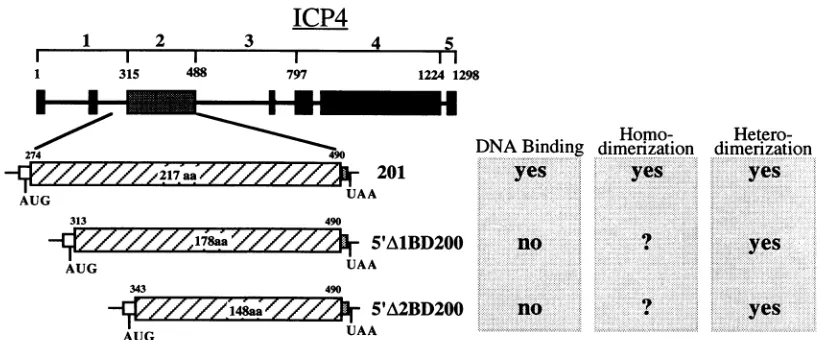
1 2 3
315 488
_
ICP4
4 5
797 1224 1298
a
*_
274 490
{;//7////17 a //////// 201
AUG UAA
313 490
{;li8aa/n//8///aal 5'A1BD200
AUG UAA
343 490
{48aa'
//xx/*
FA5'A2BD200AUG UAA
[image:10.612.100.510.73.243.2]DNABinding
FIG. 7. Summary of the DNA-binding and dimerization properties oftheICP4deletionmutants. Seethe legend toFig. I fordetails.
onstratedtoretain theabilitytobind DNAasadimer (43). As
anticipated, protein sequence analysis of the ICP4
34-amino-acid stretch failed to reveal any similarity with known
dimer-ization motifs and did not allow any prediction of secondary
structure. Furthermore, a synthetic peptide spanning the
34-amino-acid stretch appeared to be mostly random coiled by circular dichroism analysis. It is therefore conceivable that additional flanking residues are required to stabilize its
con-formation.
Our studies do not rule out the possibility that other
sequencesoutside amino acid residues343 to376can contrib-ute to the stabilization of the dimeric state of the native protein. Other authors (35, 44) proposed that the first 90 residues ofthefull-length polypeptide facilitate the formation of ICP4 oligomers, which appear as multiple electrophoretic
variants of ICP4-DNA complexes. Although we did not test
this hypothesis directly, the 201 protein expressed in bacteria did form higher-order oligomers at high protein
concentra-tions(Fig. 4B, right panel), probablyas aresultofnonspecific
protein-protein orprotein-DNAinteractions. Indeed, theuse
ofdiluted or invitro-translated proteinsaswell asincreasing
concentrations of salt or nonspecific competitor DNA in the
assay resulted in the disappearance of these multiple oligo-mericforms (data not shown). Also, no multimerization was
observed in gel shift assays in which shorter oligonucleotide
probes containing the ICP4 DNA-binding sitewere used, and
only the dimeric form was evident in solution by both gel
filtration andprotein-protein cross-linking.
The main difference between the ICP4-LFB1 hybrid polypeptides and the corresponding ICP4 truncationswasthat
the chimeric proteins could freely exchange monomers in solution, whereas the ICP4 mutants, like the native HSV-1 protein(36, 38), could heterodimerize onlyuponcotranslation. We propose that amino acid residues within the C-terminal
portion of region 2 (410 to490) contribute tothisincrease in the stability of the truncated ICP4 dimers. This hypothesis is basedonourobservation that the chimeric protein5'A2(410)/
LFB1, containing amino acids 343 to 410 of the ICP4
se-quence, retained theabilitytoexchangemonomersin solution,
whichindicated that the region between residues 376 and 410
does not augment the stability of the dimer, whereas that
delineated by residues 410 to490 clearlydoes.
Whether dimerization is strictly necessary for binding of
ICP4 to DNAisa question that has been debated byseveral authors. The first consideration is that the ICP4 consensus
sequencedoesnotexhibit credible palindromesorrepeatsthat would be recognized by the two identical subunits of the dimeric transcription factor. In thecaseofgDpromotersiteII,
animperfectaxis ofsymmetryhas beenidentified between the 5' portion of the consensus site andan inverted sequence at
the 3' side of the footprint (29). However, deletion ofoneof thetwohalves of the imperfectpalindromicregioncaused no
change in the mobility of the retarded complexes. These findingswereconfirmedbythe data ofMichael and Roizman
(21), who showed that site II and other nonpalindromic
sequencesbound thesameICP4 species. We obtained similar results when testing the 201 mutant protein for binding to
DNA probes of identical length containing either the gDIIsite
orthe ICP4 high-affinity site (datanot shown). Furthermore,
our results with the truncated ICP4 proteins synthesized in vitro suggested thatonlyoneDNA-bindingsubunit within the dimeric protein is sufficient toestablishahigh-affinity
interac-tion with the ICP4 consensus sequence. Thus, in spite of the
complete loss of DNA-binding activity observed with the homodimeric truncated molecules lacking region 1 (5'A
1BD200 and5'A2BD200),their cotranslation withafunctional
ICP4 variantgenerated heterodimeric forms that boundDNA. If ICP4 need not be a dimer in order to bind to its high-affinity site, whichmost likely results in the repression of transcription, the significanceof ICP4 dimerizationmostlikely relates to some other function of the protein. As a good
correlation exists between mutations in region 2 that impair DNAbinding and the loss ofsomeofthe ICP4trans-activation functions (5, 27, 28, 37) and since region 2 appears to be responsible for bothhigh- and low-affinity bindingtoDNA, it is temptingtospeculate that dimerization maybe a
prerequi-site for trans activation by the protein. Several low-affinity DNA-binding siteswerefound inthevicinity of early and late
ICP4-activatedpromoters(12,14, 23, 26, 34, 41). As these sites donotcontainanapparentconsensus sequence,it is likely that
aninteraction ofan ICP4monomerwith these sites would be
too labile to be functional. It would thus need tobe further stabilized,mostlikelyby interaction with other proteins bound in the vicinity with high affinity. Alternatively, were ICP4 to
bind as a dimer, it could effectively double the number of
contacts with the DNA and thus stabilize the interaction.
However, no evidence in favor of this hypothesis has as yet
beenprovided.
Alternatively, as indicatedby previous results (38, 39), the
dimeric state could represent a necessary constraint on the
1
m
J.VlIROL.
on November 9, 2019 by guest
http://jvi.asm.org/
conformation of a domain of ICP4 which participates in
protein-protein interactions required for activating transcrip-tion. These studies indicated that the stable expression of a truncated ICP4 peptide lacking region 4 and therefore unable totransactivate viral genes (5, 35) led to increased resistance to infection with HSV-1 both in tissue culture and in transgenic mice(38, 39). This trans-dominant inhibition was attributed to the formation of nonfunctional heterodimers between the truncated and wild-type ICP4 proteins. As these heterodimers could be isolated from cells and demonstrated in gel shift
experiments to be capable of DNA binding, their failure to transactivatewas ascribed to the conformational alteration in the C-terminal region, genetically implicated as important for trans activation.
Construction of a recombinant ICP4 gene lacking sequences encodingamino acids 343 to 376 should yield a protein that is unable to dimerize. Such a protein ought to provide us with invaluable information about the real role of dimerization in thefunction of this important HSV-1 transcriptional regulator. These experiments are in progress.
ACKNOWLEDGMENTS
We areparticularlygrateful to Bernard Roizman for the gift of the HSV-1BamHI-Yfragment-containing plasmid pRB113 and to Roger Everettfor thegenerousgift of plasmidpT7I1OX. We are grateful to Angela Carbonetti and Andrea Pucci for help in purification of the bacterialproteins. We also thank Riccardo Cortese and Roger Everett for many helpful discussions, Philippe Neuner for oligonucleotide
synthesis,YvesCully for photographic assistance, and Fabio Palombo, Petra Neddermann, Licia Tomei, and Raffaele De Francesco for
criticalreading of the manuscript. REFERENCES
1. Amster-Choder, O., and A. Wright. 1992. Modulation of the dimerization of atranscriptional antiterminator protein by
phos-phorylation. Science257:1395-1398.
2. Baohua, G., R.Rivera-Gonzales, C. A. Smith, and N. A. De Luca. 1993.Herpes simplex virus infected cell polypeptide 4
preferen-tiallyrepressesSpi-activatedover basal transcription from its own
promoter. Proc. Natl. Acad. Sci. USA 90:9528-9532.
3. DeLuca, N. A., A. M. McCarthy, and P. A. Schaffer. 1985. Isolation andcharacterization of deletion mutants of herpes simplex virus type 1 in the gene encoding immediate-early regulatory protein ICP4. J. Virol.56:558-570.
4. De Luca, N. A., and P. A.Schaffer.1985. Activation of immediate-early, immediate-early, and late promoters by temperature-sensitive and wild-type forms of herpes simplex virus type 1 protein ICP4. Mol. Cell. Biol. 5:1997-2008.
5. DeLuca, N. A., and P. A. Schaffer. 1988. Physical and functional domains of the herpes simplex virus transcriptional regulatory
protein ICP4. J. Virol. 62:732-743.
6. Di Donato, J. A., J. R. Spitzner, and M. T. Muller. 1991. A
predictivemodel for DNArecognition by the herpes simplex virus
protein ICP4. J. Mol. Biol. 219:451-470.
7. Dixon, R. A. F., and P. A.Shaffer. 1980. Fine-structure mapping and functional analysis of temperature-sensitive mutants in the gene encoding the herpes simplex virus type 1 immediate-early
protein VP175. J. Virol.36:189-203.
8. Everett, R D., NL Elliot, G.Hope,andA.Orr. 1991. Purification of the DNAbinding domain of herpes simplex virus type 1 immediate-early
proteinVmwl75as ahomodcimerand extensive mutagenesis of its DNA recognition site. Nucleic Acids Res.19:4901-4908.
9. Everett, R D., T. Paterson, and M. Elliott. 1990. The major transcriptional regulatoryprotein of herpes simplex virus type 1 includes aproteaseresistantDNAbinding domain. Nucleic Acids Res. 18:4579-4585.
10. Faber, S. W.,and K. W.Wilcox. 1986. Association of the herpes simplex virus regulatory protein ICP4 with specific nucleotide sequences.Nucleic Acids Res. 14:6067-6083.
11. Faber,S. W.,and K. W.Wilcox. 1988. Association of the herpes
simplex virus regulatory protein ICP4 withsequencesspanning the
ICP4 genetranscription initiationsite. Nucleic Acids Res. 16:555-570.
12. Flanagan, W. M.,A. G. Papavassiliou, M. Rice, L. B. Hecht, S. J. Silverstein, and E. K. Wagner. 1991. Analysis of the herpes simplex virus type 1 promoter controlling the expression
of
UL38,a true late gene involved in capsid assembly. J. Virol. 65:769-786. 13. Hu, J. C., E. K. O'Shea, P.
S.
Kim, andR T. Sauer. 1990. Sequencerequirements for coiled-coils: analysiswith X repressor-GCN4leucine zipper fusions. Science250:1400-1403.14. Imbalzano, A. N., A. Shepard, and N. De Luca. 1990.Functional relevance of specific interactions between herpes simplex virus type 1 ICP4and sequences from the promoter-regulatorydomain of the viralthymidine kinase gene. J. Virol. 64:2620-2631. 15. Kattar-Cooley, P., and K. W. Wilcox. 1989. Characterization of the
DNA-binding properties of herpes simplex virus regulatory pro-tein ICP4. J. Virol. 63:696-704.
16. Kristie, T. M., and B. Roizman. 1986.a4, the major regulatory protein of herpes simplex virus type 1, is stable and specifically associated with the promoter-regulatory domains of a genes and of selected otherviral genes. Proc. Natl. Acad. Sci. USA 83:3218-3222.
17. Kristie, T. M., and B. Roizman. 1986. DNA-binding site of major regulatory protein a4 specifically associated with promoter-regu-latorydomains of a genes of herpes simplex virus type 1. Proc. Natl. Acad. Sci. USA83:4700-4704.
18. Latchman, D.S. 1991. DNA binding by transcription factors, p. 177-205. In Eukaryotic transcription factors. Academic Press,
London.
19. McGheoch, D. J., A. Dolan, S. Donald, and D. H. K. Brauer. 1986. Complete DNA sequence of the short repeat region in the genome of herpes simplexvirus type 1. Nucleic Acids Res. 14:1727-1744. 20. Metzler, D. W., and K. W. Wilcox. 1985. Isolation of herpes simplexvirus regulatory protein ICP4 as a homodimeric complex. J. Virol. 55:329-337.
21. Michael, N., and B. Roizman. 1989. Binding of the herpes simplex virus major regulatory protein to viral DNA. Proc. Natl. Acad. Sci. USA 86:9808-9812.
22. Michael, N., and B. Roizman. 1993. Repression of the herpes simplex virus 1a4 gene by its gene product occurs within the context of the viral genome and is associated with all three identified cognate sites. Proc. Natl. Acad. Sci. USA90:2286-2290. 23. Michael, N., D. Spector, P. Mavromara-Nazos, T. M. Kristie, and B. Roizman. 1988. The DNA-binding properties of the major regulatory proteina4of herpes simplex virus. Science 239:1531-1534.
24. Muller, M. T. 1987. Binding of the herpes simplex virus type 1 gene product ICP4 to its own transcription start site. J. Virol. 61:858-865.
25. Nicosia, A., P. Monaci, L. Tomei, R. De Francesco, M. Nuzzo, H. Stunnenberg, and R Cortese. 1990. A myosin-like dimerization helix and an extra-large homeodomain are essential elements of the tripartite DNA binding structure of LFB1. Cell 61:1225-1236. 26. Papavassiliou, A. G., and S. J. Silverstein. 1990. Interaction of cell and virus proteins with DNA sequences encompassing the pro-moter/regulatory and leader regions of the herpes simplex virus
thymidine kinase gene. J. Biol. Chem. 265:9402-9412.
27. Paterson, T., and R. D. Everett. 1988. Mutational dissection of the HSV-1 immediate-early protein Vmwl75 involved in transcrip-tional transactivation and repression. Virology 166:186-196.
28. Paterson, T., and R D. Everett. 1988. The regions of the herpes simplex virus type 1 immediate early protein
Vmwl75
required for site specific DNA binding closely correspond to those involved in transcriptional regulation. Nucleic Acids Res. 16:11005-11025. 29. Pizer, L. I., R. D. Everett, D. G. Tedder, M. Elliott, and B. Litman.1991. Nucleotides within both proximal and distal parts of the consensus sequence are important for specific DNA recognition by the herpes simplex virus regulatory protein ICP4. Nucleic Acids Res. 19:477-483.
30. Preston, C. M. 1979. Control of herpes simplex virus type1 mRNA synthesis in cells infected with wild-type virus or the temperature-sensitive mutant tsK. J. Virol. 29:275-284.
31. Preston, C. M. 1979. Abnormal properties of an immediate-early
on November 9, 2019 by guest
http://jvi.asm.org/
3820 GALLINARI ET AL.
polypeptide in cells infected with the herpessimplex virustype1
mutant tsK.J. Virol.32:357-369.
32. Resnick, J., B. A. Boyd, and M. L.Haffey. 1989. DNAbinding by
the herpes simplex virus type I ICP4 protein is necessary for efficient down regulation of the ICP0promoter. J.Virol.
63:2497-2503.
33. Roizman, B., and A. E. Sears. 1990. Herpes simplex virusesand
theirreplication,p. 1795-1841.InB.N.Fields,D.M.Knipe,etal. (ed.), Virology, 2nd ed., vol. 2. Raven Press, New York. 34. Romanelli, M. G., P. Mavromara-Nazos, D. Spector, and B.
Roizman. 1992. Mutational analysis of the ICP4binding sites in
the 5' transcribed noncoding domains of theherpes simplex virus U, 49.5-y gene.J.Virol.66:4855-4863.
35. Shepard,A.A., andN. A.DeLuca. 1989.Intragenic complemen-tationamongpartial peptides of herpes simplex virus regulatory protein ICP4.J.Virol. 63:1203-1211.
36. Shepard, A. A., and N. A. De Luca. 1991. Activities of
het-erodimers composed of DNA-binding and transactivation-defi-cient subunits of the herpes simplex virus regulatoryprotein ICP4. J.Virol. 65:299-307.
37. Shepard, A. A., A. N. Imbalzano, and N. A. De Luca. 1989.
Separation of primary structural componentsconferring
autoreg-ulation,transactivation, and DNA-binding properties tothe
her-pessimplex virustranscriptional regulatoryproteinICP4. J.Virol. 63:3714-3728.
38. Shepard, A. A., P. Tolentino, and N. A. De Luca. 1990. trans
inhibition of herpes simplex virus transcriptional regulatory
pro-tein ICP4by heterodimer formation. J. Virol. 64:3916-3926. 39. Smith, C.A., and N.A. De Luca. 1992.Transdominant inhibition
of herpes simplex virus growth in transgenic mice. Virology 191:581-588.
40. Studier, F. W., A. H. Rosenberg, J. J. Dunn, and J. W.Dubendorff.
1990. Use of the T7 RNA polymerase to direct expression of clonedgenes.MethodsEnzymol. 185:60-89.
41. Tedder, D.G., R. D. Everett, K. W. Wilcox, P. Beard, andL. I.
Pizer.1989.ICP4-binding sites in thepromoterandcoding regions
of theherpessimplex virus gDgenecontributetoactivation of in vitro transcription by ICP4. J. Virol. 63:2510-2520.
42. Tomei, L., R. Cortese, and R. De Francesco. 1992. A POU-A related region dictates DNA bindingspecificity ofLFBI/HNF1 by orienting the two XL-homeodomains in the dimer. EMBO J. 11:4119-4129.
43. Tyler,J. K.,and R. D. Everett.1993.The DNAbinding domain of
the varicella-zoster virusgene62 proteininteracts withmultiple
sequences which are similar to the binding site of the related
protein of herpes simplex virustype1.Nucleic Acids Res. 21:513-522.
44. Wu, C., and K. W. Wilcox. 1990. Codons 262 to490 from the
herpes simplex virus ICP4 gene are sufficient to encode a
se-quence-specificDNA-binding protein. Nucleic Acids Res. 18:531-538.
J. VIROL.