Copyright © 1997, American Society for Microbiology
Localized Sequence Heterogeneity in the Long Terminal Repeats of
In Vivo Isolates of Equine Infectious Anemia Virus
WENDY MAURY,1* SYLVIA PERRYMAN,2J. LINDSAY OAKS,3BRIAN K. SEID,1 TIMOTHY CRAWFORD,3TRAVIS MCGUIRE,3ANDSUSAN CARPENTER4
Department of Microbiology, University of South Dakota, Vermillion, South Dakota 570691; Laboratory of
Persistent Viral Diseases, Rocky Mountain Laboratories, Hamilton, Montana 598402; Department of
Veterinary Microbiology and Pathology, Washington State University, Pullman, Washington 991643;
and Department of Microbiology, Immunology, and Preventive Medicine,
Iowa State University, Ames, Iowa 500114
Received 25 March 1996/Accepted 28 March 1997
The role of in vivo long terminal repeat (LTR) sequence variation of the lentivirus equine infectious anemia virus (EIAV) has not been explored. In this study, we investigated the heterogeneity found in the LTR sequences from seven EIAV-seropositive horses: three horses with clinical disease and four horses without any detectable signs of disease. LTR sequences were targeted in this study because the LTR U3 enhancer region of tissue culture-derived isolates has been identified as one of the few hypervariable regions of the EIAV genome. Furthermore, LTR variation may regulate EIAV expression in vivo. Both intra- and interanimal sequence variations were investigated. The intra-animal variation was low in seropositive, healthy horses (on average 0.44%). Intra-animal variation was consistently higher in clinically ill horses (0.99%), suggesting that greater numbers of quasispecies of EIAV are present when active virus replication is ongoing. Interanimal compari-sons of consensus sequences generated from each horse demonstrated that the enhancer region is a hotspot of sequence variation in vivo. Thirty-seven of the 83 nucleotides that compose the U3 enhancer region were variable between the different in vivo-derived LTRs. The remainder of the LTR that was analyzed was more conserved, 8 of 195 nucleotide positions being variable. Results of electrophoretic mobility shift assays demonstrated that some nucleotide substitutions that occurred in the enhancer region eliminated or altered transcription factor binding motifs that are known to be important for EIAV LTR expression. These data suggested that the selective pressures exerted on the EIAV LTR enhancer sequences are different from those exerted on the remainder of the LTR. Our findings are consistent with the possibility that enhancer sequence hypervariability can alter expression of the virus in tissue macrophages and therefore contribute to clinical disease in infected horses.
Equine infectious anemia virus (EIAV) is the only lentivirus that causes an acute, fulminant disease that is usually con-trolled to become a quiescent, persistent infection. Clinical disease present during periods of frank viremia is distinguished by fever and thrombocytopenia (13, 14, 24). Episodes of vire-mia can occur during subsequent months and can cause chronic anemia, ventral edema, and general wasting. In most horses, the viremia and accompanying clinical disease are eventually controlled, resulting in an aviremic, clinically quies-cent, seropositive, carrier status. The host’s immune response clearly plays an important role in controlling viremia (28, 48); however, it is possible that the viral sequence variation is also responsible for the clinical and carrier states. Viral sequence differences present during these different states have not been explored.
High rates of mutation as reflected in sequence heterogene-ity are found in both in vivo- and in vitro-passaged isolates of human immunodeficiency virus types 1 and 2 (HIV-1 and -2) and simian immunodeficiency virus (SIV) (5, 20, 22, 41, 57, 59). While the highest levels of sequence heterogeneity occur in envelope sequences of HIV (5, 7, 41, 54), other regions of the genome also contain substantial amounts of variation, and this variation appears to be randomly distributed throughout the
genome (1, 34, 37). The high rates of genomic mutation are primarily attributed to the lack of proofreading activity of the lentivirus reverse transcriptase (RT) (23, 25, 43, 45). Higher rates of nucleotide misincorporation by HIV RT may occur compared with rates found for murine or avian type C RT (3, 36). Similar high rates of misincorporation have been docu-mented in in vitro assays for other lentiviral RTs, such as SIV (35) and EIAV (4, 6) RTs.
While all lentiviral RTs appear to have high rates of nucle-otide misincorporation, the actual level of in vivo sequence variation in nonprimate lentiviruses has been poorly studied. This is primarily because few independent field samples of nonprimate lentiviruses have been isolated directly from in-fected animals and characterized. More field isolates of the sheep virus visna-maedi virus and the related virus caprine arthritis-encephalitis virus than of any other nonprimate len-tivirus have been characterized. Between 0.5 and 38% diver-gence in nucleotide sequence within the envelope gene has been reported between different in vivo visna isolates (8, 50). A single study investigating intertissue variation of the visna pol gene within a single sheep found between 2.3 and 8.1% vari-ation in nucleotide sequence between different clones (31). These findings with visna virus suggest that high levels of vari-ation can be found both within and between infected sheep. Only recently has a single virulent EIAV isolate from an acutely infected horse been sequenced (47). This isolate was compared with a fibroblast-adapted isolate of EIAV and found to have 3 to 5% nucleotide variation in the env, S1 (tat), and S3 * Corresponding author. Mailing address: Department of
Microbi-ology, University of South Dakota, Vermillion, SD 57069. Phone: (605) 677-6681. E-mail: wmaury@charlie.usd.edu.
4929
on November 9, 2019 by guest
http://jvi.asm.org/
(rev) open reading frames and the long terminal repeat (LTR). Whether this level of variation occurs between different, inde-pendent in vivo isolates of EIAV is not known.
In this study, we analyzed the LTR sequences present in seven seropositive horses: three horses with acute clinical dis-ease and four carriers. A comparison of consensus sequences generated from each horse demonstrated that the U3 enhancer region was hypervariable. Many of the nucleotide changes were found to modify transcription factor motifs that are known to be important for EIAV LTR expression.
MATERIALS AND METHODS
Virus strains and horse infections.Three of the four EIAV-seropositive, clinically healthy horses (carriers) used in this study had field infections that were identified serologically by agar gel immunodiffusion (Coggins testing). Because these were field infections, these strains of EIAV have not been previously characterized. The first horse was identified on a ranch in southwestern Mon-tana. This mare, which never exhibited any overt clinical signs of disease, was closely monitored and remained seropositive for approximately 5 years prior to necropsy. The second animal was a healthy, seropositive mule from Oklahoma for which the clinical history was unknown. The third was a recently (within 6 to 8 weeks) infected horse from a ranch in southern Idaho where an EIAV outbreak occurred. This horse was never clinically ill, although herdmates died or were ill due to EIAV infection. The fourth carrier was a pony experimentally infected with a tissue culture-adapted strain of EIAV, WSU5 (44). WSU5 virus was derived from the Wyoming (WY) strain of EIAV by adapting the WY strain to replicate in equine dermal fibroblasts. Virus was then backpassaged through three horses. At each of the three passages, the plasma that was passaged was taken from the experimentally infected horse during a febrile episode. Virus was isolated from plasma of the third horse and biologically cloned in vitro to generate WSU5. The pony used in this study that was infected with WSU5 had a single clinical episode which was very mild and had been clinically healthy for 1.5 years prior to necropsy.
The three horses used in this study that were clinically ill with EIAV at necropsy were infected with three different strains of EIAV. For the WY strain infection, 103horse infectious units of the virulent WY strain of EIAV was experimentally injected into a 4-month-old foal. Euthanasia and necropsy took place 15 days postinfection during the initial disease episode consisting of fever and thrombocytopenia. A second horse (Texas) was experimentally inoculated at Washington State University with a strain that had been identified in an EIAV outbreak in 1968 in Texas. This strain (TX strain) had been experimentally passaged in vivo once prior to inoculation of the horse in this study. Tissue samples were harvested approximately 3 weeks postinfection during the first disease episode. The third acutely infected horse was experimentally infected with whole blood from a naturally infected, acutely ill horse from Connecticut. Peripheral blood mononuclear cells (PBMCs) were taken from the experimen-tally infected horse during the first documented febrile episode. All three strains of virus that resulted in acutely ill horses appeared to be highly virulent causing febrile episodes.
DNA extraction, RT-PCR, and PCR amplification.Upon euthanasia, tissues were taken and rapidly frozen on dry ice. Tissues were stored at270°C until use. Briefly, DNA was extracted from the tissues as follows: minced pieces of tissue were incubated overnight at 37°C with 500ml of lysis buffer (100 mM NaCl, 10 mM Tris [pH 8.0], 25 mM EDTA, 0.5% sodium dodecyl sulfate, 0.1 mg of proteinase per ml). DNA was extracted repeatedly with phenol, phenol-chloro-form, and chloroform-isoamyl alcohol. DNA was precipitated with sodium ace-tate and 2.5 volumes of ethanol, centrifuged, dried, and resuspended in 100ml of sterile, distilled water.
RT-PCR was performed with pelleted viral particles from cultured superna-tants of PBMCs of the horse infected with the Connecticut (CT) strain of virus. Single-stranded cDNA was synthesized from mRNA extracted from lysed viral particles obtained following ultracentrifugation (60,0003g for 45 min) of cul-tured supernatants as previously described (38). The following antisense, EIAV-specific oligonucleotides were used during reverse transcription: LTR, AGTGC CCAATTGTCAG; gp90, CCTCAACACGTACAGAGTTA; and gp46, GAAT TTTTTCTTCAGGTAAC.
One to 5ml of DNA or cDNA was used for each PCR amplification, and each reaction was initiated by hot start at 94°C to reduce the amplification of non-specific bands. If a single round of amplification occurred, the PCR amplification consisted of a total of 40 cycles: 1 cycle of 10 min at 94°C, 30 s at 47°C, and 1.5 min at 72°C; 38 cycles of 30 s at 94°C, 30 s at 47°C, and 1.5 min at 72°C; 1 cycle of 30 s at 94°C, 30 s at 47°C, and 10 min at 72°C. Nested PCR was performed in some cases to amplify LTR sequences. Nesting used two rounds of 30 cycles each: 1 cycle of 10 min at 94°C, 30 s at 47°C, and 1.5 min at 72°C; 28 cycles of 30 s at 94°C, 30 s at 47°C, and 1.5 min at 72°C; 1 cycle of 30 s at 94°C, 30 s at 47°C, and 10 min at 72°C.
The primer pairs used to test for the presence of EIAV gp90 sequences in all tissues were (i) 5552 (TGCTTAGCAGGAACTACTGG) and 5903C9(CCTCA ACACGTACAGAGTTA) and (ii) 5910 (CGAACACAGCGGAATATTGG
GG) and 6393C9(TGTACCTGGTTTGTTTCATA). The primer pairs used to test for the presence of EIAV gp46 sequences were (i) 6497 (AGAATAAGGA ACCAAAGCTT) and 6990C9 (GTTGACTCATTTAAATGTCCCC) and (ii) 6874 (GGAAAGACAACAGGTAGAGG) and 7729C9(TACTATAATTACTA GTCCCCT). The primer pairs used to test for the presence of EIAV LTR sequences were (i) Xho 7606 (GGTTTTCTCGAGGGGTTTTATAAATG) and Xba 323C9(TCTAGAGTAGGATCTCGAACA) and (ii) 7333 (CCCAAGAA GGAACTCTCG; located in the 39end of envelope open reading frame) and MunIC9(AGTGCCCAATTGTCAG). All EIAV oligonucleotides are numbered beginning with the first nucleotide of the 59LTR of the provirus, and C9denotes the antisense primer. Southern blot analysis was performed on PCR amplifica-tions of gp90, gp46, and LTR sequences by using gel-purified,32P-labeled, EIAV sequence-specific DNA probes 200 to 600 nucleotides in length.
All amplified LTR sequences, except LTR sequences from the spleen tissue of the foal infected with the WY strain of virus, were cloned into a TA cloning vector, pCRII (Invitrogen) or pGEM-T (Promega), and 4 to 10 clones were sequenced from each tissue from each horse. LTR sequences amplified from the spleen tissue of the foal infected with the WY strain of virus were sequenced directly from the PCR product; thus, this LTR sequence was not used for the intra-animal variation analysis. LTR sequences from each tissue from each horse were amplified independently and served as independent controls for sequences derived from each of the other tissues from that same horse. In addition, LTR sequences were amplified on at least two separate occasions from one or more tissues from each horse except the CT isolate, where starting material was limited and a single set of RT-PCR amplifications was possible.
EMSAs.Electrophoretic mobility shift assays (EMSAs) were performed as previously described (38). Briefly, nuclear extracts (NEs) from the fibroblast cell lines which support EIAV replication, Cf2Th (10) and FEA (56), or primary adherent equine macrophages were extracted from cells as described by Dignam et al. (16). Double-stranded oligonucleotides were labeled with32P-labeled de-oxynucleoside triphosphate and incubated with approximately 10mg of NE in a 20-ml volume for 30 min at room temperature in the presence of 90 mM KCl, 1 mM Mg2Cl, and 4mg of poly(dI-dC). Blunt-ended, unlabeled 400-fold-excess oligonucleotides (200mg) were used as competitors to determine the specificity of binding of NEs. EMSAs using oligonucleotides 105 (AGGCAACTAAACCG CAATATCCTGT) and 105T (AGGCAACTAAACTGCAATATCCTGT) were run on a 5% acrylamide gel in 13Tris-glycine buffer (16, 50). EMSAs using oligonucleotides 136 (TCAATATAGTTCCGCATTTGTGA) and 136GAA (TC AATATAGTGAAGCATTTGTGA) were run on a 5% acrylamide gel in 13 Tris-borate-EDTA buffer.
RESULTS
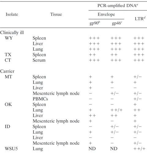
PCR amplification of EIAV sequences from horses. The EIAV sequence heterogeneity present in vivo was explored by analyzing the viral sequences from three clinically ill and four healthy, seropositive, carrier horses. Proviral sequences from tissue DNA from all of the carriers and from two of the acutely ill horses were used for these studies. From the horse infected with the Connecticut strain of virus, analysis was performed on virion RNA extracted from supernatants of in vitro-cultured PBMCs taken during the first clinical episode. Proviral DNA from tissues was primarily used for analysis during these stud-ies because viremia can be, but is not always (29), undetectable in carriers (13, 24, 26). Tissues from the infected horses were tested for the presence of viral sequences from Env gp90, Env gp45, and the LTR (Table 1). Two different sets of gp90 and gp45 primers were used on tissue DNAs to reduce the possi-bility of negative results stemming from template-primer mis-matches. DNAs were scored positive if amplification was con-sistently positive in assays using at least one set of primers. DNAs which were variably positive for EIAV sequences in assays using the same primer set were scored as such. Upon PCR amplification, DNA fragments containing sequences from gp90, gp45, and LTR were detectable by ethidium bro-mide staining from all tissue DNA tested from clinically ill horses. In contrast, proviral sequences from DNA of carrier tissues were usually detectable only upon Southern blot anal-ysis of the amplified material or were not detectable at all. In addition to the results shown, a number of other LTR and envelope PCR primer pairs were tested on the DNA from carrier tissues, with similar results (data not shown). The lim-ited ability to amplify the EIAV sequences from the carriers suggested either that few to no copies of proviral sequences
on November 9, 2019 by guest
http://jvi.asm.org/
were present in these horses or that the primers used could not amplify EIAV sequences due to sequence mismatches. The first alternative is more likely since different sets of primer pairs gave similar amplification results. Low levels of proviral sequences in tissues from the carriers are consistent with pre-vious findings that these horses have low viral loads (26).
Only some tissues from each carrier had detectable levels of proviral sequences following a single round of 40 PCR ampli-fication cycles and Southern blotting. Lung, spleen, liver, and mesenteric lymph node samples were each positive for EIAV sequences in some carriers but not others. Lung samples were the most consistently positive sources of proviral sequences, but even from this tissue, not all viral sequences were detected from all carriers. It appeared that the level of proviral se-quences in tissues from the horse infected with the Idaho (ID) strain of virus was at the threshold of detection, since the spleen, lung, and lymph node DNAs were only sporadically positive and liver samples were never positive.
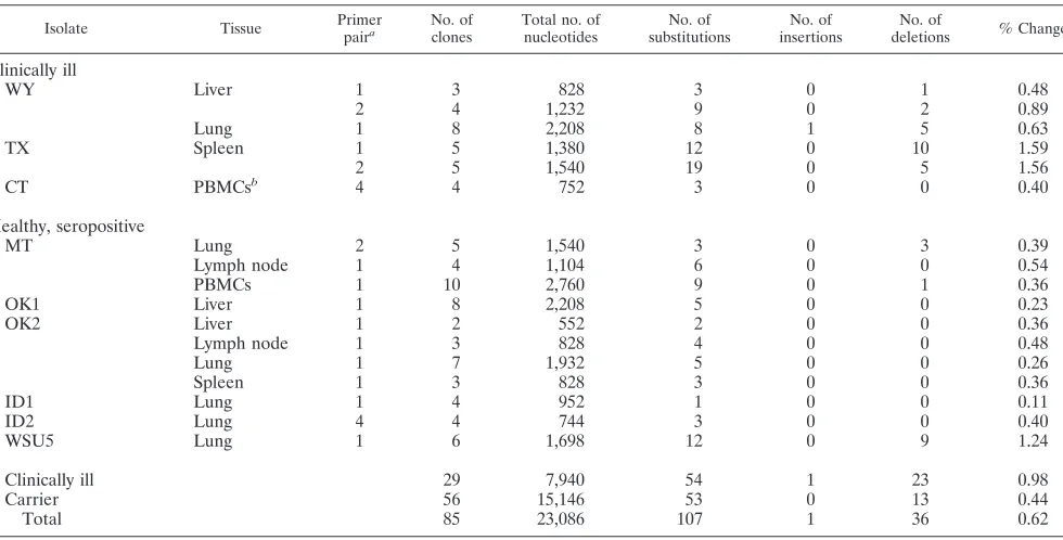
LTR nucleotide variation within individual horses.To de-termine the level of EIAV LTR variation present within each individual animal, the sequences from multiple LTR clones derived from different tissues of each animal were compared
(Table 2). The average nucleotide substitution frequency found between different tissue LTR clones in carriers was 0.44% and ranged from 0.23 to 1.24%. The nucleotide substi-tution frequency found in horses with clinical disease was con-sistently higher, averaging 0.98% and ranging from 0.40 to 1.56% variation. Most nucleotide alterations were found to be either single base pair substitutions or deletions. A single nu-cleotide insertion in a single clone was identified. In several of the carriers, nucleotide changes were observed at such a low frequency that nucleotide misincorporation by Taq polymerase could not be excluded as the cause of the heterogeneity (18, 21).
Two distinct LTR sequences were present in the carriers from Oklahoma and from Idaho. The LTR clones from these animals were not found to be a gradation of sequence variation between these two distinct LTR sequences but, instead, were sets of LTRs which clustered around one of the two distinct sequences. For purposes of analysis, these LTRs were charac-terized as two separate isolates and were designated (i) Okla-homa 1 (OK1) and OK2 and (ii) ID1 and ID2.
EIAV sequence variation between horses was primarily lo-calized to enhancer sequences.It was of interest to investigate EIAV LTR variation not only within each animal but also between different EIAV-infected horses to identify regions of variation within the LTR. A consensus sequence of the ampli-fied clones from each horse was generated by using the mul-tiple sequence alignment program from the Genetics Com-puter Group (GCG; Madison, Wis.), and nucleotide positions that varied between the consensus LTRs were identified (Fig. 1). A similar analysis was performed for seven in vivo-derived HIV-1 LTRs that have been previously published (1, 2, 39). These HIV LTRs were obtained directly from patient tissues or PBMCs. The overall percentage of nucleotide positions where base pair changes occurred was 16.3% in the EIAV LTRs. We found that 24.8% of the nucleotide positions varied between the HIV LTRs amplified from seven different pa-tients; however, one of the HIV isolates was dramatically dif-ferent from the other isolates at the U3-R border, and if this LTR was not included in the analysis, the nucleotide variation within the HIV LTR was found to be similar to that of EIAV, at 16.6%. Despite the relative equivalence of nucleotide changes between the EIAV and HIV LTRs, the distributions of those changes within the LTRs were dramatically different. Changes were found throughout the HIV LTR, with the high-est density of changes found in upstream U3 sequences, and at the U3-R border. One region of the HIV LTR that was con-served was the enhancer region containing two NF-kB sites. In contrast to the location of variation found in HIV, most of the variable nucleotides in the EIAV LTR were found within the enhancer region. Of the 83 nucleotides that compose the cen-tral enhancer region of the EIAV LTR (11, 12, 38, 52), 44.6% of the nucleotide positions were variable between the different in vivo-derived LTRs. The variation found in the remainder of the EIAV LTR was only 4.7%, suggesting that the selective pressures influencing the nucleotide sequence of the enhancer sequences were different from those influencing the rest of the EIAV LTR.
[image:3.612.58.298.89.338.2]Nucleotide changes altered transcription factor binding mo-tifs. The enhancer sequences of EIAV are composed of a number of different transcription factor binding motifs, and the presence of specific motifs can vary between different tissue culture isolates (9, 15). To characterize the sequence variation that occurred in the transcription factor motifs of the in vivo isolates, the consensus sequence of the enhancer region of each in vivo isolate was analyzed and compared to LTR en-hancer regions obtained from in vitro virus cultures in primary TABLE 1. Detection of PCR-amplified viral DNA from
tissues of seropositive horses
Isolate Tissue
PCR-amplified DNAa
Envelope
LTRd
gp90b gp46c
Clinically ill
WY Spleen 111 111 111
Liver 111 111 111
Lung 111 111 111
TX Spleen 11 11 111
CT Serum 111 111 111
Carrier
MT Spleen 1 1 1/2
Lung 1 1 1
Liver 1 2 2
Mesenteric lymph node 2 1/2 1/2
PBMCs 2 2 1/2
OK Spleen 2 2 1
Lung 1 11/1 11
Liver 11 11 1
Mesenteric lymph node 1 2 1
ID Spleen 2 1/2 1/2
Lung 1 1/2 1/2
Liver 2 2 2
Mesenteric lymph node 1 2 1/2
WSU5 Lung ND ND 11/1
a2, negative; 1/2, variably positive by Southern blotting; 1, positive by
Southern blotting only; 11, weakly positive by ethidium bromide staining; 111, strongly positive by ethidium bromide staining; ND, not determined.
bThe primer pairs used to test for the presence of EIAV gp90 sequences in all
tissues were (i) 5552 (TGCTTAGCAGGAACTACTGG) and 5903C9(CCTCA ACACGTACAGAGTTA) and (ii) 5910 (CGAACACAGCGGAATATTGG GG) and 6393C9(TGTACCTGGTTTGTTTCATA). All EIAV oligonucleotides are numbered beginning with the first nucleotide of the 59LTR of the provirus.
cThe primer pairs used to test for the presence of EIAV gp46 sequences were
(i) 6497 (AGAATAAGGAACCAAAGCTT) and 6990C9 (GTTGACTCATT TAAATGTCCCC) and (ii) 6874 (GGAAAGACAACAGGTAGAGG) and 7729C9(TACTATAATTACTAGTCCCCT).
dThe primer pairs used to test for the presence of EIAV LTR sequences were
(i) Xho 7606 (GGTTTTCTCGAGGGGTTTTATAAATG) and Xba 323C9(TC TAGAGTAGGATCTCGAACA) and (ii) 7333 (CCCAAGAAGGAACTCT CG) and MunIC9(AGTGCCCAATTGTCAG). U3 sequences from RT-PCR-amplified samples were RT-PCR-amplified by using Xho 7606 and MunIC9.
on November 9, 2019 by guest
http://jvi.asm.org/
macrophages or fibroblasts (Fig. 2). The LTRs obtained from clinically ill horses had a characteristic enhancer sequence that was highly conserved between the different horses and differ-ent from the overall consensus sequence. These enhancer se-quences were similar to that previously published for a virulent Wyoming strain (47). All LTRs from horses with clinical dis-ease contained an ACCT or AACT at positions296 to 293
[image:4.612.64.554.80.329.2]that altered sequences within the binding motif for the tran-scription factor PEA-2. PEA-2 in fibroblast NEs has been shown to interact with tissue culture-derived EIAV LTRs (11). To test whether these nucleotide changes influenced the bind-ing of PEA-2 to the site, EMSAs were performed. In these studies, fibroblast NEs were used since they interact with the EIAV LTR PEA-2 site (11) but equine macrophage NEs do
[image:4.612.67.548.499.667.2]FIG. 1. Schematic diagram of the heterogeneity found at nucleotide positions in in vivo-derived LTRs from EIAV and HIV. Each vertical hatch mark represents an LTR nucleotide position where heterogeneity occurred. LTR deletions and insertions were not included in this analysis. A consensus sequence of each of the seven EIAV LTRs was generated and aligned by using the GCG multiple alignment program. Representative clones of HIV LTRs taken from the PBMCs of four patients (39) and consensus sequences obtained from a number of tissues from three other patients (1, 2) were aligned by using the GCG multiple alignment program. All HIV isolates used for this analysis were from clade B (42). Overall nucleotide position variations were 16.3% for EIAV and 24.8% for HIV. One of the HIV LTRs (1) dramatically differed from the other six at the U3-R border. If this isolate was excluded from the analysis, the HIV LTR had an overall nucleotide position variation of 16.6%.
TABLE 2. LTR sequence heterogeneity in horses with clinical disease and healthy, seropositive carriers
Isolate Tissue Primerpaira No. ofclones Total no. ofnucleotides substitutionsNo. of insertionsNo. of deletionsNo. of % Change
Clinically ill
WY Liver 1 3 828 3 0 1 0.48
2 4 1,232 9 0 2 0.89
Lung 1 8 2,208 8 1 5 0.63
TX Spleen 1 5 1,380 12 0 10 1.59
2 5 1,540 19 0 5 1.56
CT PBMCsb 4 4 752 3 0 0 0.40
Healthy, seropositive
MT Lung 2 5 1,540 3 0 3 0.39
Lymph node 1 4 1,104 6 0 0 0.54
PBMCs 1 10 2,760 9 0 1 0.36
OK1 Liver 1 8 2,208 5 0 0 0.23
OK2 Liver 1 2 552 2 0 0 0.36
Lymph node 1 3 828 4 0 0 0.48
Lung 1 7 1,932 5 0 0 0.26
Spleen 1 3 828 3 0 0 0.36
ID1 Lung 1 4 952 1 0 0 0.11
ID2 Lung 4 4 744 3 0 0 0.40
WSU5 Lung 1 6 1,698 12 0 9 1.24
Clinically ill 29 7,940 54 1 23 0.98
Carrier 56 15,146 53 0 13 0.44
Total 85 23,086 107 1 36 0.62
aPrimer pair 1 consisted of oligonucleotides Xho 7606 (GGTTTTCTCGAGGGGTTTTATAAATG) and Xba 323C9(TCTAGAGTAGGATCTCGAACA), which,
excluding the primer sequences, resulted in amplification of 276 bp of LTRs from WY, TX, MT, and ID samples, 238 bp from ID1, and 283 bp from WSU5; primer pair 2 consisted of 7333 (CCCAAGAAGGAACTCTCGCT) and Xba 323C9, which amplified 308 bp of LTR sequences from WY, TX, and MT samples. Oligonu-cleotide pairs 3 and 4 were used to amplify U3 sequences from virion RNA from the CT sample, using RT-PCR and nested PCR performed on ID DNA. Primer pair 3 consisted of Xho 323 and MunIC9(AGTGCCCAATTGTCAG), and pair 4 consisted of 7333 and MunIC9, resulting in 188 bp from CT and 186 bp from ID samples.
bVirion RNA was obtained from the supernatant of primary PBMC cultures of the CT horse.
on November 9, 2019 by guest
http://jvi.asm.org/
not (38). As shown in Fig. 3, fibroblast NEs specifically inter-acted with oligonucleotide 105, which contained an intact PEA-2 site. The presence of the T residue at position293 in oligonucleotide 105T eliminated the binding. The absence of the PEA-2 site in all of the LTRs obtained from horses with clinical disease suggests that this site is not involved in causing disease. The nucleotide changes in the PEA-2 site within the LTRs from two of the acutely ill horses (CT and TX samples) resulted in not only an abrogation of the PEA-2 site but also the introduction of a c-myb site (CAACTG). However, in EM-SAs using NEs from macrophages or fibroblasts, no binding of this site was detected (data not shown). In addition to these upstream alterations, LTRs isolated from clinically ill horses were found to contain three PU.1 (Spi-1) sites and a CREB site. This CREB site had previously been identified as an AP-1 site, but recent EMSAs with fibroblast NEs have demonstrated that the CREB family protein ATF-1 binds to this site within the EIAV LTR (38a).
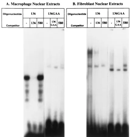
The consensus sequences of the enhancer regions isolated from carriers differed from each other as well as from the overall consensus sequence. The enhancer region isolated from the OK1 LTR had all three PU.1 motifs (GTTCC) re-placed with the nucleotide sequence GTGAA. PU.1 sites have been demonstrated to be important for EIAV LTR transcrip-tion in primary macrophages (38), the cell type infected during acute infection (51). It would be predicted that the absence of the PU.1 motifs in this LTR would result in this enhancer region functioning poorly in primary macrophages. In EMSAs, the band retardation pattern of equine macrophage NEs was altered when the PU.1 motif was mutated from GTTCC to GTGAA (Fig. 4A). The strong, specific binding found with the PU.1 motif was eliminated by the GTGAA mutation. In a related series of experiments, the ability of the
PU.1-contain-ing and the GTGAA-containPU.1-contain-ing oligonucleotides to interact with fibroblast cell line NEs was tested (Fig. 4B). Fibroblast NEs did not specifically bind to the PU.1 motif; however, the GTGAA-containing oligonucleotide did specifically interact with the fibroblast NEs, indicating that a novel protein that did not interact with the PU.1 motif-containing oligonucleotide was interacting with the GTGAA-containing oligonucleotide. Upon overexposure of the autoradiograph of the GTGAA-containing oligonucleotide shown in Fig. 4A, a band of similar size was also retarded by equine macrophage NEs (data not shown). It is not known what nuclear protein(s) interacted with which sequences within the GTGAA-containing oligonucleo-tide, but this finding suggested that the transcription factor motif changes observed in the OK1 LTR may result in the ability of this isolate to replicate in fibroblasts.
[image:5.612.83.534.85.322.2]The second LTR sequence derived from the Oklahoma car-rier (OK2) contained a single GTGAA motif that replaced the central PU.1 site. The remaining two PU.1 sites were intact, but the CREB site was abolished. The Montana carrier con-tained an LTR sequence that was identical to a previously characterized enhancer sequence from a fibroblast-adapted isolate, MA-1 (9). This LTR contained three PU.1 sites as well as octamer, PEA-2, and CREB sites. The consensus sequence from the WSU5 LTR clones contained a seven-nucleotide in-sert in the center of the enhancer region which altered a PU.1 site and introduced an octamer site. ID1 contained a 48-nu-cleotide deletion that encompassed most of the enhancer re-gion. It would be predicted that this LTR would have little or no transcriptional activity. The ID2 LTR contained a more intact enhancer sequence with two PU.1 sites, an octamer site, and a CREB site. Interestingly, five of six of the carrier con-sensus LTRs (MT, OK1, WSU5, OK2, and ID2) retained the PEA-2 site that was absent in all the horses with acute disease. FIG. 2. Enhancer region of the EIAV LTR from the PCR-amplified in vivo samples and from tissue culture isolates. The in vivo LTRs shown are represented by consensus sequences generated by multiple sequence alignments of the sequences of all the clones from each animal. The in vitro isolates are representative clones of that isolate. The overall consensus sequence for the EIAV LTR enhancer region is shown at the bottom. Transcription factor motifs that are known to be present in the EIAV enhancer are underlined in the consensus sequence. Only the nucleotides that differ from the consensus sequence are shown. Dashes represent regions of the LTR that are deleted. The 39dashes in macrophage-derived LTR, eial3, represent unsequenced nucleotide positions rather than deletions.
on November 9, 2019 by guest
http://jvi.asm.org/
In addition to the nucleotide changes found within the en-hancer region, the LTRs of OK1, MT, and ID had a G-to-A change at position138 and a G-to-T change at position148 within R (data not shown). It is not known if these changes result in functional LTR alterations.
Nucleotide variation within the enhancer region was also observed between individual LTR clones from a single carrier but not from an acutely ill horse. As shown in Fig. 5, individual clones from Wyoming (clinically ill) and Montana (carrier) were examined for the level and location of variation. En-hancer sequences of clones derived from separate, indepen-dent PCR amplifications of lung, liver, and spleen from the clinically ill horse infected with the WY strain of virus were found to be completely conserved. Interestingly, a hotspot of variation that was unique to the WY strain-derived LTRs was located upstream from the R-U5 border. The relevance of this variation is not known. In contrast, enhancer sequences from the Montana carrier contained variation within the enhancer region (10 of 1,577 nucleotides analyzed) as well as in other regions of the LTR (12 of 3,667 nucleotides analyzed). This finding indicated that nucleotide variation within the enhancer
region could be found both between different LTR clones obtained from a single carrier as well as from different carriers but not from clinically ill animals.
DISCUSSION
[image:6.612.93.259.67.382.2]This study investigated the LTR sequence heterogeneity present in EIAV-infected horses. A comparison of consensus sequences generated for each of the in vivo-derived LTRs demonstrated that a region within U3 was hypervariable. The hypervariation coincided with sequences known to function as the transcriptional enhancer (11, 17, 38, 52). Not counting LTR enhancer region insertions and deletions, 44.6% of the nucleotide positions (37 of 83) were heterogeneous within the U3 enhancer region, whereas the remainder of the LTR ana-lyzed had 4.7% variation (9 of 193). Only the central 276 nucleotides of the LTR were analyzed in this study, since the primer sequences used during the amplification prevented de-tection of variation in the 59 and 39LTR regions which were overlapped by the primers. The hypervariability of the LTR enhancer region found between the different horses has pre-viously been noted in comparisons of in vitro-derived isolates (9, 15, 46). Our findings indicate that high levels of enhancer region variation between different isolates also occur in vivo.
FIG. 3. EMSAs using a blunt-end, double-stranded,32P-labeled
oligonucle-otide containing the PEA-2 sequence of the EIAV LTR (oligonucleoligonucle-otide 105) or a C-to-T alteration at position293 (oligonucleotide 105T). The C-to-T mutation which eliminated PEA-2 binding is found in all EIAV isolates obtained from clinically ill horses. (A) Oligonucleotides used in the EMSAs. (B) EMSAs dem-onstrating specific binding of NEs from Cf2Th cells to oligonucleotide (Oligo)
105 but no binding to the mutant oligonucleotide (105T). Cf2Th is one of a
number of fibroblast cell lines that support replication of tissue culture-derived isolates of EIAV (9, 47). Excess unlabeled competitor (Comp) oligonucleotides were used to demonstrate binding specificity. Oligonucleotides 91, 105, and 111 contain a PEA-2 site; oligonucleotides 116, 580, and 1566 do not contain an intact PEA-2 site.
FIG. 4. EMSAs using blunt-end, double-stranded,32P-labeled
oligonucleo-tides containing either the GTTCC motif that is the antisense core motif for the transcription factor, PU.1, or a GTGAA motif that replaces the PU.1 sites in the LTR from the carrier from Oklahoma. (A) EMSAs using primary adherent macrophage NEs demonstrated specific binding of oligonucleotide 136 (TCAA TATAGTTCCGCATTTGTGA). This pattern of binding was not observed with the GTGAA-containing oligonucleotide; however, specific retardation of a band higher in the gel was observed upon overexposure of the EMSA gel, using oligonucleotide 136GAA (TCAATATAGTGAAGCATTTGTGA) (data not shown). (B) EMSAs using fibroblast (FEA) NEs did not demonstrate specific binding to the PU.1 motif but did display specific binding to oligonucleotide 136GAA. FEA is a feline fibroblast cell line that supports replication of tissue culture-adapted strains of EIAV (56). Excess unlabeled competitor oligonucle-otides were used to demonstrate binding specificity. Competitor oligonucleotide 136GAA competed specifically for binding of the larger, retarded complex. Competitor oligonucleotide 580 (GGGTGAATACCATACAGACA) did not compete for binding even though a GTGAA motif was present within the oligonucleotide. This finding indicated that the nucleotides surrounding the GTGAA motif of 136GAA were also required for the binding of this novel factor.
on November 9, 2019 by guest
http://jvi.asm.org/
[image:6.612.328.542.335.560.2]FIG. 5. Multiple sequence alignment of LTR-containing clones. (A) LTRs derived from a clinically ill horse infected with the virulent WY strain of EIAV. Individual LTR clones were sequenced from DNA amplification of liver and lung tissue. LTR from infected spleen tissue was sequenced directly from amplified material. Suffixes A and B denote independent PCR amplifications. Consens, consensus. (B) LTR clones were obtained from a healthy, seropositive horse from Montana from lung, mesenteric lymph node (lymnode), and PBMCs (pbmc). PBMCs were activated with either bacterially derived lipopolysaccharide (lps) or phytohemagglutinin (pha) for 5 days prior to DNA isolation, and the clones derived are noted as such.
on November 9, 2019 by guest
http://jvi.asm.org/
Sequence variation in HIV-1 is attributed to high rates of nucleotide misincorporation by HIV RT (23, 25, 43, 45). Sim-ilar rates of misincorporation have been reported for the EIAV RT molecule in in vitro assays (4, 6). Thus, if RT mis-incorporation were solely responsible for the sequence varia-tion found in the EIAV LTR, it might be expected that, as in HIV-1, random variation throughout the EIAV LTR would occur. Yet, clearly this was not the case. The localization of the nucleotide variation within the EIAV LTR suggests that the sequence heterogeneity was not simply the result of random misincorporations of nucleotides by RT, but that selection pressures directed specifically toward enhancer sequences were different from those directed toward the rest of the EIAV LTR, which was highly conserved between different isolates.
A closer comparison of the LTR sequences from the three acutely ill horses demonstrated that enhancer sequences from these LTRs were relatively homogeneous and similar to the LTR sequence of a virulent strain of EIAV reported previously (47). This finding suggested that viral virulence may correlate with the presence of specific sequences within the enhancer region. In contrast to findings for the acutely ill horses, the enhancer regions from the healthy, seropositive carriers dem-onstrated hypervariability. The changes which occurred were unique and characteristic for each horse. Two of the four carriers were found to have two distinct LTR enhancer se-quences which for the purposes of analysis were treated as separate LTR isolates. In the mule from Oklahoma, the two LTR sequences were related in sequence. Thus, one of the sequences may be derived from the other. However, it should be noted that no LTR sequences or quasispecies which were intermediate in sequence between the two LTRs were found in this animal, and intermediate quasispecies might be expected if one strain of LTR was a derivative of the other (57). OK1 LTR sequences were found only in the liver, whereas OK2 LTR sequences were found in all tissues tested. This finding sug-gested that OK1 arose within the liver during the infection, perhaps from OK2-type sequences, and had not spread to other organs. Alternatively, OK1-type sequences had a prefer-ential liver tropism and consequently were not found in other tissues. Two distinct LTR sequences were also observed in DNA from the Idaho horse. These LTRs were not closely related, which suggested that this horse may have been in-fected with multiple strains of virus.
Many of the nucleotide changes found in the U3 enhancer region of carriers resulted in changes of transcription factor motifs, suggesting the intriguing possibility that these changes are in part responsible for the carrier status of these animals. For instance, a loss or modification of at least one PU.1 site was observed in many of the consensus sequences derived from the carriers. PU.1 sites have previously been shown to be im-portant for expression of the LTR in primary equine macro-phages (38). The potentially decreased levels of transcriptional activity resulting from transcription factor motif alterations may in turn reduce virus replication and virus load. These characteristics would be consistent with previous phenotypic descriptions of the carrier state of EIAV where viremia is not detectable and the proviral load in tissues is low (26). Thus, the carrier state may result not only from immunologic control of the virus (28, 48) but also from alterations in EIAV LTR sequences that reduce virus expression. In addition, the data generated from our EMSAs suggested the possibility that cell tropism changes also result from in vivo transcription factor motif alterations. LTR alterations have been demonstrated to change the cell tropism of murine leukemia virus (32, 53). In a similar manner, transcription factor motif changes may alter the cell tropism of EIAV from macrophage tropic during the
clinical phase to fibroblast (or another, yet unidentified cell type) tropic during the carrier phase.
While enhancer sequence hypervariability was found be-tween the different horses, intrahorse sequence variation was lower than that reported for other lentiviruses and suggested that limited genetic drift was occurring during the infection of each horse. As much as 10% nucleotide heterogeneity between clones within an individual animal has been reported for pri-mate lentiviruses (40, 41, 59) and visna virus (8, 50). Levels of EIAV intrahorse sequence variation were more similar to that found in macaque monkeys 170 days after inoculation with an SIV molecular clone (27). Low levels of HIV-2 sequence het-erogeneity within two different individuals have also been re-ported, suggesting that primate lentivirus sequence heteroge-neity can be limited during a natural infection (19). It should be noted that both the SIV and HIV-2 studies investigated early time points during infection, and several primate lentivi-rus studies have shown that sequence heterogeneity is usually limited during early infection (37, 59). The clinically ill horses were sampled early in infection, and thus the early sampling times may account for the low level of variation found between the sequences obtained from these horses. However, some of the carriers had been infected for more than 5 years, and thus early sampling times cannot account for the absence of LTR sequence variation found in the carriers. In addition, it is possible that the lower level of nucleotide substitutions found in EIAV compared with the primate lentiviruses is in part due to the presence in EIAV of the enzyme dUTPase (33, 55, 58), which is not present in primate lentiviruses. The absence of dUTPase activity has been shown to enhance mutation rates in feline immunodeficiency virus (30) and in herpes simplex virus (49). In carriers, the low levels of sequence heterogeneity may result from the absence of viremia and virus spread within these animals. In addition, it cannot be excluded that the low level of sequence variation found within the carriers may be due to the amplification of a limited number of proviral se-quences found in the tissues of these animals.
ACKNOWLEDGMENTS
We thank Vincent Emery for providing compiled HIV LTR se-quences, Keith Weaver for critical evaluation of the manuscript, Anne Shoemaker for technical help, and Kristy Anderson and Glen Malchow for graphic arts assistance.
This study was supported in part by the following grants: NIH CA72063 (W.M.), NIH AI24291 (T.M.), NIH AI30025 (S.C.), NIH HL46651 (T.C.), and NIH AI01255 (J.L.O.).
REFERENCES
1. Ait-Khaled, M., and V. C. Emery. 1994. Phylogenetic relationship between human immunodeficiency virus type 1 (HIV-1) long terminal repeat natural variants present in the lymph node and peripheral blood of three HIV-1-infected individuals. J. Gen. Virol. 75:1615–1621.
2. Ait-Khaled, M., J. E. McLaughlin, M. A. Johnson, and V. Emery. 1995. Distinct HIV-1 long terminal repeat quasispecies present in nervous tissues compared to that in lung, blood and lymphoid tissues of an AIDS patient. Curr. Sci. 9:675–683.
3. Bakhanashvili, M., and A. Hizi. 1992. Fidelity of the RNA-dependent DNA synthesis exhibited by the reverse transcriptases of human immunodeficiency virus types 1 and 2 and of murine leukemia virus: mispair extension frequen-cies. Biochemistry 31:9393–9398.
4. Bakhanashvili, M., and A. Hizi. 1993. Fidelity of DNA synthesis exhibited in vitro by the reverse transcriptase of the lentivirus equine infectious anemia virus. Biochemistry 32:7559–7567.
5. Balfe, P., P. Simmonds, A. Ludlam, J. O. Bishop, and A. J. L. Brown. 1990. Concurrent evolution of human immunodeficiency virus type 1 in patients infected from the same source: rate of sequence change and low frequency of inactivating mutations. J. Virol. 64:6221–6233.
6. Borroto-Esoda, K., and L. R. Boone. 1991. Equine infectious anemia virus and human immunodeficiency virus DNA synthesis in vitro: characterization of the endogenous reverse transcriptase reaction. J. Virol. 65:1952–1959.
on November 9, 2019 by guest
http://jvi.asm.org/
7. Breuer, J., N. W. Douglas, N. Goldman, and R. S. Daniels. 1995. Human immunodeficiency virus type 2 (HIV-2) env gene analysis: prediction of glycoprotein epitopes important for heterotypic neutralization and evidence for three genotype clusters within the HIV-2a subtype. J. Gen. Virol. 76: 333–345.
8. Carey, N., and R. G. Dalziel. 1994. Sequence variation in the gp135 gene of Maedi visna virus strain EV1. Virus Genes 8:115–123.
9. Carpenter, S., S. Alexandersen, M. J. Long, S. Perryman, and B. Chesebro. 1991. Identification of a hypervariable region in the long terminal repeat of equine infectious anemia virus. J. Virol. 65:1605–1610.
10. Carpenter, S., and B. Chesebro. 1989. Change in host cell tropism associated with in vitro replication of equine infectious anemia virus. J. Virol. 63:2492– 2496.
11. Carvalho, M., and D. Derse. 1993. Physical and functional characterization of transcriptional control elements in the equine infectious anemia virus promoter. J. Virol. 67:2064–2074.
12. Carvalho, M., M. Kirkland, and D. Derse. 1993. Protein interactions with DNA elements in variant equine infectious anemia virus enhancers and their impact on transcriptional activity. J. Virol. 67:6586–6595.
13. Cheevers, W. P., and T. C. McGuire. 1985. Equine infectious anemia: im-munopathogenesis and persistence. Rev. Infect. Dis. 7:83–88.
14. Clabough, D. L., D. Gebhard, M. T. Flaherty, L. E. Whetter, S. T. Perry, L.
Coggins, and F. J. Fuller.1991. Immune-mediated thrombocytopenia in horses infected with equine infectious anemia virus. J. Virol. 65:6242–6251. 15. Derse, D., R. Carroll, and M. Carvalho. 1993. Transcriptional regulation of
equine infectious anemia virus. Semin. Virol. 4:61–68.
16. Dignam, J. D., P. L. Martin, B. S. Shastry, and R. F. Roeder. 1983. Eukary-otic gene transcription with purified components. Methods Enzymol. 101: 582–599.
17. Dorn, P. L., and D. Derse. 1988. cis- and trans-acting regulation of gene expression of equine infectious anemia virus. J. Virol. 62:3522–3526. 18. Eckert, K. A., and T. A. Kundel. 1991. DNA polymerase fidelity and the
polymerase chain reaction. PCR Methods Appl. 1:17–24.
19. Gao, F., L. Yue, A. T. White, P. G. Pappas, J. Barchue, A. P. Hanson, B. M.
Greene, P. M. Sharp, and B. H. Hahn.1992. Human infection by genetically diverse SIV-sm-related HIV-2 in West Africa. Nature 358:495–498. 20. Goodenow, M., T. Huet, W. Saurin, S. Kwok, J. Sninsky, and S.
Wain-Hobson.1989. HIV-1 isolates are rapidly evolving quasispecies: evidence for viral mixtures and preferred nucleotide substitutions. J. Acquired Immune Defic. Syndr. 2:344–352.
21. Hengen, P. N. 1995. Fidelity of DNA polymerases for PCR. Trends Biochem. Sci. 20:324–325.
22. Holland, J. J., J. C. De La Torre, and D. A. Steinhauer. 1992. RNA virus populations as quasispecies. Curr. Top. Microbiol. Immunol. 176:1–20. 23. Hubner, A., M. Kruhoffer, F. Grosse, and G. Krauss. 1992. Fidelity of human
immunodeficiency virus type 1 reverse transcriptase in copying natural RNA. J. Mol. Biol. 223:595–600.
24. Issel, C. J., and L. Coggins. 1979. Equine infectious anemia: current knowl-edge. J. Am. Vet. Med. Assoc. 174:727–733.
25. Ji, J., and L. A. Loeb. 1994. Fidelity of HIV-1 reverse transcriptase copying a hypervariable region of the HIV-1 env gene. Virology 199:323–330. 26. Kim, C. H., and J. W. Casey. 1994. In vivo replicative status and envelope
heterogeneity of equine infectious anemia virus in an inapparent carrier. J. Virol. 68:2777–2780.
27. Kodama, T., K. Mori, T. Kawahara, D. J. Ringler, and R. C. Desrosiers. 1993. Analysis of simian immunodeficiency virus sequence variation in tis-sues of rhesus macaques with simian AIDS. J. Virol. 67:6522–6534. 28. Kono, Y. 1969. Viremia and immunological responses in horses infected with
equine infectious anemia virus. Natl. Inst. Anim. Health Q. 9:1–9. 29. Langemeier, J. L., S. J. Cook, R. F. Cook, K. E. Rushlow, R. C. Montelaro,
and C. J. Issel.1996. Detection of equine infectious anemia viral RNA in plasma samples from recently infected and long-term inapparent carrier animals by PCR. J. Clin. Microbiol. 34:1481–1487.
30. Lerner, D. L., P. C. Wagaman, T. R. Phillips, O. Prospero-Garcia, S. J.
Henriksen, H. S. Fox, F. E. Bloom, and J. H. Elder.1995. Increased mutation frequency of feline immunodeficiency virus lacking functional deoxyuridine-triphosphatase. Proc. Natl. Acad. Sci. USA 92:7480–7484.
31. Leroux, C., S. Vuillermoz, J. F. Mornex, and T. Greenland. 1995. Genomic heterogeneity in the pol region of ovine lentiviruses obtained from bron-choalveolar cells of infected sheep from France. J. Gen. Virol. 76:1533–1537. 32. Li, Y., E. Golemis, J. W. Hartley, and N. Hopkins. 1987. Disease specificity of nondefective Friend and Moloney murine leukemia viruses is controlled by a small number of nucleotides. J. Virol. 61:693–700.
33. Lichtenstein, D. L., K. E. Rushlow, R. F. Cook, M. L. Raabe, C. J. Swardson,
G. J. Kociba, C. J. Issel, and R. C. Montelaro.1995. Replication in vitro and in vivo of an equine infectious anemia virus mutant deficient in dUTPase activity. J. Virol. 69:2881–2888.
34. Louwagie, J., F. E. McCutchan, M. Peeters, T. P. Brennan, E. Sanders-Buell,
G. A. Eddy, G. van der Groen, K. Fransen, G. M. Gershy-Damet, R. Deleys, et al.1993. Phylogenetic analysis of gag genes from 70 international HIV-1
isolates provides evidence for multiple genotypes. AIDS 7:769–780. 35. Manns, A., H. Konig, M. Baier, R. Kurth, and F. Grosse. 1991. Fidelity of
reverse transcriptase of the simian immunodeficiency virus from African monkey. Nucleic Acids Res. 19:533–537.
36. Mansky, L. M., and H. M. Temin. 1996. Lower in vivo mutation rate of human immunodeficiency virus type 1 than that predicted from the fidelity of purified reverse transcriptase. J. Virol. 69:5087–5094.
37. Markham, R. B., X. Yu, H. Farzadegan, S. C. Ray, and D. Vlahov. 1995. Human immunodeficiency virus type 1 env and p17gag sequence variation in polymerase chain reaction-positive, seronegative injection drug users. J. In-fect. Dis. 171:797–804.
38. Maury, W. 1994. Monocyte maturation controls expression of equine infec-tious anemia virus. J. Virol. 68:6270–6279.
38a.Maury, W. J. Unpublished data.
39. Michael, N. L., L. D’Arcy, P. K. Ehrenberg, and R. R. Redfield. 1994. Naturally occurring genotypes of the human immunodeficiency virus type 1 long terminal repeat display a wide range of basal and tat-induced transcrip-tional activities. J. Virol. 68:3163–3174.
40. Monken, C. E., B. Wu, and A. Srinivasan. 1995. High resolution analysis of HIV-1 quasispecies in the brain. AIDS 9:345–349.
41. Murphy, E., B. Korber, M. C. Georges-Courbot, B. You, A. Pinter, D. Cook,
M. P. Kieny, A. Georges, C. Mathiot, F. Barre-Sinoussi, et al.1996. Diversity of V3 region sequences of human immunodeficiency viruses type 1 from the Central African Republic. AIDS Res. Hum. Retroviruses 9:997–1006. 42. Myers, G., B. Korber, B. H. Hahn, K.-T. Jeang, J. W. Mellors, F. E.
Mc-Cutchan, L. E. Henderson, and G. N. Pavlakis.1995. Human retroviruses and AIDS 1995. Theoretical Biology and Biophysics, Los Alamos, N.Mex. 43. Nowak, M. 1990. HIV mutation rate. Nature 347:522.
44. O’Rourke, K. I., L. E. Perryman, and T. C. McGuire. 1988. Antiviral, anti-glycoprotein and neutralizing antibodies in foals with equine infectious ane-mia virus. J. Gen. Virol. 69:667–674.
45. Patel, P. H., and B. D. Preston. 1994. Marked infidelity of human immuno-deficiency virus type 1 reverse transcriptase at RNA and DNA template ends. Proc. Natl. Acad. Sci. USA 91:549–553.
46. Payne, S. L., J. Rausch, K. Rushlow, R. C. Montelaro, C. Issel, M. Flaherty,
S. Perry, D. Sellon, and F. Fuller.1994. Characterization of infectious mo-lecular clones of equine infectious anemia virus. J. Gen. Virol. 75:425–429. 47. Perry, S. T., M. T. Flaherty, M. J. Kelley, D. L. Clabough, S. R. Tronick, L.
Coggins, L. Whetter, C. R. Lengel, and F. Fuller.1992. The surface envelope protein gene region of equine infectious anemia virus is not an important determinant of tropism in vitro. J. Virol. 66:4085–4097.
48. Perryman, L. E., K. I. O’Rourke, P. H. Mason, and T. C. McGuire. 1988. Immune responses are required to terminate viremia in equine infectious anemia lentivirus infection. J. Virol. 62:3073–3076.
49. Pyles, R. B., and R. L. Thompson. 1994. Mutations in accessory DNA replicating functions alter the relative mutation frequency of herpes simplex virus type 1 strains in cultured murine cells. J. Virol. 68:4514–4524. 50. Sargan, S. R., I. D. Bennet, C. Cousens, D. J. Roy, B. A. Blacklaws, R. G.
Dalziel, N. J. Watt, and I. McConnell.1991. British isolate of maedi visna virus. J. Gen. Virol. 72:1893–1903.
51. Sellon, D. C., S. T. Perry, L. Coggins, and F. J. Fuller. 1992. Wild-type equine infectious anemia virus replicates in vivo predominantly in tissue macrophages, not in peripheral blood monocytes. J. Virol. 66:5906–5913. 52. Sherman, L., A. Yaniv, H. Lichtman-Pleban, S. R. Tronick, and A. Gazit.
1989. Analysis of regulatory elements of the equine infectious anemia virus and caprine arthritis-encephalitis virus long terminal repeats. J. Virol. 63: 4925–4931.
53. Short, M. K., S. A. Okenquist, and J. Lenz. 1987. Correlation of leukemo-genic potential of murine retroviruses with transcriptional tissue preference of the viral long terminal repeats. J. Virol. 61:1067–1072.
54. Starcich, B. R., B. H. Hahn, G. M. Shaw, P. D. McNeely, S. Modrow, H. Wolf,
E. S. Parks, W. P. Parks, S. F. Josephs, R. C. Gallo, and F. Wong-Staal.1986. Identification and characterization of conserved and variable regions in the envelope gene of HTLV III/LAV, the retrovirus of AIDS. Cell 45:637–648. 55. Steagall, W. K., M. D. Robek, S. T. Perry, F. J. Fuller, and S. L. Payne. 1995. Incorporation of uracil into viral DNA correlates with reduced replication of EIAV in macrophages. Virology 210:302–313.
56. Stephens, R. M., D. Derse, and N. R. Rice. 1990. Cloning and characteriza-tion of cDNAs encoding equine infectious anemia virus Tat and putative Rev proteins. J. Virol. 64:3716–3725.
57. Tao, B., and P. N. Fultz. 1995. Molecular and biological analyses of quasi-species during evolution of a virulent simian immunodeficiency virus, SIVsmmPBj14. J. Virol. 69:2031–2037.
58. Threadgill, D. S., W. K. Steagall, M. T. Flaherty, F. Fuller, S. Perry, K. E.
Rushlow, F. J. LeGrice, and S. Payne.1993. Characterization of equine infectious anemia virus dUTPase: growth properties of a dUTPase-deficient mutant. J. Virol. 67:2592–2600.
59. Wolfs, T. F., G. Zwart, M. Bakker, and J. Goudsmit. 1992. HIV-1 genomic RNA diversification following sexual and parenteral virus transmission. Vi-rology 189:103–110.


