R E S E A R C H
Open Access
Development of metabolic and inflammatory
mediator biomarker phenotyping for early
diagnosis and triage of pediatric sepsis
Beata Mickiewicz
1, Graham C. Thompson
2, Jaime Blackwood
3, Craig N. Jenne
4,5, Brent W. Winston
5,6,7,
Hans J. Vogel
1, Ari R. Joffe
3*and for the Alberta Sepsis Network
Abstract
Introduction:The first steps in goal-directed therapy for sepsis are early diagnosis followed by appropriate triage. These steps are usually left to the physician’s judgment, as there is no accepted biomarker available. We aimed to determine biomarker phenotypes that differentiate children with sepsis who require intensive care from those who do not.
Methods:We conducted a prospective, observational nested cohort study at two pediatric intensive care units (PICUs) and one pediatric emergency department (ED). Children ages 2–17 years presenting to the PICU or ED with sepsis or presenting for procedural sedation to the ED were enrolled. We used the judgment of regional pediatric ED and PICU attending physicians as the standard to determine triage location (PICU or ED). We performed metabolic and inflammatory protein mediator profiling with serum and plasma samples, respectively, collected upon presentation, followed by multivariate statistical analysis.
Results:Ninety-four PICU sepsis, 81 ED sepsis, and 63 ED control patients were included. Metabolomic profiling revealed clear separation of groups, differentiating PICU sepsis from ED sepsis with accuracy of 0.89, area under the receiver operating characteristic curve (AUROC) of 0.96 (standard deviation [SD] 0.01), and predictive ability (Q2) of 0.60. Protein mediator profiling also showed clear separation of the groups, differentiating PICU sepsis from ED sepsis with accuracy of 0.78 and AUROC of 0.88 (SD 0.03). Combining metabolomic and protein mediator profiling improved the model (Q2=0.62), differentiating PICU sepsis from ED sepsis with accuracy of 0.87 and AUROC of 0.95 (SD 0.01). Separation of PICU sepsis or ED sepsis from ED controls was even more accurate. Prespecified age subgroups (2–5 years old and 6–17 years old) improved model accuracy minimally. Seventeen metabolites or protein mediators accounted for separation of PICU sepsis and ED sepsis with 95 % confidence.
Conclusions:In children ages 2–17 years, combining metabolomic and inflammatory protein mediator profiling early after presentation may differentiate children with sepsis requiring care in a PICU from children with or without sepsis safely cared for outside a PICU. This may aid in making triage decisions, particularly in an ED without
pediatric expertise. This finding requires validation in an independent cohort.
* Correspondence:ari.joffe@albertahealthservices.ca
3Division of Pediatric Critical Care Medicine, Department of Pediatrics,
University of Alberta, 4-546 Edmonton Clinic Health Academy; 11405 87 Avenue, Edmonton, AB T6G 1C9, Canada
Full list of author information is available at the end of the article
© 2015 Mickiewicz et al.Open AccessThis article is distributed under the terms of the Creative Commons Attribution 4.0 International License (http://creativecommons.org/licenses/by/4.0/), which permits unrestricted use, distribution, and reproduction in any medium, provided you give appropriate credit to the original author(s) and the source, provide a link to the Creative Commons license, and indicate if changes were made. The Creative Commons Public Domain Dedication waiver (http://creativecommons.org/publicdomain/zero/1.0/) applies to the data made available in this article, unless otherwise stated.
Mickiewiczet al. Critical Care (2015) 19:320
Introduction
Sepsis is a leading cause of mortality in children world-wide [1]. In children age ≥1 year in the United States, sepsis is the second most common cause of death [2, 3]. The incidence of sepsis in children is increasing. Re-searchers in one study found an 81 % increase in hospitalizations for severe sepsis between 1995 and 2005 [4–6]. In addition, sepsis may lead to significant physical, neuropsychological, and neurocognitive mor-bidity in survivors [7–10]. For example, in a recent multicenter study, investigators found that 34 % of survivors of severe sepsis had a decline in their func-tional status at 28 days, and 18 % were determined to have a “poor” functional outcome (moderate, severe, or vegetative disability) [10].
Early, appropriate antibiotic therapy, fluid resuscita-tion, and vasoactive support are associated with im-proved outcomes after sepsis in a highly time-dependent manner [11–13]. The first steps in this therapy are the early diagnosis of sepsis followed by the appropriate stratification of patients (e.g., admission to a hospital ward for observation or to a more intensive monitoring environment in a critical care unit). These first steps are left to the physician’s judgment, as there is currently no accepted, accurate biomarker available to help in making this decision [13–15]. Physicians’ judgment in pediatric emergency departments (EDs) with experienced physi-cians and in tertiary pediatric intensive care units (PICUs) is likely quite different from that in smaller cen-ters without extensive experience with sepsis in children [16]. The vast majority (>80 %) of children requiring emergency care present to an ED that does not have this specialized pediatric expertise [16, 17]. This problem has led to“great interest in developing diagnostic and strati-fication biomarkers for sepsis” [18]. A systems biology approach to biomarker phenotyping of the systemic re-sponse to sepsis has the potential to provide diagnostic and patient stratification profiles that inform clinical decision-making [19]. Unlike more proximal genomic and transcriptomic analysis, untargeted metabolomics and targeted proteomics reflect the downstream systems-level metabolic processes and pathways at play, defining specific phenotypes and shedding light on underlying patho-physiology [20, 21].
In previous work, our group has found that metabolo-mic modeling in adults accurately differentiated 39 pa-tients with septic shock papa-tients from 20 intensive care unit (ICU) control patients and that in children, it differ-entiated 58 PICU patients with septic shock from 39 PICU control patients [22, 23]. In adults, combining metabolomic and protein mediator data more accurately differentiated patients with septic shock from ICU con-trols [24]. In these studies, our group demonstrated promising metabolic biomarker profiles for diagnosing
septic shock in ICU patients. In the present study, we asked different questions:
1. Is there a biomarker-defined phenotype that can dif-ferentiate children with sepsis who require intensive care from those who do not?
2. Does combining metabolic profiling with an analysis of protein mediators improve modeling?
3. Are there a limited number of biomarkers that may be used for targeted phenotyping in future studies?
As there is currently no objective gold standard for making this decision in the clinic, we used the judgment of experienced regional pediatric ED and PICU attend-ing physicians as the standard for triage location of care. This standard is the same as that used for telemedicine and pediatric transport systems, where the expertise at the specialized center is relied upon [16].
Methods Ethical approval
This study was approved by the Health Research Ethics Board of the University of Alberta (Pro00008797) and the Conjoint Health Research Ethics Board of the University of Calgary (Ethics ID 23426).
Patient cohorts PICU sepsis cohort
The Alberta Sepsis Network (ASN) prospectively en-rolled all eligible children up to age 17 years admitted to the only two PICUs in the Province of Alberta, Canada, with a diagnosis of sepsis between April 2010 and October 2013.Sepsiswas defined as the systemic inflammatory re-sponse syndrome (SIRS) caused by a suspected or proven bacterial or fungal infection [25], with antibiotics ordered and an arterial and/or central venous line in place. Pa-tients not expected to survive ≥24 h, refusing intubation or vasoactive infusions (i.e., palliative care), or already having had severe sepsis for ≥48 h (defined as sepsis with cardiovascular dysfunction, acute respiratory dis-tress syndrome, or two other organ dysfunctions) [25] were excluded. Demographic, infection and severity-of-illness variables (including pediatric logistic organ dysfunction [PELOD] and Pediatric Risk of Mortality [PRISM III] scores) were recorded prospectively [26, 27]. Blood was drawn as soon as possible on the day the pa-tient met the eligibility criteria, using deferred consent. If consent was subsequently refused, the blood was not used and the patient was not enrolled. Patients were divided into the predefined 2–5-year-old and 6–17-year-old age groups on the basis of suspected pathophysiology, as done by others [23, 25], with microbiologically confirmed sepsis (positive culture from a normally sterile site, including blood, cerebrospinal fluid, peritoneal fluid, or tissue) or pneumonia without microbiological confirmation (SIRS with chest infiltrate suggestive of pneumonia). We did not
consider sputum or endotracheal aspirates as sterile site cultures or require them as confirmatory of pneumonia, and it is not current practice to perform bronchoalveolar lavage in patients with suspected pneumonia. The site of infection was defined as that diagnosed by the attending medical team.
ED sepsis cohort
The ASN prospectively enrolled all children age ≤17 years admitted to one pediatric ED in Alberta with a diagnosis of sepsis.Sepsiswas defined as SIRS caused by a suspected or proven bacterial or fungal infection [25], with antibiotics and blood culture ordered. Patients who were admitted to the PICU from the ED were included in the PICU sepsis cohort and not in the ED sepsis co-hort. Basic demographic information was recorded, and blood was drawn as soon as possible on the day the pa-tient met the eligibility criteria after informed consent was obtained. The site of infection was defined as that diagnosed by the attending medical team.
ED control cohort
In the same pediatric ED, all previously healthy children age ≤17 years admitted for a procedure and without an infection (i.e., no history of fever within 2 weeks and no clinical evidence of infection) were prospectively enrolled. These children had traumatic lacerations, fractures and/or dislocations, or foreign body removal requiring intraven-ous sedation and/or analgesia. Blood was drawn after con-sent was obtained, and basic demographic information was recorded.
Sample collection and preparation
Samples were obtained via an existing arterial or central venous catheter (PICU sepsis cohort) or an intravenous tube insertion or blood culture draw (ED cohorts). Sample processing was performed as described in Additional file 1.
Proton nuclear magnetic resonance spectroscopy and metabolite concentration profiling
Proton nuclear magnetic resonance (1H-NMR) spectra were acquired on a Bruker AVANCE-II 600 MHz spec-trometer (Bruker BioSpin, Milton, ON, Canada). All spectra were randomly ordered for untargeted profiling to avoid progressive bias. Untargeted profiling involves identification of different compounds by their character-istic spectral signature using information that is stored in an external metabolite reference database [28–30]. Detailed methods are described in Additional file 1.
Protein mediator profiling
Quantification of targeted protein mediators (cytokines, chemokines, and acute-phase proteins involved in inflam-mation) was done using validated Luminex bead-based
multiplexing assays according to manufacturer’s instruc-tions (Luminex, Austin, TX, USA). Detailed methods are described in Additional file 1.
Statistical modeling
Multivariate statistical analysis was performed using the SIMCA-P+ software (v12.0.1; Umetrics, Umeå, Sweden) [19, 31–35]. All metabolites or protein mediators with >50 % missing values were excluded from analysis. Data preprocessing (median fold change normalization, logarithmic transformation, centering, and unit variance scaling) was first conducted separately for the metabolic and protein mediator datasets and then for the combined dataset [33]. An unsupervised principal component ana-lysis (PCA) was performed to obtain an overview of the multivariate dataset and to identify and exclude outliers that could seriously disturb the supervised models [31]. Outliers are defined as those samples that are situated outside the 95 % confidence interval of the Hotelling’sT2 distribution (elliptical or spherical area in the score scat-terplot) [31]. Then, supervised partial least squares dis-criminant analysis (PLS-DA) and orthogonal PLS-DA (OPLS-DA) models were developed to determine the best class discrimination (PICU sepsis, ED sepsis, and ED con-trol) based on the preprocessed original data [31–35]. The OPLS-DA method was applied to models including only two classes. The OPLS-DA models for the combined data-sets were based on potentially relevant metabolites and protein mediator data selected using the variable import-ance to projection (VIP), and only those variables with VIP >1 were chosen [31]. In supervised analysis,R2Y (the percentage of variation explained by the model) and Q2 (the predictive ability of the model) metrics were calcu-lated using a sevenfold cross-validation method [31, 36]. Additionally, the OPLS-DA models were validated by cal-culating coefficient of variation-analysis of variance p values and the receiver operating characteristic curve (ROC) (Metz ROC Software; University of Chicago, Chicago, IL USA) [37, 38]. The sensitivity, specificity, and accuracy were determined on the basis of sample class prediction during sevenfold cross-validation (Y-predcv) using SIMCA-P+ software. To describe specific biopatterns for 2–17-year-old children using combined NMR and protein mediator data, the OPLS-DA regression coefficients were calculated and metabolites and/or protein mediators with significant changes in concen-tration (p <0.05) were considered as the most import-ant variables [31].
Results
Description of the cohorts
The demographics, sites of infection, and severity-of-illness measures for each age and category cohort are given in Table 1. Children meeting ASN eligibility were
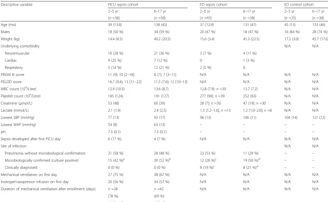
Table 1Description of the three cohorts of patients
Descriptive variable PICU sepsis cohort ED sepsis cohort ED control cohort
2–5 yr 6–17 yr 2–5 yr 6–17 yr 2–5 yr 6–17 yr
(n=36) (n=58) (n=43) (n=38) (n=25) (n=38)
Age (mo) 39 (13.8) 138 (45) 37 (12.9) 131 (47) 45 (13) 133 (46)
Males 18 (50 %) 34 (59 %) 20 (47 %) 18 (47 %) 16 (64 %) 28 (74 %)
Weight (kg) 14.4 (4.5) 40.2 (20.5) 15.6 (3.4) 41.3 (22.5) 17.2 (3.0) 45.7 (17.6)
Underlying comorbidity N/A N/A
Neuromuscular 10 (28 %) 21 (36 %) 3 (7 %) 4 (11 %)
Cardiac 9 (25 %) 7 (12 %) 0 1 (3 %)
Respiratory 5 (14 %) 12 (21 %) 2 (5 %) 0
PRISM III score 11 (9); 10 [2–18] 8 (7); 7 [3–11] N/A N/A N/A N/A
PELOD score 16.7 (9.4); 13 [11–22] 11.5 (7.6); 12 [10–13] N/A N/A N/A N/A
WBC count (109/Litre) 12.9 (10.3) 13.6 (8,7) 12.8 (7.9);n=39 13.7 (7.2) N/A N/A
Platelet count (109/Litre) 185 (124) 191 (127) 277 (99);n=39 252 (93) N/A N/A
Creatinine (μmol/L) 53 (48) 60 (39) 28 (7);n=30 47 (19);n=30 N/A N/A
Lactate (mmol/L) 2.1 (1.9) 2.4 (2.5) 1.3 [1.2–1.6];n=13 1.2 [1.0–2.0];n=8 N/A N/A
Lowest SBP (mmHg) 77 (13) 92 (17) 96 (13) 106 (11) 104 (14) 121 (12)
Lowest MAP (mmHg) 54 (8) 63 (13) – – – –
pH 7.3 (0.1) 7.3 (0.1) – – – –
Sepsis developed after first PICU day 6 (17 %) 4 (7 %) N/A N/A N/A N/A
Site of infection N/A N/A
Pneumonia without microbiological confirmation 21 (58 %) 28 (48 %) 23 (53 %) 11 (29 %) – –
Microbiologically confirmed (culture positive) 15 (42 %)a 30 (52 %)b 12 (28 %)c 19 (50 %)d – –
Clinically diagnosed 0 (0 %) 0 (0 %) 8 (19 %)c 8 (21 %)d – –
Mechanical ventilation on first day 27 (75 %) 38 (67 %) N/A N/A N/A N/A
Inotrope/vasopressor infusion on first day 20 (56 %) 34 (57 %) N/A N/A N/A N/A
Duration of mechanical ventilation after enrollment (days) n=28 n=42 N/A N/A N/A N/A
(78 %) (69 %)
10 [5–13] 6 [3–8]
Mickiewic
z
et
al.
Critical
Care
(2015) 19:320
Page
4
of
Table 1Description of the three cohorts of patients(Continued)
PICU length of stay after enrollment (days) <2 days: 2 (6 %) <2 days: 4 (7 %) Hospital stay Hospital stay N/A N/A
8 [3–15] 7 [3–10] 4 [3, 5];n=20 4 [3, 6];n=19
RRT 4 (11 %) 2 (4 %) 0 0 N/A N/A
ECLS therapy 6 (17 %) 4 (7 %) 0 0 N/A N/A
PICU mortality 1 (3 %) 1 (2 %) 0 0 0 0
Abbreviations:ECLSextracorporeal life support,MAPmean arterial pressure,N/Anot applicable,PELODpediatric logistic organ dysfunction,PICUpediatric intensive care unit,PRISMPediatric Risk of Mortality,RRTrenal replacement therapy,SBPsystolic blood pressure;WBCwhite blood cells
Results are given asn(%) or mean (SD) or median [IQR]. a
Sites of infection included meningitis (n=2), bacteremia (n=7), empyema (n=4), mediastinitis (n=1), and peritonitis (n=3) b
Sites of infection included meningitis (n=9), bacteremia (n=12), empyema (n=3), peritonitis (n=7), and fasciitis (n=1) c
Sites of infection included microbiologically confirmed meningitis (n=3), bacteremia (n=1), peritonitis (n=2), urinary tract infection (n=4), andStreptococcus pyogenesthroat culture (n=2) and clinically diagnosed otitis media with draining ear or mastoiditis (n=3), cellulitis (n=2), cervical adenitis (n=1), and other (n=2)
d
Sites of infection included microbiologically confirmed bacteremia (n=3), peritonitis (n=7), urinary tract infection (n=4), otitis media withS. pyogenesthroat infection (n=5), clinically diagnosed otitis media with mastoiditis (n=2), osteomyelitis (n=1), pelvic abscess (n=1), toxic shock syndrome (n=1), and other (3)
Mickiewic
z
et
al.
Critical
Care
(2015) 19:320
Page
5
of
prospectively entered into the PICU-ASN database after deferred consent was obtained (refusal rate of 22 %). Of 205 patients in the PICU-ASN database, those under 24 months of age (n=63), with missing data (n=3), or with-out clear bacterial sepsis (unknown, viral or other causes of SIRS) were excluded from this PICU sepsis nested co-hort. The PICU sepsis cohort patients had PELOD and PRISM III severity-of-illness scores comparable to other PICU sepsis trial patients [39, 40]. Most PICU patients were ventilated (for about 1 week) and received support-ive vasoactsupport-ive infusions on the first day of sepsis. A sig-nificant number required renal replacement therapy and extracorporeal life support during their PICU stay. The average PICU length of stay after sepsis was about 1 week. The ED sepsis cohorts showed comparable demo-graphics, except for having fewer comorbidities and bet-ter values for platelet count and systolic blood pressure. The ED control cohorts had proportionately more male subjects. Only 3 (3 %) of 94 of the PICU sepsis cohort were admitted to the PICU from the ward; these 3 pa-tients had blood for analysis drawn in the ED, and none were outliers in the models.
2–17-year-old children
Ninety-four PICU sepsis, 81 ED sepsis, and 63 ED con-trol patients were included. A total of 58 metabolites and 48 protein mediators were recognized and quanti-fied for each patient (Additional file 1: Table E1). Totals of 1.12 % and 2.01 % missing values were observed in the NMR and protein mediator datasets, and these were randomly distributed.
Profiling
Metabolomic profiling
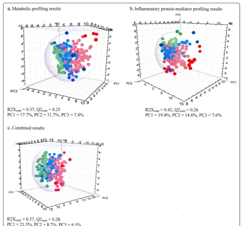
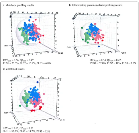
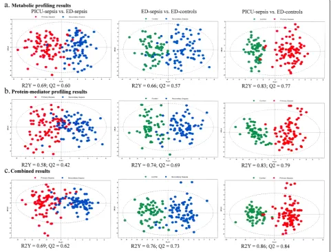
Three principal components (PCs) were calculated via cross-validation to build the PCA model, with good data grouping, explaining the following percentages of vari-ation: PC1 17.7 %, PC2 11.7 %, and PC3 7.4 % (Fig. 1a and Additional file 2: Figure E7a). There were nine out-liers comprising 6 (6 %) in the PICU sepsis cohort and 3 (4 %) in the ED sepsis cohort. These outliers were ex-cluded from subsequent analyses. A supervised PLS-DA showed that the three different cohorts were well clus-tered, with specific metabolic profiles for each. The model showed excellent goodness of fit (cumulative R2Y =0.56) and goodness of prediction (cumulativeQ2=0.47) (Fig. 2a). The OPLS-DA method was applied to compare metabolic variance in patient groups consisting of only two classes. The score scatterplots for each statistical analysis show clear separation of groups, with high values for the R2Y and Q2parameters (Fig. 3a and Table 2). The predictive accuracy statistics for differentiating PICU sepsis from ED sepsis (Table 2) show accuracy of 0.89 and area under the ROC (AUROC) of 0.96 (standard deviation [SD] 0.01).
Protein mediator profiling
PCA revealed three PCs explaining the following per-centages of variation: PC1 19.4 %, PC2 14.6 %, and PC3 7.6 % (Fig. 1b and Additional file 2: Figure E7b). A total of 20 outliers comprising 13 (14 %) in the PICU sepsis cohort, 4 (5 %) in the ED sepsis cohort, and 3 (5 %) in the ED control cohort were excluded from further ana-lyses. The PLS-DA model shows that the three different cohorts are reasonably well clustered, with anR2Y cumu-lative score of 0.54 and a Q2 cumulative score of 0.47 (Fig. 2b). The score scatterplots for each OPLS-DA statis-tical analysis show clear separation of groups, with high values for theR2Y andQ2parameters (Fig. 3b and Table 2). The predictive accuracy statistics for differentiating PICU sepsis from ED sepsis (Table 2) showed accuracy of 0.78 and AUROC of 0.88 (SD 0.03), which were not as high as the values for NMR metabolomics.
Combined results
When PCA was performed, a three-PC model explained the following percentages of variation: PC1 21.5 %, PC2 8.7 %, and PC3 6.5 % (Fig. 1c and Additional file 2: Figure E7c). There were eight outliers comprising seven (7 %) in the PICU sepsis cohort and one (1 %) in the ED sepsis co-hort. These outliers were excluded from subsequent ana-lyses. A supervised PLS-DA shows that the three different cohorts are well clustered, with excellent model descrip-tive values: cumuladescrip-tive R2Y 0.63 and cumulative Q20.56 (Fig. 2c). The score scatterplots for each OPLS-DA statis-tical analysis show clear separation of the groups, with high values for the R2Y and Q2parameters (Fig. 3c and Table 2). The predictive accuracy statistics for differentiat-ing PICU sepsis from ED sepsis (Table 2) show accuracy of 0.87 and AUROC 0.95 (SD 0.01). This model had very similar accuracy statistics compared with NMR alone; however, the sensitivity was higher (0.90 vs 0.86), the spe-cificity was lower (0.85 vs 0.91), and goodness of predic-tion was higher (Q2
0.62 vs 0.60).
Models without outliers excluded
To confirm that the outliers detected in the PCA models (and subsequently excluded) did not bias the results of the supervised analyses, we recalculated the PLS-DA and OPLS-DA models including all outliers. This had only minor influence on the discriminative and predict-ive ability of the models (Additional file 1: Table E2). For example, for the combined dataset model differentiating PICU sepsis from ED sepsis, R2Y =0.67 and Q2 =0.63 compared with the model with outliers excluded where R2
Y =0.69 andQ2=0.62.
Age subgroups
Full details of the results for the separate metabolomic and protein mediator analyses and the combined analyses
in the 2–5-year-old and the 6–17-year-old children are given in Additional files 1 and 2. A summary of the results is shown in Table 2.
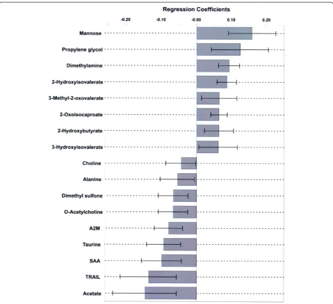
Most meaningful metabolites and protein mediators Using the combined data, a total of 14 metabolites and 3 protein mediators defined the most significant differences responsible for the separation between PICU sepsis and ED sepsis cohorts in the OPLS-DA model (Fig. 4).
Discussion
The main findings of this nested cohort comparison of three groups of patients ages 2–17 years with or without sepsis include the following. First, our metabolomic ana-lysis using 1H-NMR spectroscopy clearly distinguished patients with sepsis requiring care in a PICU (n =94) from those with sepsis in the ED (n=81) and those with-out sepsis in the ED (controls, n =63) never requiring care in a PICU, with few outliers excluded from the
Fig. 1Principal component analysis (PCA) results for the 2–17-year-old cohorts based on the preprocessed original data (where each point represents one patient).aMetabolomic profiling data.bInflammatory protein mediator profiling data.cCombined biomarker profiling data. The three-dimensional PCA score scatterplots show the distribution of observations (red dots, pediatric intensive care unit sepsis patients;blue dots, emergency department [ED] sepsis patients;green dots, ED controls) in the three-dimensional space formed by principal components PC1, PC2, and PC3. The PCs are the lines in the multivariable dimensional space (variables: metabolites, protein mediators) that best approximate the observations in the least squares sense. The sphere describes the 95 % confidence interval of the Hotelling’sT2distribution
[image:7.595.50.541.84.544.2]model (6 % of the PICU sepsis cohort). Second, protein mediator profiling also distinguished between these groups; however, there were more outliers that had to be excluded from these analyses, limiting the accuracy of this method alone. Third, combining metabolomic and protein mediator data produced relatively few outliers (7 % of the PICU sepsis cohort), AUROC of 0.95 (SD 0.01), and the strongest model (Q2
=0.62), being more inform-ative than either dataset alone. Fourth, the prespecified
age subgroups resulted in only marginally more accurate models, but at the price of smaller patient numbers, and more outliers were excluded from model development. Fifth, a small group of 17 metabolites and protein medi-ators accounted for the separation of PICU sepsis and ED sepsis cohorts with 95 % confidence. Taken together, our data suggest that development of a laboratory test using these findings to help make diagnostic and triage decisions in children with sepsis is feasible.
Fig. 2Partial least squares discriminant analysis (PLS-DA) for the 2–17-year-old cohorts based on the preprocessed original data.aMetabolomic profiling data.bInflammatory protein mediator profiling data.cCombined biomarker profiling data. The three-dimensional PLS-DA score scatterplots show the distribution of observations (red dots, pediatric intensive care unit [PICU] sepsis patients;blue dots, emergency department [ED] sepsis patients;green dots, ED controls) in the three-dimensional space formed by PLS components (PLS1, PLS2, and PLS3). During the model construction, a discriminant plane (PLS component) was found in which the projected observations were well separated according to the class (PICU sepsis, ED sepsis, ED controls). The sphere describes the 95 % confidence interval of the Hotelling’sT2distribution
[image:8.595.55.541.84.548.2]These results are important for several reasons. First, they demonstrate proof of concept for using biomarker phenotyping in a clinical environment, generating infor-mation on patient biology that can guide clinicians’ diag-nosis and triage decisions [19]. Second, the potential of this biomarker phenotyping has been demonstrated in a disease where decisions are time-sensitive [11–13]. In sepsis, poor decisions can mean the difference between survival and death and between survival with or without significant functional and/or neurocognitive sequelae [7–13]. For example, in meningococcemia, the symptoms in the first 4–6 h are non-specific, and many children are initially misdiagnosed by their physicians [41]. Third, these results were obtained in children, and regionalization of clinical experience in pediatric EDs and PICUs leaves most hospitals where children present without the special-ized expertise to make timely diagnostic and triage deci-sions [16, 17]. Biomarker phenotyping accurately tracked the clinical decisions made by the specialized pediatric
physicians, differentiating a PICU cohort that had high need for ventilation and vasoactive infusions and a pro-longed PICU length of stay from an ED cohort that did not require the specialized and costly care of a PICU. Fourth, by using this discovery and systems biology–based approach, we identified a limited number of metabolites and protein mediators of interest that may realistically lead to development of a point-of-care decision aid and inform future research into the mechanisms of severe sepsis in children.
Whether the 17 metabolites and mediators of interest cause or are the result of manifestations of sepsis cannot be determined on the basis of the observational design of our study. Nevertheless, overall, they suggest broad changes in metabolic and inflammatory processes induced by severe sepsis, identified together as a specific biopattern to inform patient triage and possible pathophysiological mechanisms (see Additional file 1). For example, the me-tabolite changes suggest PICU sepsis–associated enhanced
Fig. 3Orthogonal partial least squares discriminant analysis for the 2–17-year-old cohorts, using metabolomic profiling (a), protein-mediator profiling (b), and combined biomarker profiling (c) data.Red dots, pediatric intensive care unit (PICU) sepsis cohort (primary sepsis);blue dots, emergency department (ED) sepsis cohort (secondary sepsis);green dots, ED control cohort
[image:9.595.58.540.92.458.2]fatty acid breakdown, ketoacidosis, and dysfunction in amino acid metabolism [30]; hepatic glycogen catabolism [42]; and disruption in glycerophospholipid and sulfur metabolism [30]. The protein mediator changes suggest altered leukocyte recruitment and function [43–45]. A representative metabolic pathway network identified the most impaired biological pathways in PICU sepsis: taurine and hypotaurine metabolism; glycine, serine, and threo-nine metabolism; aminoacyl–transfer RNA biosynthesis; pyruvate metabolism; and, in 6–17-year-olds, arginine and proline metabolism [46]. This is similar to the most perturbed pathways in a previous PICU septic shock (vs PICU control) cohort [23].
This study has some limitations. It was performed at two centers, and the ED cohorts were from one center, possibly limiting the generalizability of the results. The number of patients included in each cohort was modest. The exact timing of the onset of sepsis in patients is un-clear, as some present at different times in the course of the systemic response. In addition, the time from pres-entation until blood was drawn was likely different in the PICU sepsis and ED sepsis cohorts. We cannot be sure if different models would be obtained if stricter cri-teria for time course were applied. The PICU and ED sepsis cohorts were broadly separated by age groups a priori, but not strictly matched for age, sex, and clinical severity, and the definition of sepsis was more stringent in the PICU cohort. The ED control group was meant to reflect children with acute stress known not to be due to infection; thus, that group included children with pre-dominantly fractures and lacerations. We did not deter-mine whether the model could differentiate PICU sepsis and ED sepsis from other non-sepsis diagnoses that may require intensive care. Nevertheless, these patients were recruited from the only two PICUs serving the Province of Alberta and much of Northern Canada, with a catch-ment population >4 million. The biomarker phenotyping was applied in the real-world setting of patients pre-senting to the hospital with sepsis, regardless of the
exact time of onset of their disease. Finally, the num-ber of patients included is the largest sample for bio-marker phenotyping in children of which we are aware [18, 19, 22, 23, 47].
We did not compare our biomarker phenotyping with existing Pediatric Early Warning Scores (PEWSs) used in the ED. We do not believe that this is a major limita-tion, for several reasons. First, most of these scores in-clude, in addition to vital signs, subjective descriptions of the level of consciousness, capillary refilling, work of breathing, and worry about clinical status [48]. The goal of our model is to allow decisions that do not rely on this subjective expertise. Second, evaluations of the existing PEWSs have concluded that they are not accurate enough to replace clinical judgment, having inadequate discriminant ability for predicting PICU admission [48–51]. A related limitation is that we did not determine whether PICU sepsis patients initially presented with obvious fluid-refractory or vasoactive-dependent sepsis making a biomarker unnecessary. We retrospectively determined timing of interventions in ASN patients admitted to one PICU and found that, in patients ventilated on day 1 of sepsis, the times from initial presentation to 20 ml/kg volume bolus or vasoactive infusions were, on average, >3 h and >8 h, respectively. This suggests that few patients were declared to have fluid-refractory or vasoactive-dependent septic shock in the first hours after pres-entation to the ED.
[image:10.595.57.540.100.239.2]A prospective validation of our findings in an inde-pendent multicenter cohort using clear definitions of sepsis upon presentation to an ED is needed. Although there is need for some caution [52, 53], we believe that developing a point-of-care test targeted at detecting the metabolites and protein mediators of interest identified here holds great promise, as recently found for gene expression mosaics [54]. This may involve enzyme-linked immunosorbent assay or novel, rapid liquid chromatography-mass spectrometry techniques [55, 56]. Table 2Accuracy results of orthogonal partial least squares discriminant analysis models
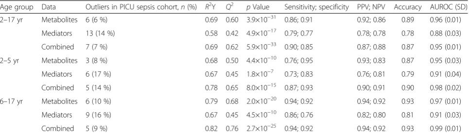
Age group Data Outliers in PICU sepsis cohort,n(%) R2Y Q2 pValue Sensitivity; specificity PPV; NPV Accuracy AUROC (SD)
2–17 yr Metabolites 6 (6 %) 0.69 0.60 3.9×10⁻³¹ 0.86; 0.91 0.92; 0.86 0.89 0.96 (0.01)
Mediators 13 (14 %) 0.58 0.42 4.9×10⁻¹⁷ 0.79; 0.77 0.78; 0.78 0.78 0.88 (0.03)
Combined 7 (7 %) 0.69 0.62 5.9×10⁻³³ 0.90; 0.85 0.87; 0.88 0.87 0.95 (0.01)
2–5 yr Metabolites 3 (8 %) 0.68 0.50 4.4×10⁻¹⁰ 0.76; 0.95 0.93; 0.83 0.87 0.95 (0.03)
Mediators 6 (17 %) 0.67 0.45 1.8×10⁻⁷ 0.73; 0.83 0.76; 0.81 0.79 0.91 (0.04)
Combined 5 (14 %) 0.78 0.65 8.0×10⁻¹⁵ 0.87; 0.93 0.90; 0.91 0.90 0.98 (0.02)
6–17 yr Metabolites 6 (10 %) 0.79 0.68 2.0×10⁻²⁰ 0.94; 0.92 0.94; 0.92 0.93 0.97 (0.01)
Mediators 9 (16 %) 0.67 0.45 4.5×10⁻¹⁰ 0.86; 0.76 0.82; 0.80 0.81 0.91 (0.03)
Combined 5 (9 %) 0.82 0.76 2.7×10⁻²⁵ 0.94; 0.92 0.94; 0.92 0.93 0.99 (0.01)
Abbreviations:AUROCarea under the receiver operating characteristic curve,NPVnegative predictive value,PICUpediatric intensive care unit,PPVpositive predictive value,SDstandard deviation
Conclusions
In children ages 2–17 years, combining metabolomic and inflammatory protein mediator profiling early after pres-entation can differentiate children with sepsis requiring care in a PICU from children with or without sepsis who can be safely cared for outside a PICU. By using this dis-covery and systems biology–based approach, we identified a limited number of metabolites and protein mediators of interest that may realistically lead to development of a point-of-care decision aid. This may aid triage decisions, particularly in EDs without pediatric expertise. This find-ing requires validation in an independent cohort.
Key messages
• In children ages 2–17 years, combining metabolomic and inflammatory protein mediator profiling on serum and plasma early after presentation can differentiate children with sepsis requiring care in a PICU from chil-dren with or without sepsis who can be safely cared for outside a PICU.
•By using this discovery and systems biology–based approach, we identified a limited number of metabo-lites and protein mediators of interest that may real-istically lead to development of a point-of-care decision aid.
Fig. 4The regression coefficient plot for the orthogonal partial least squares discriminant analysis model differentiating pediatric intensive care unit (PICU) sepsis from emergency department (ED) sepsis cohorts in children ages 2–17 years (Fig. 3c). Positive values of the coefficients indicate increased concentrations in the PICU sepsis cohort samples, and negative values indicate a decrease in concentration in the PICU sepsis cohort samples, compared with the ED sepsis cohort samples. Only statistically significant metabolites and protein mediators are shown (p<0.05).A2M
α-macroglobulin,SAAserum amyloid A,TRAILtumor necrosis factor-related apoptosis-inducing ligand
[image:11.595.58.538.87.524.2]Additional files
Additional file 1:Supplemental descriptions of methods, statistical modeling, results of subgroups, discussion of the importance of the metabolites and protein mediators of interest (biomarkers) identified, and supplemental references.This file also includes two tables.Table E1.The metabolites (non-targeted1H NMR spectroscopy
and metabolite concentration profiling) and protein mediators (targeted bead-based multiplex assay) detected and quantified.Table E2.Comparison of statistical measures calculated for the supervised OPLS-DA models without excluding outliers from the results presented in the main text of the article. (PDF 307 kb)
[image:12.595.305.534.206.738.2]Additional file 2:Results of the metabolomic and protein mediator biomarker phenotyping in the two age subgroups.This file contains six figures depicting the results for the age subgroups and a seventh figure showing the loading plots that demonstrate which metabolites and/or inflammatory protein mediators contribute most to each component in the PCA models for the age 2–17-year-old cohort. (PDF 4020 kb)
Abbreviations
ASN:Alberta Sepsis Network; AUROC: area under the receiver operating characteristic curve; ECLS: extracorporeal life support; ED: emergency department;1H-NMR: proton nuclear magnetic resonance; ICU: intensive care
unit; MAP: mean arterial pressure; NPV: negative predictive value; OPLS-DA: orthogonal partial least squares discriminant analysis; PC: principal component; PCA: principal component analysis; PELOD: pediatric logistic organ dysfunction score; PEWS: Pediatric Early Warning Score; PICU: pediatric intensive care unit; PLS-DA: partial least squares discriminant analysis; PPV: positive predictive value; PRISM: Pediatric Risk of Mortality; ROC: receiver operating characteristic curve; RRT: renal replacement therapy; SBP: systolic blood pressure; SD: standard deviation; SIRS: systemic inflammatory response syndrome; VIP: variable importance to projection; WBC: white blood cells.
Competing interests
This study was supported by the Alberta Innovates-Health Solutions team grant to the Alberta Sepsis Network. BM, BWW, and HJV hold patent US 20140205591 A1 (Metabolite Biomarkers for Diagnosis and Prognosis of Pediatric Septic Shock). None of the other authors has any competing interests or other financial disclosures to make. The funding agency had no role in the design and conduct of the study; the collection, management, analysis, or interpretation of the data; the preparation, review, or approval of the manuscript; or the decision to submit the manuscript for publication.
Authors’contributions
ARJ, BM, GT, JB, CNJ, BWW, and HJV contributed to study conception and design, acquisition and interpretation of data, and revision of the manuscript critically for intellectual content. ARJ drafted the manuscript. BM revised the statistics section of the manuscript and drafted part of the Discussion section. CNJ revised the Methods section and drafted part of the Discussion section. BM and HJV performed the statistical analysis. All authors approved the final version of the manuscript and agree to be accountable for all aspects of the work. ARJ had full access to all the data in the study and takes responsibility for the integrity of the data and the accuracy of the data analysis.
Acknowledgments
We thank the following for their help with this project: Josee Wong and the Critical Care Epidemiologic and Biologic Tissue Resource for specimen handling, Derrice Knight for her organizational skills as the project manager for the Alberta Sepsis Network, Mandy Tse and the Snyder Translational Laboratory in Critical Care Medicine for protein mediator analysis, and the pediatric research coordinators who managed the consent process and data collection and entry.
Author details
1
Bio-NMR Center, Department of Biological Sciences, University of Calgary, Calgary, AB, Canada.2Division of Emergency Medicine, Department of Pediatrics, University of Calgary, Calgary, AB, Canada.3Division of Pediatric Critical Care Medicine, Department of Pediatrics, University of Alberta, 4-546
Edmonton Clinic Health Academy; 11405 87 Avenue, Edmonton, AB T6G 1C9, Canada.4Calvin, Phoebe and Joan Snyder Institute for Chronic Diseases, University of Calgary, Calgary, AB, Canada.5Department of Critical Care Medicine, University of Calgary, Calgary, AB, Canada.6Department of Medicine, University of Calgary, Calgary, AB, Canada.7Department of Biochemistry and Molecular Biology, University of Calgary, Calgary, AB, Canada.
Received: 7 May 2015 Accepted: 12 August 2015
References
1. Liu L, Johnson HL, Cousens S, Perin J, Scott S, Lawn JE, et al. Global, regional, and national causes of child mortality: an updated systematic analysis for 2010 with time trends since 2000. Lancet. 2012;379:2151–61. 2. Watson RS, Carcillo JA. Scope and epidemiology of pediatric sepsis. Pediatr
Crit Care Med. 2005;6(3 Suppl):S3–5.
3. Watson RS, Carcillo JA, Linde-Zwirble WT, Clermont G, Lidicker J, Angus DC. The epidemiology of severe sepsis in children in the United States. Am J Respir Crit Care Med. 2003;167:695–701.
4. Hartman ME, Linde-Zwirble W, Angus DC, Watson RS. Trends in the epidemiology of pediatric severe sepsis. Pediatr Crit Care Med. 2013;14:686–93.
5. Dombrovskiy VY, Martin AA, Sunderram J, Paz HL. Rapid increase in hospitalization and mortality rates for severe sepsis in the United States: a trend analysis from 1993 to 2003. Crit Care Med. 2007;35:1244–50. 6. Martin GS, Mannino DM, Eaton S, Moss M. The epidemiology of sepsis in
the United States from 1979 through 2000. N Engl J Med. 2003;348:1546–54. 7. Als LC, Nadel S, Cooper M, Pierce CM, Sahakian BJ, Garralda ME.
Neuropsychologic function three to six months following admission to the PICU with meningoencephalitis, sepsis, and other disorders: a prospective study of school-aged children. Crit Care Med. 2013;41:1094–103. 8. Bronner MB, Knoester H, Sol JJ. An exploratory study on quality of life and
psychological and cognitive function in pediatric survivors of septic shock. Pediatr Crit Care Med. 2009;10:636–42.
9. Conlon MP, Breatnach C, O’Hare BP, Mannion DW, Lyons BJ. Health related quality of life after prolonged pediatric intensive care unit stay. Pediatr Crit Care Med. 2009;10:41–4.
10. Farris RWD, Weiss NS, Zimmerman JJ. Functional outcomes in pediatric severe sepsis: further analysis of the researching severe sepsis and organ dysfunction in children. A global perspective trial. Pediatr Crit Care Med. 2013;14:835–42.
11. Weiss SL, Fitzgerald JC, Balamuth F, Alpern ER, Lavelle J, Chilutti M, et al. Delayed antimicrobial therapy increases mortality and organ dysfunction duration in pediatric sepsis. Crit Care Med. 2014;42:2409–17.
12. Beck V, Chateau D, Bryson GL, Pisipati A, Zanotti S, Parrillo JE, et al. Timing of vasopressor initiation and mortality in septic shock: a cohort study. Crit Care. 2014;18:R97.
13. Dellinger RP, Levy MM, Rhodes A, Annane D, Gerlach H, Opal SM, et al. Surviving Sepsis Campaign: international guidelines for management of severe sepsis and septic shock. Crit Care Med. 2013;41:580–637.
14. Fischer JE. Physicians’ability to diagnose sepsis in newborns and critically ill children. Pediatr Crit Care Med. 2005;6(3 Suppl):S120–5.
15. Wacker C, Prkno A, Brunkhorst FM, Schlattmann P. Procalcitonin as a diagnostic marker for sepsis: a systematic review and meta-analysis. Lancet Infect Dis. 2013;13:426–35.
16. Committee on the Future of Emergency Care in the United States Health System, Board on Health Care Services, Institute of Medicine. Emergency care for children: growing pains. Washington, DC: The National Academies Press; 2007.
17. Canadian Institute for Health Information (CIHI). Emergency departments and children in Ontario. Ottawa, ON, Canada: CIHI; April 2008 https:// secure.cihi.ca/free_products/aib_apr24_08_en.pdf. Accessed 21 August 2015. 18. Kaplan JM, Wong HR. Biomarker discovery and development in pediatric
critical care medicine. Pediatr Crit Care Med. 2011;12:165–73. 19. Nicholson JK, Holmes E, Kinross JM, Darzi AW, Takats Z, Lindon JC.
Metabolic phenotyping in clinical and surgical environments. Nature. 2012;491:384–91.
20. Kiehntopf M, Nin N, Bauer M. Metabolism, metabolome, and metabolomics in intensive care: is it time to move beyond monitoring of glucose and lactate? Am J Resp Crit Care Med. 2013;187:906–7.
21. Banoei MM, Donnelly SJ, Mickiewicz B, Weljie A, Vogel HJ, Winston BW. Metabolomics in critical care medicine: a new approach to biomarker discovery. Clin Invest Med. 2014;37:E363–76.
22. Mickiewicz B, Duggan GE, Winston BW, Doig C, Kubes P, Vogel HJ, et al. Metabolic profiling of serum samples by 1H nuclear magnetic resonance spectroscopy as a potential diagnostic approach for septic shock. Crit Care Med. 2014;42:1140–9.
23. Mickiewicz B, Vogel HJ, Wong HR, Winston BW. Metabolomics as a novel approach for early diagnosis of pediatric septic shock and its mortality. Am J Respir Crit Care Med. 2013;187:967–76.
24. Mickiewicz B, Tam P, Jenne CN, Leger C, Wong J, Winston BW, et al. Integration of metabolic and inflammatory mediator profiles as a potential prognostic approach for septic shock in the intensive care unit. Crit Care. 2015;19:11.
25. Goldstein B, Giroir B, Randolph A, International Consensus Conference on Pediatric Sepsis. International pediatric sepsis consensus conference: definitions for sepsis and organ dysfunction in pediatrics. Pediatr Crit Care Med. 2005;6:2–8.
26. Leteurtre S, Martinot A, Duhamel A, Proulx F, Grandbastien B, Cotting J, et al. Validation of the paediatric logistic organ dysfunction (PELOD) score: prospective, observational, multicentre study. Lancet. 2003;362:192–7. A published erratum appears in. Lancet. 2006;367:902.
27. Pollack MM, Patel KM, Ruttimann UE. PRISM III: an updated Pediatric Risk of Mortality score. Crit Care Med. 1996;24:743–52.
28. Nicholson JK, Foxall PJ, Spraul M, Farrant RD, Lindon JC. 750 MHz1H and 1H–13C NMR spectroscopy of human blood plasma. Anal Chem.
1995;67:793–811.
29. Weljie AM, Newton J, Mercier P, Carlson E, Slupsky CM. Targeted profiling: quantitative analysis of1H NMR metabolomics data. Anal Chem.
2006;78:4430–42.
30. Wishart DS, Knox C, Guo AC, Eisner R, Young N, Gautam B, et al. HMDB: a knowledgebase for the human metabolome. Nucleic Acids Res. 2009;37(Database issue):D603–10.
31. Eriksson L, Johansson E, Kettaneh-Wold N, Trygg J, Wikström C, Wold S. Multi- and megavariate data analysis. Part I: basic principles and applications. Umeå, Sweden: Umetrics; 2006. p. 425.
32. Eriksson L, Johansson E, Kettaneh-Wold N, Trygg J, Wikström C, Wold S. Multi- and megavariate data analysis. Part II: advanced applications and method extensions. Umeå, Sweden: Umetrics; 2006. p. 307.
33. van den Berg RA, Hoefsloot HC, Westerhuis JA, Smilde AK, van der Werf MJ. Centering, scaling, and transformations: improving the biological information content of metabolomics data. BMC Genomics. 2006;7:142. 34. Trygg J, Holmes E, Lundstedt T. Chemometrics in metabonomics. J
Proteome Res. 2007;6:469–79.
35. Madsen R, Lundstedt T, Trygg J. Chemometrics in metabolomics: a review in human disease diagnosis. Anal Chim Acta. 2010;659:23–33.
36. Picard RR, Cook DR. Cross-validation of regression models. J Am Stat Assoc. 1984;79:575–83.
37. Fawcett T. An introduction to ROC analysis. Pattern Recognit Lett. 2006;27:861–74.
38. Zweig MH, Campbell G. Receiver-operating characteristic (ROC) plots: a fundamental evaluation tool in clinical medicine. Clin Chem. 1993;39:561–77.
39. Choong K, Bohn D, Fraser DD, Gaboury I, Hutchison JS, Joffe AR, et al. Vasopressin in pediatric vasodilatory shock: a multicenter randomized controlled trial. Am J Respir Crit Care Med. 2009;180:632–9. 40. Menon K, Ward RE, Lawson ML, Gaboury I, Hutchison JS, Hebert PC. A
prospective multicenter study of adrenal function in critically ill children. Am J Respir Crit Care Med. 2010;182:246–51.
41. Thompson MJ, Ninis N, Perera R, Mayon-White R, Phillips C, Bailey L, et al. Clinical recognition of meningococcal disease in children and adolescents. Lancet. 2006;367:397–403.
42. Taguchi T, Yamashita E, Mizutani T, Nakajima H, Yabuuchi M, Asano N, et al. Hepatic glycogen breakdown is implicated in the maintenance of plasma mannose concentration. Am J Physiol Endocrinol Metab. 2005;288:E534–40. 43. Cicarelli DD, Vieira JE, Benseñor FEM. Comparison of C-reactive protein and
serum amyloid A protein in septic shock patients. Mediators Inflamm. 2008;2008:631414. doi:10.1155/2008/631414.
44. Dalli J, Norling LV, Montero-Melendez T, Federici Canova D, Lashin H, Pavlov AM, et al. Microparticle alpha-2-macroglobulin enhances pro-resolving responses and promotes survival in sepsis. EMBO Mol Med. 2014;6:27–42.
45. Tian Y, Tao T, Zhu J, Zou Y, Wang J, Li J, et al. Soluble tumor necrosis factor related apoptosis inducing ligand level as a predictor of severity of sepsis and the risk of mortality in septic patients. PLoS One. 2013;8, e82204. 46. Xia J, Mandal R, Sinelnikov I, Broadhurst D, Wishart DS. MetaboAnalyst
2.0—a comprehensive server for metabolomic data analysis. Nucleic Acids Res. 2012;40:W127–33.
47. Fanos V, Caboni P, Corsello G, Stronati M, Gazzolo D, Noto A, et al. Urinary
1
H-NMR and GC-MS metabolomics predicts early and late onset neonatal sepsis. Early Hum Dev. 2014;90 Suppl 1:S78–83.
48. Seiger N, Maconochie I, Oostenbrink R, Moll HA. Validity of different pediatric early warning scores in the emergency department. Pediatrics. 2013;132:e841–50.
49. Gold DL, Mihalov LK, Cohen DM. Evaluating the Pediatric Early Warning Score (PEWS) system for admitted patients in the pediatric emergency department. Acad Emerg Med. 2014;21:1249–56.
50. Breslin K, Marx J, Hoffman H, McBeth R, Pavuluri P. Pediatric Early Warning Score at time of emergency department disposition is associated with level of care. Pediatr Emer Care. 2014;30:97–103.
51. Bradman K, Borland M, Pascoe E. Predicting patient disposition in a paediatric emergency department. J Paediatr Child Health. 2014;50:E39–44. 52. Kentsis A, Lin YY, Kurek K, Calicchio M, Wang YY, Monigatti F, et al.
Discovery and validation of urine markers of acute pediatric appendicitis using high-accuracy mass spectrometry. Ann Emerg Med. 2009;55:62–70. 53. Kentsis A, Ahmed S, Kurek K, Brennan E, Bradwin G, Steen H, et al. Detection
and diagnostic value of urine leucine-richα-2-glycoprotein in children with suspected acute appendicitis. Ann Emerg Med. 2012;60:78–83.e1. 54. Wong HR, Cvijanovich NZ, Anas N, Allen GL, Thomas NJ, Bigham MT, et al.
Developing a clinically feasible personalized medicine approach to pediatric septic shock. Am J Respir Crit Care Med. 2015;191:309–15.
55. Tüdős AJ, Besselink GAJ, Schasfoort RBM. Trends in miniaturized total analysis systems for point-of-care testing in clinical chemistry. Lab Chip. 2001;1:83–95.
56. Bell C, George C, Kicman AT, Traynor A. Development of a rapid LC-MS/MS method for direct urinalysis of designer drugs. Drug Test Anal. 2011;3:496–504.
Submit your next manuscript to BioMed Central and take full advantage of:
• Convenient online submission
• Thorough peer review
• No space constraints or color figure charges
• Immediate publication on acceptance
• Inclusion in PubMed, CAS, Scopus and Google Scholar
• Research which is freely available for redistribution
Submit your manuscript at www.biomedcentral.com/submit