This is a repository copy of Nerve tissue engineering using blends of
poly(3-hydroxyalkanoates) for peripheral nerve regeneration.
White Rose Research Online URL for this paper:
http://eprints.whiterose.ac.uk/90581/
Version: Accepted Version
Article:
Lizarraga-Valderrama, L.R., Nigmatullin, R., Taylor, C. et al. (4 more authors) (2015) Nerve
tissue engineering using blends of poly(3-hydroxyalkanoates) for peripheral nerve
regeneration. Engineering in Life Sciences, 15 (6). 612 - 621. ISSN 1618-0240
https://doi.org/10.1002/elsc.201400151
[email protected] https://eprints.whiterose.ac.uk/
Reuse
Unless indicated otherwise, fulltext items are protected by copyright with all rights reserved. The copyright exception in section 29 of the Copyright, Designs and Patents Act 1988 allows the making of a single copy solely for the purpose of non-commercial research or private study within the limits of fair dealing. The publisher or other rights-holder may allow further reproduction and re-use of this version - refer to the White Rose Research Online record for this item. Where records identify the publisher as the copyright holder, users can verify any specific terms of use on the publisher’s website.
Takedown
If you consider content in White Rose Research Online to be in breach of UK law, please notify us by
Research Article
Nerve Tissue Engineering Using Blends of Poly(hydroxyalkanoates) for Peripheral Nerve
Regeneration.
Lorena R. Lizarraga-Valderrama1
Rinat Nigmatullin1
Caroline Taylor 2
John W. Haycock 2
Frederik Claeyssens 2
Jonathan C. Knowles 3 4
Ipsita Roy 1
1
Applied Biotechnology Research Group, University of Westminster, London, United Kingdom.
2
Department of Materials Science & Engineering, Kroto Research Institute, Sheffield, UK.
3
Division of Biomaterials and Tissue Engineering, UCL Eastman Dental Institute, London, UK.
4
Department of Nanobiomedical Science & BK21 Plus NBM Global Research Center for
Regenerative Medicine, Dankook University, Cheonan, Republic of Korea
Correspondence: Dr. Ipsita Roy. [email protected]. Applied Biotechnology Research
Group, University of Westminster, 115 New Cavendish Street, W1W 6UW, London, UK.
Keywords: Biocompatibility, In vitro test, Nerve regeneration, Neuronal cells,
Poly(hydroxyalkanoates).
Abbreviations: COLI1, collagen type I 1; ECM2, extracellular matrix; LPS3,
National Collection of Industrial and Marine Bacteria; NGCs, nerve guidance conduits; NTE,
nerve tissue engineering, PGA, polyglycolic acid; PHAs, poly(hydroxyalkanoates); PLCL,
poly(DL-lactide- -caprolactone); PNI, peripheral nerve injury, P(3HB),
poly(3-hydroxybutyrate); P(3HB-co-3HHx), poly(3-hydroxybutyrate-co-3-hydroxyhexanoate); PVA,
polyvinyl alcohol; RGD, L-arginine-glycine-L-aspartic acid; Rq, root mean square roughness;
Practical application
The standard treatment for peripheral nerve repair is the nerve autograft, which has several
limitations including donor site morbidity, scar tissue invasion, scarcity of donor nerves,
inadequate return of function and aberrant regeneration. Although artificial nerve guidance
conduits (NGCs) made from various biomaterials have been clinically approved, they have not
been able to overcome these limitations and can induce scar tissue and release compounds that
are detrimental to the nerve regeneration process. Therefore, in this study, blends of
poly(hydroxyalkanoates) (PHAs), that have not been used before in nerve tissue engineering
were analysed for their potential use in the manufacture of multi-channel and electrospun
NGCs. PHAs displayed properties that could overcome some of the limitations of the available
NGCs such as controllable surface erosion, lower acidity of their degradation products after
biodegradation and longer-lasting stability compared to their synthetic counterparts.
Abstract
The only types of poly(hydroxyalkanoates) (PHAs) that have been explored for their use in
nerve regeneration are poly-3-hydroxybutyrate P(3HB) and
poly(3-hydroxybutyrate-co-3-hydroxyhexanoate) (P(3HB-co-3HHx)). However, the regeneration displayed by these PHAs is
still inferior to that displayed in using autologous nerve grafting. The aim of this work was to
study PHA blends as resorbable biomaterials for their use in the manufacture of nerve guidance
conduits (NGCs). PHA blend films with varying ratios of
poly(3-hydroxyoctanoate)/poly(3-hydroxybutyrate) (P(3HO)/P(3HB)) were produced using the solvent-casting method. Neat
films of P(3HO) and P(3HB) along with 25:75, 50:50 and 75:25 blend films of
(P(3HO)/P(3HB) were characterised with respect to their chemical, material and biological
properties in order to evaluate them as potential base materials for nerve tissue engineering. In
the surface analysis the blends exhibited the highest values of roughness compared with the neat
films. The DSC values of the blends confirmed that P(3HO) and P(3HB) formed immiscible
blends. FTIR and XRD analysis of the blends showed a decrease in the chrystallinity with the
increase of the proportion of P(3HO). However, an increase in the stiffness of the blends was
observed when the proportion of P(3HB) increased. Although all of the blends were
biocompatible with NG108-15 neuronal cells, the 25:75 P(3HO)/P(3HB) blend showed not only
significantly better support for the growth and differentiation of these cells, but also displayed
1 Introduction
Peripheral nerve injuries (PNI) affect about 2.8% of trauma patients, many of who suffer
life-long disability [1]. Peripheral nerves are able to repair when the injuries present a gap of less
than 5mm to bridge [2, 3]. For injuries resulting in nerve damage with gaps of more than 5mm,
treatment is most commonly attempted using autologous nerve graft repair [4, 5]. When nerve
damage is even more extreme and gaps exceed 3cm, allografts, vascularized nerve grafts and
nerve grafts without vessels are used [5]. Peripheral nerve repair using nerve autografts has
several limitations including donor site morbidity, scar tissue invasion, scarcity of donor nerves,
inadequate return of function and aberrant regeneration[5, 6]. Currently there are several
clinically approved artificial nerve guidance conduits (NGCs) made from various biomaterials
that have overcome some of the limitations of these nerve autografts. Conversely, NGCs made
from synthetic materials can trigger immune responses, induce scar tissue and can release
compounds that are detrimental to the nerve regeneration process [5].
During the past two decades a large variety of materials including nano-structured materials and
biochemical factors have been explored in attempts to improve the quality of nerve conduits,
and currently there are several commercial nerve conduits approved by U.S Food and Drug
Administration (FDA) and Conformit Europe (CE) [2]. All of the models currently available
take the form of a simple hollow tube with a single lumen. They possess no internal
substructure, are made from either synthetic or natural materials and are available in different
designs and sizes [7]. The materials that have been used for their manufacture include
poly(DL-lactide- -caprolactone) (PLCL), polyglycolic acid (PGA), polyvinyl alcohol (PVA), collagen
type I (COLI) and extracellular matrix (ECM). Furthermore, a large diversity of materials have
been used experimentally to produce nerve guidance conduits such as aliphatic polyesters,
polylactic acids, polycaprolactones, polyurethanes, silicones, collagens, glycoproteins,
polypeptides, poly(hydroxyalkanoates) (PHAs), polysaccharides, proteins and acellular or
PHAs possess great potential as materials for use in the manufacturing of NGCs to assist axonal
regeneration. Their prominent properties such as: controllable surface erosion; variability in
material properties; lower acidity of degradation products and longer stability compared to their
synthetic counterparts are all of special interest in this field.
Currently, P(3HB) and P(3HB-co-3HHx) are the only type of PHA that have been explored for
their use in nerve regeneration. P(3HB) conduits have been shown to repair nerve gaps of 10mm
[8, 9, 10, 11, 12] and 40mm [13, 14] in rat sciatic nerves and rabbit peroneal nerves
respectively. Hollow P(3HB-co-3HHx) conduits have also been used to bridge 10mm defects in
rat sciatic nerves [15]. Although these studies showed low level of inflammatory infiltration and
suitable reabsorption time for nerve repair, the regeneration obtained was not statistically
comparable with the regeneration obtained by using autologous nerve grafting.
The aim of this work was to investigate PHA blends as resorbable biomaterials for their use in
the manufacture of NGCs. Mechanical, physical and chemical properties of P(3HO), P(3HB)
and their blends were characterized. The biocompatibility of these materials with NG108-15
neuronal cells was also studied. As P(3HO) displays mechanical properties similar to those of
the peripheral nerve and P(3HB) have shown to be biocompatible to neuronal cells these PHAs
were chosen for evaluation as improved materials for nerve tissue engineering.
2 Materials and Methods
2.1 Production and extraction of P(3HO) and P(3HB)
Production, extraction and purification of P(3HO) were performed as described previously [16].
The extraction method used to extract P(3HO) was dispersion of chloroform and sodium
hypochlorite [16]. Production, extraction and purification of P(3HB) were carried out as
2.2 Film preparation
Films of P(3HO), P(3HB) and three different blends of P(3HO)/P(3HB) were prepared using the
solvent casting method. The PHAs were dissolved in chloroform in order to obtain a total
polymer concentration of 5 w/v%. The P(3HO)/P(3HB) blends were prepared in ratios of 75:25,
50:50 and 25:75 by dissolving the required amounts of polymers in 10 mL of chloroform. After
polymer dissolution, the solutions were homogenised by sonication and then cast in 6-cm glass
petri dishes. The films were air dried for two weeks and produced in triplicate to obtain a total
of fifteen films with varying thicknesses of 0.09 – 0.15mm.
2.3 Scanning electron microscopy (SEM) of the films
Surface topography of the films was analysed using a FEI XL30 Field Emission Gun Scanning
Electron Microscope (Eindhoven, the Netherlands). All the samples were previously
sputter-coated with a 20 nm film of palladium using a Polaron E5000 sputter coater. The operating
pressure of the sputter coating was 5x10⁻⁵ bar with a deposition current of 20 mA for a duration
of 1m 30s. The images were then recorded at different magnifications at 5kV using the FEI
software.
2.4 Surface wettability of the films
The static contact angle of the films was carried out as described previously [27].
2.5 Profilometric surface analysis
The surface roughness of the films was analysed using a Sony Proscan 1000 Laser Profilometer
(Tokyo, Japan). The laser used was model 131A, which has a measuring range of 400 m, a
resolution of 0.02 m and a maximum output of 10 mW. Scans of 0.5 mm2 were obtained from
each sample. Nine random coordinates were selected from each specimen in order to measure
2.6 X-Ray diffraction analysis
Crystallization analysis of the films was performed using a Br̈ker D8 Advance diffractometer
in flat plate geometry, using Ni filtered Cu Ka, radiation. Data was collected from 10 to 40°
with a primary beam slit size of 0.6 mm. A Br̈ker Lynx Eye silicon strip detector was used and
a step size of 0.02° and a count time of 0.1 s per step.
2.7 Fourier transform infrared spectroscopy (FTIR) of the films
The FTIR of the P(3HO), P(3HB) and (P3HO)/P(3HB) blend films was performed as described
previously [16].
2.8 Static tensile test of the films
Mechanical analysis of the films was conducted using a Perkin Elmer Dynamic Mechanical
Analyser 7 (Norwalk, USA). The sample dimensions were 1.66 mm - 2.05 mm in width; 5mm -
6mm in length and had a thickness of 0.05 mm - 0.18 mm. The load was set within a range of 1
nN – 6000 mN with a rate of 200 mN/min-1 at 24°C. The mechanical properties were
determined using the software. This included analysis of Young’s modulus, ultimate tensile
strength and elongation at break.
2.9 DSC
Thermal analysis of neat polymers and their blends was conducted as described previously [27].
2.10 NG108-15 neuronal cell culture
NG108-15 neuronal cells were obtained from The European Collection of Cell Cultures
(ECACC) and grown in Dulbecco’s Modified Eagle Medium (DMEM) under a humidified
atmosphere of 5% CO2 at 37°C. The DMEM was supplemented with 10% (v/v) foetal calf
serum, 1% (w/v) glutamine, 1%(w/v) penicillin/streptomycin, and 0.5% (w/v) amphotericin B.
trypsinized and 3x10⁴ cells were seeded directly onto the PHA film samples within 12-well
plates in 3 mL of DMEM. The cultures were maintained for 4 days, with half of the medium
being removed and replaced with fresh serum-free DMEM on day 2 to trigger experimental
differentiation. NG-108-15 neuronal cells were used between passages 10 and 20.
2.11 Live / dead measurement of NG-108-15 neuronal cells
After growing cells for 4 days, the culture medium was removed and replaced with fresh
serum-free DMEM containing 0.0015% (w/v) propidium iodide (Invitrogen) and 0.001% (w/v) Syto-9
(Invitrogen) at 37°C/5% CO2 for 15 min. After washing with PBS (x3), the cells were imaged
with an upright Zeiss LSM 510 confocal microscope. A helium-neon laser was used for the
detection of propidium iodide ( ex= 536 nm/ em=617 nm) while an argon-ion laser was used
for Syto λ ( ex= 4λ4 nm/ em=515 nm). Three fields-of-view were imaged containing 20-500
cells per sample, so as to express the data as a percentage of live versus dead cells ± SEM.
Quantification of live and dead cells was performed using the software Image J.
2.12 Immunolabelling of NG108-15 neuronal cells
To assess the differentiation of neuronal cells, samples were inmunolabelled using III-tubulin
as the primary antibody and with Alexa Fluor® 488 goat anti-mouse IgG as the secondary
antibody. Sample films containing cultures of NG108-15 neuronal cells previously washed with
PBS (x3), were fixed with 4% (v/v) paraformaldehyde for 20 mins, then permeabilized with
0.1% (v/v) Triton X-100 for 20 mins, before being washed with PBS (x3). Unreactive binding
sites were blocked with 3% (w/v) bovine serum albumin (BSA) with the cells being incubated
overnight with mouse anti III-tubulin antibody (1:1000) (Promega, USA) diluted in 1% BSA
at 4°C. Cells were then washed three times with PBS before being incubated with Alexa Fluor®
488 goat anti-mouse IgG antibodies (1:200 in 1 % BSA) (Sigma Aldrich) for 90 min. After
washing the cells once with PBS, 4’, 6-diamidino-2-phenylindole dihydrochloride (DAPI)
for 15 mins at room temperature before being washed again with PBS (x3). Cells were then
imaged using an upright Zeiss LSM 510 confocal microscope. Nuclei ware visualised by two
photon excitation using a Tiμsapphire laser (716 nm) for DAPI ( ex = 358 nm/ em = 461 nm).
For imaging the neuronal cell body and neurites of NG108-15 cells, a helium-neon laser
(543nm) was used to detect the Alexa Fluor® 488 goat anti-mouse IgG ( ex= 58λ nm/ em=615
nm). The differentiated cells were then counted using ImageJ software, identified as neuronal
cells expressing neurites.
2.13 Statistical analysis
Statistical analysis was conducted using Graph Pad Prism 6 software. A Shapiro - Wilk and
Bartlett’s test was previously performed to verify the normality and homogeneity of the data
respectively. To analyse the difference between data, a one-way ANOVA test (p < 0.05) was
conducted followed by Turkey’s post-test (p < 0.05). Data was reported as mean ± SEM.
3 Results
3.1 Scanning electron microscopy of PHA films
Scanning electron microscopy images of the films were obtained in order to compare their
surface morphology. The P(3HO) film (Fig. 1A) displayed the presence of pores with sizes
ranging from 0.1 m to 5 m. The 75μ25 (Fig. 1B) and 25μ75 (Fig. 1D) blends presented smaller
and less abundant pores (0.1 m to 3 m) compared to the P(3HO) film. Conversely, pores were
not detected in the 50:50 blend (Fig. 1C), which showed protrusions uniformly distributed on
the surface. The P(3HB) film (Fig. 1E) displayed the smoothest and most homogenous surface
without the presence of pores. It was observed that the presence of P(3HO) in the blends
3.2 Profilometric surface analysis
The root mean square roughness (Rq) of the films was determined using a laser profilometer.
The roughness of P(3HO) and P(3HB) films were significantly different (3.69 ± 0.20nm versus
2.60 ± 0.09nm, p < 0.05 ). Although the P(3HO) film was the most porous, its roughness (3.69
± 0.20 nm) was not significantly different to the roughness of the 75:25, 50:50 and 25:75 blends
(4.00 ± 0.15nm, 4.23 ± 0.37nm, 4.16 ± 0.25nm, p > 0.05). On the other hand, statistical analysis
showed that the roughness of P(3HB) (2.60 ± 0.09 nm) was significantly different to that of the
blends 25:75, 50:50 and 75:25 (4.00 ± 0.15nm, 4.23 ± 0.37nm, 4.16 ± 0.25nm, p < 0.05). As
expected, the neat P(3HB) film presented the lowest value of roughness, which correlates with
its surface having the smoothest appearance of all the films, as can be observed in the SEM
analysis (Fig. 1F).
3.3 X - Ray diffraction analysis
The solvent-casted films were characterised by wide-angle X-ray diffraction spectroscopy. The
neat P(3HB) film exhibited two intense peaks at 2 values of 13.5° and 16.4° (Fig. 2A). The
peak positions correspond to the reported values in previous studies of P(3HB) [18, 19]
crystalline structure where they were assigned to the (020) and (110) planes of the orthorhombic
unit cell. Moreover, a series of peaks were observed in 2 range between 18° and 34°. These
peaks are most likely attributed to the crystalline lattice planes of (021), (120), (111) and (101).
In contrast to neat P(3HB), the diffraction pattern of P(3HO) was characterised by the presence
of a broad amorphous halo located around 2 =20°. However, in the P(3HO) diffractogram the
amorphous halo was superposed with a series of diffraction peaks around 17°, 19° and 21°.
Thus a significant fraction of semi-crystalline P(3HO) was in amorphous phase. X-ray
diffractograms of all the blends showed intense peaks of P(3HB) (020) and (110) planes
P(3HO) was 75%. However, peaks corresponding to P(3HO) crystallites were not detected in
the diffractograms of the blends. Also the weak amorphous halo was observed only in the 75:25
blend while other blend compositions did not show P(3HO) amorphous phase. Comparing the
spectrum of the pure P(3HB) with those of the blends, it was seen that peak positions
corresponding to P(3HB) were constant. This indicated that the P(3HB) unit cell did not change
in the blends.
3.4 DSC
A representative thermogram of aged P(3HO)/P(3HB) blends (Fig. 2B) shows the presence of
two endothermic events with peak temperatures around 50 °C and above 150 °C corresponding
to the melting temperatures (Tm) of P(3HO) and P(3HB) respectively. After erasing the thermal
history and material cooling at 20°C/min, neither material including neat P(3HO) showed the
lower temperature melting event. Thus such cooling conditions do not allow crystallisation of
P(3HO). The blending of these two polymers did not have any effect on the Tm of P(3HB).
These PHAs have distinctive glass transitions (Tg) namely -39 and 2 °C for P(3HO) and P(3HB)
respectively. Blends of these polymers were characterised by the presence of two glass
transition events. Similar to melting, positions of both glass transitions were not affected by the
presence of the second polymer. Interestingly, cold crystallisation was observed for the blends
with high P(3HO) content (50:50 and 75:25) as shown by the relatively narrow exothermal
peaks between 40 to 70 °C. In contrast, the cold crystallisation of neat P(3HB) and the 25:75
blend were barely detectable, they manifested as slight depreciation of the baseline at
temperatures above 60 °C lasting until the endothermic melting of the P(3HB) crystals. It
appears that P(3HB) crystallises to the same degree as P(3HB) in blends with P(3HO) content
up to 50%. Specific enthalpy of fusion ( Hf ) had slight fluctuations for P(3HB), 25:75, 50:50
blends, namely 73.4, 74.4, 71.6 J per gram of P(3HB) respectively. However, it significantly
decreased to 28.8 J per gram of P(3HB) for the 75:25 blend.
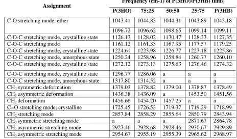
3.5 FTIR of the films
The makers or signatures commonly used to rapidly detect and identify PHAs are the bands of
the C=O group stretch and ester C-O-C group stretch. Furthermore, C-O-C stretch vibrations are
also useful not only for the identification of PHAs but also for the determination of the
crystallinity index (CI) [20]. In the infrared spectra of PHAs, C=O stretch bands are detected in
the region 1719-1744 cm-1, whereas ester C-O-C stretch bands are observed in the range of
1160-1300 cm-1. Although in the FTIR spectra, the above bands are the ones most commonly
used to analyse PHAs, the peaks of other detectable groups confirming the presence of PHA
molecules such as CH2 and CH3 were also identified and are presented in the table 1 [21, 22,
23].
In figure 2C, it was observed that the intensity of the peaks was higher when the concentration
of P(3HB) in the blends was increased. As absorbance (A) is proportional to the concentration
of the molecules in the sample, the higher A values observed in the films containing P(3HB)
could indicate a higher number of molecules of P(3HB) compared to P(3HO), regardless of
blend composition. The P(3HO) film showed the carbonyl band at 1725.45 cm-1, whereas the
P(3HB) film showed it at 1718.99 cm-1 (Fig. 2C, Table I). The 75:25, 50:50, 25:75
P(3HO)/P(3HB) blends showed their carbonyl bands at 1726.53 cm-1, 1719.37 cm-1 and 1719.29
cm-1 respectively. The carbonyl bands presented by both P(3HO) and the P(3HO)/P(3HB) 75:25
blend at around 1725 cm-1 are characteristic of the crystalline phase of medium chain length
PHAs (mcl-PHAs). Similarly, the C=O stretch band at around 1719 cm-1 presented by the
P(3HB) film and both the 50:50 and 25:75 P(3HO)/P(3HB) blends are attributed to the
crystalline phase of short chain length PHAs (scl-PHAs).
In the region 1126-1317 cm-1 multiple C-O-C corresponding to the amorphous and crystalline
state of the polymers are observed (Fig. 2C, Table I). The C-O-C stretch bands around 1160cm-1
and 1180 cm-1 are most common C-O-C fingerprints studied in the FTIR analyses of PHAs.
These bands were similar for the P(3HO), 75:25 and 50:50 P(3HO)/P(3HB) films (1161.12cm-1;
P(3HO) and 25:75 P(3HO)/P(3HB) films (1179.25cm-1; 1177.57cm-1). The bands around
2900cm-1 correspond to the stretching vibration of the C-H aliphatic groups of the polymer
backbones including the side chains. As expected, these bands were stronger for P(3HO)
compared with P(3HB) due to the longer aliphatic chains present in P(3HO) molecules (Fig. 2C,
Table I).
3.6 Surface wettability
Surface wettability of the PHA films was analyzed by measuring the water contact angle.
Wettability describes how easy a fluid spreads or adheres across a solid surface. A high contact
angle signifies low wettability whereas a low contact angle means high wettability. When the
contact angle between distilled water and the surface of a solid substrate is less than 90°, the
material is said to be hydrophilic or wet. When the angle is greater than 90°, the material is
called hydrophobic or water-repellent [24]. The water contact angle of the P(3HO)/P(3HB)
films decreased as the content of P(3HO) decreased ((P(3HO), 103.56 ± 0.95; 75:25, 94.41 ±
1.16; 50:50, 84.40 ± 0.70; 25:75, 77.36 ± 0.81; P(3HB), 69.69 ± 1.63) (Fig. 2). This is due to the
long aliphatic chains present in P(3HO) that constitute its hydrophobic character. The statistical
analyses showed that the differences in water contact angle between all the films
P(3HO)/P(3HB) were significant (p-value < 0.05). As the water contact angle of both the
P(3HO) and the 75:25 P(3HO)/P(3HB) films were greater than 90°, they are considered to be
hydrophobic in nature. By contrast, the water contact angles of the 50:50, 25:75 blend films and
the P(3HB) films were less than 90° and are therefore hydrophilic.
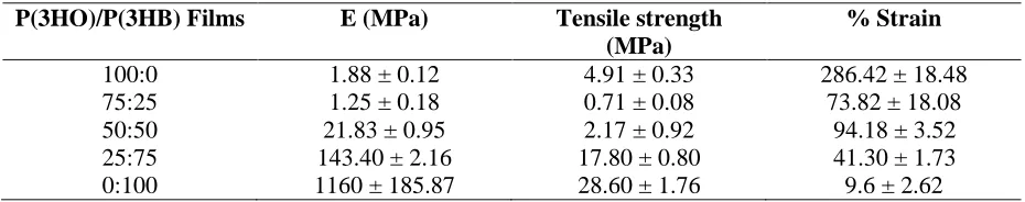
3.7 Static tensile test of the films
Mechanical properties of the films were measured through the static tensile test. Young’s
Modulus (E values), tensile strength and elongation at break of the different films are shown in
Table IV. Young’s modulus, a measure of the stiffness of materials, was determined by
maximum load that a material can sustain during the test, whereas the elongation at break is the
ratio between the final length before breakage and the initial length of the specimen. The E
values of the blends and P(3HO) were significantly different to P(3HB) E value (p-value <
0.05). The tensile strength values were all significantly different when compared against each
other excepting P(3HO) and the blend 75:25 of which difference was not significant. All the
values of percentage strain were significantly different to P(3HO) percentage strain (p-value <
0.05). When the percentage strain values of the blends were compared against each other the
only significant difference was found between the blends 50:50 and 25:75. When the % strain
of P(3HB) was compared with the blends the only significant difference found was when
compared with the blend 50:50. Stiffness of the blends increased when increasing the content of
P(3HB) excepting for the 75:25 P(3HO)/P(3HB) blend. In the other hand, the pliability of the
blends decreased when increasing the P(3HB) content.
3.8 Live/dead measurement of NG-108-15 neuronal cells
A live/dead cell measurement was conducted in order to compare the attachment and survival of
NG-108-15 neuronal cells on the P(3HO)/P(3HB) films using PCL and glass as controls. In
Figure 4, representative confocal images of the cells grown in the different substrates can be
seen. Images (C) and (D) correspond to the 50:50 and 25:75 blends respectively and displayed
the highest density of cells compared with the other images.
The percentage of live cells grown on P(3HO), 75:25, 50:50, 25:75 and P(3HB) films was 80.54
± 6.26%, 89.73 ± 3.68%, 95.12 ± 1.02%, 95.59 ± 1.23%, 88.40 ± 2.99% and on PCL was 89.29
± 3.06%. This was higher when compared with glass substrate (62.68 ± 3.15%) (Fig 4G).
Statistical analysis showed that the difference in the percentage of live cells was significant for
all the substrates including PCL when compared with that of glass (p < 0.05). The difference in
the percentage of live cells between the 75:25, 50:50, 25:75 P(3HO)/P(3HB) and PCL
substrates was not significant (p > 0.05). The only significant difference in the percentage of
blend (95.59 ± 1.23%) were compared (p < 0.05). Hence, the percentage of live cells obtained
using the P(3HO)/P(3HB) 25:75 film was significantly higher than that obtained with P(3HB).
In Figure 4H, the average number of cells in the different substrates is shown. The 50:50 and
25:75 blends were associated with the highest number of adhered neuronal cells per view
(385.11 ± 77.69 cells and 456.00 ± 75.67 cells respectively). No significant differences were
found in the number of adhered neuronal cells on 50:50 and 25:75 polymer blends. Statistical
analysis demonstrated that the number of cells on the 50:50 and 25:75 blends were significantly
higher than the other substrates, including PCL and glass. The difference in the number of live
cells found between the P(3HO), 75:25, P(3HB), PCL and glass substrates was not significant
(23.78 ± 5.96, 84.56 ± 19.92, 54.22 ± 14.04, 27.78 ± 4.07, 19.00 ± 2.52).
3.9 Neurite outgrowth assessment
NG108-15 neuronal cells were immunolabelled with the anti- III-tubulin antibody to assess
neurite outgrowth on the substrates. The protein -III-tubulin is considered a neuron-specific
marker as this molecule is expressed in neuronal cell bodies, dendrites, axons and axonal
terminations. Therefore, this protein is widely used as an indicator of neuronal cell
differentiation. Figure 5 (A-F) shows the confocal images of neuronal cells grown on each of
the substrates, where neurite outgrowth can clearly be observed. However, two important
characteristics of neuronal cells grown on the 25:75 blend, P(3HB) and PCL films were both the
presence of several neurite-bearing neurons and the appearance of longer neurites compared
with those cells grown on the P(3HO), 25:75 and 50:50 P(3HO)/P(3HB) blends.
Figure 5G shows the percentage of cells containing neurites on each substrate. Statistical
analysis showed no significant difference in the percentage of cells with neurites for the
P(3HO), 25:75, 50:50, 25:75, P(3HB) (91.42 ± 2.99; 80.90 ± 11.39; 86.15 ± 5.76; 99.30 ±
0.40; 97.98 ± 0.73), PCL (97.00 ± 1.30) and glass (81.85 ± 9.64), (p < 0.05). Fig. 6H, shows
the number of cells with neurites in the different substrates. There was no significant difference
(280.00 ± 147.31), 25:75 (854.00 ± 243.02) and P(3HB) (591.00 ± 159.34) (p < 0.05). However,
the number of cells with neurites on 25:75 blend was significantly higher compared to the
P(3HO) and 50:50 blends (p < 0.05).
Discussion
The presence of two glass transitions and melting events in P(3HO)/P(3HB) blends, which
occurs at the same temperatures as neat polymers (Fig. 2B) indicates that the PHAs used in this
study were immiscible in both amorphous and crystalline phase. The independence of specific
enthalpy of fusion of P(3HB) for blends with P(3HO) content up to 50% indicated that P(3HB)
crystallises as separate phase in the presence of P(3HO). Although the aged blends show
melting of P(3HO) crystals, the crystallisation was slower than P(3HB) and occurred when
crystallisation of main part of P(3HB) was complete. Apparently the presence of mcl-PHA, a
polymer with lower glass transition temperature, accelerates the kinetics of P(3HB)
crystallisation. This was observed in the P(3HB) non-isothermal crystallisation, which was
characterised by cold crystallisation for blends containing up to 50% of P(3HO). Such influence
of mcl-PHA on the crystallisation of P(3HB) could be due to the increase of polymer chain
mobility in the system containing polymer in rubbery state. However, in the 75:25 blend
crystallisation of P(3HB) was significantly suppressed.
Crystallisation of P(3HB) was also confirmed by XRD. Although intensities of the peaks in the
diffractograms of P(3HO)/P(3HB) blends decreased with the increase of P(3HO) content their
positions were essentially the same as the peaks on the diffractogram of P(3HB). The peaks
characteristic for P(3HO) were not detectable even in the blend with the highest P(3HO)
content. That might imply that P(3HO) co-crystallised with P(3HB). However, considering that
DSC experiment showed two separate melting events exactly matching the melting
temperatures of the neat polymers, it is unlikely that these two PHAs could co-crystallise. One
might suggest that crystallites of P(3HO) did not develop sufficiently in the blends in order to
The surface of the P(3HB) film was found to be smooth whereas the surface of the P(3HO) film
was porous. The smooth surface observed in P(3HB) film was similar to that characterized by
Wang et al. (2012), where the degradation of P(3HB) by polyhydroxybutyrate depolymerase
was investigated. Conversely, this smooth appearance contrasts with the porous morphology of
a P(3HB) film characterised by Kai et al. (2003) in their study of P(3HB-co-3HHx)/P(3HB)
blends. Although the surfaces of P(3HO) films have shown to be smooth and non-porous in
previous studies [16, 27] it is worth noting that there is a lack of information available relating
to the characterisation of P(3HO) for comparison.
The smooth appearance shown in the SEM images of P(3HB) film was in accordance with the
profilometric analysis in which this film presented the lowest root square mean roughness. The
higher Rq values of P(3HO) compared to that of P(3HB) might be attributed to its porous
surface. The higher Rq values of the 75:25, 50:50 and 25:75 P(3HO)/P(3HB) blend films
compared to the P(3HO) and P(3HB) film corroborated the phase separation process detected in
the blend films by DSC analysis. Furthermore, it has been shown that when smooth blend films
are produced using a wide compositional range, the polymers have a high level of compatibility
[28]. Most of the Rq values of P(3HO) and P(3HB) films found in the available literature have
been obtained using atomic force microscopy (AFM) and not by profilometric analysis. As it
has been found that there is a discrepancy in the resultant Rq values between these two
techniques, comparisons with these values were not feasible [29].
The crystalline phases of P(3HO) and P(3HB) were also detected in all the films by FTIR. The
bands around 1725 cm-1 and 1719 cm-1 correspond to the C=O stretching vibrations of the
crystalline phase of P(3HO) and P(3HB) respectively. When mcl-PHAs are in the crystalline
phase, the band of the carbonyl group is detected at 1728 cm-1, whereas in the amorphous phase
this band is detected at 1743 cm-1. When the scl-PHAs are in the amorphous phase the C=O
group band is detected at 1740 cm-1 [30]. The peaks detected in the FTIR spectra of all the films
It has been shown that the forces affecting the wettability in a solid substrate are the surface
tension of the substrate, the surface tension of the liquid and the interfacial tension. The
hydrophobicity of blend materials can change due to compositional variations and the
arrangement of polymer molecules in surface layers. When polymer films are formed from
polymer solutions the solution interface is exposed to the hydrophobic air environment. Thus,
polymeric molecules in the surface may re-orientate their hydrophobic groups towards the
surface of the material, resulting in a less wettable surface. The neat P(3HO) film was the most
hydrophobic one. This polymer has a longer aliphatic chain per monomer unit with four more
methyl groups compared with P(3HB). These methyl groups may have rotated towards the
hydrophobic interface through chain rotation. This orientation is more favourable energetically
and decreases the surface free energy [31]. Therefore, the observed decrease in the wettability
of the films as the P(3HO) content increased demonstrates the higher number of hydrophobic
chains present in the films.
The Young’s modulus, tensile strength and elongation at break values obtained for the P(3HO)
and 50:50 blend differed slightly from the mechanical properties determined for similar films
previously examined [27]. The higher Young’s modulus values obtained in this study for the
P(3HO) and 50:50 blend could be the result of variations in the molecular weight of the
polymers. It has been shown that lower molecular weights of the same polymer increase
Young’s modulus and tensile strength values [32]. Therefore, these results suggest that the
molecular weight of the P(3HO) used in this study could be higher than that used by Basnett et
al. (2013). It is well known that cultivation conditions can affect the molecular weight of PHAs.
Tomizawa et al. (2011) and Agus et al. (2012) showed that the molecular weight of P(3HB)
decreased with an increase in culture time and temperature. The mechanical properties of
P(3HB) obtained in this study agreed with values obtained in similar studies [35]. The Young’s
modulus of the 75:25 P(3HO)/P(3HB) blend (1.25 ± 0.18MPa) was the most similar to that of
peripheral nerves in rats (0.58MPa) [36], suggesting that it could be a good base material for the
blend (17.80 ± 0.80MPa and 41.30 ± 1.73% respectively) were the most similar to that of a
rabbit peripheral nerve, which has in situ tensile strength and in situ % strain of 11.7MPa and
38.5% respectively [37]. Therefore, the 25:75 P(3HO)/P(HB) blend would provide the
appropriate resistance and elasticity that NGCs require to provide adequate flexibility at the site
of implantation.
The inferior cell attachment observed in the P(3HO) and 75:25 P(3HO)/P(3HB) films could be
related to higher levels of endotoxins in these films when compared with the 50:50 and 25:75
P(3HO)/P(3HB) films. P(3HO) was produced using the gram-negative bacteria P. mendocina,
which contain endotoxins (lipopolysaccharides) within their cell wall. A higher level of
lipopolysaccharides is expected with the increased concentration of the P(3HO) in the blend. In
previous work, the endotoxicity of P(3HO), extracted using the dispersion method, was 4.3
EU/mL [16]. Considering that the United States Pharmacopeia permits a maximum of 20EU per
medical device, further efforts of purification are needed to remove more endotoxins from
P(3HO), such as the use of activated charchoal or soxhlet extraction [38]. The superior
biocompatibility displayed in the 50:50 and 25:75 blends could be attributed to the higher
roughness values featured in these films and their lower hydrophobicity. It is well known that
cell attachment is enhanced by the roughness, porosity and hydrophilicity. However, in the
neurite outgrowth test, the 50:50 blend presented a very low number of cells with neurites
compared with the 25:75 blend, which supported the highest number of cells containing
neurites. Therefore, these findings indicate that the 25:75 blend support significantly better the
growth and differentiation of NG108-15 neuronal cells. Biocompatibility studies of NG108-15
neuronal cells with poly(hydroxyalkanoates) have only been performed on P(3HB) substrates.
P(3HB) has demonstrated high biocompatibility not only with NG108-15 neuronal cells [39] but
also with neuronal cells in animal models [8, 9, 10, 11, 12, 13, 14]. Armstrong et al. (2007) have
used NGC made from P(3HB) as a substrate to investigate the effect of Schwann cells (SC) on
The growth of NG108-15 neuronal cells was characterized by their irregular distribution in
various layers on all substrates showing a random migration of cells. Neuronal migration is
highly dependant on the expression of cell adhesion proteins, which can also be involved in
neuronal differentiation. In neuronal cells, including NG108-15, different families of proteins
regulate cell-cell and cell-substrate interactionsμ the Efh family, - -hydrolase fold family, and
three families of CAMs; the inmunoglubolin (Ig) superfamily CAMs, cadherins and integrins
[40, 41, 42, 43, 44, 45]. It has been shown that cell adhesion to polymeric surfaces such as PHA
films is mediated mainly by integrins through the interactions between proteins and the
polymers [46, 47]. Proteins coming from the serum, the surrounding medium or those produced
by the cells could be adsorbed into the surface of the films and recognized by integrins through
the Arg-Gly-Asp (RGD) sequence which is present in a considerable number of proteins.
In summary, although all of the P(3HO)/P(3HB) blends and P(3HO) were able to support
neuronal growth, only the 25:75 P(3HO)/P(3HB) and P(3HB) films displayed suitable
properties for supporting better neurite extension. Although P(3HO) and the 75:25
P(3HO)/P(3HB) blend presented suitable stiffness for the manufacture of NGCs, their
biocompatibility was found to be inferior to that of the other blends. As the 25:75
P(3HO)/P(3HB) blend displayed not only tensile strength and % strain similar to that of the
peripheral nerve, but also presented superior biocompatibility compared with the other
substrates tested. Thus, the 25:75 P(3HO)/P(3HB) blend proved to be the most appropriate base
material for the manufacture of NGCs.
Acknowledgements
The authors would like to thank to the University of Westminster and Department of Materials
Science and Engineering (Kroto Research Institute, University of Sheffield, UK) and
NEURIMP, the Framework 7 project funded by the EC, for providing the facilities, materials
and funding for this research work. The authors thankfully acknowledge the reliable assistance
Palmer (UCL Eastman Dental Institute, London, UK) for the material characterisation and Dr.
Nicola Mordan (UCL Eastman Dental Institute, London, UK) for the SEM analysis of the films.
References
[1] Midha, R, Emerging techniques for nerve repair: nerve transfers and nerve guidance tubes.
Clin. Neurosurg. 2006, 53, 185-190.
[2] Jiang, X., Shawn, H. L., Mao, H-Q., Chew, S. Y., Current applications and future
perspectives of artificial nerve conduits. Exp. Neurol. 2010, 223, 86-101.
[3] Schmidt, C. E., Leach, J. B., Neural tissue engineering: strategies for repair and
regeneration. Annu. Rev. Biomed. Eng. 2003, 5, 293-347.
[4] De Ruiter, G. C. W., Malessy, M. J. A., Yaszemski, M. J., Windebank, A. J. et al.,
Designing ideal conduits for peripheral nerve repair. Neurosurg. Focus. 2009, 26, 1-14.
[5] Babu, P., Behl, A., Chakravarty, B., Bhandari, P. S. et al., Entubulation techniques in
peripheral nerve repair. Ind. J. Neurot. 2008, 5, 15-20.
[6] Huang, W., Begum, R., Barber, T., Ibba, V. et al., Regenerative potential of silk conduits in
repair of peripheral nerve injury in adult rats. Biomaterials. 2012, 33, 59-71.
[7] Bell, J. H. A., Haycock, J. W., Next generation nerve guides - materials, fabrication, growth
factors and cell delivery. Tissue Eng, 2012, 18, 116-128.
[8] Hart, A. M., Wiberg, M., Terenghi, G., Exogenous leukaemia inhibitory factor enhances
nerve regeneration after late secondary repair using a bioartificial nerve conduit. Br. J. Plast.
Surg. 2003, 56, 444-450.
[9] Hazari, A., Wiberg, M., Johansson-Rudén, G., Green, C. et al., A resorbable nerve conduit
as an alternative to nerve autograft in nerve gap repair. Br. J. Plast. Surg. 1999, 52, 653-657.
[10] Mosahebi, A., Woodward, B., Wiberg, M., Martin, R. et al., Retroviral labelling of
Schwann cells: in vitro characterization and in vivo transplantation to improve peripheral nerve
[11] Mosahebi, A., Fuller, P., Wiberg, M., Terenghi, G., Effect of allogeneic Schwann cell
transplantation on peripheral nerve regeneration. Exp. Neurol. 2002, 173, 213-223.
[12] Mosahebi, A., Wiberg, M., Terenghi, G., Addition of fibronectin to alginate matrix
improves peripheral nerve regeneration in tissue-engineered conduits. Tissue Eng. 2003, 9,
209-218.
[13] Mohanna, P. N., Terenghi, G., Wiberg, M., Composite PHB-GGF conduit for long nerve
gap repair: a long-term evaluation. Scand. J. Plast. Reconstr. Surg. Hand Surg. 2005, 39,
129-137.
[14] Young, R. C., Wiberg, M., Terenghi, G., Poly-3-hydroxybutyrate (PHB): a resorbable
conduit for long-gap repair in peripheral nerves. Br. J. Plast. Surg. 2002, 55, 235-240.
[15] Bian Y-Z., Wang Y., Aibaidoula, G., Chen G-Q. et al., Evaluation of
poly(3-hydroxybutyrate-co-3-hydroxyhexanoate) conduits for peripheral nerve regeneration,
Biomaterials. 2009, 30, 217-225.
[16] Rai, R., Yunos, D. M., Boccaccini, A. R., Knowles, J. C. et al., Poly-3-hydroxyoctanoate
P(3HO), a medium chain length polyhydroxyalkanoate homopolymer from Pseudomonas
mendocina. Biomacromolecules. 2011,12, 2126-2136.
[17] Valappil, S.P., Peiris, D., Langley, G.J., Herniman, J. M. et al., Polyhydroxyalkanoate
(PHA) biosynthesis from structurally unrelated carbon sources by a newly characterized
Bacillus spp. J. Biothechnol. 2007. 475-487.
[18] Capitan, M. J., Rueda, D. R., Ezquerra, T.A., Inhibition of the crystallization in nanofilms
of poly(3-hydroxybutyrate). Macromolecules. 2004, 37, 5653-5659.
[19] Yokouchi, M., Chatani, Y., Tadokoro, H., Teranishi, K. et al., Structural studies of
polyesters: Molecular and crystal structures of optically active and racemic poly (
-hydroxybutyrate). Polymer. 1973, 14, 233-288.
[20] Galego, N., Rozsa, C., Sánchez R., Fung, J. et al., Characterization and application of
[21] Noda, I., Dowrey, A. E., Haynes, J. L., Marcott, C., Group frequency assignments for
major infrared bands observed in common synthetic polymers, in: Marck, J. I. (Ed.), Physical
Properties of Polymers Handbook, Springer Science & Business Media, Otawa 2007, pp.
405-406.
[22] Sato, H., Murakami, R., Padermshoke, A., Hirose, F. et al., Infrared spectroscopy studies of
CH···O hydrogen bondings and thermal behavior of biodegradable poly(hydroxyalkanoate).
Macromolecules. 2004, 37, 7203-7213.
[23] Wu, Q., Tian, G., Wu, Q., Sun, S. Q. et al., Study of Microbial
Poly(hydroxybutyrate-co-hydroxyhexanoate) using two-dimensional fourier-transform infrared correlation spectroscopy.
J Appl Polym Sci. 2001, 82, 934-940.
[24] Förch, R., Schönherr, H., Jenkins, A. T. A. Appendix C: Contact Angle Goniometry, in:
Förch, R., Schönherr, H., Jenkins, A. T. A. (Ed.), Surface Design: Applications in Bioscience
and Nanotechnology, Wiley-VCH Verlag GmbH & Co. KGaA, Weinheim 2009, pp. 471
[25] Wang, Y., Li, F., Wang, Z-y., Liu, D-b. et al., Purification and properties of an extracellular
polyhydroxybutyrate depolymerase from Pseudomonas mendocina DSWY0601. Chem. Res.
Chinese. U. 2012, 28, 459-464.
[26] Kai, Z., Ying, D., Guo-Qiang, C., Effects of surface morphology on the biocompatibility of
polyhydroxyalkanoates, Biochem. Eng. J. 2003, 16, 115-123.
[27] Basnett, P., Ching, K. Y., Stolz, M., Knowles, J. C., Novel
Poly(3-hydroxyoctanoate)/Poly(3-hydroxybutyrate) blends for medical applications, React. Funct.
Polym. 2013, 73, 1340-1348.
[28] Gutmann, J. S., Müller-Buschbaum, P., Schubert, D. W., Stribeck, N. et al., Roughness
correlations in ultra-thin polymer blend films. Physica B: Condensed Matter. 2000, 283, 40-44.
[29] Poon, C. Y., Bhushan, B., Comparison of surface roughness measurements by stylus
[30] Randriamahefa, S., Renard E., Guérin P., Langlois, V., Fourier transform infrared
spectroscopy for screening and quantifying production of PHAs by Pseudomonas grown on
sodium octanoate. Biomacromolecules. 2003, 4, 1092-1097.
[31] Menzies, K. L., Jones, L., The impact of contact angle on the biocompatibility of
biomaterials. Optom. Vis. Sci. 2010, 87, 387-99.
[32] Al-Nasassrah M. A., Podczeck, F., Newton, J. M., The effect of an increase in chain length
on the mechanical properties of polyethylene glycols. Eur. J. Pharm. Biopharm. 1998, 46, 31–
38.
[33] Tomizawa S., Hyakutake, M., Saito, Y., Agus, J. et al., Molecular weight change of
polyhydroxyalkanoate (PHA) caused by the PhaC subunit of PHA synthase from Bacillus
cereus YB-4 in recombinant Escherichia coli. Biomacromolecules. 2011, 12, 2660-2666.
[34] Agus, J., Kahar, P., Hyakutake, M, Tomizawa, S. et al., Unusual change in molecular
weight of polyhydroxyalkanoate (PHA) during cultivation of PHA-accumulating Escherichia
coli, Polym. Degrad. Stabil. 2010, 95, 2250-2254.
[35] Misra, S. K., Valappil, S. P., Roy, I., Boccaccini, A. R., Polyhydroxyalkanoate
(PHA)/inorganic phase composites for tissue engineering applications. Biomacromolecules.
2006, 7, 2249-2258.
[36] Borschel, G. H., Kia, K. F., Kuzon, W. M. Jr, Dennis, R. G. Mechanical properties of
acellular peripheral nerve. J. Surg. Res. 2003, 114, 133-139.
[37] Rydevik, B. L., Kwan, M.K., Myers, R.R., Brown, R. A., Triggs, K.J., Woo, S. L., and
Garfin, S. R. An in vitro mechanical and histological study of acute stretching on rabbit tibial
nerve. J. Orthop. Res. 1990, 8, 694-701.
[38] Wampfler, B., Ramsauer, T., Rezzonico, S., Hischier, R. et al., Isolation and Purification of
Medium Chain Length Poly(3-hydroxyalkanoates) (mcl-PHA) for Medical Applications Using
[39] Armstrong, S. J., Wiberg, M., Terenghi, G., Kingham, P. J., ECM molecules mediate both
schwann cell proliferation and activation to enhance neurite outgrowth. Tissue Eng. 2007, 13,
2863-2870.
[40] Binns, K. T., Taylor, P. P., Sicheri, F., Pawson, T. et al., Phosphorylation of tyrosine
residues in the kinase domain and juxtamembrane region regulates the biological and catalytic
activities of Eph receptors. Mol. Cell. Biol. 2000, 20, 4791-4805.
[41] De Jaco, A., Comoletti, D., Kovarik, Z., Gaietta, G. et al., A Mutation linked with autism
reveals a common mechanism of endoplasmic reticulum retention for the , -hydrolase fold
protein family. J. Biol. Chem. 2006, 281: 9667-9676.
[42] Tojima, T., Yamane, Y., Takahashi, M., Ito, E., Acquisition of neuronal proteins during
differentiation of NG108-15 cells. Neurosci. Res. 2000, 37, 153-161.
[43] Charness, M. E., Safran, R. M., Perides G., Ethanol inhibits neural cell-cell adhesion. J.
Biol. Chem. 1994, 269, 9304-9309.
[44] Hynes, R. O., Lander, A. D., Contact and adhesive specificities in the associations,
migrations, and targeting of cells and axons. Cell. 1992, 68, 303-322.
[45] Hynes, R. O, Integrins: Bidirectional, Allosteric Signaling Machines. Cell. 2002, 110,
673-687.
[46] Lee, J. W., Kim, Y. H., Park, K. D., Jee, K. S. et al., Importance of integrin 1-mediated
cell adhesion on biodegradable polymers under serum depletion in mesenchymal stem cells and
chondrocytes. Biomaterials. 2004, 25, 1901-1909.
[47] Cargill, R. S. 3rd, Dee, K. C., Malcolm, S., An assessment of the strength of NG108-15
Table 1. Assignments of the peaks in the FTIR spectra of the blends P(3HO)/P(3HB)
Assignment Frequency (cm-1) of P(3HO)/P(3HB) films
P(3HO) 75:25 50:50 25:75 P(3HB)
C-O stretching mode, ether 1043.41 1044.83 1044.31 1043.89 1043.18
1096.72 1096.62 1098.65 1099.14 1099.11 C-O-C stretching mode, crystalline state 1126.13 1128.02 1130.47 1128.33 1127.35 C-O-C stretching mode 1161.12 1161.33 1167.95 1177.57 1179.25 C-O-C stretching mode, crystalline state 1224.61 1223.98 1226.77 1227.18 1225.86 C-O-C stretching mode, amorphous state 1250.24 1258.96 1258.84 1260.77 1260.10 C-O-C stretching mode, crystalline state 1272.12 1273.13 1275.63 1276.46 1274.32
C-O-C stretching mode, crystalline state 1296.77 1286.06 a a a C-O-C stretching mode, amorphous state 1317.80 1314.52 a a a CH3 symmetric deformation 1379.03 1378.82 1379.00 1378.87 1378.49 CH3 asymmetric deformation 1436.38 1436.09 a 1453.50 1451.56
CH2 deformation 1456.66 1454.20 1457.25 a a
C=O stretching mode, crystalline 1725.45 1726.53 1719.37 1719.29 1718.99 CH2 stretching mode 2857.84 2858.29 2855.64 2850.79 2843.94
CH3 symmetric stretching mode a a a 2871.67 2864.78
CH2 asymmetric stretching mode 2927.46 2928.68 2928.46 2930.67 2929.89 CH3 asymmetric stretching mode 2954.67 2955.19 2955.39 2965.62 2968.97
a)
Table 2. Mechanical analysis of the P(3HO)/P(3HB) films
P(3HO)/P(3HB) Films E (MPa) Tensile strength
(MPa)
% Strain
100:0 1.88 ± 0.12 4.91 ± 0.33 286.42 ± 18.48
75:25 1.25 ± 0.18 0.71 ± 0.08 73.82 ± 18.08
50:50 21.83 ± 0.95 2.17 ± 0.92 94.18 ± 3.52
25:75 143.40 ± 2.16 17.80 ± 0.80 41.30 ± 1.73
Figure legends
Figure 1. Characterization of the surface topography of P(3HO)/P(3HB) films by scanning
electron microscopy analysis. (A) P(3HO); (B) 75:25; (C) 50:50; (D) 25:75, (E) P(3HB) and (F)
25:75 blend at 500 x. The films P(3HO) (A), 75:25 (B) and 25:75 (D) showed porous
topography whereas the P(3HB) presented smooth surface. The surface observed on the blend
50:50 (C) presented protrusions uniformly without pores. The figure F taken with a higher
magnification (500 x) of the 25:75 blend shows the porous structure of this blend.
Figure 2. P(3HO); 75:25 P(3HO)/P(3HB), 50:50 P(3HO)/P(3HB), 25:75
P(3HO)/P(3HB), P(3HB). A) XRD spectra of P(3HO)/P(3HB) films showing crystalline
(sharp peaks) and amorphous (broad peaks) phases of the polymers. As the content of P(3HB)
increased in the blend the number the peaks increased. B) DSC thermograms of neat polymers
and their blends. Dotted line is a representative thermogram of the first heating scan of aged
75:25 blend. Solid lines are thermograms of the second heating run. C) FTIR spectra of
P(3HO)/P(3HB) films showing the characteristic peaks of P(3HO) and P(3HB). The FTIR
spectra of the P(3HO) and 75:25 blend showed peaks in similar shift positions. Similarly the
films 25:75 and P(3HB) presented peaks at similar frequencies.
Figure 3. Static water contact angle of P(3HO)/P(3HB) films. The water contact increased as
the content of P(3HO) increased in the blend. The water contact angle was significantly
different for all the blends (mean ± SEM, n = 9 independent experimentsP < 0.05).
Figure 4. Confocal micrographs of NG108-15 neuronal cells labelled with propidium iodide
(red) and Syto-9 (green) after four days in culture on P(3HO)/P(3HB) films and PCL. (A)
P(3HO) ; (B) 75:25; (C) 50:50; (D) 25:75; (E) P(3HB) and F) PCL. Cell growth was randomly
oriented on each of the flat substrates. (G) Live/dead analysis of neuronal cells on the
blends and PLC was higher in comparison to glass (control) (mean ± SEM, n = 9 independent
experiments *P < 0.05. Percentage of live neuronal cells on P(3HO) was lower compared to the
25:75 blend (mean ± SEM, n = 9 independent experiments #P < 0.05. (H) Number of live cells
on the P(3HO)/P(3HB) blends, PCL and glass (control). The number of live cells on P(3HO),
75:25, P(3HB), PCL and glass decreased in comparison to the 50:50 and 25:75 blends (mean ±
SEM, n = 9 independent experiments **P < 0.05 compared to 50:50, ##P < 0.05 compared to
25:75.
Figure 5. Confocal micrographs of NG108-15 neuronal cells inmunolabelled for beta-III
tubulin after four days in culture on P(3HO)/P(3HB) films and PCL. (A) P(3HO); (B) 75:25;
(C) 50:50; (D) 25:75; and (E) P(3HB) and F) PCL. Neurite outgrowth was randomly oriented on
each of the flat substrates. (G) Percentage of cell with neurites on the P(3HO)/P(3HB) blends,
PCL and glass (control). Percentage of neurites on all the blends, PLC and glass was similar
(mean ± SEM, n = 6 independent experiments,P < 0.05. (H) Number of cells with neurites on
the P(3HO)/P(3HB) blends, PCL and glass (control). The number of cell with neurites on
P(3HO) and 50:50 was lower in comparison to 25:75 blend (mean ± SEM, n = 6 independent