This is a repository copy of
Continuous hyperfractionated accelerated radiotherapy -
Escalated dose (CHART-ED): A phase i study
.
White Rose Research Online URL for this paper:
http://eprints.whiterose.ac.uk/93414/
Version: Accepted Version
Article:
Hatton, M.Q.F., Hill, R., Fenwick, J.D. et al. (6 more authors) (2015) Continuous
hyperfractionated accelerated radiotherapy - Escalated dose (CHART-ED): A phase i
study. Radiotherapy and Oncology, 118 (3). pp. 471-477. ISSN 0167-8140
https://doi.org/10.1016/j.radonc.2015.11.015
Article available under the terms of the CC-BY-NC-ND licence
(https://creativecommons.org/licenses/by-nc-nd/4.0/)
[email protected] https://eprints.whiterose.ac.uk/
Reuse
This article is distributed under the terms of the Creative Commons Attribution-NonCommercial-NoDerivs (CC BY-NC-ND) licence. This licence only allows you to download this work and share it with others as long as you credit the authors, but you can’t change the article in any way or use it commercially. More
information and the full terms of the licence here: https://creativecommons.org/licenses/
Takedown
If you consider content in White Rose Research Online to be in breach of UK law, please notify us by
Continuous Hyperfractionated
Accelerated RadioTherapy
–
Escalated
Dose (CHART-ED): A Phase I study
MQF Hatton, MBChB, MSc, FRCP, FRCR
Dept Clinical Oncology, Weston Park Hospital, Sheffield
R Hill BSc (hons)
CaCTUS - Cancer Clinical Trials Unit, NHS National Services, Scotland
John Fenwick MA MSc PhD MIPEM
Department of Oncology, University of Oxford,
S Morgan MA, MB BS, MRCP, FRCR
Dept Clinical Oncology, City Hospital, Nottingham
P Wilson BSc, MBChB(hons), MRCP, FRCR
Dept Clinical Oncology, Bristol Haematology and Oncology Centre
P Atherton MBBS MRCP FRCR
Dept Clinical Oncology, Northern Centre for Cancer Treatment, Freeman, Hospital, Newcastle
J Dickson MB, ChB; MSc; MD; MRCP; FRCR
Dept Clinical Oncology, Mount Vernon Hospital, Middlesex
K Murray BSc (hons), MSc
CaCTUS - Cancer Clinical Trials Unit, NHS National Services, Scotland
J Paul BSc (hons)
Abstract
Introduction
Patients who present with locally advanced inoperable non-small cell lung cancer (NSCLC) may be
suitable for radical radiotherapy. A randomised trial of 563 patients compared CHART and
conventional radical radiotherapy (60Gy/30f) given over 6 weeks and suggested that CHART resulted
in a 9% improvement in 2-year survival1. RT dose escalation for both conventional and CHARTWEL
(CHART-WeekEndLess) - fractionation schedules is feasible with modern 3-dimensional CT-based
planning techniques and we initiated a phase I CHART dose escalation study in 2009. Methods
Patients with WHO performance status 0-2 histologically confirmed, inoperable, stage I-III non-small
cell lung cancer were recruited into an open phase I dose escalation trial. Three cohorts of six patients
were recruited sequentially. Total dose was escalated from standard CHART radiotherapy of 54
Gy/36f/12 days to 57.6Gy (2 x 1.8 Gy fractions on day 15, Group 1), 61.2Gy (4 x 1.8 Gy fractions on
days 15-16, Group 2) and 64.8Gy (6 x 1.8 Gy fractions on days 15-17, Group 3).
Results
Between April 2010 and May 2012, 18 patients were enrolled from 5 UK centres and received
escalated dose radiotherapy. 14 were male, 16 squamous cell histology and 12 were stage IIIA or IIIB.
The median age was 70 years and baseline characteristics were similar across the three dose cohorts.
One patient did not start escalated radiotherapy but all remaining patients completed their planned
radiotherapy schedules. Of these 9 patients have died to date with a median survival of 2 years across
the three cohorts. Grade 3 or 4 adverse events (fatigue, dysphagia, nausea and anorexia – classified
according to the National Cancer Institute Common Terminology Criteria for Adverse Events
(CTCAE) version 4.0) were reported in 6 patients but the pre-specified dose limiting toxicities (grade 4
early oesophagitis; grade 3 cardiac, spinal cord and pneumonitis) were not observed.
Conclusions
CHART remains a radiotherapy schedule in routine use across the UK and in this dose escalation study
no dose limiting toxicities were observed. We feel the dose of 64.8Gy / 42f / 17 days should be taken
forward into further clinical trials. The sample size used in this study was small so we plan a
randomised phase II study that includes other radiotherapy schedules to confirm safety and select an
accelerated sequential chemo-radiotherapy schedule to take into phase III studies.
Introduction
Lung cancer is the leading cause of cancer mortality worldwide with approximately 38,000
new cases diagnosed annually in the UK alone2. Non-Small Cell Lung Cancer (NSCLC)
accounts for approximately 80% of all lung cancers. The majority of cases are inoperable at
presentation due to medical co-morbidity (stages I, II and III NSCLC), or tumour extent
(stages III and IV NSCLC). Although many patients with stage I-III disease can still be
treated radically using radiotherapy the 5 year survival from lung cancer in the UK has
changed little (from 3% to 8%) over the last 60 years. The poor outcome can be related to
both failure to eradicate local disease and the development of distant metastases.
Bronchoscopic re-evaluation after radical thoracic RT has demonstrated persistent tumour in
85% of cases3. Successful local control has been found to correlate with improved survival,4.
Approaches to achieve better local control include the acceleration of the RT schedule, the
addition of radio-sensitizers and dose escalation4-6.
Concurrent chemo-radiotherapy (CTRT) (chemotherapy and RT given at the same time) is the
standard of care in stage III NSCLC4 with median survival rates of approximately 21 months.
However, the majority of patients are not suitable for this treatment based on poor
performance status and co-morbidities7. The alternative treatment offered to patients who are
unsuitable for concurrent CTRT is sequential CTRT (chemotherapy given prior to RT), but
local control rates are inferior when compared to concurrent CTRT which is reflected in
worse survival rates4. Alternative strategies to achieve improved local control by intensifying
the local anti-tumour effect are needed.
In the UK the Continuous Hyper-fractionated Accelerated Radiation Therapy (CHART) trial
intensified local treatment by accelerating the RT course. The investigators delivered 54Gy
using 1.5Gy fractions 3 times per day for 12 consecutive days (including weekends) and
demonstrated an overall survival advantage when compared with the standard fractionation
regimen in use at that time (60Gy in 30 daily fractions over 6 weeks)1. There was a 9%
absolute improvement in 2 year survival (29% v 20%; p=0.004) for CHART with no evidence
of a difference in acute or long-term toxicity. This result is supported by an individual patient
data meta-analysis6 confirming that CHART and other intensified schedules which accelerate
or hyper-fractionate improve overall survival as compared to conventional fractionation, with
an absolute benefit of 3% at 5 years. CHART is currently recommended as the standard
radical RT schedule for treating NSCLC in the UK8. Despite this improvement in overall
survival, persistent local disease remains the main cause of death in patients who received
RT dose escalation for conventional and the accelerated CHARTWEL fractionation schedules
was shown to be feasible with 3-dimensional CT-based planning techniques9-10. The initial
dose escalation studies delivered additional daily fractions of radiotherapy and the recently
completed RTOG 0617 11 study has shown no benefit for dose escalation with a prolonged
treatment schedule. This study compared 60 to 74Gy and reported patients in the higher dose
arm suffered higher local relapse rates and inferior survival compared to the control arm.
The RTOG study result is refocusing interest on acceleration and hypo-fractionation and
techniques that avoid prolongation of the overall treatment time are attractive as they reduce
the impact of accelerated tumour clonogen proliferation, which becomes clinically relevant
for NSCLC at least 3-4 weeks after initiation of radiotherapy12. Studies have shown that dose
escalation using 3-D conformal hypo-fractionated radiotherapy is feasible13,14 and the
application of extreme hypo-fractionation associated with stereotactic ablative radiotherapy
(SABR) has delivered impressive local control when used for early NSCLC patients15.
However, when treating central tumours the toxicity rates associated with SABR were
excessive16. Due to the short overall duration of the standard CHART schedule, it is possible
to dose escalate by introducing additional days of treatment and still complete therapy before
the period of accelerated tumour repopulation is expected to begin. This study reports dose
escalation beyond standard CHART performed in a stepped approach using additional twice
Methods and materials
Patients and study design
When the study was designed a pragmatic decision was taken not to give the additional
radiotherapy fractions outside the standard working hours for the radiotherapy departments.
Radiobiological advice guided us to use twice daily 1.8 Gy fractions estimating that the
maximum tolerated dose for oesophagus would be around 65 Gy given over 18 days.
Patients had to have histologically or cytologically confirmed stage I-III non-small cell lung
cancer, with disease deemed inoperable disease by a Lung Cancer Multi-Disciplinary Team
(MDT) with input from Thoracic Surgeon, or operable but the patient refuses surgery.
Patients had to be previously untreated with chemotherapy or radiotherapy, have ECOG
performance status 0 or 1, a life expectancy of at least 6 months, be free of any malignancy
likely to interfere with protocol treatment or comparisons, have adequate respiratory function
(i.e. forced expiratory volume in one second (FEV1) or transfer factor (DLCO) of greater than
40% predicted), and be considered suitable for CHART.
Pre-trial entry, patients had an up to date clinical assessment of eligibility, which included
PET-CT, pulmonary function tests, ECG, Haematology and biochemistry tests (with a brain
scan if required) performed within 42 days of trial registration. Baseline patient characteristics
were collected, and then participating centres telephoned the Scottish Clinical Trials Research
Unit, who allocated patients to one of the treatment cohorts. Patients attended for CHART
radiotherapy planning as soon as possible after registration and on confirmation that the plan
would meet the normal tissue dose constraints the patient attended for treatment verification
and to consent to enter the trial.
Radiotherapy Treatment Planning
All patients were 3D-conformally planned and treated in the supine position, with arms
supported above the head with an external immobilisation device. A single phase technique
was used, without elective nodal irradiation. A planning CT scan using continuous 2.5 - 5mm
slices was acquired throughout the entire volume of both lungs in the treatment position.
Treatment was planned with full information from bronchoscopy, CT-PET and, if performed,
mediastinoscopy or thoracotomy and 3D conformal RT planning used inhomogeneity
correction. Treatment planning aimed to optimise the dose distribution to allow dose
escalation and dose volume histograms (DVH) were constructed for the planning target
volume (PTV), oesophagus, heart, whole lung minus gross tumour volume (GTV), and spinal
Gross Tumour Volume (GTV) included the primary tumour mass and involved nodes, defined
as nodes with short axis > 10 mm or showing increased uptake on PET-CT. Clinical Target
Volume (CTV) is arrived by expansion of GTV by a 5 mm. Planning Tumour Volume (PTV)
is arrived by expansion of CTV by a 10 mm margin (15 mm margin in the cranial caudal
direction). Dose was calculated using type B algorithms and prescribed to the ICRU
reference point. The specified dose constraints were -
CTV Minimum dose > 95% of prescribed dose
PTV V (95% of prescribed dose) > 90%
Cord Maximum dose < 44Gy
Whole Lung-GTV V (20Gy) < 35%
Oesophagus Maximum dose < 105% of prescribed dose
Heart V (100% of prescribed dose) < 30%
V (50% of prescribed dose) < 50%
Simulation and electronic portal images (EPIs) were used to confirm the accuracy of
treatment to within 5mm with set-up should be adjusted according to local guidelines when
discrepancy was ≥5mm.
Radiotherapy Quality Assurance
Radiotherapy Quality Assurance (QA) was a requirement for all participating centres, and
was administered by a central QA group. The QA process built on that developed for the
INCH17 and CONVERT18 trials in collaboration with the NCRI QA group based at Mount
Vernon Hospital. Prior to entering patients centres had to complete a questionnaire covering
immobilisation and planning imaging, planning parameters, commissioning the treatment
planning system, and treatment delivery facilities. Each clinician had to submit a patient plan
and accreditation was only granted once the QA group had approved the plan. Subsequently
each local radiotherapy team had to complete a delineation exercise and each centre was
visited in order to perform dosimetric and portal imaging QA using phantoms.
Statistical analysis
The dose escalation schedule was -
Group 1 57.6Gy in 38 fractions, treating 8 hours apart on day 15 (CHART
plus 2 x 1.8 Gy fractions on day 15)
Group 2 61.2Gy in 40 fractions, treating 8 hours apart on days 15-16 (CHART
plus 4 x 1.8 Gy fractions on days 15-16)
Group 3 64.8Gy in 42 fractions, treating 8 hours apart on days 15-17 (CHART
Six patients were recruited sequentially into each Group, providing that the following dose
limiting toxicity (DLT, classified according to the National Cancer Institute Common
Terminology Criteria for Adverse Events (CTCAE) version 4.0) were not met at the previous
dose level:
Pneumonitis: Grade 3 or above: More than one patient (out of six)
Cardiac Toxicity: Grade 3 or above: More than one patient (out of six)
Spinal Cord Toxicity: Grade 3 or above: More than one patient (out of six)
Oesophagitis (Early): Grade 4 or above: More than one patient (out of six)
The maximum tolerated dose (MTD) was defined as the dose level below which > 1/6 or >
2/12 patients experience a DLT. If these DLT rates are not observed then the top dose will be
recommended.
When 6 patients had been recruited into each of the groups, review ensured that the maximum
oesophageal dose lay within +/- 5% of the prescribed dose in at least 4 patients before
recruitment proceeded to the next dose level. If dose limiting levels of toxicity were reported
for one patient, a further 6 patients were recruited at that dose level to expand the group.
Recruitment was interrupted following completion of each group, until at least 4 patients had
been followed up for 2 months to ensure that any early oesophageal reactions had settled. If
one or more patients developed late pulmonary toxicity of Grade 3 or higher then recruitment
into the CHART-ED study would end.
Data were analysed on an intention to treat basis regardless of any deviation from the
protocol. Survival was measured from the date of first treatment until the date of death from
any cause, with surviving patients censored at the date of their last assessment.
Trial Governance
The trial was approved by the Oxford A Medical Research Ethics Council (MREC), and all
patients provided written informed consent. The trial was conducted in compliance to the
principles outlined in the Medical Research Council Good Clinical Practice (MRC GCP)
Results
Nineteen patients were recruited into the study, one received the standard CHART dose and
fractionation but did not receive the planned dose escalation on day 15. This patient in cohort
three, suffered an exacerbation of COPD as the CHART schedule was being completed. A
further patient was recruited to this cohort and all other patients received the planned
radiotherapy treatment on schedule. The baseline demographical details are recorded in Table
1, and radiotherapy planning details summarised in table 2.
No patient experienced the pre-specified dose limiting toxicities though 6 patients did report
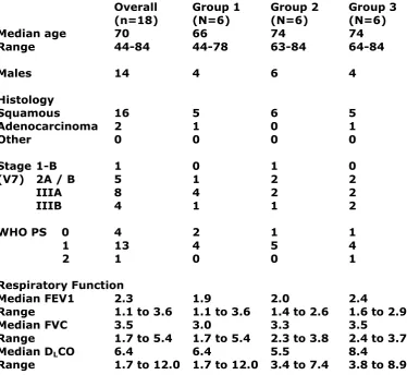
grade 3 – 4 adverse events (fatigue 3, dysphagia 3, anorexia 3 and nausea 1). Figure 1 shows
the doses received by the oesophagus and acute oesophagitis was documented in 17 of the
patients treated, generally grade I – II but three patients required iv hydration (Grade 3
oesophagitis). Late oesophageal toxicity has been limited to grade I/II in 5 patients. Grade I
pulmonary pneumonitis / fibrosis was documented (clinically or radiologically) in 12 patients
across the three cohorts and no cardiac or spinal cord toxicity was reported though one
cardiac arrest was documented 12 months after completion of radiotherapy.
A tumour response was documented in 11/18patients (61%) across the cohorts (complete
response in 5/18 (28%) patients and a further 5 patients had stable disease on post-treatment
CT scans. After a median follow up time of 21 months (range 1 – 36) 8 patients had relapsed,
3 with only loco-regional progression.Nine patients were alive and had their data censored
when the study closed; 8 patients dying of disease related causes during the study and the
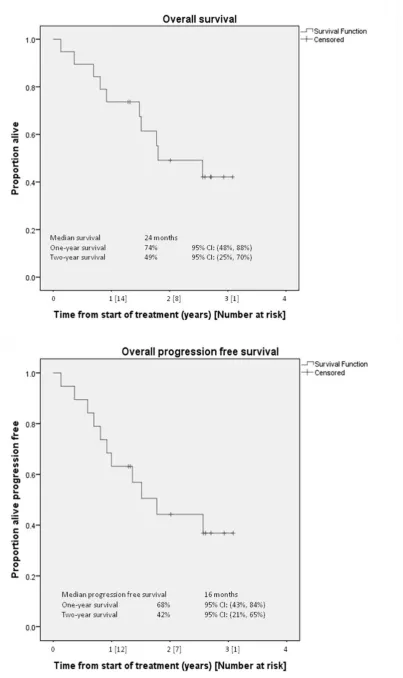
remaining patients death was not disease related. Overall 2 year survival was 49% and 42% of
patients were alive and progression free at 2 years. Overall survival curves are shown in
Figure 2 with no significant differences seen between the three cohorts.
Limitations
As there were no dose limiting toxicities reported the study did not need to expand any of the
dose cohorts which limited recruitment to 19 patients during the 25 months the study was
open. This is a much smaller number than the number of eligible patients seen in the centres
recruiting to the study giving the potential for selection bias to influence some of the
Discussion
Despite considerable effort over the last few decades, there has been little improvement in
survival for NSCLC when compared to other sites such as breast and colorectal cancers.
Many reasons are documented for patients not receiving radical surgery or concurrent
chemo-radiotherapy treatment7 but in the vast majority it is due to co-morbidity, insufficient
respiratory function or poor performance status. Stereotactic ablative body radiotherapy
(SABR) has been developed as a treatment option for early NSCLC and is providing proof of
principle evidence for accelerated dose escalation. Over the past 5 years the published
evidence for SABR has increased19,20 and consistently shows impressive local control rates of
around 90% at 5 years, with evidence that this contributes to improved outcomes across a
population21. However, there is little published randomised trial data comparing SABR with
‘conventionally’ fractionated radiotherapy, and the data presented has yet to show superior outcomes with SABR22.
Van Baardwijk et al have performed a systematic review that extends the SABR information
by including data from high dose ‘conventional’ radiotherapy series delivered using schedules lasting around 4 weeks15. These accelerated ‘conventional’ treatments also report good local
control in stage I disease with a local relapse figure of 13%, and we should remember the
median survival of 25.2 months documented for CHART treatment of stage 1A and 1B
disease23 is consistent with that reported in these publications. The significant toxicities seen
when SABR was used for more central tumours16 is an additional reason to continue to
develop accelerated schedules for the treatment of stage III disease where mediastinal
radiotherapy will need to be given.
Meta-analysis has confirmed that accelerated or hyper-fractionated radiotherapy schedules6
improve local control and survival when compared to conventionally fractionated treatments
(64-66Gy in 32-33 fractions), with a Hazard Ratio of 0.88 equating to a 2.5% improvement in
5 year survival. Recent NICE (National Institute for Health and Clinical Excellence)
guidance8 perceives CHART as a “gold standard” radiotherapy schedule and Din et al24
showed that CHART results can be reproduced in daily practice with the short schedule time
being popular with patients 99% of whom complete treatment and less than 1% suffering
grade 4 / 5 toxicity. However, the fact remains that 61% of patients who received CHART
died with persistent local disease1.
Therefore, it is important to explore dose escalation using both CHART and other
radiotherapy schedules. The recently completed RTOG 061711 study used a standard arm of
60Gy in 30 fractions and reported no benefit from dose escalation to 74Gy in 37 daily
survival compared to the control arm. While we can take the median survival of over 24
months in the standard arm to reflect contemporary practice in terms of patient selection
(routine use of PET) and the availability of third and fourth line therapies for relapsed disease,
we have to conclude that dose escalation that extends the treatment schedule is very unlikely
to lead to any significant gains in outcomes.
However, radiobiological modelling suggests that dose escalation to improve the Tumour
Control Probability (TCP) is likely to be more effective if the overall treatment time is fixed
rather that the dose per fraction25. While designing this study we applied similar TCP
modelling26 to tentatively predict 30 month local progression free survival when treating a
tumour size of around 150 cc. TCPs of 14% and 20% were calculated for 60 Gy given
conventionally over 6 weeks and for CHART, the CHART study itself reporting 12 and 18%
local control at 3 years1. These calculations also suggested that the CHART-ED schedule
delivering 64.8 Gy would achieve a TCP of 47%, which compares favourably to the TCP of
42% calculated for concurrent chemo-radiotherapy delivering 66 Gy using conventional
fractionation. Since the radiobiological modelling suggested that the 64.8 Gy dose would have
a significant effect on local control probability we took the decision not to escalate further
even though the study did not reach a MTD for the oesophagus.
The Meta-analysis by LePechoux et al 6 indicated the benefit of accelerated fractionation is
not confined to the CHART schedule and dose escalation of other schedules should lead to
significantly higher rates of local control than those seen with conventional fractionation.
However, the results from the CHARTWEL study where the dose was escalated to 60 Gy
failed to show improvement in local control feeding through to any overall survival benefits27.
The difference in outcomes reported for CHART and CHARTWEL could be a matter of
statistical chance, but factors relating to the overall length of the treatment schedule may also
be in play as induction chemotherapy was routinely given prior to radiotherapy in the
CHARTWEL study 27. In addition, subgroup analysis28 from this study does point toward
improved outcomes for the accelerated schedule in populations of patients with the larger
tumours where you might expect to see the most benefit.
When this study was designed, volume effects had not been demonstrated as clearly for
oesophagitis as for pneumonitis. We used a volumetric dose constraint for lung, limiting V20Gy
to a maximum of 35% to control the rates of pneumonitis and lung fibrosis29,30, and relied on
the incrementally increasing (57.6-64.8 Gy) prescribed dose limit to control oesophageal
toxicity. A time factor has been demonstrated for early mucosal reactions31,32, and we
calculated the extension of CHART to the more protracted 18 day schedule should allow
schedule was also limited to 44 Gy to allow for incomplete repair of normal tissue between
RT fractions, a concern for schedules delivering three fractions each day.
The majority of patients included in our study have locally advanced NSCLC, for which
neo-adjuvant, concomitant and adjuvant chemotherapy have well documented survival benefits.
Consequently combined treatment is commonly recommended for this group of patients.
Meta-analysis established the benefits and toxicities for conventionally fractionated
radiotherapy, and the comparison of sequential with concurrent treatment produces a hazard
ratio in favour of the concurrent treatment (0.84, 95% CI 0.74 – 0.95)4 and median survivals
of 24 months would now be expected in the PET staged population11.
It needs to be remembered that the potential toxicity from the concurrent approach can be
significant, with population based studies showing that performance status, age and
co-morbidities exclude a high number of patients from the concurrent form of treatment7. The
feasibility of adding induction chemotherapy to CHART has also been demonstrated by the
INCH trial17, results from which suggest this approach is associated with less toxicity than
concurrent schedules.
It is in this group of patients, unsuited to concurrent chemo-radiotherapy, that the approach of
sequential chemo-radiotherapy using dose escalated, accelerated radiotherapy schedules can
be studied further. The UK has a depth of experience in the use of accelerated fractionations,
and in addition to CHART-ED dose escalation to 64.8 Gy, similar studies for a dose-escalated
30 fraction five week concurrent CRT schedule (IDEAL-CRT)33, and 4 week sequential CRT
schedules (I-START, and Iso-IMRT) are recruiting or has just completed their recruitment.
Our TCP modeling yields similar calculated TCP values (40-50%) when each of these
schedules are used to deliver sequential CRT. We aim to carry out a randomized phase II trial
that compares these schedules which will give some comparative data on the hypo- vs
hyperfractionated approach for acceleration and aims to identifying the most appropriate dose
Used as a single modality treatment the CHART schedule remains a strong alternative to
conventionally fractionated regimes for patients unsuitable for chemotherapy. The dose
escalated CHART schedule is feasible and we saw no dose limiting toxicities up to a dose of
64.8 Gy in 42 fractions over 18 days. Given with sequential chemotherapy this schedule could
be developed as an alternative in patients unable to undergo concurrent chemo-radiotherapy.
We plan to take the CHARTED schedule into randomized phase II studies against dose
escalated accelerated sequential chemo-radiotherapy schedules to establish the optimum
method of delivering these accelerated radiotherapy regimens in the multi-modality treatment
setting.
ACKNOWLEDGEMENTS
References
1. Saunders M, Dische S, Barrett A, et al. Continuous, hyperfractionated, accelerated radiotherapy (CHART) versus conventional radiotherapy in non-small cell lung cancer: mature data from the randomised multicentre trial. CHART Steering committee. Radiother Oncol. 1999;52(2):137-148. 2. Lung cancer and smoking statistics - key facts. Cancer Research UK Website.
http://info.cancerresearchuk.org/cancerstats/types/lung
3. Perez CA, Pajak TF, Rubin P et al. Long-term observations of the patterns of failure in patients with unresectable non-oat cell carcinoma of the lung treated with definitive radiotherapy. Report by the Radiation Therapy Oncology Group. Cancer 1987;59:1874-1881
4. Aupérin A, Le Péchoux C, Rolland E, et al. Meta-analysis of concomitant versus sequential radio-chemotherapy in locally advanced non-small-cell lung cancer. J Clin Oncol 2010;28(13):2181-90 5. Kong FM, Ten Haken RK, Schipper MJ et al. High-dose radiation improved local tumor control
and overall survival in patients with inoperable/unresectable non-small-cell lung cancer: Long-term results of a radiation dose escalation study. Int J Radiat Oncol Biol Phys 2005;63:324-333 6. LePechoux C, Mauguen A, Baumann M, et al. Hyperfractionated or Accelerated Radiotherapy in
Lung Cancer: An Individual Patient Data Meta-Analysis. JCO 2012;30:2788-2797.
7. De Ruysscher D, Botterweck A, Dirx M, et al. Eligibility for concurrent chemotherapy and radiotherapy of locally advanced lung cancer patients: a prospective, population-based study. Ann Oncol 2009; (1):98-102.
8. National Collaborating Centre for Acute Care. The diagnosis and treatment of lung cancer,
update. National Collaborating Centre for Acute Care, London, 2011,
www.nice.org.uk/guidance/CG121.
9 Machtay M, Bae K, Movsas B et.al. Higher biologically effective dose of radiotherapy is associated with improved outcomes for locally advanced Non Small Cell Lung Carcinoma treated with chemoradiation: An analysis of the Radiation Therapy Oncology Group. Int. J. Radiation
Oncology Biol. Phys. 2012:82(1)425–434.
10. Saunders MI, Rojas A, Lyn BE et al. Experience with dose escalation using CHARTWEL (Continuous Hyperfractionated Accelerated Radiotherapy Week End Less) in non-small cell lung cancer. Br J Cancer 1998;78(10):1323-8
11 Bradley JD, Paulus R, Komaki R et al. Standard-dose versus high-dose conformal radiotherapy
with concurrent and consolidation carboplatin plus paclitaxel with or without cetuximab for patients with stage IIIA or IIIB non-small-cell lung cancer (RTOG 0617): a randomised, two-by-two factorial phase 3 study. Lancet Oncol 2015; 16(2):187-99
12 Fowler JF and Chappell RJ. Non small cell lung tumors repopulate rapidly during radiation therapy [Letter to the editor]. Int J Radiat Oncol Biol Phys 2000;46:516-517
13. Thirion P, Holmberg O, Collins CD et.al. Escalated dose for non-small cell lung cancer with accelerated hypofractionated three dimensional conformal radiation therapy. Radiother Oncol. 2011 71;163-6.
14. Belderbos J, De Jeager K, Heemsbergen W et.al. Final results of a phase I/II dose escalation trial in non small cell lung cancer using three-dimensional conformal radiotherapy. Int J Radiat Oncol Biol Phys 2006 66:126-34.
15 Baardwijk A, Tome WA, Elmpt W, et.al. Is high dose stereotactic body radiotherapy (SBRT) for stage I Non Small Cell lung Cancer (NSCLC) overkill? A systematic review. Radiother Oncol 2012;105;145-9.
16 Timmerman, R., R. McGarry, C. Yiannoutsos, et al., Excessive toxicity when treating central tumors in a phase II study of stereotactic body radiation therapy for medically inoperable early-stage lung cancer. J Clin Oncol, 2006;24:4833-9.
17.
Hatton M, Lyn E, Nankivell M et.al. Induction chemotherapy and Continuous Hyperfractionated
Accelerated Radiotherapy (CHART): the MRC INCH randomised trial. Int J Radiat Oncol Biol Phys 2011;81:712-18
18. CONVERT Concurrent ONce-daily VErsus twice-daily RadioTherapy: A 2-arm randomised controlled trial of concurrent chemo-radiotherapy comparing twice-daily and once-daily radiotherapy schedules in patients with limited stage small cell lung cancer (SCLC) and good performance status. ISRCTN 91927162.
19. Munshi A, Krishnatry S, Banerjee S et al. Stereotactic conformal radiotherapy in Non small cell lung cancer – An overview. Clinical Oncology 2012;24(8):556-68.
21. Palma D, Visser O, Lagerwaard FJ et al. Impact of introducing stereotactic lung radiotherapy for elderly patients with stage I non-small-cell lung cancer: a population-based time-trend analysis. J Clin Onc, 2010. 28(35): p5153-9
22. Nyman J, Hallqvist A, Lund JA, et.al. SPACE - A randomized study of SBRT vs conventional fractionated radiotherapy in medically inoperable stage I NSCLC. Radiotherapy and Oncology 111suppl 1 S232;2014
23. Bradshaw AG, Esler C, Roy AEF et.al, Continuous Hyperfractionated Accelerated Radiotherapy (CHART) for NSCLC: Experience from nine UK centres. Radiotherapy and Oncology 111suppl 1 S35;2014
24. Din OS, Lester J, Cameron A et al. The routine use of Continuous, Hyper-fractionated, Accelerated Radiotherapy (CHART) for Non-Small Cell Lung Cancer: A five centre experience. Int J Radiat Oncol Biol Phys 2008; 72:716-722.
25. Mehta M, Scrimger R, Mackie R, et al. A new approach to dose escalation in non-small cell lung cancer. Int. J. Radiat. Oncol., Biol., Phys. 2001; 49:23-33
26. Fenwick JD, Nahum AE, Malik MI, Eswarz CV, Hatton MQ, Laurence VM, Lester JF, Landau DB. Escalation and Intensification of Radiotherapy for Stage III Non-small Cell Lung Cancer: Opportunities for Treatment Improvement. Clinical Oncology 21:343-360;2009
27. Baumann, M., Herrmann, T., Koch, R. et al. Final results of the randomised phase III CHART-WEL trial (ARO 97-1) comparing hyper-fractionated accelerated vs conventionally fractionated radiotherapy in non-small-cell lung cancer (NSCLC). Radiotherapy and Oncology100;76-85:2011.
28. Soliman M, Yaromina A, Appold S, Zips D, Reiffenstuh C, Schreiber A, Thames HD, Krause
Ma, Baumann M. GTV differentially impacts locoregional control of non-small cell lung cancer
(NSCLC) after different fractionation schedules: Subgroup analysis of the prospective randomized
CHARTWEL trial. Radiotherapy and Oncology 2013:106;299–304.
29. Seppenwoolde Y, Lebesque J V, de Jaeger K, et al. Comparing different NTCP models that predict the incidence of radiation pneumonitis Int J Radiat Oncol Biol Phys.2003;55:724-735
30. SJ Clenton, PM Fisher, J Conway, P Kirkbride, MQ Hatton. The use of lung dose volume histograms in predicting post-radiation pneumonitis after non-conventionally fractionated radiotherapy for thoracic carcinoma Clinical Oncology 17:599-603;2005
31 Bentzen S M, Sauders M I and Dische S 2002 From CHART to CHARTWEL in Non-small Cell Lung Cancer: Clinical Radiobiological Modelling of the Expected Change in Outcome Clin Oncol 2002;14:372-381
32. Fenwick JD. Lawrence GP, Malik Z, et al. Early mucosal reactions during and after head-and-neck radiotherapy: dependence of treatment tolerance on radiation dose and schedule durations. Int. J. Radiat. Oncol., Biol., Phys. 2008; 71: 625-634
33. Landau. D.B, Khan. I, Baker.A et.al IDEAL-CRT: Isotoxic Dose Escalation and Acceleration in Lung Cancer Chemo-Radiotherapy. A phase I/II trial of dose-escalated radiotherapy and concurrent chemotherapy in patients with stage II or III non-small cell lung cancer. Journal of Thoracic Oncology 8 Suppl 2, S190 2013
34. A phase I/II trial of isotoxic accelerated radiotherapy in the treatment of patients with non-small cell lung cancer. ISRCTN74841904
35. Isotoxic Intensity Modulated Radiotherapy (IMRT) in stage III non small cell lung cancer
Table 1
Demographic details for patients recruited to the CHART-ED study.
Overall Group 1 Group 2 Group 3
(n=18) (N=6) (N=6) (N=6)
Median age 70 66 74 74
Range 44-84 44-78 63-84 64-84
Males 14 4 6 4
Histology
Squamous 16 5 6 5
Adenocarcinoma 2 1 0 1
Other 0 0 0 0
Stage 1-B 1 0 1 0
(V7) 2A / B 5 1 2 2
IIIA 8 4 2 2
IIIB 4 1 1 2
WHO PS 0 4 2 1 1
1 13 4 5 4
2 1 0 0 1
Respiratory Function
Median FEV1 2.3 1.9 2.0 2.4
Range 1.1 to 3.6 1.1 to 3.6 1.4 to 2.6 1.6 to 2.9
Median FVC 3.5 3.0 3.3 3.5
Range 1.7 to 5.4 1.7 to 5.4 2.3 to 3.8 2.4 to 3.7
Median DLCO 6.4 6.4 5.5 8.4
Table 2
Radiotherapy planning data for patient who received a dose escalated
schedule
Group 1 (n=6)
Group 2 (n=6)
Group 3 (n=6)
All patients (n=18)
% coverage of Mean 89.4 93.6 97.4 93.5
PTV by 95% Std dev 8.6 7.4 1.7 7.1
isodose Maximum 97.5 100.0 99.5 100.0
Minimum 74.7 79.5 94.9 74.7
% whole lung Mean 25.3 23.2 27.6 25.4
V20 (Gy) Std dev 7.2 5.0 5.7 6.0
Maximum 33.6 29.7 34.5 34.5
Minimum 13.1 16.2 21.3 13.1
Maximum Mean 34.6 33.4 36.2 34.7
dose to spinal Std dev 8.0 6.5 5.6 6.5
cord (Gy) Maximum 39.6 43.5 42.1 43.5
Minimum 18.9 25.2 26.4 18.9
Gross tumour Mean 87.7 84.5 94.4 88.9
volume (GTV) (cm3)
Std dev
59.1 73.0 77.9 66.3
Maximum 192.0 219.0 212.6 219.0
Minimum 18.5 29.0 18.0 18.0
Planning Mean 495.5 485.7 521.7 500.9
target volume Std dev 206.7 234.0 188.3 198.4
(PTV) (cm3) Maximum 795.0 897.0 733.0 897.0
Minimum 208.0 281.0 278.0 208.0
Oesophageal
dose +/- 5% of 5 patients 5 patients 5 patients
54.0 57.6 61.2 64.8 68.4 -5
0 5 10 15 20 25 30 35 40
V95%
(%)
[image:18.595.79.492.104.253.2]prescribed dose (Gy)
Figure 1
Plots showing the percentages of outlined oesophagus receiving doses in excess of (a) 95% and (b) 100% of the prescribed dose for the 18 patients treated on CHART-ED
N
volumes for multiple patients lie at or very close to zero).
54.0 57.6 61.2 64.8 68.4
0 5 10 15 20 25 30 35
V100%
(%)