R E S E A R C H
Open Access
One particular
Anaplasma phagocytophilum
ecotype infects cattle in the Camargue,
France
Thibaud Dugat
1, Agnès Leblond
2,3, Nicolas Keck
4, Anne-Claire Lagrée
5, Isabelle Desjardins
3, Aurélien Joulié
2,3,
Sophie Pradier
6, Benoit Durand
7, Henri-Jean Boulouis
5and Nadia Haddad
5*Abstract
Background:Anaplasma phagocytophilumis a zoonotic tick-borne pathogen responsible for granulocytic anaplasmosis, a mild to a severe febrile disease that affects man and several animal species, including cows and horses. In Europe,I. ricinusis the only proven vector for this pathogen, but studies suggest that other tick genera and species could be involved in its transmission. Our objective was to assess the presence and genetic diversity of
A. phagocytophilumin domestic animals and different tick species from the Camargue region, located in the south of France.
Methods:A total of 140 ticks and blood samples from 998 cattle and 337 horses were collected in Camargue and tested for the presence ofA. phagocytophilumDNA bymsp2quantitative real-time PCR. Molecular typing with four markers was performed on positive samples.
Results:Anaplasma phagocytophilumDNA was detected in 6/993 (0.6%) cows, 1/20 (5%)Haemaphysalis punctata, 1/57 (1.75%)Rhipicephalus pusillus, and was absent in horses (0%). All cattleA. phagocytophilumpresented a profile identical to anA. phagocytophilumvariant previously detected inDermacentor marginatus,Hyalomma marginatum, andRhipicephalusspp.in Camargue.
Conclusions:Our results demonstrate that one particularA. phagocytophilumvariant infects cattle in Camargue, whereI. ricinusis supposed to be rare or even absent.Dermacentor marginatus,Rhipicephalusspp. andHyalomma
spp., and possibly other tick species could be involved in the transmission of this variant in this region.
Keywords:Anaplasma phagocytophilum, Camargue, Cattle, Horse,Haemaphysalis punctata,Rhipicephalus pusillus
Background
Anaplasma phagocytophilum is a tick-borne intragranu-locytic alpha-proteobacterium that can infect many mammalian species. It is known to be the causative agent of tick-borne fever (TBF) in domestic ruminants, a disease with significant economic impact in Europe, and of equine granulocytic anaplasmosis (EGA) in horses in
both the USA and Europe [1].Anaplasma
phagocytophi-lum also infects cats, dogs and humans and causes
human granulocytic anaplasmosis (HGA), an emerging zoonotic disease in Asia, the USA, and Europe.
F1
Anaplasma phagocytophilum epidemiological cycles are complex and involve different ecotypes, vectors, and mammalian host species. To date, these epidemiological cycles are poorly understood, especially in Europe.
Known arthropod vectors of A. phagocytophilum are
ticks belonging to the genusIxodes:I. ricinusin Europe, I. scapularisin eastern USA,I. pacificusandI.
spinipal-pis in the western USA, and I. persulcatusin Asia and
Russia [1]. However, several studies suggest that other
tick genera and species could be involved in A.
phago-cytophilum transmission, but their vector competence
is yet to be proven [2]. For example, I. trianguliceps
could play the role of an A. phagocytophilum vector
in an independent epidemiological cycle in the UK and central Europe, involving different rodent species * Correspondence:nadia.haddad@vet-alfort.fr
5UMR BIPAR, Université Paris-Est, Ecole Nationale Vétérinaire d’Alfort, Maisons-Alfort, France
Full list of author information is available at the end of the article
as reservoir hosts [3, 4]. In addition, it has been suggested thatRhipicephalusspp. could be involved inA. phagocyto-philum transmission in the French Camargue region, a
1500 km2marshland area in the south of France, and from
whereI. ricinusis supposed to be absent [5, 6]. Here, live-stock is almost exclusively constituted of a local fighting bull breed, the Camargue breed, raised in extensive systems, which exposes them to arthropod bites. It is the same for the Camargue horses, which are also bred exten-sively. Even though up to 26% of horses have been found
seropositive for A. phagocytophilum [7], live bacteria or
bacterial DNA have never been detected in the animals. In addition, Camargue cattle have never been investigated for A. phagocytophilum infection. For these reasons, the
presence/absence and the epidemiological cycle(s) of A.
phagocytophilumin Camargue should be investigated. Our objective was to assess the presence and
gen-etic diversity of A. phagocytophilum in domestic
ani-mals and different tick species in Camargue. To achieve this, 140 ticks, and blood samples from 998 cattle (Bos taurus) and 337 horses (Equus caballus) were collected in Camargue and tested for the
pres-ence of A. phagocytophilum DNA by quantitative
real-time PCR targeting the msp2 gene. Positive
sam-ples were then molecularly typed via four sequences
located within the genes ankA, pleD, msp4 and typA,
among those genes used for MLST by Chastagner et al. [5, 8].
Methods
Study area
Camargue is a 1500 km2 area in the south of France,
from where I. ricinus has never been collected to date.
Camargue is a region of marshlands where herds are al-most exclusively constituted of a local fighting bull breed and a local horse breed raised in extensive conditions. Three French administrative departments extend into this area: Bouches-du-Rhône, Gard, and Hérault.
Animal sampling
In 2015, blood samples from 337 horses (269 located in Bouches-du-Rhône, 47 in Gard, and 21 in Hérault) were collected. Blood from horses was preferentially collected
near areas where ticks positive for A. phagocytophilum
were detected in previous studies [5]. Ticks (engorged or not) feeding on horses was also collected during the same time. One-hundred and forty ticks were found on examined horses, 108 in Bouches-du-Rhône, 19 in Gard, and 13 in Hérault (Additional file 1: Table S1). Between 2013 and 2015, blood samples from 998 cattle (i.e. 10% of the 10,000 French Camargue cattle; 235 in Gard and 765 in Hérault), older than 24 months and belonging to the Camargue or Brave breeds, were collected (Fig. 1). These blood samples from cattle had been collected in a
previous study unrelated to A. phagocytophilum, which
explains why no tick sample was associated with these cattle samples. Moreover, collecting ticks on bovines in
[image:2.595.57.538.85.358.2]this region could be very hazardous as they are bred for fighting.
Tick identification
Ticks were morphologically identified to the species level following Pérez-Eid’s recommendations [9].
DNA extraction
For DNA extraction, the NucleoSpin® Blood QuickPure kit (Macherey-Nagel, Bethlehem, USA) (blood samples and engorged ticks), or the NucleoSpin® Tissue kit
(Macherey-Nagel) (non-engorged ticks) were used
according to manufacturer’s instructions. DNA extracts
were then stored at -20 °C prior to testing.
Quantitative real-time PCR
Anaplasma phagocytophilum DNA was detected by
qPCR, targeting a 77 bp fragment of the major surface
protein 2(msp2) gene as previously described by Courtney et al. [10]. To confirm the presence ofA. phagocytophilum
DNA, each msp2 qPCR-positive sample was also tested
for another gene specific to A. phagocytophilum, with
qPCR targeting a fragment of theankAandgroEL genes,
with the primers designed by Dugat et al. [11].
For each sample, qPCR targeting the bovine, equine
and ticks glyceraldehyde-3-phosphate dehydrogenase
(gapdh) gene was also performed to demonstrate the efficiency of DNA extraction and the absence of PCR inhibitors in the sample, using the primers designed
[12, 13]. All samples had to be gapdh PCR-positive to
be included in subsequent analyses.
For ankA, groEL and gapdh, qPCR assays were per-formed using the Maxima SYBR Green qPCR Master Mix (2×) Kit (Thermo Fisher Scientific,
Villebon-sur-Yvette, France) in a 25 μl total reaction volume, with
Master Mix at a 1× final concentration, 0.3μM of each
primer and 5μl of purified DNA. Negative controls were
included in each run. qPCR cycling was performed on the LightCycler480 Multiwell Plate 96 system (Roche, Basel, Switzerland) as follows: 95 °C for 10 min, then 40 cycles of 10 s at 95 °C, 30 s at 60 °C and 30 s at 72 °C. The signal emitted was detected at the end of each annealing-extension step. A threshold was automatically set, and the threshold cycle value (Ct) was consequently determined. All real-time PCR reactions were performed in duplicate.
Genotyping
Samples positive for A. phagocytophilum were initially
typed using the eight loci selected from a recently
devel-oped - MLST adapted for A. phagocytophilum [5, 8].
These loci were the following: ankA, groESL, gyrA,
msp4,pleD,polA,recG,typA.
Sequencing results were analysed using Bioedit soft-ware version 7.2.5 (Ibis Biosciences, Carlsbad, USA).
Each sequence was deposited in GenBank (KU859923–
KU859946). Nucleotide sequences were then aligned to all the sequences available in GenBank, including those obtained by Chastagner et al. [5] (GenBank accession
numbers: JX197073–JX197368) using the programme
MEGA7 (Molecular Evolutionary Genetics Analysis Ver-sion 7.0.18) [14]. Gene sequences were aligned using ClustalW while applying the IUB matrix.
Results
Tick identification
A total of 140 ticks were collected from horses: 5
Der-macentor reticulatus(3.5%), 20Haemaphysalis punctata
(14.2%), 4 Haemaphysalis sp. (2.8%), 57 Rhipicephalus
pusillus(40.4%), 19Rhipicephalus sanguineus (13.5%), 1 Rhipicephalus turanicus(0.7%), and 34Rhipicephalussp. (24.2%). None of the 140 collected ticks belonged to the genusIxodes.
Detection ofA. phagocytophilumDNA
For domestic animals, 993/998 (99.5%) cow samples and
269/337 (79.8%) horse samples gave positivegapdhPCR
results and were included in subsequent analyses. 6/993
(0.6%, 95% CI: 0.2–1.3%) cow samples were msp2
PCR-positive. These six positive cows belonged to the same herd, located in Hérault, and all cows had been born in
this herd. Nomsp2amplification was observed in any of
the 269 horses tested (0%, 95% CI: 0–1.4%) (Table 1,
Additional file 1: Table S1). Statistically, cattle were not
more frequently infected by A. phagocytophilum than
horses (Fisher’s exact test,P= 0.35).
For the tick samples, 137/140 (97%) weregapdh
PCR-positive and were included in subsequent analyses. Only
2/137 (1.5%, 95% CI: 0.2–5.2%) weremsp2PCR-positive:
[image:3.595.306.539.581.731.2]oneR. pusillusmale and oneH. punctatamale (Table 1, Additional file 1: Table S1). These two ticks were
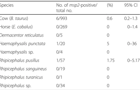
Table 1Prevalence ofA. phagocytophiluminfection for each animal species
Species No. ofmsp2-positive/ total no.
(%) 95% CI
Cow (B. taurus) 6/993 0.6 0.2–1.3
Horse (E. cabalus) 0/269 0 0–1.4
Dermacentor reticulatus 0/5 0
Haemaphysalis punctata 1/20 5 0–36
Haemaphysalissp. 0/4 0
Rhipicephalus pusillus 1/57 1.75 0–5.17
Rhipicephalus sanguineus 0/19 0
Rhipicephalus turanicus 0/1 0
collected from two different horse farms next to the town of Arles (Bouches-du-Rhône).
The eight msp2 PCR-positive samples were also
posi-tive forankAandgroELqPCRs.
Genotyping
In the positive H. punctata and R. pusillus, DNA
quantity was too low to enable complete genotyping. However, genotyping could be performed on the six positive cow samples. All eight genes included in this technique gave positive PCR results, but positive
sequen-cing results were generated only for four loci: ankA,
pleD, msp4, and typA. The sequence quality of groESL, gyrA, recG and polA loci was too poor to be analysed properly. Subsequently, too little sample DNA remained to perform a second round of genotyping. All six cow samples presented 100% identity at these four loci. The sequences of these genes were then aligned to all the se-quences available on GenBank, including those obtained by Chastagner et al. [5]. For all four loci, the six positive
cow samples shared 100% identity with the single A.
phagocytophilumgenotype identified in 40Rhipicephalus
spp., D. marginatus and H. marginatum [5]. However,
when comparing the msp4 sequences, the Camargue
genotype was located in a different cluster than A.
phagocytophilum variants from cattle living in several other French areas [14]. Moreover, a significant part of
the available sequences of A. phagocytophilum from
humans in different areas in the USA (HGE1, HZ, NY18 and Webster) were also located in this cluster, as well as one sequence from a horse living in the USA (Additional file 2: Table S2). Apart one sequence from a red deer in Slovenia and one sequence from a donkey in Italy, they were the only non-Camargue samples to belong to this cluster.
Discussion
Anaplasma phagocytophilum can infect many mamma-lian species worldwide and is known to be the causative agent of TBF and EGA, two diseases with high economic impact in Europe [1]. On this continent,I. ricinus is the
main vector, and to date the only proven vector, of A.
phagocytophilum. In the present study, we investigated for the first time the presence and genetic diversity ofA. phagocytophilum both in ticks and domestic animals in
Camargue, a 1500 km2area in the south of France from
where I. ricinus is supposed to be absent, due to
unfavourable ecosystem conditions for this species. To our knowledge, this study is the first large-scale
screening of A. phagocytophilum in cattle from a
par-ticular French region and is the first to reportA. phago-cytophilum DNA in cows from Camargue. Six on 998 cows, which all belonged to the same herd, were found positive forA. phagocytophilum. Our results are consistent
with those obtained by Torina et al. [15] in Sicily (5/374 cows, 1.3%, 95% CI: 0.4–3.1%), a region in whichI. ricinus
is rare, whereasDermacentor marginatus,H. marginatum
and Rhipicephalus spp. are commonly collected. Our results demonstrate thatA. phagocytophiluminfects cattle in Camargue.
However, this region is considered aI. ricinus-free area: indeed, noI. ricinuswere collected in our study or during several studies conducted between 2007 and 2010 [16].
Moreover,A. phagocytophilumDNA has already been
de-tected inR. bursa,R. sanguineus,R. turanicus,R. pusillus, D. marginatus and H. marginatum in Camargue [5].
Taken together, these results indicateA. phagocytophilum
is most likely transmitted by the vector(s) other than I.
ricinusin this region. Thus we investigated the presence ofA. phagocytophilumin different tick genera and species collected in Camargue. At the level of our sampling, only
two ticks were qPCR-positive: one R. pusillus male and
oneH. punctatamale. This is the first report ofA. phago-cytophilum in R. pusillus, whereas H. punctata has
re-cently been suspected to be a vector of A.
phagocytophilum in Spain [17]. This species could then
also potentially transmit A. phagocytophilum to cows in
Camargue. However, the high number ofA.
phagocytophi-lum-infected tick species found in Camargue from
previ-ous studies raises questions about their vector
competence. Many of these ticks could have acquired A.
phagocytophilum (or only its DNA) by passively feeding on an infected animal, without then being able to transmit the pathogen. Unfortunately, due to the low quantity of tick sample DNA, we were not able to determine the
genotype ofA. phagocytophilumpresent in these ticks and
compare it to the genotype present in the cows typed in our study. However, it is noteworthy that the profile of the six A. phagocytophilum cow samples for the four tested
genes (ankA, pleD,msp4 andtypA) was identical to that
of A. phagocytophilum from 40 Rhipicephalus spp., D. marginatus and H. marginatum sampled in Camargue during a previous study [5]. This profile was shared by ticks that had been collected throughout the three French administrative departments of Camargue, which covers
1500 km2. In addition, this genotype has never been
de-tected in ticks and animals from other regions in France. These observations strongly suggest that only one variant, which is transmitted by one or several tick species, infects cows in Camargue in an epidemiological cycle independ-ent fromI. ricinus. This“specialisation” could have led to
decreased A. phagocytophilum diversity in Camargue,
resulting in the circulation of this single variant.
between the samples from Camargue and the human Webster profile, the only American human sample that could be tested by MLVA [6]. The sequence identity, for both markers, of samples originating from regions as far apart from one another (Camargue and the USA) could be due to homoplasy.
None of the 269 studied horses was infected byA.
pha-gocytophilum, whereas in prior studies occurring between 2001 and 2010, 8.6 to 26% of examined horses were sero-positive [7, 16, 18]. In this context, the absence of any positive PCR result in horses in our study was a priori sur-prising as 42 out of the 337 horses tested in the present study displayed clinical signs compatible with equine granulocytic anaplasmosis. This could be explained by the fact that most of the diseased horses received imidocarb.
This treatment was administered because Theileria equi
infection is the main cause of fever in horses in this area. Imidocarb is also commonly used for the treatment of cat-tle anaplasmosis. Thus, the use of this babesicid molecule could most likely explain the negative PCR results.
Finally, the reservoir host(s) of the Camargue vari-ant must be identified. This unique and stable varivari-ant suggests that it could be adapted to a restricted num-ber of reservoir host(s) and/or vector(s). This variant
belongs to the ankA cluster I, which mostly contains
variants obtained from humans, dogs, cats, and horses, and several variants obtained from domestic
and wild ruminants [5, 8]. Haemaphysalis punctata
females are known to have a trophic preference for
both wild and domestic ruminants, and R. pusillus for
rabbits [9]. For these reasons, wild ruminants and
rabbits should be investigated as potential A.
phagocy-tophilum reservoirs in Camargue, even if there is
some doubt about the vector competence of H.
punc-tate and R. pusillus. Furthermore, it also remains to be demonstrated that the same unique variant is also present in these ticks.
Due to these observations, prevalence studies should be continued in both ticks and domestic animals throughout the coming years in order to determinetheA.
phagocytophi-lumvector competence of the tick species present in
Cam-argue, the reservoir host of A. phagocytophilum in
Camargue, the level of infection in cattle and horses and the clinical impact of disease in these species, and the zoo-notic potential of this variant.
Conclusion
In conclusion, our results strongly suggest that one
particular A. phagocytophilum variant infects cows in
Camargue, an area whereI. ricinusis supposed to be rare
or even absent. A variant that presented the same profile based on our four markers was already identified in Rhipi-cephalus spp., D. marginatus, H. marginatum, H. punc-tata and R. pusillusby Chastagner et al. [5]. These ticks
could be involved inA. phagocytophilumtransmission in
this particular region, but additional studies are needed before confirming this theory. The vertebrate and inverte-brate actors of this epidemiological cycle must now be confirmed or identified to develop appropriate surveil-lance measures. Finally, the zoonotic potential of this vari-ant should also be investigated.
Additional files
Additional file 1: Table S1.Characteristics and qPCR results for the samples used in this study. (XLSX 67 kb)
Additional file 2: Table S2.Phylogenetic tree ofmsp4sequences ofA. phagocytophilumavailable in the GenBank database. (PDF 26 kb)
Abbreviations
EGA:Equine granulocytic anaplasmosis;gapdh: Glyceraldehyde-3-phosphate dehydrogenase; HGA: Human granulocytic anaplasmosis; MLST: Multilocus sequence typing; msp2: Major surface protein 2; qPCR: Quantitative real-time PCR; TBF: Tick-borne fever
Acknowledgements
The authors thank Valérie Poux for tick identification, Severine Barry for tick DNA extraction, Elsa Jourdain for scientific support and Christelle Gandouin, Corinne Bouillin, and Joanne Befort for their technical support. This work was performed within the Laboratory of Excellence (Labex) of Integrative Biology of Emerging Infectious Diseases (IBEID).
Funding
This work was supported by the Alfort National Veterinary School (ENVA), by the French Agency for Food, Environmental and Occupational Health and Safety (Anses) and by the French National Institute for Agricultural Research (INRA). TD was funded by Anses.
Availability of data and materials
The data supporting the conclusions of this article are included within the article and Additional file 1.
Authors’contributions
TD contributed to experimental design, to genotyping data analysis, was responsible for laboratory work and drafted the manuscript. AL, AJ, SP and ID were responsible for horse and tick sample collection, tick identification, and were involved in critical revision of the manuscript. NK was responsible for cattle sample collection and was involved in critical revision of the manuscript. ACL participated in laboratory work as well as critical revision of the manuscript. BD helped with genotyping analysis and critical revision of the manuscript. HJB and NH were responsible for the conception and contributed to experimental design and to drafting the manuscript. All authors read and approved the final manuscript.
Ethics approval and consent to participate
The domestic animals used in this study met the definition of“farm animals”, which are not currently covered by French regulations
(Décret2013–118 dated the 1st February 2013, from the French Ministry of Agriculture). The owners of the animals provided permission for studies to be performed on animal samples collected during sanitary investigations. Samples were de-identified so that breeding farms remained anonymous.
Consent for publication Not applicable.
Competing interests
Publisher’s Note
Springer Nature remains neutral with regard to jurisdictional claims in published maps and institutional affiliations.
Author details
1UMR BIPAR, Université Paris-Est, Agence nationale de sécurité sanitaire de l’alimentation, de l’environnement et du travail, Laboratoire de santé animale, Maisons-Alfort, France.2UR 0346 Épidémiologie Animale, INRA, Saint Genès Champanelle, France.3Equine Department, VetAgroSup, Marcy L’Etoile, France.4Laboratoire Départemental Vétérinaire de l’Hérault, Montpellier, France.5UMR BIPAR, Université Paris-Est, Ecole Nationale Vétérinaire d’Alfort, Maisons-Alfort, France.6IHAP, Université de Toulouse, INRA, ENVT, Toulouse, France.7Unité d’Épidémiologie, Université Paris-Est, Agence Nationale de Sécurité Sanitaire de l’alimentation, de l’environnement et du travail, Laboratoire de Santé Animale, Maisons-Alfort, France.
Received: 14 December 2016 Accepted: 24 July 2017
References
1. Dugat T, Lagrée A-C, Maillard R, Boulouis H-J, Haddad N. Opening the black box ofAnaplasma phagocytophilumdiversity: current situation and future perspectives. Front Cell Infect Microbiol. 2015;61
2. Stuen S, Granquist EG, Silaghi C.Anaplasma phagocytophilum -a widespread multi-host pathogen with highly adaptive strategies. Front Cell Infect Microbiol. 2013;3(31)
3. Blaňarová L, Stanko M, Carpi G, Miklisová D, Víchová B, Mošanský L, et al. DistinctAnaplasma phagocytophilumgenotypes associated withIxodes triangulicepsticks and rodents in Central Europe. Ticks Tick-Borne Dis. 2014;5(6):928–38.
4. Bown KJ, Lambin X, Ogden NH, Begon M, Telford G, Woldehiwet Z, et al. DelineatingAnaplasma phagocytophilumecotypes in coexisting, discrete enzootic cycles. Emerg Infect Dis. 2009;15:1948–54.
5. Chastagner A, Bailly X, Leblond A, Pradier S, Vourc’h G. Single genotype of Anaplasma phagocytophilumidentified from ticks, Camargue. France Emerg Infect Dis. 2013;19:825–6.
6. Dugat T, Chastagner A, Lagrée A-C, Petit E, Durand B, Thierry S, et al. A new multiple-locus variable-number tandem repeat analysis reveals different clusters forAnaplasma phagocytophilumcirculating in domestic and wild ruminants. Parasit Vectors. 2014;7:439.
7. Leblond A, Pradier S, Pitel P, Fortier G, Boireau P, Chadoeuf J, et al. Enquête épidémiologique sur l’anaplasmose équine (Anaplasma phagocytophilum) dans le sud de la France. Rev Sci Tech Off Int Epizoot. 2005;24:899–908. 8. Chastagner A, Dugat T, Vourc HG, Verheyden H, Legrand L, Bachy V, et al.
Multilocus sequence analysis ofAnaplasma phagocytophilumreveals three distinct lineages with different host ranges in clinically ill French cattle. Vet Res. 2014;45:114.
9. Pérez-Eid C. Les tiques, identification, biologie, importance médicale et vétérinaire. TEC&DOC; 2007.
10. Courtney JW, Kostelnik LM, Zeidner NS, Massung RF. Multiplex real-time PCR for detection ofAnaplasma phagocytophilumandBorrelia burgdorferi. J Clin Microbiol. 2004;42:3164–8.
11. Dugat T, Loux V, Marthey S, Moroldo M, Lagrée A-C, Boulouis H-J, et al. Comparative genomics of first available bovineAnaplasma phagocytophilum genome obtained with targeted sequence capture. BMC Genomics. 2014;15:973.
12. Bougarn S, Cunha P, Gilbert FB, Meurens F, Rainard P. Technical note: validation of candidate reference genes for normalization of quantitative PCR in bovine mammary epithelial cells responding to inflammatory stimuli. J Dairy Sci. 2011;94:2425–30.
13. Kayis SA, Atli MO, Kurar E, Bozkaya F, Semacan A, Aslan S, et al. Rating of putative housekeeping genes for quantitative gene expression analysis in cyclic and early pregnant equine endometrium. Anim Reprod Sci. 2011;125:124–32.
14. Tamura K, Stecher G, Peterson D, Filipski A, Kumar S. MEGA6: Molecular Evolutionary Genetics Analysis version 6.0. Mol Biol Evol. 2013;30:2725–9.
15. Torina A, Alongi A, Naranjo V, Estrada-Peña A, Vicente J, Scimeca S, et al. Prevalence and genotypes ofAnaplasmaspecies and habitat suitability for ticks in a Mediterranean ecosystem. Appl Environ Microbiol. 2008;74:7578–84. 16. Leblond A, Chastagner A, Pradier S, Bailly X, Masseglia S, Vourc’h G. La
prévalence de l’anaplamose dans le sud de la France. Bull Épidémiologique Santé Anim Aliment. 2012:30–1.
17. Palomar AM, Portillo A, Santibáñez P, Mazuelas D, Roncero L, García-Álvarez L, et al. Detection of tick-borneAnaplasma bovis,Anaplasma
phagocytophilumandAnaplasma centralein Spain. Med Vet Entomol. 2015;29:349–53.
18. Tilliette B. Anaplasmose granulocytaire équine: enquête
sero-épidémiologique dans le sud est de la France en 2007 [Veterinary thesis]. Ecole Nationale Vétérinaire d’Alfort; 2008.
• We accept pre-submission inquiries
• Our selector tool helps you to find the most relevant journal
• We provide round the clock customer support
• Convenient online submission
• Thorough peer review
• Inclusion in PubMed and all major indexing services
• Maximum visibility for your research
Submit your manuscript at www.biomedcentral.com/submit