R E S E A R C H
Open Access
Multiplication of the waterborne pathogen
Cryptosporidium parvum
in an aquatic biofilm
system
Wan Koh
1*, Peta L Clode
2, Paul Monis
3and RC Andrew Thompson
1Abstract
Background:In natural aquatic environments biofilms are known to act as environmental reservoirs for Cryptosporidium parvumoocysts. However, the fate of these oocysts within biofilms has yet to be determined. Methods:This study aimed to identify if biofilms have the ability to support the multiplication ofCryptosporidium by measuring the change in parasite number over time using quantitative polymerase chain reaction (qPCR) and detecting the possible extracellular developmental stages using a combination of confocal microscopy and immunolabelling techniques.Pseudomonas aeruginosabiofilm flow cell systems were established andC. parvum oocysts were constantly supplied over a six day period.
Results:A significant (P < 0.001) increase inCryptosporidiumwas detected as the biofilm matured, with the total number ofC. parvummultiplying 2–3 fold during this period. With this, variousCryptosporidiumdevelopmental stages (sporozoites, trophozoites, type I and II meronts) were identified from the biofilm.
Conclusion:This is the first study demonstrating that biofilms not only serve as an environmental reservoir for oocysts, but are also capable of supporting the multiplication ofCryptosporidiumover time in an aquatic environment.
Keywords:Cryptosporidium, Biofilms, Extracellular multiplication, Water, qPCR, Fluorescence
Background
Cryptosporidium parvumis a zoonotic waterborne pathogen
found worldwide [1], with Cryptosporidium oocyst levels
commonly monitored in urban water distribution systems [2]. If oocysts are ingested by a suitable host, which includes humans and livestock [3], the oocyst undergoes several cycles of multiplication via both asexual (sporozoites, trophozoites) and sexual processes (merozoites and microgametes) [4]. Historically, it has always been assumed that
Cryptosporidium, like other apicomplexans, can only multiply intracellularly by forming an extracytoplasmic parasitophorous vacuole in host intestinal cells and that they are not able to multiply outside of the host [5].
There are, however, an increasing number of in-vitro
studies, both in cell and cell-free [6-12] cultures,
demonstrating that Cryptosporidium may not be an
obligate intracellular parasite and can in fact multiply extracellularly. This capacity to multiply both intracellularly and extracellularly may reflect the fact thatCryptosporidium
is closely related to gregarine protozoa [9,11,13], which can also multiply by either means [14]. The ability of
Cryptosporidium to multiply extracellularly also indicates that they are able to salvage nutrients required for multiplication from their environment, despite lacking mitochondria and associated metabolic capabilities [15]. Independent of the mechanisms, it is clear that the surrounding environment plays an important role in shaping the life cycle of Cryptosporidium.
Biofilms are highly efficient and stable ecosystems [16] that are formed mainly by bacteria [17-19], creating a favourable micro-environment that can support the survival and growth of other micro-organisms under prolonged periods of environmental stress [16]. Biofilms have been shown to serve as an environmental reservoir for
Cryptosporidium oocysts in aquatic environments [20-22] and may be responsible for the occurrence of sporadic * Correspondence:w.koh@murdoch.edu.au
1
School of Veterinary and Life Sciences, Murdoch University, South Street, Murdoch, WA 6150, Australia
Full list of author information is available at the end of the article
Cryptosporidium outbreaks [23]. Therefore, there is a
current need to better understand Cryptosporidium
behaviour in biofilm environments, especially in relation to water distribution systems [18,22] and to investigate whether
Cryptosporidium oocysts captured within biofilms can utilise this nutrient rich micro-environment to survive and multiply.
The aim of this study was to investigate whether biofilms can support the multiplication ofCryptosporidiumin aquatic environments. Model flow cell biofilm systems were developed and confocal laser scanning microscopy coupled with image analysis was used to quantitatively compare biofilm thickness between pure biofilm cultures and biofilms exposed toCryptosporidiumoocysts. Quanti-tative PCR and a combination of confocal microscopy and
immunolabelling were used to monitor Cryptosporidium
within the biofilm system over a six day period.
Methods
Bacterial strains and media
Wild typePseudomonas aeruginosabacteria (PA01) were
used to establish the biofilms for this study. Before
incu-bating into the biofilm flow cell system, Pseudomonas
cultures were maintained onPseudomonasagar
(Beckon-Dickson) at 37°C. A 10% solution of tryptic soy broth (Beckon-Dickson) was used as the flow-through media in all biofilm experiments.
Parasite isolation and purification
C. parvumcattle genotype (Swiss cattle C26) oocysts were obtained from the Institute of Parasitology, University of Zurich and were subsequently passaged through and purified from ARC/Swiss mice as described by Meloni and Thompson [24]. Purified oocysts were stored
in 1 x phosphate buffered saline (PBS) with antibiotics (10,000 U penicillin G and 0.01 g streptomycin; Sigma) at 4°C before use. Oocysts used in biofilm experiments were less than 4 weeks old and were decontaminated with 2% household bleach at room temperature. Identical batches of oocysts were used for parallel control experiments.
Flow cell biofilm systems
Flow cell biofilm systems were set up as described by
Werner et al. [25], except that here, the system was
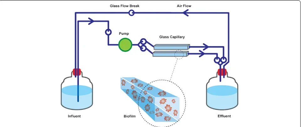
modified to be a fully closed system with no air intake, to prevent air contamination (Figure 1). Instead, two capillary flow cells (20 cm in length) were run in parallel, and a silicon tube was attached between the influent and effluent to ensure a smooth air flow. Hence, no bubbles were observed in the liquid flow. Five-litre glass bottles were used for both influent and effluent media. All experiments were performed at room temperature under sterile and dark environmental conditions.
P. aeruginosa inoculum with a turbidity equivalent to that of one McFardland standard was prepared from
Pseudomonasagar and 1 ml of inoculum was transfered to the flow cells as described by Werner et al. [25]. To allow bacterial attachment to the surface of the flow tube, no flow was initiated for the first 24 h. Decontaminated oocysts were injected into the influent medium and the flow initiated and continued (60 ml h-1) for one, three or six days. Biofilms exposed toCryptosporidiumoocysts are hereafter referred to asCryptosporidium–exposed biofilm samples.
[image:2.595.59.539.510.713.2]The volume of influent medium and the number of introduced oocysts were adjusted according to the duration of the experiment (number of days). The experiment was designed such that 5 l of 10% tryptic
soy broth was sufficient for a three-day experiment. The number of oocysts introduced in the influent medium was calculated so that the biofilms received 1 × 10⁶oocysts every 24 h. In order to avoid air contamin-ation, the oocysts were introduced at the beginning of the experiment (for all experiments), and during an influ-ent medium change after three days (for the six day experiment). Two controls were set up simultaneously:
1) No biofilm was developed. Decontaminated oocysts were added into the flow system at similar rates, but without any biofilm established within the flow system. This sample is presented as the biofilm-free control. 2) No oocysts were added. Biofilms were grown in the
flow system, without the introduction of
Cryptosporidiumoocysts. This sample is presented as the biofilm-only control.
All experiments utilised two flow tubes simultaneously for each treatment and were repeated three times.
Biofilm thickness
Film Tracer calcein red orange (Invitrogen, Australia) was used to stain the biofilm for confocal laser scanning microscopy imaging and analysis. The biofilms were stained according to the protocol provided with the stain. Briefly, calcein red orange stain was introduced into the living biofilm within the flow tube using a peristaltic pump at a rate of 60 ml h-1and incubated in the dark for 60 min at room temperature. Calcein red orange stain was similarly flushed from the system and the biofilms incu-bated with sterile water. Confocal images of biofilms were acquired from the flow cells directly using 576 nm and 590 nm laser excitation/emission wavelengths. Average biofilm thickness was determined from three regions - the left, right and middle area - of the flow cell. For both biofilm-only andCryptosporidium-exposed biofilms, serial
sections in the xy plane were obtained at 0.44 μm
(one day old biofilm) and 2–10μm intervals (three and six day old biofilms) along thez-axis, and the z-stack image was then analysed with COMSTAT II [26].
DNA extraction
Following thickness measurements, biofilms were dis-persed by incubation in 500 nmol l-1sodium nitriporusside (Sigma Aldrich, NSW, Australia) overnight, after which the flow cells were gently washed with 1 x PBS several times to completely detach the biofilms from the flow cell surface [27]. The cell suspensions were further washed several times with 1 x PBS to remove any residual nitric oxide. Biofilms from the flow cell were resuspended in 1 x PBS to a final volume of 400μl. To quantify the total number of parasites present at the end of each experimental period,
the dispersed biofilms from the effluent were also collected and resuspended to a final volume of 2 ml.
Aliquots of flow cell biofilms (100 μl) and effluent
biofilms (500 μl) were used for DNA extraction. The
freeze-thaw DNA extraction method was used for all sam-ples with the process being repeated 12 times to release
DNA fromCryptosporidium. The resulting DNA
suspen-sion was further purified as described by the Promega Genomic DNA extraction kit protocol, except that the cell lysis step was modified from 15 min to 30 min incubation to allow complete lysing of biofilm aggregates. The purified
DNA was eluted in a final volume of 50 μl in the DNA
suspension buffer provided with the kit.
Quantitative polymerase chain reaction (qPCR)
The DNA-based technique of quantitative polymerase chain reaction (qPCR) was utilised to quantify the numbers of Cryptosporidium within one, three and six day old biofilms. All qPCR reactions were performed on a Qiagen Rotor Gene 2 system. Standard curves were constructed using five genomic DNA triplicates extracted from a known number of oocysts and serially diluted at 1:10 dilution ratio,
calibrated to correspond from 100 to 105 oocysts. C.
parvumspecific primers used in this study were designed to target the glyceraldehydes-3-phosphate dehydrogenase
(GAPDH) gene. The qPCR reactions contained 12.5 μl
of Promega GoTaq SYBR Green master mix, 2.5 mmol l-1
of bovine albumin serum, 10μmol l-1primers specific forC. parvum (forward primer 5′ATCAAGCCGTTAAGGAA
GCA3′, reverse primer 5′AAATCGGTCGAGACGACA
TC3′) and 5μl of DNA to a final volume of 25μl. Thermal cycling was performed at 95°C for 2 min to activate Go-Taq polymerase then the amplification and annealing conditions involved 40 cycles of 15 s at 94°C and 1 minute at 60°C. Data was collected during the annealing step of each cycle. After amplification, a melting curve analysis was performed to determine the specificity of the PCR product. The PCR products were incubated for 15 sec at 55°C and the temperature was increased to 95°C with a ramp rate of 0.1°C sec-1.
As the concentration of DNA in the biofilm samples was too high, DNA dilution was performed to increase
the qPCR efficacy of detecting Cryptosporidium in the
biofilm system. Generally, the dilution factors for detecting
Cryptosporidium were 1:100 in one and three day old
Cryptosporidium-exposed biofilms and 1:1000 for six day oldCryptosporidium-exposed biofilms. Biofilm-free and six day old biofilm-only samples (negative controls) were di-luted according to the respective dilution factor of the
bio-film waste samples. The total number of Cryptosporidium
comparable baseline of the number of Cryptosporidium
parasites within the system, independent of the biofilm.
Statistical analyses
All experimental data are presented as mean ± the stand-ard error of the mean. Analyses were performed using GraphPad Prism, version 5 (GraphPad Software, San Diego, CA). One-way ANOVA analysis of variance was performed to determine whether there were significant differences in (i) thickness between one, three and six day old biofilm only controls (ii) thickness between one, three and six day old Cryptosporidium-exposed biofilms, (iii)
amount of Cryptosporidium parasites detected from one,
three and six day old Cryptosporidium-exposed biofilms
(flow cell). Two-way ANOVA analysis of variance was used to determine whether there were significant
differ-ences in (i) thickness between Cryptosporidium-exposed
biofilms and biofilm-only controls, (ii) the total
num-ber of Cryptosporidium detected at the completion of
experimental and biofilm-free experiments.
Immunolabelling
As a significant increase in Cryptosporidium DNA was
detected in six day oldCryptosporidium–exposed biofilms, immunolabelling withCryptosporidium-specificSporo-a-glo
antibody was performed on this sample to examine the
possible Cryptosporidium developmental stages present
within the biofilm. This antibody has been shown to label
various Cryptosporidium developmental stages such as
sporozoites, trophozoites, type I/II meronts and type I/II merozoites [7,28,29]. An aliquot of six day old dispersed biofilm suspension (20μl) was fixed with 2.5% paraformal-dehyde in PBS for 20 min at 4°C. The cells were
pelleted and incubated with 200 μl of blocking buffer
(6% BSA + 10% rat serum in 1 x PBS) for 1 h at room temperature, then washed with PBS (pH 7.4, 2 times, 5 min) and incubated with polyclonal primary antibody,Sporo-a-glo
(Waterborne Inc) (1:100 in blocking buffers) for 2 h at 37°C. The samples were then washed again with PBS
(twice for 6 min) and incubated with 40 nmol l-1
quantum dot 655 anti-rat secondary antibody (catalogue no Q-11621MP, Invitrogen) for 2 h at room temperature. The cells were then rinsed 3 times with 1 x PBS and resuspended to a final volume of 100μl. For comparison, unexcysted oocysts and six day old biofilm-only samples were also similarly stained.
Experimental controls included six day old Cryptospor-idium-exposed biofilms and excysted Cryptosporidium
oocysts labelled with (i) primary antibody only or (ii) secondary antibody only.
Confocal microscopy
For examination of Cryptosporidium biofilms samples by
confocal microscopy, an aliquot of the immunolabeled
suspension was immobilised and attached to coverslips using 0.01% poly-L-lysine (Sigma, USA). Prior to coverslip coating, coverslips were rinsed in 100% ethanol to remove surface dirt and any contamination and air dried in a laminar hood for 20 min. Coverslips were coated with poly-L-lysine for 20 min at room temperature and were then washed with sterile water to remove excess poly-L-lysine. After being air dried in the laminar flow, the experimental sample was then transferred onto the coverslip for attach-ment and incubated in a humidified box for 20 min to prevent dehydration. The sample was then analysed directly by confocal microscopy (Leica SP2 inverted) using the exci-tation and emission wavelength of FITC at 488/525 nm. Oil immersion was used for magnifications above 40x.
Ethical approval
The animal work in this study was reviewed and approved by the Animal Ethics Committee of Murdoch University, Australia (Permit number: R2310/10).
Results
Comparison of biofilm thickness: biofilm-only vs.
Cryptosporidium-exposed biofilm
Biofilm thickness in biofilm-only and Cryptosporidium
[image:4.595.306.538.438.654.2]-exposed biofilm experiments was determined using COMSTAT II [26]. For biofilm-only experiments, one day old biofilms were found to be 0.7 ± 0.4μm thick (Figure 2), indicating that the biofilm was still developing and could
therefore be described as an immature biofilm. After three days the thickness of the biofilm had increased
to 21 ± 3 μm, but this was not significantly different
(P > 0.05) from the one day old biofilms (Figure 2). However, a significant (P < 0.05) increase in biofilm thickness was observed in six day old biofilms (105 ± 12μm) (Figure 2). This biofilm thickness fulfills the definition
of a mature biofilm as defined by Davies et al. [30],
and hence was considered a mature biofilm.
When Cryptosporidium oocysts were introduced
into the biofilm system, one day old Cryptosporidium
-exposed biofilms formed immature biofilms that were
30 ± 3 μm thick (Figure 2). Unlike biofilm-only
experi-ments, a significant (P < 0.05) increase in biofilm thickness
was observed in Cryptosporidium-exposed biofilms after
only three days (121 ± 4 μm) (Figure 2). The thickness of
these three day old Cryptosporidium-exposed biofilms
(~100μm) could be classified as the penultimate stage of biofilm development [30,31] and was considered to be a mature biofilm. No significant (P > 0.05) increase in the thickness of this mature biofilm was observed from day
three to day six in Cryptosporidium-exposed biofilms,
with six day old biofilms found to be 136 ± 7 μm
thick (Figure 2).
All Cryptosporidium-exposed biofilms were thicker than biofilm-only controls, but the level of signifi-cance varied. Although both biofilm-only controls and
Cryptosporidium–exposed biofilms formed immature biofilms on day one and mature biofilms by day six,
Cryptosporidium-exposed biofilms were always signifi-cantly (P < 0.01) thicker than the biofilm-only controls. In addition, by day three,Cryptosporidium-exposed biofilms had formed mature biofilms and thus had matured signifi-cantly (P < 0.0001) faster than the comparable three day old immature biofilms in the biofilm-only control.
Association ofCryptosporidiumwith biofilms
Overall, qPCR revealed that the number ofCryptosporidium
parasites retained within the biofilm increased significantly (P < 0.05) (Figure 3) and continually over time, as the biofilm increased in thickness and matured. When
one and three day old Cryptosporidium-exposed biofilms
were compared, a significant (P < 0.05) increase in both parasite number (Figure 3) and biofilm thickness (Figure 2) was observed over time. However, even when the
Cryptosporidium-exposed biofilm had reached matur-ity and no longer underwent any increase in thickness (day three) (Figure 2), a significant (P < 0.05) increase in parasite numbers continued to be observed until day 6 (Figure 3).
Similarly, for the total number of parasites in the flow system (i.e. both those retained within the biofilm + those recovered from the effluent), an increase in the total
number of Cryptosporidium was observed in both one
and three day old Cryptosporidium-exposed biofilms, but these were not significantly (P > 0.05) different to biofilm-free controls (Figure 4). However, after six days, Crypto-sporidium-exposed biofilms contained 2–3 fold more
[image:5.595.306.539.88.277.2]Cryptosporidium than that of the biofilm-free controls,
Figure 3Quantitative analysis ofCryptosporidiumnumber within 1, 3 and 6 days oldCryptosporidium-exposed biofilms.The number (mean ± standard error) ofCryptosporidiumretained withinPseudomonas
biofilms after one, three, and six days of exposure (n= 6 for each time period), as determined using a DNA-based qPCR approach. Significance was determined by a one-way ANOVA analysis (* P < 0.05).
Figure 4Comparison of the total number ofCryptosporidium present inCryptosporidium-exposed biofilms and biofilm-free control systems.Graph showing the total number
(mean ± standard error) ofCryptosporidium(retained in biofilm + effluent) determined at the end of each experiment (one, three and six day) for
Cryptosporidium-exposed biofilms (black bar) (n= 3) andCryptosporidium
[image:5.595.306.539.442.634.2]which reflected a highly significant (P < 0.001) increase in parasite numbers when a biofilm was present (Figure 4).
The significant increase in Cryptosporidium numbers
within the biofilm flow system over the six day period
implies that Cryptosporidium was not simply captured
and accumulated but underwent multiplication within the biofilm system.
Confocal microscopy
The six day old Cryptosporidium-exposed biofilm
sample (flow cell) was further analysed using confocal microscopy to examine the possible developmental
stages of Cryptosporidium present within the biofilm.
Clusters of Cryptosporidium and bacteria cells were
frequently observed after biofilm dispersion, thus we
differentiated Cryptosporidium from the bacterial
bio-film using specific fluorescence immunolabelling of
[image:6.595.61.538.263.693.2]Cryptosporidium developmental stages. Oocyst excystation was observed to have occurred with the released of slender shaped sporozoites (Figure 5A). Comma-shaped sporozo-ites with a rounded posterior end and a pointed tapered anterior end were also visualised, indicating that trophozo-ite transformation had begun (Figure 5B). Circular shaped (2 × 2μm) trophozoites were identified indicating completion of the trophozoite transformation process (Figure 5C). Trophozoites represent a transitional stage
from sporozoites and merozoites to meronts [3]. In addition to trophozoites, two different types of meronts similar to those description by Hijjawi et al. [8] were also observed (Figure 5D-E). The grape-like aggregated merozoites (small with circular to oval shapes, approximately 1 × 1μm) conform to type I meront (Figure 5D) while the aggregation of many spindle-shaped merozoites with pointed ends con-form to type II meront (0.5 × 1μm) (Figure 5E). The pres-ence of type I/II meronts indicated that Cryptosporidium
gametogony process had been initiated.
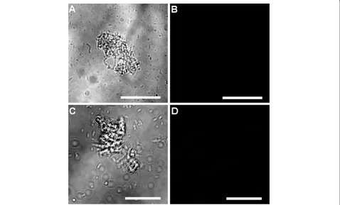
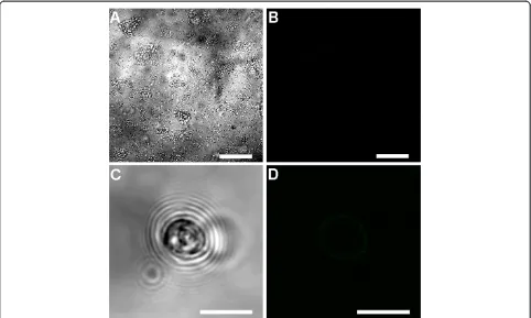
No fluorescent signal was observed in the control samples (six day oldCryptosporidium-exposed biofilms and excysted oocysts) where either the primary or the secondary antibody had been omitted (Figures 6 and 7). Fully labelled six day old biofilm-only control samples also displayed no
fluorescence (Figure 8A-B). In addition, Sporo-a-glo,
which is a Cryptosporidium-specific developmental stage antibody, binds only very weakly to unexcysted oocysts (Figure 8C-D). From this, we conclude that the abundant fluorescent expression observed within the biofilm
samples derives from actively growing Cryptosporidium,
rather than simply from accumulated unexcysted or degenerate oocysts. These observations are consistent with the qPCR data and show that oocysts were not
simply retained within the biofilms, and had under-gone the multiplication process extracellularly, as evidenced by the observation of several developmental and transitional stages.
Discussion
[image:7.595.60.538.396.685.2]This is the first study to demonstrate a significant increase in Cryptosporidium numbers over time within a biofilm system, highlighting that biofilms can readily provide a suitable environment for not only the retention, but also the multiplication ofCryptosporidiumparasites, in aquatic environments. Previous studies [32,33] have shown that the number of oocysts retained within biofilms remained constant while oocysts were continually supplied to the biofilm system. However, the apparent discrepancy between these results and those presented here can be explained by the difference in the type of biofilm and methods used to de-tect Cryptosporidium. In studies by Wolyniak et al.[32,33], natural biofilms were used with filter sterilised creek water used as the medium. Therefore, when compared to our artificial Pseudomonas biofilms, their biofilms would have a different community structure and nutrient levels. In addition, through the use of the qPCR technique, our analyses quantified not only the oocysts within the system
Figure 6Confocal observation of six day oldCryptosporidium-exposed biofilm control samples. A)Six day oldCryptosporidiumbiofilms labelled with only primary antibody seen under transmitted light;B)corresponding confocal image, showing no immunolabeling.C)Six day old
but also other Cryptosporidium life stages that were produced through multiplication.
The significant increase in Cryptosporidium DNA in
six day oldCryptosporidiumexposed biofilm was further
supported by confocal microscopy observation. Cell free culture studies by Hijjawi et al. [7,8], Karanis et al.
[10] and Zhang et al. [12] support our observation that
Cryptosporidium can multiply extracellularly, and that encapsulation within a host cell is not essential for multi-plication to occur. Due to the difficulty of identifying
Cryptosporidiumfrom a large background of bacteria from biofilms, not all Cryptosporidium stages were identified.
Nevertheless, several key developmental stages representing both asexual and sexual reproduction were observed, including sporozoites, trophozoites, and types I and II meronts. The observation of morphological changes of Cryptosporidium sporozoites agrees with previous
[image:8.595.57.539.88.522.2]in-vivo andin-vitroculture observations [34-36], including sporozoites becoming oval shaped during the trophozoite transformation process. Although previous studies by Petry et al. [37] and Matsubayashi et al. [38] suggested these changes were due to aged sporozoites that could not multiply in the nutrient-limited cell free culture environment, our observation of subsequent
Cryptosporidiumdevelopment stages of the life cycle, such as types I and II meronts (based upon the developmental stage descriptions by Hijawi et al. [8]) within six day old biofilm provides evidence that these sporozoites were not simply aged sporozoites. The misinterpretations by these authors [37,38] have also been clearly defended and clari-fied by Karanis and Aldeyarbi [39]. Furthermore, the envi-ronments that liberated sporozoites are exposed to in aquatic biofilms are nutrient rich micro-environments that
could allow Cryptosporidium to salvage their metabolite
needs to fuel their high rate of growth and multiplication. This ability to multiply either intracellularly or extracellu-larly [6,8-12] suggests that Cryptosporidium i) is capable of extracting the nutrients required for growth and multiplication from the surrounding environment, ii) is not an obligate intracellular parasite, and iii) may be physiologically as well as genetically similar to the closely-related gregarines [40,41]. Further high resolution imaging [6,29,42,43] and flow cytometry-based studies [44-46] are now needed to fully characterise any additional life stages that may be produced within biofilms.
Interestingly, the presence of Cryptosporidium was also shown to significantly affect biofilm development and maturation. Cryptosporidium-exposed biofilms were found
to form mature biofilms significantly faster than biofilms forming without exposure toCryptosporidium. Consistent with this, Singletonet al. [47] also showed that biofilms that contain both prokaryotic and eukaryotic cells often formed extensive dense and thick mature biofilms. As the biofilm sloughs off after growing to a certain density, we are unable to conclusively determine if Cryptosporidium detected in the effluent was a result of cells sloughing off with the biofilm, or free floating cells. It is likely to be both. Although it is not possible to determine what proportion of the increase in the mature biofilm thickness was due to increases in the number of bacteria or ofCryptosporidium, our qPCR analyses demonstrated that even very immature biofilms were capable of capturing and accumulating
Cryptosporidiumoocysts. These may be incidentally incor-porated into cell clusters during the biofilm aggregation process. Additionally, during transformation from an immature to a mature biofilm, matrix and water channels that form on the biofilm surface may have enhanced the adhesion of oocysts and also encased both parasites and bacteria, trapping those that were already retained within the biofilm [32,48]. However, while the thickness and maturation rate of the biofilm
[image:9.595.57.539.89.378.2]was affected by Cryptosporidium, the two factors were
not strongly correlated, thus biofilm thickness cannot be reliably used as an indicator of the number of
Cryptosporidium residing within the biofilm, a finding also concluded by other studies [32,33,49,50].
Conclusion
In conclusion, this study shows that biofilms not only serve as an environmental reservoir for oocysts, but are
also capable of supporting Cryptosporidium
multiplica-tion in an aquatic environment. The presence of biofilm on the pipes of water systems may pose a public health threat as Cryptosporidium residing within biofilms may increase in quantity over time, before being released into the water supply. With this, authorities should take into consideration the ability of Cryptosporidium to multiply within biofilms in aquatic environments when designing preventive measures to controlCryptosporidium contamin-ation in water distribution systems.
Competing interests
The authors declare that they have no competing interests.
Authors’contributions
WK, PLC, PM and RCAT designed the study; WK, PLC, PM and RCAT implemented the study; WK managed the data; WK, PLC and RCAT analysed and interpreted the data; WK wrote the paper. WK, PLC, and RCAT supervised the different phases of the study. All authors read, revised and approved the final manuscript.
Acknowledgements
The authors thank the Australian Research Council for financial support and acknowledge the facilities, and the scientific and technical assistance of the Australian Microscopy & Microanalysis Research Facility at the Centre for Microscopy, Characterisation & Analysis, The University of Western Australia, a facility funded by the University, State and Commonwealth Governments. We also thank Mr. John Murphy for the confocal microscopy technical assistance.
Author details
1
School of Veterinary and Life Sciences, Murdoch University, South Street, Murdoch, WA 6150, Australia.2Centre for Microscopy, Characterisation and Analysis, The University of Western Australia, 35 Stirling Hwy, Crawley, WA 6009, Australia.3South Australian Water Corporation, 250 Victoria Square, Adelaide, SA 5000, Australia.
Received: 6 August 2013 Accepted: 15 September 2013 Published: 19 September 2013
References
1. Putignani L, Menichella D:Global distribution, public health and clinical impact of the protozoan pathogenCryptosporidium.Interdiscip Perspect Infect Dis2010. doi: 10.1155/2010/753512.
2. Rose JB:Environmental ecology ofCryptosporidiumand public health implications.Annu Rev Public Health1997,18:135–161.
3. Thompson RCA, Olson ME, Zhu G, Enomoto S, Abrahamsen MS, Hijjawi NS:
Cryptosporidiumand cryptosporidiosis.Adv Parasitol2005,59:77–158. 4. Borowski H, Clode PL, Thompson RCA:Active invasion and/or
encapsulation? A reappraisal of host-cell parasitism byCryptosporidium. Trends Parasitol2008,24:509–516.
5. Tzipori S, Griffiths JK:Natural history and biology ofCryptosporidium parvum.Adv Parasitol1998,40:5–36.
6. Borowski H, Thompson RCA, Armstrong T, Clode PL:Morphological characterization ofCryptosporidium parvumlife-cycle stages in an in vitro model system.Parasitology2010,137:13–26.
7. Hijjawi N, Estcourt A, Yang R, Monis P, Ryan U:Complete development and multiplication ofCryptosporidium hominisin cell-free culture.Vet Parasitol
2010,169:29–36.
8. Hijjawi NS, Meloni BP, Ng’anzo M, Ryan UM, Olson ME, Cox PT, Monis PT, Thompson RCA:Complete development ofCryptosporidium parvumin host cell-free culture.Int J Parasitol2004,34:769–777.
9. Hijjawi NS, Meloni BP, Ryan UM, Olson ME, Thompson RCA:Successful in vitro cultivation ofCryptosporidium andersoni: evidence for the existence of novel extracellular stages in the life cycle and implications for the classification ofCryptosporidium.Int J Parasitol2002,32:1719–1726. 10. Karanis P, Kimura A, Nagasawa H, Igarashi I, Suzuki N:Observations on
Cryptosporidiumlife cycle stages during excystation.J Parasitol2008,
94:298–300.
11. Rosales MJ, Cordón GP, Moreno MS, Sánchez CM, Mascaró C:Extracellular like-gregarine stages ofCryptosporidium parvum.Acta Trop2005,
95:74–78.
12. Zhang L, Sheoran AS, Widmer G:Cryptosporidium parvumDNA replication in cell-free culture.J Parasitol2009,95:1239–1242.
13. Carreno RA, Martin DS, Barta JR:Cryptosporidiumis more closely related to the gregarines than to coccidia as shown by phylogenetic analysis of apicomplexan parasites inferred using small-subunit ribosomal RNA gene sequences.Parasitol Res1999,85:899–904.
14. Leander BS, Harper JT, Keeling PJ:Molecular phylogeny and surface morphology of marine aseptate gregarines (Apicomplexa):Selenidium spp. andLecudinaspp.J Parasitol2003,89:1191–1205.
15. Abrahamsen MS, Templeton TJ, Enomoto S, Abrahante JE, Zhu G, Lancto CA, Deng M, Liu C, Widmer G, Tzipori S,et al:Complete genome sequence of the apicomplexan,Cryptosporidium parvum.Science2004,304:441–445. 16. Declerck P, Behets J, Margineanu A, van Hoef V, De Keersmaecker B, Ollevier F:Replication ofLegionella pneumophilain biofilms of water distribution pipes.Microbiol Res2009,164:593–603.
17. Dunne WM:Bacterial adhesion: seen any good biofilms lately? Clin Microbiol Rev2002,15:155–166.
18. Fisher I, Angles M, Chandy J, Cox P, Warnecke M, Kastl G, Jegatheesan V:
Biofilms - a sticky situation for drinking water?Water2000,27:33–37. 19. Wingender J, Flemming HC:Biofilms in drinking water and their role as
reservoir for pathogens.Int J Hyg Environ Health2011,214:417–423. 20. Helmi K, Skraber S, Gantzer C, Willame R, Hoffmann L, Cauchie H-M:
Interactions ofCryptosporidium parvum,Giardia lamblia, vaccinal poliovirus type 1, and bacteriophages phiX174 and MS2 with a drinking water biofilm and a wastewater biofilm.Appl Environ Microbiol2008,
74:2079–2088.
21. Searcy KE, Packman AI, Atwill ER, Harter T:Capture and retention of Cryptosporidium parvumoocysts byPseudomonas aeruginosabiofilms. Appl Environ Microbiol2006,72:6242–6247.
22. Angles ML, Chandy JP, Cox PT, Fisher IH, Warnecke MR:Implications of biofilm-associated waterborneCryptosporidiumoocysts for the water industry.Trends Parasitol2007,23:352–356.
23. Howe AD, Forster S, Morton S, Marshall R, Osborn KS, Wright P, Hunter PR:
Cryptosporidiumoocysts in a water supply associated with a cryptosporidiosis outbreak.Emerg Infect Dis2002,8:619–624. 24. Meloni BP, Thompson RC:Simplified methods for obtaining purified
oocysts from mice and for growingCryptosporidium parvumin vitro. J Parasitol1996,82:757–762.
25. Werner E, Roe F, Bugnicourt A, Franklin MJ, Heydorn A, Molin S, Pitts B, Stewart PS:Stratified growth inPseudomonas aeruginosabiofilms. Appl Environ Microbiol2004,70:6188–6196.
26. Heydorn A, Nielsen AT, Hentzer M, Sternberg C, Givskov M, Ersboll BK, Molin S:Quantification of biofilm structures by the novel computer program COMSTAT.Microbiology2000,146(Pt 10):2395–2407.
27. Barraud N, Storey MV, Moore ZP, Webb JS, Rice SA, Kjelleberg S:Nitric oxide-mediated dispersal in single- and multi-species biofilms of clinically and industrially relevant microorganisms.Microb Biotechnol
2009,2:370–378.
28. Boxell A, Hijjawi N, Monis P, Ryan U:Comparison of various staining methods for the detection ofCryptosporidiumin cell-free culture. Exp Parasitol2008,120:67–72.
29. Edwards H, Andrew Thompson R, Koh WH, Clode PL:Labeling surface epitopes to identifyCryptosporidiumlife stages using a scanning electron microscopy-based immunogold approach.Mol Cell Probes2012,
30. Davies DG, Parsek MR, Pearson JP, Iglewski BH, Costerton JW, Greenberg EP:
The involvement of cell-to-cell signals in the development of a bacterial biofilm.Science1998,280:295–298.
31. Sauer K, Camper AK, Ehrlich GD, Costerton JW, Davies DG:Pseudomonas aeruginosadisplays multiple phenotypes during development as a biofilm.J Bacteriol2002,184:1140–1154.
32. Wolyniak EA, Hargreaves BR, Jellison KL:Retention and release of Cryptosporidium parvumoocysts by experimental biofilms composed of a natural stream microbial community.Appl Environ Microbiol2009,
75:4624–4626.
33. Wolyniak EA, Hargreaves BR, Jellison KL:Seasonal retention and release of Cryptosporidium parvumoocysts by environmental biofilms in the laboratory.Appl Environ Microbiol2010,76:1021–1027.
34. Umemiya R, Fukuda M, Fujisaki K, Matsui T:Electron microscopic observation of the invasion process ofCryptosporidium parvumin severe combined immunodeficiency mice.J Parasitol2005,91:1034–1039. 35. Lumb R, Smith K, O’Donoghue PJ, Lanser JA:Ultrastructure of the
attachment ofCryptosporidiumsporozoites to tissue culture cells. Parasitol Res1988,74:531–536.
36. Current WL, Reese NC:A comparison of endogenous development of three isolates ofCryptosporidiumin suckling mice.J Protozool1986,33:98–108. 37. Petry F:Structural analysis ofCryptosporidium parvum.Microsc Microanal
2004,10:586–601.
38. Matsubayashi M, Ando H, Kimata I, Nakagawa H, Furuya M, Tani H, Sasai K:
Morphological changes and viability ofCryptosporidium parvum sporozoites after excystation in cell-free culture media.Parasitology2010,
137:1861–1866.
39. Karanis P, Aldeyarbi HM:Evolution ofCryptosporidiumin vitro culture.Int J Parasitol2011,41:1231–1242.
40. Alarcón ME, Huang CG, Tsai YS, Chen WJ, Dubey AK, Wu WJ:Life cycle and morphology of Steinina ctenocephali (Ross 1909) comb. nov.
(Eugregarinorida: Actinocephalidae), a gregarine of Ctenocephalides felis (Siphonaptera: Pulicidae) in Taiwan.Zool Stud2011,50:763–772. 41. Leander BS:Marine gregarines: evolutionary prelude to the
apicomplexan radiation?Trends Parasitol2008,24:60–67.
42. Jirku M, Valigurova A, Koudela B, Krizek J, Modry D, Slapeta J:New species of Cryptosporidium tyzzer, 1907 (Apicomplexa) from amphibian host: morphology, biology and phylogeny.Folia Parasitol (Praha)2008,55:81–94. 43. Valigurová A, JirkůM, Koudela B, Gelnar M, Modrý D,Šlapeta J:
Cryptosporidia: Epicellular parasites embraced by the host cell membrane.Int J Parasitol2008,38:913–922.
44. King BJ, Hoefel D, Lim SP, Robinson BS, Monis PT:Flow cytometric assessment of distinct physiological stages withinCryptosporidium parvumsporozoites post-excystation.Parasitology2009,136:953–966. 45. Valdez LM, Dang H, Okhuysen PC, Chappell CL:Flow cytometric detection
ofCryptosporidiumoocysts in human stool samples.J Clin Microbiol1997,
35:2013–2017.
46. Vesey G, Griffiths KR, Gauci MR, Deere D, Williams KL, Veal DA:Simple and rapid measurement ofCryptosporidiumexcystation using flow cytometry.Int J Parasitol1997,27:1353–1359.
47. Singleton S, Treloar R, Warren P, Watson GK, Hodgson R, Allison C:Methods for microscopic characterization of oral biofilms: Analysis of colonization, microstructure, and molecular transport phenomena.Adv Dent Res1997,
11:133–149.
48. Rickard AH, Gilbert P, High NJ, Kolenbrander PE, Handley PS:Bacterial coaggregation: an integral process in the development of multi-species biofilms.Trends Microbiol2003,11:94–100.
49. Okabe S, Kuroda H, Watanabe Y:Significance of biofilm structure on transport of inert participates into biofilms.Water Sci Technol1998,
38:163–170.
50. Okabe S, Satoh H, Kindaichi T:A polyphasic approach to study ecophysiology of complex multispecies nitrifying biofilms. Methods Enzymol2011,496:163–184.
doi:10.1186/1756-3305-6-270
Cite this article as:Kohet al.:Multiplication of the waterborne pathogenCryptosporidium parvumin an aquatic biofilm system.Parasites & Vectors20136:270.
Submit your next manuscript to BioMed Central and take full advantage of:
• Convenient online submission
• Thorough peer review
• No space constraints or color figure charges
• Immediate publication on acceptance
• Inclusion in PubMed, CAS, Scopus and Google Scholar
• Research which is freely available for redistribution