Multiscale modeling methods
in biomechanics
Pinaki Bhattacharya
*
and Marco Viceconti
More and more frequently, computational biomechanics deals with problems where the portion of physical reality to be modeled spans over such a large range of spatial and temporal dimensions, that it is impossible to represent it as a single space–time continuum. We are forced to consider multiple space–time continua, each representing the phenomenon of interest at a characteristic space–time scale. Multiscale models describe a complex process across multiple scales, and account for how quantities transform as we move from one scale to another. This review offers a set of definitions for this emerging field, and pro-vides a brief summary of the most recent developments on multiscale modeling in biomechanics. Of all possible perspectives, we chose that of themodeling intent, which vastly affect the nature and the structure of each research activity. To the
purpose we organized all papers reviewed in three categories: ‘causal
confirmation,’where multiscale models are used as materializations of the causa-tion theories; ‘predictive accuracy,’ where multiscale modeling is aimed to improve the predictive accuracy; and ‘determination of effect,’ where multiscale modeling is used to model how a change at one scale manifests in an effect at another radically different space–time scale. Consistent with how the volume of computational biomechanics research is distributed across application targets, we extensively reviewed papers targeting the musculoskeletal and the cardiovas-cular systems, and covered only a few exemplary papers targeting other organ systems. The review shows a research subdomain still in its infancy, where causal confirmation papers remain the most common.© 2017 The Authors.WIREs Sys-tems Biology and Medicinepublished by Wiley Periodicals, Inc.
How to cite this article:
WIREs Syst Biol Med2017, e1375. doi: 10.1002/wsbm.1375
INTRODUCTION
A
s per March 2016, PubMed indexed 2180 papers including the word ‘multiscale’ in the title, and 5457 anywhere in the PubMed record. While thefirst of these papers was published in 1979, it is only in the last ten years that the biomedical research community has started to think across scales (Figure 1). Biome-chanics research follows similar trends.The aim of this study is to provide a systematic review of the multiscale modeling methods reported
so far in biomechanics research. It also aims to offer a set of candidate definitions for this emergingfield. As a lot of multiscale biomechanics involves either the musculoskeletal or the cardiovascular system, we will systematically review these two specific areas. However, we will also provide an overview of other interesting applications.
De
fi
nitions
The definition of scale varies widely depending on the context; in its simplest instance, it can be defined in term of grain and extent, both in space and time. The grain can be defined as largest value between the lower limit of spatial/temporal resolution allowed by the instrumentation, and the smallest/fastest feature of interest to be observed. Similarly, the extent can be defined as the smallest value between the upper
*Correspondence to: p.bhattacharya@sheffield.ac.uk
Department of Mechanical Engineering and INSIGNEO Institute forin silicoMedicine, University of Sheffield, Sheffield, UK
Conflict of interest: The authors have declared no conflicts of
limit of spatial/temporal resolution (i.e., the volume of interest in a four-dimensional space) and the size of the largest/slowest feature of interest to be observed. Resolution is defined as‘the smallest inter-val of a measured signal that will still cause a change in the measurement result.’1 In a perfect world, we would not need to worry about scales, because we would be free from the‘curse of resolution.’1Because our ability to resolve quantities in space and time is limited, ‘to explore from the infinitely small to the infinitely large with a finite resolution we need scales.’1
Most engineering theories avoid this complexity through one fundamental, and often implicit, assumption: scale separation. In a steel beam the microstructure grain size is 105 times smaller than the beam length. This means that every small portion of the macrostructure contains thousands of micro-structural elements, whose properties can be described statistically within that small volume. In other words, we do not need to have a detailed description of the microstructure to calculate the mechanical behavior of the macrostructure, but only its average properties as they manifest at the macro-scopic scale. In every biological material of interest in biomechanics research, the degree of scale separation is much smaller, typically around 102, so making this assumption is much less safe.
From a computational point of view, it is fre-quently more convenient to build a multiscale model not as a monolithic object, but rather as the orches-tration of multiple models, each typically capturing the causal relation at a given space–time scale. In this case we refer to each single-scale model as a hypomo-del, and to their orchestration as ahypermodel.2
Also in some implementations it is more con-venient to separate the models that capture the causal
relation at each space–time scale, from those that capture the transformation on the quantities involved from one scale to another (sometimes referred to as
homogenization, when they transform from small to large, and particularization, when they transform from large to small). In this case, we callcomponent
models those capturing single-scale causation, and relation models those capturing the scale transformation.
For the purpose of this study, we define a model as any causal quantitative relation M between an input set I and an output set O, so that:
O0 = M Ið Þ
In the physical and natural sciences M captures some knowledge about nature; such knowledge can be phenomenological (purely based on induction, i.e., exclusively on experimental observations), or mechanistic (based on deduction, i.e., on theoretical reasoning), although in practice ‘both phenomeno-logical and mechanistic approaches are inherently present in any model.’3 The variable I represents a set of necessarily measurable quantities, whereas O0 is the prediction of a set of desirably measurable quantities O.
As most of biomechanical models tend to be complex, most often M(I) is not computable in closed form, and we need to resort to some numerics N:
O00= N M Ið ð ÞÞ
O00 differs from the true value O for three reasons: (1) the approximation due to N; (2) the aleatory uncertainty associated with the measurement of I (and if possible O); and (3) theepistemic uncertainty
associated with the model M. We use verification,
uncertainty quantification (also called sensitivity analysis), and validation (when a measure of O is available) methods respectively to estimate these three sources of predictive inaccuracy.4
Structure
[image:2.594.56.272.70.207.2]The biomechanical multiscale modeling literature is highly heterogeneous, and presents regularities only if you compare papers dealing with the same prob-lem. This makes any attempt to review the overall field quite challenging. In the following we will review the relevant literature along two dimensions. First, we divide papers by organ system; we review in detail papers targeting the musculoskeletal and the cardiovascular systems, by far the most popular in the literature. However, we offer only an overview, FIGURE 1| Incidence of multiscale papers indexed in PubMed
without any pretence of being exhaustive, for the other organ systems.
The second and more important dimension we used in this review is that of the modeling intent, expressed in terms of operational motivations. Reviewing the literature, we found that of all differ-ences this is the most profound, as it severely infl u-ences the difficulty of the challenge involved. But what are the operational motivations that require the building of models where I and O are defined at radi-cally different space–time scales? We have identified three common motivations:
1. Causal confirmation. When we need a causal explanation of why O is observed given I, and O and I are defined on different space–time scales, the experimental testing of causal hypotheses can be quite challenging. In these cases, multiscale models are used as materiali-zations of the causation theories, whose falsifi -cation is attempted by measuring a large set of I and O values (possibly independently), and then by comparing the range of observed O values with the range of O00values predicted by the model for the range of I values. If the range of O and O00 differ considerably, this means that the causal theory the model materializes is not compatible with the observations, which ‘falsifies’such a theory.
2. Predictive accuracy. In all problems where we need to know the quantity O with a given accuracy, O is difficult to measure directly, but other measurable quantities are known to be causally related to O, we can use that causal knowledge to build a model that predicts O given some other measurable quantities I. In many cases, we can obtain accurate predictions using a causal knowledge entirely defined with a single space–time scale. However, in some cases the only way to obtain the necessary accuracy is to stop assuming scale separation, and extend the predictive model to account for causation across multiple scales.
3. Determination of effect. In other problems, the need for a multiscale model does not come from the desire of improving the predictive accuracy of O, but rather from the need to develop a predictor of the effect of quantity I defined at the scale S1 on the quantity O defined at the scale S2.
As these motivations are fairly different, and this reflects deeply on the approach used to develop and
evaluate multiscale models in the following, we will review the literature by highlighting which of these three aims is pursued in each paper.
The vast majority of the body of multiscale modeling research has been motivated bycausal
con-firmation. In the following we briefly review these studies, and emphasize the relatively fewer studies which went beyond causal confirmation and were motivated bypredictive accuracyordetermination of effectin their modeling efforts.
MULTISCALE MODELS OF THE
MUSCULOSKELETAL SYSTEM
The musculoskeletal system is considered herein to comprise of the following tissue types: bone, carti-lage, skeletal muscle (i.e., excluding smooth muscle and cardiac muscles), tendon, and ligament. Without any pretence to being exhaustive, we highlight below recent advances relating to bone mechanics, bone adaptation and remodeling, fracture healing, skeletal muscle remodeling, electromechanical behavior of skeletal muscle, tendon mechanics, tendon remodel-ing, and the mechanics of tendon under conditions of homeostasis and pathological mineralization.
Causal Con
fi
rmation
Bone Mechanics
Bone Adaptation and Remodeling
The process of natural bone adaptation (bone remod-eling) is driven by underlying cellular processes which are in turn influenced by biochemical and mechano-sensitive activation. In modeling bone adaptation, one approach is to explicitly account for bone remodeling although mathematical models of bone remodeling are themselves relatively untested.14 Various authors adopted a multiscale modeling approach,15–17implementing analytical scale-bridging relationships from the mineral constituent scale through the bone tissue scale along with a cell-scale bone remodeling algorithm. Due to challenges in mea-suring bone remodeling activity parameters in a speci-men specific manner, the above model predictions relating to the evolution of bone mineral content with time could only be compared against a micro finite element simulation with identical initial bone mineral content and cellular activity parameters. Hambli18 introduced a multiscale model that coupled a finite element model at the whole bone scale to a neural network surrogate model at the tissue scale which was trained using micro FE simulations on high-resolution image-based models of cancellous bone. Thus, the mechanobiology approach to bone adapta-tion has yielded only causal confirmation to date.
A different approach to bone adaptation is based on the hypothesis that local microarchitecture of bone is governed by a material redistribution problem that seeks to simultaneously minimize material used, maxi-mize resistance to applied loading, and control bone surface area and permeability. This approach—which is blind to the details of the cell-scale bone remodeling process—was first proposed in Fyhrie et al.19 Coelho
et al.20recently advanced this approach by implement-ing a multiscale model where at selected locations of the bone, microscale material redistribution problems were solved given the state of stress transferred from the bone scale due to physiological activity (normal walking and stair climbing). The resulting steady state periodic microstructures were used to compute the apparent scale density and orthotropic elastic stiffness tensor components. The predicted bone density distri-butions, and the power law relationship between pre-dicted local bone density and prepre-dicted local elastic tensor components agreed in general with those reported in literature, suggesting that the causal theory the model represented was at least compatible with the range of available observations. Thus, this line of research has also yielded only causal confirmation.
Fracture Healing
Multiscale models of osteogenesis in order to predict fracture healing are another line of research where
predictive accuracy is yet to be demonstrated. The multiscale model of Carlier et al.21 combined earlier
models of DII4/Notch signaling at the intracellular scale22and a bio-regulatory framework of angiogen-esis.23 At the intracellular process time-scale, the
model predicts tip cell movement and sprout forma-tion. At the tissue-level time scale, the integrated effect of angiogenesis and associated transport of molecules regulates differentiation and proliferation of various cell types (e.g., mesenchymal stem cells, chondrocytes, osteoblasts, and fibroblasts) toward bone formation. The multiscale model is exercised with input obtained from literature sources and its predictions qualitatively match experimental observa-tions of temporal evolution of bone, cartilage and fibrous tissue types in a rodent model. This model was enhanced with a more detailed oxygen budget model,24 in order to improve the qualitative agree-ment of the model predictions of cartilage tissue tem-poral evolution for a large defect in a rodent model with the available experimental observations. In Car-lier et al.,25 the model predictions showed qualitative agreement with regard to spatial distribution of tissue types and steady-state union/nonunion outcomes.
Skeletal Muscle Remodeling
Similar to the mechanobiology approach to bone remodeling, the remodeling and adaptation of skele-tal muscle has received considerable attention (see the recent review by Wisdom et al.26). Although the microscale architecture of the muscle and the process of force generation in the muscle (active/passive) are reasonably well understood, the mechanosensitivity of the muscular remodeling process is largely phe-nomenologically defined.27,28 Zöllner et al.29
obtained causal confirmation for a multiscale model that predicted the shortening of the gastrocnemius muscle as a result of remodeling induced by high-heeled footwear use. In their model, the apparent scale muscle length was a function of cellular scale sarcomere number. The evolution of sarcomere num-ber with time was dependent on physical activity level represented by a strain-threshold (cf. physical activity level is represented by a strain energy density threshold in bone remodeling).
Tendon Homeostasis
Biochemical and biomechanical factors that affect Achilles tendon homeostasis were reviewed by Smith
and various regimes of tendon repair that included or excluded inflammatory response. Maceri et al.31
implemented a multiscale model for the tendon to predict: (1) its mechanical response; (2) its remodel-ing in response to physical activity; and (3) strains within a coupled muscle model in response to coupled neuromuscular excitation. At the tendon-scale, lumped-parameter models were used to describe viscous response and strain-dependent elas-tic response in the tendon. The parameters at the ten-don scale were determined from homogenization of properties at the tissue-scale (e.g., fiber aspect ratio, fiber curvature, and fiber tangent modulus). Tissue-scale properties were derived from persistence length, contour length, kink dimension, and end-to-end ref-erence length at the molecular scale. The study explores causal confirmation for the proposed multi-scale model by comparing predictions with range of values reported in literature.
Mineralized Tendon Mechanics
Avian tendon tissue is known to mineralize under physiological conditions. Yet, as a partially minera-lized soft tissue mineraminera-lized turkey leg tendon (MTLT) serves as a model to understand pathologi-cal mineralization of human tendon tissue. Spiesz
et al.32 modeled the indentation modulus of a micro-porous collagen fibril array at the tissue-scale by employing a Mori–Tanaka homogenization of the nanoscale variables fibril volume fraction, the min-eral distribution between fibrils and extra-fibrillar matrix and the degree of mineralization. Fibril vol-ume fraction and mineral volvol-ume fraction were meas-ured in the same study from two distinct tissue zones
(circumferential and interstitial) each from a tendon sample. The parameter controlling mineral distribu-tion between fibrils was varied within the range of values in literature. Other model parameter values were taken from literature. Distinct mineral distribu-tion parameter values for circumferential and intersti-tial tissue regions were found tofit satisfactorily the measured indentation moduli. In a later study,33 the
same group found that using a single average value of the mineral distribution parameter resulted in the variation of microindentation moduli explained by the tissue-scale model to be higher in the circumfer-ential zone of the tendon (R2= 0.231) than in the interstitial zone (R2 = 0.003). An independent
meas-urement of the mineral distribution parameter is needed to better validate the model.
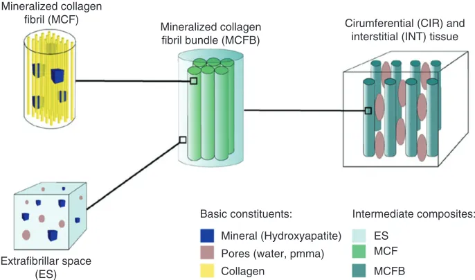
Tiburtius et al.34 identified separate
circumfer-ential and interstitial tissue zones (Figure 2) and employed homogenization methods (to bridge across length-scales) and hierarchical organization along the lines proposed by Spiesz and coworkers.32,33 Tibur-tiuset al.34experimentally determined microporosity, degree of mineralization, and acoustic impedance for a number of tendon samples. Model parameter values not directly measured were varied in the range reported in literature to determine their influence on the sensitivity of the tissue-scale stiffness tensor for each sample. The stiffness tensor was used to derive an effective acoustic impedance. The study found that the multiscale model captured the separation of acoustic impedances between circumferential and interstitial tissue zones over the range of measured mineral volume fraction in fibril bundles. Further-more, the variation of computed acoustic impedance
Mineralized collagen fibril (MCF)
Mineralized collagen fibril bundle (MCFB)
Extrafibrillar space (ES)
Basic constituents: Intermediate composites:
ES MCF
MCFB Mineral (Hydroxyapatite)
Pores (water, pmma) Collagen
[image:5.594.135.478.496.698.2]Cirumferential (CIR) and interstitial (INT) tissue
matched the variation of measured acoustic impedance—in both order of magnitude and trend— over the range of measured mineral volume fraction in fibril bundles, thus confirming the causal basis in their model.
Open Problems Motivated by Predictive
Accuracy
For each multiscale model in musculoskeletal (MSK) biomechanics that is motivated by causal confi rma-tion there exists an open problem that is motivated by predictive accuracy (PA), which has a stricter demand in terms of model validation. We offer one open problem each for the six applications discussed in this section:
MSK_PA1. Bone mechanics: A multiscale model to predict the elastic anisotropy as measured in a given bone tissue specimen, using structural and composition information taken at lower scales from the same specimen
MSK_PA2. Bone remodeling: A multiscale model to predict the evolution of bone mineral content as measured in a given bone volume, using bone remodeling activity parameters measured on the same specimen
MSK_PA3. Fracture healing: A multiscale model to predict tip cell movement and sprout formation as measured within a bone fracture site volume, using intracellular and tissue scale parameters measured on the same specimen
MSK_PA4. Skeletal muscle remodeling: A multiscale model to predict the shortening of the gastrocne-mius muscle as measured on a subject, using sar-comere scale parameters measured on the same subject
MSK_PA5. Tendon homeostasis: A multiscale model to predict whole tendon remodeling as measured on a subject, using tissue and molecular scale measured on the same subject
MSK_PA6. Mineralized tendon mechanics:
A multiscale model to predict the tissue stiffness tensor measured on a tendon specimen, using structural and composition information obtained on the same specimen
Predictive Accuracy
Bone Mechanics
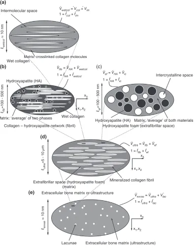
Predictive accuracy of multiscale modeling of bone mechanics was demonstrated in the work of
Hellmich and coworkers35, where the bone material is considered a hierarchically organized composite of hydroxyapatite (HA) crystals, collagen and water (Figure 3). The elastic properties of the basic consti-tuents were shown to be ‘universal’ and were deter-mined from separate experiments. Hellmich et al.35 showed that this multiscale model can predict bone stiffness given only the information on volume frac-tion of each constituent. The study assessed the pre-dictive accuracy of the multiscale model by comparing individually the experimentally measured stiffness for multiple cortical and trabecular bone specimens with the stiffness predicted using their multiscale model.
Fritsch et al.36 showed that the multiscale model could use the identical specimen-specific infor-mation of volume fraction of the constituents and accurately predict mass density of bone regions smal-ler than the tissue scale, for example, extracellular and extravascular bone regions. At the same time, considering the inclusions in the bone composite to possess a given distribution of orientations, Fritsch
et al. successfully assessed the predictive accuracy of the model with respect to tissue scale elastic anisot-ropy, thus directly answering the problem MSK_PA1 posed earlier. The same group, in a follow-up paper37 added to the above multiscale model a description of postelastic response at the micrometer scale: brittle rupture of collagen cross-links and an ideal plastic yielding of the mineral crystals. Satisfac-tory predictive accuracy of tissue-scale strength was reported considering cortical bone regions from human and bovine long bones. Eberhardsteiner
et al.38included in the above model nanoscale sliding of mineral crystals over water layers in order to explain observed viscoelasticity of wet and dry bone tissue specimens and assessed the model accuracy against specimen-specific experimental observations.
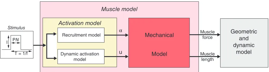
Skeletal Muscle Electromechanics
electrical stimulation. The relative contraction of given musclefiber is assumed to result in an identical relative contraction in each component sarcomere.
The sarcomere model42determines the stiffness and force generated by a sarcomere as a consequence of the applied electrical stimulation and relative
contraction. The model homogenizes across the whole-muscle/muscle-fiber length scales by assuming identical mechanics for all muscle fibers in a given state (contracting, relaxing, or completely relaxed). The predictive accuracy of the recruitment model was tested against published experimental
Intermolecular space
(a)
(b)
(d)
(e)
(c)
Intercrystalline space Wet collagen
Hydroxyapatite (HA)
Hydroxyapatite (HA)
Hydroxyapatite foam (extrafibrillar space)
Extrafibrillar space (hydroxyapatite foam) (matrix)
Extracellular bone matrix or ultrastructure
Extracellular bone matrix (ultrastructure)
Extravascular bone material Lacunae Collagen – hydroxyapatite network (fibril) Matrix: ‘average’ of two phases
Matrix: ‘average’ of both materials Wet collagen
Mineralized collagen fibril
Matrix. crosslinked collagen molecules
x3
x1,x2
x3
x1,x2
x3
x1,x2
ℓwetcol
ù
10 nm
ℓexvas
ù
10 nm
ℓfib
=100 - 500 nm
ℓultra
=5 - 10
µ
m
ℓef
=100 - 500 nm
Vwetcol = Vcol + Vim
Vfib = VHA + Vwetcol
1 = fcol + fim
1 = fHA + fwetcol Vef = VHA + Vic
1 = fHA + fic
Vultra = Vfib + Vef
1 = ffib + fef
Vexvas = Vultra + Vlac
[image:7.594.113.509.72.577.2]1 = fultra + flac
FIGURE 3 | Micromechanical representation of bone material by means of afive-step homogenization procedure. (Reprinted with permission
measurements.43It was also possible to find a single set of parameters for the multiscale model that could predict the whole muscle scale force in a rabbit knee experiment under two different electrical stimulation conditions. Finally, a different set of model para-meters could also be found corresponding to the pre-diction of whole muscle scale forces in patients with spinal-cord injury.44
Tendon Tissue Mechanics
As a resolution to the problem MSK_PA6 posed ear-lier, Szczesny and Elliot, in two consecutive papers,45,46 reported predictive accuracy for their multiscale model of a tendon fascicle comprising dis-continuous, staggered, crimped fibrils. A probability distribution informed the number offibrils that were fully un-crimped at a given level of stretch in the ten-don; the load supported by each fibril was taken to be nonzero only in the un-crimped state; the fibril load was transferred to an interfibrillar interface pos-sessing a perfectly plastic response initiated at incipi-ent sliding. The model was analyzed using microstructural parameter values (radius, length and volume fraction of fibrils) from the literature and micromaterial parameters (interfibrillar interface plastic stress, fibril modulus and fibril stretch distri-bution) from experiments conducted by Szczesny
et al.46 The model predicted accurately the ratio of strains in the fibril and the tendon over a range of tendon-strains in rat-tail tendon fascicles.46Over this range of tendon strain, the tendon undergoes plastic unloading and the plastic response was satisfactorily captured due to thefibril-scale plasticity included the multiscale model. The results suggested that it was the plasticity of the interfibrillar interface rather than that of thefibril itself, which resulted in the accurate prediction.45 The study considered an elastic regime of the interfibrillar interface up to afinite sliding dis-tance, but showed that the resulting elastoplastic
micromechanics improved the prediction only up to relatively small tendon strain values.45
Open Problems Motivated by
Determination of Effect
As before, we pose open problems in multiscale mod-eling in musculoskeletal biomechanics motivated by
determination of effect(DE).
MSK_DE1. Bone mechanics: A multiscale model that predicts better than a single-scale model the bone tissue elastic anisotropy
MSK_DE2. Skeletal muscle electromechanics: A mul-tiscale model that predicts better than a single-scale model the force–length relationship in a muscle
MSK_DE3. Tendon tissue mechanics: A multiscale model that predicts better than a single-scale model the stiffness tensor of a tendon tissue
Summary
The discussion in this section showed that most of the work in multiscale modeling of musculoskeletal biomechanics has been motivated by causal confi r-mation. Of the three categories defined in this article,
causal confirmation imposes the least demand on model validation. Thus, by simply raising the bar on validation to the next level, we posed six open pro-blems motivated by predictive accuracy. Two of the six problems were found to have been answered. Table 1 summarizes the multiscale musculoskeletal biomechanics models, that were motivated by predic-tive accuracy. Reflecting on the four problems that remain open (MSK_PA 2–5), it is evident that multi-scale modeling of musculoskeletal biomechanics pro-blems involving cellular remodeling/adaptation processes remains a challenge. Finally, we offered
Geometric and dynamic
model
Muscle model
Activation model Stimulus
Recruitment model
Dynamic activation model I PN
T = 1/f
Muscle force
Muscle length
Mechanical
Model u
[image:8.594.54.523.69.196.2]α
three additional open problems motivated by deter-mination of effect.
MULTISCALE MODELS OF THE
CARDIOVASCULAR SYSTEM
In this section, we consider research on the cardio-vascular system, for example, studies on heart rate and blood flow. Yet, as the vascular system pene-trates other organs and organ systems (e.g., brain and lungs), we also include in the discussion studies that seek to model the interaction between vascula-ture and other organs or organ systems. An excep-tion to this rule is the interaction between vasculature and the musculoskeletal system, which has already been discussed in the foregoing section.
Causal Con
fi
rmation
Blood Flow Interaction with Blood Vessel Walls
The understanding of the interaction between blood flow and the blood vessel walls is essential for several medical problems such as aneurysms and atheroscle-rosis.47Although the dysregulation of blood flow or arterial endothelial function is typically restricted to a local region, it influences and is influenced by the flow in regions far away from the site of the pathol-ogy. Twenty years ago, Dubini et al.48 proposed a
geometrically multiscale approach to model the mul-tiphysics problem of fluid structure interaction, to which others have added to subsequently.49–51In this
approach, the local site of interest is modeled in three dimensions using patient-specific geometry—an area where much progress has been made in the past dec-ade.52For theflow–structure interaction in the three-dimensional (3D) model both monolithic50and segre-gated53 coupling algorithms have been developed.
The 3D model is coupled to a 0D (electrical circuit analogy) or a 1D (network of segments) model of the circulation system, supplemented by proper condi-tions specified at the interface of the different models.54–56 The ongoing research focus in this area is on method development motivated by achieving causal confirmation, and much work remains with regard to model validation.47,52
Blood Flow Interaction with Blood Coagulation
Blood coagulates in response to a rupture of a blood vessel that can potentially lead to loss of blood. Coagulation is effected by platelets, which adhere to the site of breakage, by sensing biochemical signals released by the endothelial cells lining the blood
vessel. As such, coagulation is a classic bio-chemo-mechanical interaction process. It is also a multiscale problem, with individual platelets on the order of microns, while a wound region is upward of the order of millimeters. Recent reviews57,58on the
state-of-the-art of multiscale modeling highlight the chal-lenges in the different approaches taken until now. Specifically, Diamond et al.57 note that top–down
approaches such as neural network models miss patient-specific features while bottom–up approaches such as systems of ordinary differential equations suf-fer from incomplete knowledge. Sophisticated multi-scale models have been developed that integrate submodels for blood flow through the blood vessel and the growing clot, platelet interactions with blood flow and the vessel wall, and for the coagulation pathway.59–64 Until now, only causal confirmation
has been achieved by these models.
Cellular Mechanics in Blood Flow
Erythrocytes, or red blood cells (RBCs), transport oxygen and other essential nutrients to the various tissues that the vasculature penetrates through in the human body.65 This transport functionality depends on the ability of an RBC to undergo large deforma-tions as it passes through the human circulatory sys-tem, an ability that is compromised in diseases such as sickle-cell anemia or malaria.65 Hence there has been a steadily growing interest in the modeling of RBC mechanics in response to surface tractions applied on their boundary. The state-of-the-art of computational approaches on this topic was recently reviewed by Liet al.66
An extreme case of blood flow through a very small opening occurs in the venous sinuses of the spleen. Salehyar et al.67 employed the above multi-scale model to investigate RBC dynamics and inter-nal stresses in the cell during this passage. They postulated that the high deformation mechanics required to pass through the slit-like sinus can be accurately captured by including the molecule-scale unfolding dynamics of the spectrin network which were therein modeled as worm-like chains and ensur-ing the intactness of intraprotein, interprotein, and protein-to-lipid linkages within the RBC. The model captured ‘infolding’ of the RBC membrane in dependence of the initial relative orientation of the RBC and the slit. Comparison with experimental evi-dence of RBC shape dynamics was not performed in the study.
Vasculogenesis and Angiogenesis
within which the vasculature penetrates. Examples of multiscale studies modeling such control within the musculoskeletal system, for example, osteogenesis and fracture healing and tendon tissue homeostasis and repair, were visited in the previous section. Along similar lines, multiscale models for vasculogen-esis and angiogenvasculogen-esis have also been developed. Using a cellular Potts modeling framework, Scianna
et al. investigated angiogenesis68 by detailing the interactions between cellular and molecular scale models, and investigated vasculogenesis69 by
detail-ing the interactions between subcellular, cellular and extracellular scale models. The multiscale models parameters in each study were determined from experimental evidence in literature, and model pre-diction qualitatively agreed with observed characteristics.
Scianna et al.68 incorporated the influence of vascular endothelial growth factor (VEGF) on cellu-lar signaling by defining a model of VEGF diffusion at the extracellular scale and models simulating subcellular processes in dependence of VEGF con-centration. Stefanini et al.70,71 focussed on the
dis-tribution of VEGF receptors, and distinguished specifically the distribution of two VEGF isoforms and their receptors in their multiscale model. Their multiscale models included compartment models for blood and tissue (normal, tumor) and intercompart-mental interactions. Stefanini et al.70 showed that
under pathological conditions the distinction between VEGF isoforms and between the local dis-tributions of their receptors influences the eventual signaling cascade. Stefanini et al.71 obtained qualita-tive causal confirmation for their model by predict-ing the clinically observed increase in plasma VEGF following the administration of a VEGF antibody. Bonilla et al.72 and Terragni et al.73 implemented deterministic and stochastic models of tumor-induced angiogenesis.
Cerebral Autoregulation in Cardiopulmonary Bypass
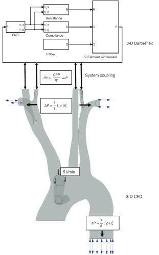
The cardiopulmonary bypass (CPB) pump or the ‘heart–lung machine’ is a device routinely used in surgery to take over the function of the heart and the lungs. Neurological malfunction leading to stroke is a common complication of the CPB technique and its prediction has long attracted research interest.74 Kaufmannet al.75 highlighted that the brain’s ability to adapt to changingflow conditions during CPB are influenced by the state of its autoregulation mechan-ism. They also incorporated a phenomenological model for autoregulation within a 3D computational fluid dynamics model for CPB and validated
experimentally the cerebral blood flow predictions. Neidlin et al.76 improved this framework by
repla-cing the phenomenological model with a 0D model of the baroreflex mechanism (Figure 5). The barore-flex model predicted subject-specific static and dynamic cerebral autoregulation behavior.76 This 0D–3D coupling model76 was further enriched by Neidlin et al.77 who included the elasticity of vessel
walls in the 3D CPB model this allowing flow– structure interaction. Model predictions of central aortic pressure and blood flow velocity through the descending aorta were compared with experimental observations reported in literature. Causal confi rma-tion for the model was established by the good corre-spondence of time-dependent haemodynamic features over one cardiac cycle.
Cardiomyocyte Mechanics
Weinberget al.78 obtained causal confirmation for a model that predicted the influence of high-frequency stimulation in cardiac myocytes on ionic current amplitude and gating dynamics. Adeniran et al.79 predicted the difference in the influence of pCa (−log10 of the calcium concentration) on force
between normal and heart failure with preserved ejection fraction cases as observed by Borbélyet al.80 Gaur et al.81 provided causal confirmation for a model that predicted stochastic Ca release processes locally in cardiac ventricular myocytes. Hand and coworkers implemented a multiscale model to study the effects of gap-junctional and ephaptic coupling on conduction,82,83 with a microscale description
near action potential wave fronts, and a macroscale description in regions far away from them.
Heart Valve Mechanics
heart valve. These macroscale strains were applied to a tissue model that was used to determine local tissue-scale strains in the different layers, and which were used to stimulate a cell-scale model. Compar-ing against experimental results reported in litera-ture, causal confirmation was achieved for multiscale predictions of cellular aspect ratio in nor-mal86 as well as bicuspid heart valves87 and of cal-cification of the aortic valve during aging.88
Open Problems Motivated by Predictive
Accuracy
In a similar manner as before, we pose open pro-blems in multiscale modeling in cardiovascular (CV) biomechanics motivated bypredictive accuracy.
CV_PA1. Blood flow interaction with blood vessel walls: A multiscale model to predict the measured bloodflow and blood vessel wall dynamics within
n_s
R R
C
C P
P
Q Q
n_p
n_s n_p n_s
n_p CNS
Resistance
Compliance
Inflow
CPP
n wіP mі=
Rn
2-Element windkessel
0-D Baroreflex
System coupling
3-D CFD 5 l/min
1
L p U2
ΔP =
2
n
1
L p U2
ΔP =
[image:12.594.137.448.72.572.2]2
a local region (e.g., surrounding an aneurysm) of the systemic circulation, using parameters meas-ured on the same circulation system, vascular tis-sue and geometry, and bloodfluid
CV_PA2. Blood flow interaction with blood coagu-lation: A multiscale model to predict the measured development of a blood clot, using the bio-chemo-mechanical parameters measured on the same platelet–blood–vessel wall system
CV_PA3. Cellular mechanics in blood flow: A multiscale model to predict the dynamics of an RBC as measured in blood flow, using the micro-mechanical parameters measured on the same RBC
CV_PA4. Vasculogenesis and angiogenesis: A multiscale model to predict measured angiogen-esis and vasculogenangiogen-esis based on measured subcel-lular, cellular and molecular scale parameters CV_PA5. Cerebral autoregulation in CPB: A
multi-scale model to predict the measured cerebral auto-regulation, using the measured elasticity of vessel walls on the same cardiac system
CV_PA6. Cardiomyocyte mechanics: A multiscale model to predict the measured electrical activity in a cardiac myocyte under high-frequency stimula-tion, using cell membrane electro-mechanical property parameters measured in the same myocyte
CV_PA7. Heart valve mechanics: A multiscale model to predict the change in cell aspect ratio during a
cardiac cycle and increase in valvular tissue calcifi -cation due to aging, using cellular and tissue-scale parameters measured in the same human heart valve
Predictive Accuracy
Cellular Mechanics in Blood Flow
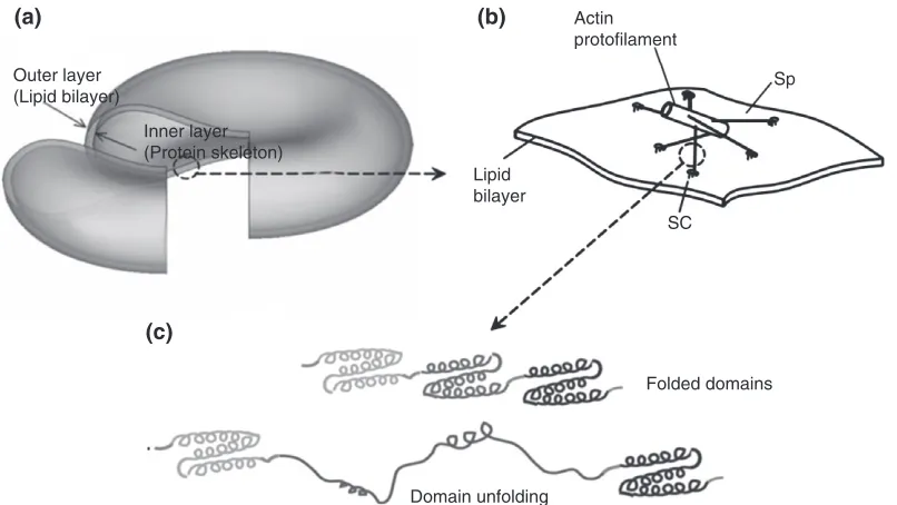
One approach to the multiscale modeling of RBC mechanics in blood flow, due to Peng et al.,89 defines three length scales (Figure 6): (1) whole cell scale; (2) junctional-complex (JC) scale; and (3) spec-trin (Sp) protein scale. The protein-scale submodel90 used a worm-like chain description that was para-meterized using measured properties of intraprotein linkages and causally confirmed experimentally observed folding/unfolding dynamics. The JC scale submodel91 describes the actin proto-filament attachments to the RBC lipid bilayer and the Sp network. Finally, in the whole cell scale model89 the RBC is defined as a closed shell constituting the protein-network/lipid bilayer membrane as above and constrained by area and enclosed volume con-servation rules. The whole cell model established causal confirmation of the dependence of RBC rest-ing shapes and its microscale properties. Addition-ally, addressing the problem CV_PA3, this model demonstrated predictive accuracy through compari-son with micropipette aspiration and cell stretching experiments.89
Outer layer (Lipid bilayer)
(a)
(c)
(b)
Inner layer (Protein skeleton)
Folded domains
Domain unfolding Actin protofilament
Sp
SC Lipid
[image:13.594.108.511.69.296.2]bilayer
Autonomic Heart Rate Regulation
In 1991, the American College of Chest Physicians/ Society of Critical Care observed that inflammation response syndrome, or sepsis, was increasingly becoming a ‘cause of morbidity and mortality, par-ticularly in elderly, immunocompromised, and criti-cally ill patients.’92 Understanding of the pathogenesis of sepsis has since increased and insights gained about its molecular basis was out-lined in a 2005 review by Tetta et al.93 Foteinou
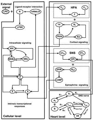
et al.94 implemented a multiscale model to probe the relationship between systemic inflammation and autonomic heart rate regulation. Their multiscale model comprised three submodels: (1) a cell-scale model of leukocytes under endotoxin challenge; (2) an organ-scale model for the heart to determine
[image:14.594.129.445.281.690.2]changes to heart rate variability in response to sys-temic inflammation; and (3) an organism-scale neuro-endocrine system model (Figure 7). Their cell-scale model is based on a previous description95 of endotoxin signaling and associated transcriptional dynamics along with a pharmacokinetics/pharmaco-dynamics model for exogenous immune-suppressive agents. At the organ scale, the rate of change of heart rate variability is considered to be a switch-like func-tion of the cellular pro-inflammatory response. At the scale of the neuroendocrine system, the model activates the influence of pro-inflammatory response on cytosol levels at the cell-scale and defines the influence of the level of epinephrine hormone (secreted by the sympathetic nervous system) on cell-scale anti-inflammatory response.
Model parameters associated with the influence of systemic inflammation signaling on cell-scale tran-scriptional response are determined from experiments,94,95 while parameters associated with the neuro-endocrine immune system are estimated. The multiscale model predicts accurately the tempo-ral evolution of cortisol concentrations and steroid active signal and the significant differences between conditions of presence and absence of immunomodu-latory drug infusion prior to endotoxemia. The model also predicts accurately heart rate variability over time and its relatively low sensitivity to immu-nomodulatory drug infusion.
Foteinou et al.96 enriched the above multiscale
model by adding the influence of sympathetic and parasympathetic nerve activities on heart rate varia-bility based on a previous model by Warner
et al.97 The additional model parameters are obtained from experiments on human subjects.96 This extended model is found to accurately predict experimentally observed temporal changes in para-sympathetic activity and heart rate. Furthermore, the model accurately predicts the significant differ-ences in both parasympathetic activity and heart rate between presence and absence of exogenous epinephrine drug infusion prior to the endotoxemic challenge.
Circulation System
The influence of the venous system on heart dynamics and circulation has attracted the interest of researchers since the late 1960s.98 Müller
et al.99 implemented a multiscale model for the
cir-culation system that comprises: (1) a network of major arteries; (2) a network of major veins; (3) -lumped-parameter models for the heart and pulmo-nary circulation; and (4) lumped parameter models for the arterioles, capillaries, and venules. For the venous submodel, model input parameters are obtained in a subject-specific manner from experi-ments. Other submodel predictions are tested against results reported in the literature. Using their multiscale model, Müller et al.99 demonstrate causal confirmation for predictions of blood flow in the aorta, blood flow in the major leg arteries, blood flow in arteries located in the neck and the head, and blood flow in systemic veins located out-side the neck and the head. Furthermore, Müller
et al.99 obtain significant predictive accuracy for subject-specific blood flow in the veins in the head and in the neck when compared against phase-contrast MRI data.
Open Problems Motivated
by Determination of Effect
In a similar manner as before, we pose open pro-blems in multiscale modeling in cardiovascular (CV) biomechanics motivated by determination of effect(DE).
CV_DE1. Cellular mechanics in bloodflow: A multi-scale model that predicts the dynamics of an RBC in bloodflow better than a single-scale model CV_DE2. Autonomic heart rate regulation: A
multi-scale model that predicts autonomic heart rate reg-ulation under endotoxemic challenge better than a single-scale model
CV_DE3. Circulation system: A multiscale model that predicts the venous system in the head and in the neck better than a single-scale model
Determination of Effect
Cellular Mechanics in Blood Flow
In a direct response to the CV_DE1 problem, Peng
et al.100 simulated the aggregated flow of RBCs in surrounding blood plasma using a coupled flow– structure interaction model. They compared their multiscale model predictions against those from a single scale model (possessing a continuum descrip-tion for the RBC membrane and excluding the detailed bilayer–skeleton architecture). This allowed
45
11 cP (Fischer et al. 1978) 18 cP (Fischer et al. 1978) 31 cP (Fischer et al. 1978) 59 cP (Fischer et al. 1978) Simulationvb = vs = 0 (13 cP)
Simulation(13 cP) Simulation(31 cP) Simulation(59 cP) Single-layer model(13 cP)
Tank-treading frequency (s
–1
)
40
35
30
25
20
15
10
5
0 200 400 600
Shear rate (s–1)
[image:15.594.316.541.451.607.2]800 1000
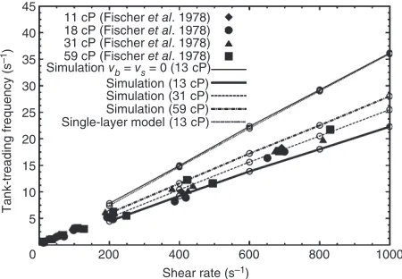
FIGURE 8 | Establishing‘determination of effect’of RBC membrane microstructural details on tank-treading dynamics of RBC in shearflow.‘Simulation’refers to the multiscale model89–91,100and ‘single-layer model’refers to a single-scale model. The multiscale model simulation with zero membrane viscosity (vb=vs= 0) retrieves
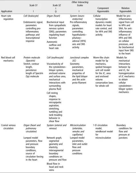
TABLE 2| Summary of Multiscale Modeling Approaches in Cardiovascular Biomechanics
Application
Scale S1 Scale S2
Other Interacting Scales Component Hypomodels Relation Hypomodels I I I O Heart rate regulation
Cell (leukocyte) Organ (heart) System (neuro-endocrine)
Cellular transcription dynamics model, models for heart rate control, and for HPA and SNS activity
Model for pro-inflammatory signal from cell to HPA and SNA, for anti-inflammatory influence of HPA and SNS on cellular processes, and for biochemical input from SNS to the heart Endotoxemic signal,
parameters controlling pro-inflammatory pathways and transcriptional response Biochemical input from sympathetic nervous system (SNS), parameters regulating heart function
Pro-inflammatory signals, parameters controlling hypothalamic– pituitary– adrenal (HPA) axis and SNS activity Autonomic
outflow and heart rate Red blood cell
mechanics
Protein molecule (Spectrin)
Cell (erythrocyte) Junctional complex (JC)
Worm-like chain model for Sp mechanics, spoked hexagon unit cell model for the JC, area and enclosed volume
conservation laws for whole cell
Models for mechanical interactions between Sp and JC, for homogenization of JC mechanics to obtain cellular cytoskeleton– bilayer system mechanics Stretch, contour length, persistence length of Spectrin (Sp) molecule
Viscoelasticity of the cytoskeleton– bilayer system, enclosed volume and surface area, mechanical interactions with surrounding plasmafluid
Geometry parameters, mechanical properties of the lipid bilayer and the actin proto-filament
Cell resting shapes, response to micropipette aspiration, stretching, tumbling and tank-treading behavior in shearflow Cranial venous
circulation
Organ (heart and pulmonary circulation)
System (arterial and venous) Microcirculation (arterioles, venules and capillaries) 1-D circulation model, windkessel model for microcirculation Boundary conditions for
flow and pressure at junctions Lumped model
parameters,flow and pressure boundary conditions, source of circulation forcing in the heart
Network graph, segmental geometry and elasticity, intersegmental boundary conditions on pressure andflow
Lumped model parameters, inlet and outlet
flow and pressure conditions
Bloodflow in head and neck veins
a‘determination of effect’of microstructural proper-ties on RBC mechanics in shear flow, particularly during tumbling and tank-treading behavior (Figure 8). The tumbling rate of the multiscale model100 was slightly higher than that of the single
scale model, the shear ratios and protein density ratios were locally different along the membrane sur-face. Tank-treading frequencies were better predicted by the multiscale model compared to the single-scale model as the former allowed for nonzero membrane viscosity.
Summary
The discussion in this section showed that most of the work in multiscale modeling of cardiovascular biomechanics has been motivated by causal confi r-mation. Based on the reviewed literature, seven open problems motivated by predictive accuracy were posed. One of these (CV_PA3) was already found answered in the application area of RBC dynamics in blood flow. This application area was found to have successfully modeled a related problem (CV_DE1) motivated by determination of effect. This leaves open two multiscale modeling problems motivated by determination of effect, related to autonomic heart rate regulation and venous circulation in the
head and the neck. Table 2 summarizes the multi-scale modeling approaches in cardiovascular biome-chanics that successfully solved problems motivated bypredictive accuracyanddetermination of effect.
MULTISCALE MODELS OF OTHER
BIOMECHANICS PROBLEMS
While a lot of multiscale modeling research targets cardiovascular or musculoskeletal biomechanics, there are some other interesting biomechanics appli-cations that are worth mentioning, without any pre-tence of being exhaustive.
In respiratory biomechanics, multiscale models are being used to provide ‘… with accurate spatial relationships between airway, vessel and the tissue to which they are tethered,’ necessary for the computa-tional analysis of ‘airway–vessel–tissue interactions such as coupling of ventilation distribution in ‘embedded’ airway models to the large deformation of the lung tissue.’101 These models are also used to
look at the transport mechanisms,102 or to investi-gate bronchoconstriction.103
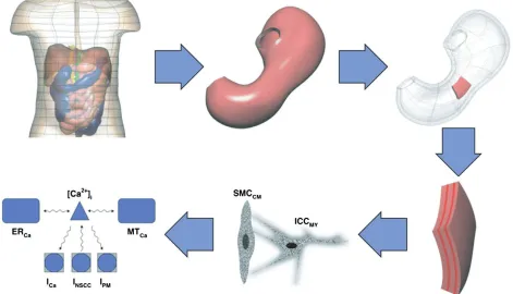
[image:17.594.72.543.412.682.2]The gastro-intestinal system is comprised of organs (stomach, small intestine, and large intestine) that differ significantly in function, organ-scale
morphology and organization at the tissue and cellu-lar scales. In order to achieve proper motility of ingested material through this complex system, mus-cular activity must be under strong spatiotemporal regulation. This regulation is achieved by gastro-intestinal electrophysiology.104Du et al. provides an
extensive review of the multiscale modeling of the gastro-intestinal tract up to 2010.105 The same authors proposed an electromechanical model for the interpretation of electrogastrograms;106 more
recently, a multiscale model was used to investigate reflux in adenocarcinoma.107 In current multiscale
models (Figure 9), muscle cell electro-physiology is modeled either by the phenomenological Alievet al.’s
model,108 or more recently, the biophysically based
model of Corrias et al.109 These models describe wave equations in transmembrane potential and slow current variables for various ion gating mechanisms occurring in the cells. Individual cell models plug into the tissue scale models providing the ionic activity induced electrical current. Tissue-scale organization is modeled depending on the organ, for example, stomach models possess three layers of smooth mus-cles (one longitudinal and two circular) separated by two ICC layers;110 whereas a small-intestine model possesses only one smooth muscle cell layer and one ICC layer.111 Tissue-scale electrophysiology
[image:18.594.57.522.69.399.2]model-ing is based on a bi-domain framework, which has FIGURE 10 | Multiscale modeling of lymphatic drainage. (Reprinted with permission from Ref 120. Copyright 2012 Elsevier)
TABLE 3| Summary of the Reviewed Literature
[image:18.594.47.527.458.531.2]previously been applied to cardiac electrophysiol-ogy112 and specifies the relationships between the
potential difference across the cell membrane and the electric potential in the extra-cellular space.
Thomas and coworkers proposed a whole mod-eling environment (SAPHIR) to model blood pressure regulation and fluid homeostasis.113Chen et al. used a multiscale model to investigate the relationship between systolic blood pressure and the pathogenesis and progression of renal diseases.114
Computational oncology is another area where multiscale modeling is being used extensively. For a general review on the topic, see Deisboecket al.115May
et al.116coupled the biomechanics of tumor–host tissues
interaction with a cellular model of cancer growth, an important determinant especially in tumors growing in regions confined by bone tissue, such as in the case of brain tumors; multiscale models are also used to investi-gate the role of angiogenesis in tumor growth,72or to better understand the effect of radiotherapy.117
Rim et al. used a three-scale model to investi-gate the transdermal diffusion of drugs.118 Adra
et al. developed an agent-based model of keratinocyte colony formation in 2D culture.119Roose and Swartz developed an extensive multiscale model of the fluid drainage from tissues through the lymphatic system (Figure 10).120 Biswas et al. developed a multiscale model of a skin mechanoreceptor, the Pacinian cor-puscle.121 Valero et al. modeled the angiogenesis
process during wound contraction.122
Another important application area is the mod-eling of organogenesis and growth processes. New-man et al. modeled the limb development process in vertebrates;123 Cox used a multiscale model to explore the mechanoregulation during dentition;124
Göktepe et al. proposed a multiscale model of the cardiac sarcomerogenesis.125
CONCLUSION
Of all possible angles we could use to structure this review, we chose that of the modeling intent, orga-nizing all reviewed papers around three distinct operational motivations: ‘Causal confirmation,’ ‘ Pre-dictive accuracy,’ and ‘Determination of effect.’ The first represents the most‘humble’ intent, among the three. In these papers multiscale modeling is used to merely show that a complex set of observations is ‘compatible’ with a mechanistic theory embodied by the multiscale model; of course‘compatible’does not mean‘true,’and even less‘accurate.’The papers that fall in the predictive accuracy category are driven by the necessity to improve predictive accuracy over sys-tems where the assumption of scale separation
applies poorly. Last, the models motivated by the need to determine the effect of processes at radically different scale are the probably the most challenging, and intellectually interesting, as they look directly at the complexity of multiscale systems.
In the survey conducted here on the literature on multiscale modeling in biomechanics, we reviewed 72 studies motivated by causal confirmation, 12 stud-ies aiming at predictive accuracy, and only one demonstrating determination of effect across scales (Table 3). Although the present review is not exhaus-tive, this relative multiplicity is representative of the body of research in the subdomain of multiscale modeling in biomechanics.
By focusing on the progress made beyond causal confirmation, we showed how in some research pro-blems, a particular scale separation schema has gained wider acceptance through validation. For those topics where a valid scale separation picture is yet to emerge, this was found to be the case typically because model input variables/parameters at the different scales could not be determined in a specimen-specific manner. This highlights focus areas for future experimental research. To clarify the road ahead, a total of 15 open problems (see Boxes 1 and 2) were posed in relation to musculo-skeletal or cardiovascular applications which, if solved, would advance the state-of-the-art of multiscale model-ing in biomechanics. Finally, this review of current research reveals that, from basic biology to medicine, multiscale modeling in biomechanics is relevant to a variety of other research areas, and is expected to become more so in the future.
BOX 1
OPEN PROBLEMS MOTIVATED BY PREDICTIVE ACCURACY
PA1. Bone remodeling: A multiscale model to
predict the evolution of bone mineral con-tent as measured in a given bone volume, using bone remodeling activity parameters measured on the same specimen
PA2. Fracture healing: A multiscale model to
predict tip cell movement and sprout forma-tion as measured within a bone fracture site volume, using intracellular and tissue scale parameters measured on the same specimen
PA3. Skeletal muscle remodeling: A multiscale
ACKNOWLEDGMENT
This study was supported by the EPSRC grant EP/K03877X/1.
REFERENCES
1. Viceconti M, Humphrey JD, Erdemir A, Tawhai M. Multiscale modelling in biomechanics.Interface Focus 2015, 5:20150003.
2. Viceconti M, Hunter P, Hose R. Big data, big knowledge: big data for personalized healthcare. IEEE J Biomed Health Inform 2015, 19:1209–1215.
3. Rodrigue N, Philippe H. Mechanistic revisions of phenomenological modeling strategies in molecular evolution.Trends Genet2010, 26:248–252.
4. Anderson AE, Ellis BJ, Weiss JA. Verification, valida-tion and sensitivity studies in computavalida-tional biome-chanics. Comput Methods Biomech Biomed Engin 2007, 10:171–184.
BOX 2
OPEN PROBLEMS MOTIVATED BY DETERMINATION OF EFFECT
DE1. Bone mechanics: A multiscale model that
predicts better than a single-scale model the bone tissue elastic anisotropy
DE2. Skeletal muscle electromechanics: A
multi-scale model that predicts better than a
single-scale model the force–length
relation-ship in a muscle
DE3. Tendon tissue mechanics: A multiscale
model that predicts better than a single-scale model the stiffness tensor of a tendon tissue
DE4. Autonomic heart rate regulation: A
multi-scale model that predicts autonomic heart rate regulation under endotoxemic chal-lenge better than a single-scale model
DE5. Circulation system: A multiscale model
that predicts the venous system in the head and in the neck better than a single-scale model
PA4. Tendon homeostasis: A multiscale model
to predict whole tendon remodeling as measured on a subject, using tissue and molecular scale measured on the same subject
PA5. Blood flow interaction with blood vessel
walls: A multiscale model to predict the
meas-ured bloodflow and blood vessel wall
dynam-ics within a local region (e.g., surrounding an aneurysm) of the systemic circulation, using parameters measured on the same circulation system, vascular tissue and geometry and
bloodfluid
PA6. Blood flow interaction with blood
coagu-lation: A multiscale model to predict the measured development of a blood clot, using the bio-chemo-mechanical parameters
meas-ured on the same platelet–blood–vessel wall
system
PA7. Vasculogenesis and angiogenesis: A
multi-scale model to predict measured angiogene-sis and vasculogeneangiogene-sis based on measured
subcellular, cellular and molecular scale
parameters
PA8. Cerebral autoregulation in CPB: A
multi-scale model to predict the measured cerebral autoregulation, using the measured elasticity of vessel walls on the same cardiac system
PA9. Cardiomyocyte mechanics: A multiscale
model to predict the measured electrical activity in a cardiac myocyte under high-frequency stimulation, using cell membrane
electro-mechanical property parameters
measured in the same myocyte
PA10. Heart valve mechanics: A multiscale
model to predict the change in cell aspect ratio during a cardiac cycle and increase in
valvular tissue calcification due to aging,
5. Aoubiza B, Crolet JM, Meunier A. On the mechanical characterization of compact bone structure using the homogenization theory.J Biomech1996, 29:1539–1547. 6. Crolet JM, Aoubiza B, Meunier A. Compact bone: numerical simulation of mechanical characteristics. J Biomech1993, 26:677–687.
7. Currey JD. Biocomposites: micromechanics of biolog-ical hard tissues. Curr Opin Solid State Mater Sci 1996, 1:440–445.
8. Eshelby JD. The determination of the elasticfield of an ellipsoidal inclusion, and related problems. Proc Math Phys Eng Sci1957, 241:376–396.
9. Hill R. A self-consistent mechanics of composite materials.J Mech Phys Solids1965, 13:213–222. 10. Mori T, Tanaka K. Average stress in matrix and
aver-age elastic energy of materials with misfitting inclu-sions.Acta Metall1973, 21:571–574.
11. Hamed E, Lee Y, Jasiuk I. Multiscale modeling of elastic properties of cortical bone. Acta Mech 2010, 213:131–154.
12. Martínez-Reina J, Domínguez J, García-Aznar JM. Effect of porosity and mineral content on the elastic constants of cortical bone: a multiscale approach. Biomech Model Mechanobiol2011, 10:309–322. 13. Sansalone V, Naili S, Bousson V, Bergot C, Peyrin F,
Zarka J, Laredo JD, Haïat G. Determination of the heterogeneous anisotropic elastic properties of human femoral bone: from nanoscopic to organ scale. J Biomech2010, 43:1857–1863.
14. Webster D, Müller R. In silico models of bone remod-eling from macro to nano-from organ to cell.WIREs Syst Biol Med2011, 3:241–251.
15. Colloca M, Blanchard R, Hellmich C, Ito K, van Rietbergen B. A multiscale analytical approach for bone remodeling simulations: linking scales from col-lagen to trabeculae.Bone2014, 64:303–313.
16. Pivonka P, Buenzli PR, Scheiner S, Hellmich C, Dunstan CR. The influence of bone surface availabil-ity in bone remodelling—a mathematical model including coupled geometrical and biomechanical reg-ulations of bone cells.Eng Struct2013, 47:134–147. 17. Scheiner S, Pivonka P, Smith DW, Dunstan CR,
Hellmich C. Mathematical modeling of postmenopau-sal osteoporosis and its treatment by the anti-catabolic drug denosumab. Int J Numer Method Biomed Eng2014, 30:1–27.
18. Hambli R. Application of neural networks andfinite element computation for multiscale simulation of bone remodeling.J Biomech Eng2010, 132:114502. 19. Fyhrie DP, Carter DR. A unifying principle relating
stress to trabecular bone morphology. J Orthop Res 1986, 4:304–317.
20. Coelho PG, Fernandes PR, Rodrigues HC. Multiscale modeling of bone tissue with surface and permeability control.J Biomech2011, 44:321–329.
21. Carlier A, Geris L, Bentley K, Carmeliet G, Carmeliet P, Van Oosterwyck H. MOSAIC: a multi-scale model of osteogenesis and sprouting angiogene-sis with lateral inhibition of endothelial cells. PLoS Comput Biol2012, 8:e1002724.
22. Bentley K, Gerhardt H, Bates PA. Agent-based simu-lation of notch-mediated tip cell selection in angio-genic sprout initialisation. J Theor Biol 2008, 250:25–36.
23. Peiffer V, Gerisch A, Vandepitte D, Van Oosterwyck H, Geris L. A hybrid bioregulatory model of angiogenesis during bone fracture healing. Biomech Model Mechanobiol2011, 10:383–395.
24. Carlier A, Geris L, van Gastel N, Carmeliet G, Van Oosterwyck H. Oxygen as a critical determinant of bone fracture healing—a multiscale model. J Theor Biol2015, 365:247–264.
25. Carlier A, van Gastel N, Geris L, Carmeliet G, Van Oosterwyck H. Size does matter: an integrative in vivo-in silico approach for the treatment of critical size bone defects. PLoS Comput Biol 2014, 10: e1003888.
26. Wisdom KM, Delp SL, Kuhl E. Use it or lose it: mul-tiscale skeletal muscle adaptation to mechanical sti-muli. Biomech Model Mechanobiol 2015, 14:195–215.
27. Haddad F, Roy RR, Zhong H, Edgerton VR, Baldwin KM. Atrophy responses to muscle inactivity. II: molecular markers of protein deficits.J Appl Phy-siol2003, 95:791–802.
28. Taber LA. Biomechanical growth laws for muscle tis-sue.J Theor Biol1998, 193:201–213.
29. Zöllner AM, Pok JM, McWalter EJ, Gold GE, Kuhl E. On high heels and short muscles: a multiscale model for sarcomere loss in the gastrocnemius mus-cle.J Theor Biol2015, 365:301–310.
30. Smith DW, Rubenson J, Lloyd D, Zheng M, Fernandez J, Besier T, Xu J, Gardiner BS. A concep-tual framework for computational models of Achilles tendon homeostasis. WIREs Syst Biol Med 2013, 5:523–538.
31. Maceri F, Marino M, Vairo G. An insight on multi-scale tendon modeling in muscle-tendon integrated behavior. Biomech Model Mechanobiol 2012, 11:505–517.
32. Spiesz EM, Roschger P, Zysset PK. Influence of min-eralization and microporosity on tissue elasticity: experimental and numerical investigation on minera-lized turkey leg tendons. Calcif Tissue Int 2012, 90:319–329.
![FIGURE 1 from 1991 to 2015. Incidence is obtained by dividing for each year thenumber of papers retrieved with the search| Incidence of multiscale papers indexed in PubMed ‘Multiscale [ALL]’ by thetotal number of papers indexed in that year.](https://thumb-us.123doks.com/thumbv2/123dok_us/7731484.162898/2.594.56.272.70.207/incidence-obtained-dividing-thenumber-retrieved-incidence-multiscale-multiscale.webp)