“AN OBSERVATIONAL STUDY TO ANALYSE THE ROLE
OF VISUAL EVOKED POTENTIAL IN VISUAL PROGNOSIS
BEFORE AND AFTER PANRETINAL
PHOTOCOAGULATION FOR DIABETIC RETINOPATHY
IN TYPE 2 DIABETIC MELLITUS”
M.S. DEGREE
BRANCH –III (OPHTHALMOLOGY)
MAY 2019
GOVT. RAJAJI HOSPITAL &
MADURAI MEDICAL COLLEGE
MADURAI
THE TAMILNADU
CERTIFICATE FROM THE DEAN
This is to certify that the dissertation entitled “AN
OBSERVATIONAL STUDY TO ANALYSE THE ROLE OF VISUAL
EVOKED POTENTIAL IN VISUAL PROGNOSIS BEFORE AND
AFTER PANRETINAL PHOTOCOAGULATION FOR DIABETIC
RETINOPATHY IN TYPE 2 DIABETIC MELLITUS” submitted by
Dr.J.DHEEBALAKSHMI, to the Faculty of Ophthalmology, The
Tamilnadu Dr.M.G.R. Medical University, Chennai in partial fulfilment of
the requirement for the reward of M.S. Degree in Ophthalmology is a
bonafide work carried out by him during the period 2016-2019.
Place: Madurai Prof.Dr.D. MARUTHUPANDIAN
Date: M.S., F.I.C.S., F.A.I.S., FAC Dean,
CERTIFICATE FROM THE HEAD OF THE DEPARTMENT
This is to certify that this dissertation entitled “AN
OBSERVATIONAL STUDY TO ANALYSE THE ROLE OF
VISUAL EVOKED POTENTIAL IN VISUAL PROGNOSIS
BEFORE AND AFTER PANRETINAL PHOTOCOAGULATION
FOR DIABETIC RETINOPATHY IN TYPE 2 DIABETIC
MELLITUS” is the bonafide original work of
Dr.J.DHEEBALAKSHMI, in partial fulfillment of the requirement for
M.S.,(Branch III) Ophthalmology examination of the Tamilnadu
Dr.M.G.R. Medical university to be held in May 2019.
Prof.Dr. K. KAVITHA M.S, D.N.B.,
Head of the Department,
ANTI- PLAGIARISM CERTIFICATE
This is to certify that this dissertation work titled “AN
OBSERVATIONAL STUDY TO ANALYSE THE ROLE OF
VISUAL EVOKED POTENTIAL IN VISUAL PROGNOSIS
BEFORE AND AFTER PANRETINAL PHOTOCOAGULATION
FOR DIABETIC RETINOPATHY IN TYPE 2 DIABETIC
MELLITUS” of the candidate Dr.J.DHEEBALAKSHMI with
Registration Number 221613102 for the done for award of Master of
Surgery Degree in the branch of Ophthalmology. I personally verified the
urkund.com website for the purpose of plagiarism check. I found that the
uploaded thesis file contained from introduction to conclusion pages and
result shows 7% of plagiarism in the dissertation.
DECLARATION
I, Dr.J.DHEEBALAKSHMI hereby solemnly declare that, this
dissertation titled “AN OBSERVATIONAL STUDY TO ANALYSE
THE ROLE OF VISUAL EVOKED POTENTIAL IN VISUAL
PROGNOSIS BEFORE AND AFTER PANRETINAL
PHOTOCOAGULATION FOR DIABETIC RETINOPATHY IN
TYPE 2 DIABETIC MELLITUS” was done by me.
I also declare that this bonafide work / a part of this work was not
submitted by me / anyone else, for any award, for Degree / Diploma to any
other University / Board either in India / abroad. This is submitted to The
Tamilnadu Dr. M. G. R. Medical University, Chennai in partial fulfillment
of the rules and regulations for the award of Master of Surgery degree
Branch -III (Ophthalmology) to be held in May 2019.
Place: Madurai (Dr.J.DHEEBALAKSHMI)
Date:
ACKNOWLEDGEMENT
I express my sincere thanks and gratitude to
Prof. Dr. D. MARUTHUPANDIAN M.S., FICS., FAIS., The Dean, GRH and
MMC Madurai for permitting me to conduct this study. I am extremely grateful
to Prof. Dr.K.KAVITHA M.S, D.N.B., HOD, Professor of Ophthalmology and
Associate Professor of Ophthalmology, GRH, MMC, Madurai, for their constant
source of support and encouragement for completing this study. I have great
pleasure in thanking my beloved guide Dr.N.PARVATHASUNDARI M.S,
DO., Associate Professor and all my Assistant Professors of Ophthalmology
department at Madurai Medical College, Madurai, for their constant source of
cheer and encouragement throughout the study.
I express my deep sense of gratitude to The Professor and HOD of
Neuromedicine, for their support to this study.
I thank the Secretary and Chairman of the Institution Ethical Committee,
GRH Madurai.
I am indebted to all the patients, paramedical staffs for their sincere
co-operation for the completion of this study.
I am grateful to my fellow post graduate colleagues for their valuable help
throughout this study.
I am extremely thankful to my family members for their constant support.
Above all I thank the almighty for guiding me with his blessings
TABLE OF CONTENTS
SERIAL
NUMBER CONTENT
PAGE NUMBER
PART I
1 INTRODUCTION 1
2 BLOOD RETINAL BARRIER 3
3 PATHOPHYSIOLOGY 7
4 NEURONAL DAMAGE IN DIABETIC
RETINOPATHY 18
5 MANAGEMENT OF DIABETIC
RETINOPATHY 22
6 PAN RETINAL PHOTOCOAGULATION 28
7 NEW LASER DELIVERY SYSTEM 37
8 PHARMACOLOGICAL MANAGEMENT 42
9 VISUAL EVOKED POTENTIAL 45
10 REVIEW OF LITERATURE 57
PART II
1 AIMS AND OBJECTIVES 62
2 MATERIALS AND METHODS 64
3 OBSERVATION AND ANALYSIS 66
4 DISCUSSION 80
5 CONCLUSION 82
6 LIMITATIONS OF THIS STUDY 82
8 ANNEXURE II- ABBREVIATIONS 90
9 ANNEXURE III- MASTER CHART 92
10 ANNEXURE IV- PROFORMA 96
11 ANNEXURE V- CONSENT FORM 100
12 ANNEXURE VI-RECEIPT FOR
PLAGIARISM 101
13 ANNEXURE VII- ETHICAL CLEARANCE
1
INTRODUCTION
Diabetic mellitus is chronic disease that increases the risk of
developing multiple medical complications. So, this patients requires
screening, long term observation for diagnosis, treatment and prognosis.
Diabetes mellitus is the major systemic cause of blindness in
western world. It contributes 4.8% of the 37 million cases of blindness
throughout the world.
Impact of Diabetic over vision can be significantly reduced by
proper glycemic control, routine ophthalmological examination and
proper treatment.
Laser photocoagulation remains the mainstay of treatment for
established diabetic retinopathy .it has been shown to reduce visual loss
due to diabetic macular edema and proliferative Diabetic Retinopathy.
DIABETIC RETINOPATHY
Retina is a ten layered sheet and most metabolically active organ in
the body which composed of neurons, photoreceptors and supporting
cells. The outer one third of retina receives its blood supply from the
choriocapillaris and the inner two third of the retina is supplied by central
2
FIGURE 1 : RETINAL LAYER
TRANSPARENCY OF RETINA
Transparency of retina should be maintained so that light should
pass through layers of retina and activate posteriorly situated
photoreceptors.
• Retinal nerve fibres are unmyelinated since myelination can block
light rays. As such, it requires more energy to more energy than
myelinated axon for maintain membrane potential.
• Vasculature density is low at inner retina .so,it is at constant state
3
• Inner retina has only few mitochondria since it can block light
transmission. so it depend on glycolysis for energy production
rather than oxidative phoporylation.
So, retina remain most metabolically active even in the low oxygen
tension.whenever oxygen demand is increased inner retina more prone
for retinopathy.
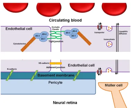
BLOOD RETINAL BARRIER
In the human brain, there is selective exchange of molecules
between blood vessel and neural parenchyma is regulated tightly by the
structure called blood-brain barrier (BBB). As a extension of CNS, retina
also has tight barrier called blood-retinal barrier (BRB).
Neural retina receives dual blood supply from retinal vessels and
choroidal vessels. Neural retina is separated from retinal vessels and
choroidal vessels by inner and outer BRB, respectively.
• Inner BRB is formed by a tight junction between adjacent retinal
vascular endothelium (resembles the BBB proper of brain)
• Outer BRB formed by a tight junction between adjacent retinal
pigment epithelium (resembles the blood-CSF barrier of brain).
• Among these two kinds of BRB, abnormality in the inner BRB
4
Inner blood-retinal barrier is consist of multiple cellular
component including endothelial cells, pericytes and Müller cells.
Pericytes ensheath the retinal microvascular endothelium and
share their common basement membrane with retinal endothelial
cells.
• Pericytes are adherant to endothelial cells by the N-cadherin
mediated adherent junction. Müller cells have spatial proximity
with endothelium cells and connect with endothelium by their
footprocesses. Each adjacent endothelial cells are interconnected
with each other through their tight junctions and act as a
functional barrier.
• Selective permeablity of molecules through the paracellular
pathway is tightly regulated by this tight junction and controlled
exchange of molecules through the transcellular pathways are
5
FIGRUE 2 : THIS PICTURE SHOWS TIGHT JUNCTION BETWEEN ENDOTHELIAL CELL OF INNER RETINAL
BARRIER.
DISTRUPTION OF INNER BRB IN DIABETIC
Protein Kinase C (PKC)
In the diabetic, higher concentration of diacylglycerol which
stimulate PKC to translocate into plasmamembrane and activate the
acquire phosphorylation activities. High blood glucose induced the
activation of PKC is associated with the diabetic retinopathy pathology.
6
retinopathy still remains unclear.PKC, especillay β-isoform is considered
as a key indicator of VEGF induced BRB disruption and retinal
vasculopathy. Recently,it is reported that PKCδ is also decrease the
expression of the endothelial tight junction (ZO-1, 2) and also vascular
hyperpermeability in diabetic retina .In addition,PKC activate occludin
phosphorylation is shown to participate in the VEGF induced vascular
leakage.
Some investigations also suggest that nitric oxide (NO) pathway
is a potential downstream target of PKC induced vascular leakage.
Advanced Glycation Endproducts (AGEs)
Hyperglycemic environment for long-term exposure results in a
non-enzymatic reaction of protein, lipid and nucleic acid to form a
irreversible products called AGEs. The clinical evidence of AGEs is well
demonstrated in diabetic patient.
In type 1 diabetic patients, glycated collagen and
carboxymethyllysine (a kind of AGEs) in skin showed correlation with
the progression of diabetic retinopathy.
Hydroimidazolone, one of the mostprominent AGEs,level in
vitreous is reported to be high in type 2 diabetic patient. AGE –RAGE
7
mediated by ROS production through AGE-RAGE interaction leads to
oxidation of DNA, membrane lipid protein peroxidation and apoptosis
pericyte cells.
In addition, AGEs stimulate the expression of growth factors
from pericyte and also inflammatory reactions which results in
disturbance of BRB function.
PATHOPHYSIOLOGY
The real mechanism of diabetic retinopathy is not known yet.still
many theories have been proposed.
Sorbital, a byproduct of aldose reductase,usually not crosses
cell wall. In diabetic mellitus concentration of aldose reductase high in
retinal pericytes.high concentration of sorbitol generate osmotic gradient
that leads to electrolyte imbalance and eventually cell damage. A
polymorphism near the transcription site of aldose reductase gene
associated with early onset development of diabetic retinopathy in type 2
diabetic patients.but, clinical trial failed to prove the efficacy of aldose
reductase inhibitor in preventing development of diabetic retinopathy.
Alternatively, another theory postulates that vasoproliferative
factors released by the retina,retinal vessels,retinal pigment epithelium
8
Experimental models suggest that neovascularization is mediated
in part by the interaction between insulin‐like growth factor (IGF‐1) and vascular endothelial growth factor (VEGF).Animal study showed IGF-1
antagonist reduced the incident of neovascularization.this is clinically
evident by worsening of diabetic retinopathy after insulin therapy. Insulin
increases serum IGF-1 while Pituitary injury decreases IGF-1 which
reverse the condition.Recently, studies more focused on VEGF role in
neovascularization. It has been demonstrated that VEGF more
concentrated in vitreous in case of PDR than those with NPDR, and
intensity of immunostaining for VEGF is proportional to the severity of
retinopathy. This theory become more evident because VEGF inhibitors
have been successful in controlling hypoxia‐induced neovascularization
in certain animal models.
Recently, erythropoietin may also contributes to progression of
retinopathy. The local concentration of erythropoietin has been found to
be much higher in patients with active proliferative retinopathy.this factor
has been more correlated than VEGF because erythropoietin inhibitors
slowed down the proliferation of endothelium and regress the retinal
neovascularization.
Other vasoactive factors includes basic fibroblast growth factor
9
increased in vitreous in PDR. these factors are strongly correlated with
inducing factors of neovascularization to ischemia.
Hyperglycemia impairs autoregulation of retinal blood flow. so, In
diabetic even minimal increase in blood volume can cause shearing force
over retinal blood vessel and results in vasoactive factors release.
Genetic factors also susceptible for retinopathy. It is evident by
first degree relatives with retinopathy have three time risk of developing
severe retinopathy.some study suggesting mechanism for this could be
platelet abnormality or hyperviscosity leads to focal attenuation and
ischemia result in neovascularization.
ANATOMICAL AND FUNCTIONAL CHANGES IN DR
Structural changes in the retinal capillary wall includes
• Pericyte loss
• Endothelial cell loss
• Basement membrane thickening
• Endothelial cell dysfunction.
All these structural abnormalities results in loss of autoregulation of
retinal blood vessels, leakage of capillaries into the extracellular space of
10
The rheological changes results from hyperglycaemia include
• Increase in fibrinogen α2 globulins and decrease in serum albumin
levels in plasma results in decreasing fibrinolysis and increased
viscosity.
• Inability of red blood cells to deform.
• Platelet aggregation.
Both structural changes of blood vessels and rheological changes
results in thrombosis and closure of the retinal capillaries results in
capillary non perfusion area. These ischemic area of retina stimulate the
production of vasoproliferative substances results in development of new
vessel formation.
Clinical features
Microaneurysms
Intraretinal hemorrhages Hard exudates
Cotton wool spots (soft exudates) Venous beading
11
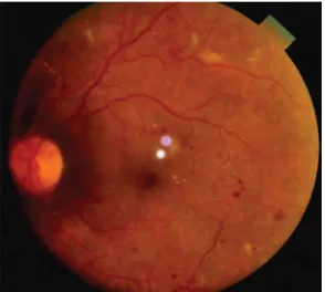
Microaneurysms
Earliest lesion detected clinically
Loss of intramural pericytes wrap and decrease in pericyte and endothelial ratio & result in weakening and outpouching of vessel
wall.
50-60 microns thick
New MA – dark red in color
[image:20.595.170.464.493.757.2] Old MA – yellow to white in color due to hyalinization. In FFA ,it appears as bright hyper fluorescent dots. Life span – months to years
12
Intraretinal hemorrhages
MA, decompensated capillaries, IRMA may cause intra retinal hemorrhage.
Superficial haemorrhage are flame shaped due to arrangement of Nerve fiber layer
dot & blot pattern seen in deep haemorrhage located in the inner deeper layers
FFA shows blocked fluorescence Hard exudates
Vascular leakage in to neuronal element gets absorbed by surrounding intact blood vessels leaving behind break down
lipid products.
yellow material with discrete margin Seen in Outer plexiform layer of the retina
Circinate pattern seen around focal leaking MA/capillaries Course -absorbed by phagocytic action of macrophages on later
phase.
Cotton wool spot
‘Soft exudate’
13
Coagulative necrosis of nerve fibre results in stasis of axoplasmic flow.
Fluffy white in appearance with ill defined margins.
FFA - Hyperfluorescent due to leakage and staining.
Intraretinal microvascular abnormalities - IRMAs
Suspected new vessels between arterioles and venules
Dilated pre-existing capillaries
Seen in areas of capillary non perfusion
Act as shunt vessels between arterioles and venules
Adjacent to ishchemic retina
Usually Budding from venous end
Multiple IRMAs indicate a state of hypoxic region and progression to a high risk of PDR (>50%)
FFA - larger than normal capillaries calliper
- Usually do not leakage from IRMA.
14
FIGURE 4: VERY SEVERE NPDR WITH VENOUS ABNORMALITIES
Venous changes
Venous beading, looping or sausage like segmentation Seen in areas of focal ischemic retina
Denotes of severe hypoxic state of the retina(40- 80% risk of PDR) FFA – seen adjacent to areas of Capillary drop out area.
New vessels
Derived from Primitive mesenchymal element.
15
Fibrocyte component – fibroglial tissue growth along with vessels form fibrovascular band
Usually situated posterior to the equator
[image:24.595.172.461.252.506.2]Shape – fronds, compact spherules, stringy pattern etc
FIGURE 5: PDR WITH NEW VESSELS
NVD
New vessel formation on the disc or within 1 DD It occurs when > 1/4th of the retina hypoxic.
Associated with preretinal/vitreous hge
16
NVE
NVE usually located along the major temporal arcades
Extraretinal hemorrhage
Subhyaloid
From NVD or NVE
Contraction of fibrovascular band elements leads to Tractional Retinal Detachment.
ETDRS Revised modified Airlie House diabetic retinopathy classification
ETDRS level
ETDRS
severity ETDRS definition
10 No
retinopathy Diabetic retinopathy absent
20 Very mild
NPDR Microaneurysms only
35 Mild NPDR Hard exudates, cotton-wool spots, and/or mild retinal hemorrhages
43 Moderate NPDR
43A:retinal hemorrhages moderate in 4 quadrant or severe in 1 quadrant
43B:mild IRMA in 1 to 3 quadrants
47 Moderate NPDR
17
ETDRS level
ETDRS
severity ETDRS definition
47C:severe retinal hemorrhage in two to three quadrants
47D:venous beading in one quadrant"
53A-D Severe NPDR
53A:≥2 level 47 characteristics 53B:severe retinal hemorrhages in 4
53C:moderate to severe IRMA in at least 1 quadrant
53D:venous beading in at least 2 quadrants"
53E Very severe
NPDR ≥2 level 53A-D characteristics
61 Mild PDR NVE <0.5 disk area in 1 or more quadrants
65 Moderate PDR
65A:NVE≥ 0.5 disk area in 1 or more quadrants
65B:NVD< 0.25-0.33 disk area
71 and 75 High-risk PDR
NVD ≥ 65B, or NVD < 65B or NVE ≥ 0.5 disk area plus VH or PRH, or VH or PRH obscuring ≥ 1 disk area
81 and 85 Advanced PDR
18
DR classified as follows:
Grade 0: absence of DR (no DR), corresponding to level 1 of the
abbreviated version of the Modified Airlie House classification (23);
Grade 1: background DR (BDR), comprising levels 2, 3, and 4;
Grade 2: pre-proliferative DR (PPDR), corresponding to level 5;
Grade 3: proliferative DR (PDR), equivalent to level 6.
NEURONAL DAMAGE IN DIABETIC RETINOPATHY
Neurodegeneration
A nerve damage in diabetic neuropathy begins immediately after
the onset of diabetes. Several investications play a role in screening
purpose such as multifocal electroretinography (ERG), flash ERG, color
vision, contrast sensitivity and short-wavelength automated perimetry.
Retinal glial cells play a vital role in maintaining normal function
of retina. In diabetic, upregulation of (GFAP) glial fibrillary acidic
protein in glial cells and selective thinning of ganglion cells and inner
plexiform layer.this results in altered potassium siphoning,GABA uptake,
glutamate excitotoxicity in glial cells leads to expression of angiogenic
19
Apoptosis
Diabetes increasing the frequency of apoptosis by inducing
proinflammatory cytokine (IL-1β) and caspase-1/IL-1β signaling
pathways results in chronic loss of retinal neurons.it is demonstrated by
presence of pyknotic bodies in histological section of diabetic retina.
Several studies suggested that the expression of proapoptotic
Protein, Bax (Bcl-2 associate X protein) is associated with degenerative
diseases which is increased in diabetic retina. The pro-apoptotic
transcription factor Forkhead box O1 (FOXO1) activates the pericyte
apoptosis through the involvement of TNF-α and AGE is the possible
mechanism for apoptosis.
Glutamate excitotoxicity
Glutamate is the excitatory neurotransmitter in the retina, but it
becomes neurotoxic when it is present in excessive amounts.
Extracellular glutamate is transported into mullers cells by glutamate
transporters
Glutamine synthase
Glutamine (non toxic amino acid)
20
• amino-3-hydroxy-5-methyl-4-isoxazolepropionic acid (AMPA) receptors
• N-methyl-D-aspartate (NMDA)
Influx of calcium and sodium in to the cells
Generation of AGE and advanced lipoxidation end product
Defective mitochondrial respiration
Formation of free radicals
Neurotoxicity
Neurodegeneration
ROLE OF NEUROTROPHIC FACTORS
Brain derived neurotrophic factors (BDNF)
• It enhances insulin sensitivity by activating several signaling
21 • Biomarker of insulin resistance.
• In hypoxic condition, BDNF enhances mullers cells to take up increased
glutamate and upregulate glutamine synthetase.
• Combine with ciliary neurotrophic factor (CNTF), it rescue
photoreceptors in retinal explants and have neuroprotective effect.
Nerve Growth Factor (NGF)
• Potent neurotrophic factor, which contributes for retinal
inflammation.
• Potent angiogenic factor in PDR.
Basic Fibroblast Growth Factor (bFGF)
• Role in survival and maturation of both glial cells and neurons.
• Mediator for regeneration after neural injury
• Potent angiogenic factor in pathogenesis of neovascularization.
Glial cell line-derived neurotrophic factor (GDNF)
• Transforming growthfactor-β (TGF-β)-related neurotrophic factor
family
• It rescue photoreceptor and muller glial cells during rtinal
22
MANAGEMENT OF DIABETIC RETINOPATHY
LASER
Light amplification by stimulated emission of radiation-when
electron shift from higher energy level to lower energy level radiation is
emitted.
Characteristics of a LASER
• Monochromatic light
• Spatial coherence
• High density of electrons
Laser effects on the eye
• Photocoagulation
• Photovapourization
• Photoablation
• Photoradiation
• Photodistruption
PHOTOCOAGULATION
When light energy is applied to targeted tissue, the light energy
is converted to heat energy. So, temperature at treated tissue raises from
37⁰ up to 50⁰c result in denaturation of tissue protein and coagulation of
23
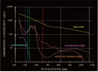
Melanin pigment absorbe light spectrum between 400 to 700nm
which is principle absorber of light in photocoagulation of trabecular
meshwork and co absorber of light in retinal pigment epithelium and
choroid.
Xanthophyll pigment more concentrated in the inner and outer
plexiform layers of retina of the macular area.it absorbs blue light
maximally and green light poorly.
Hemoglobin absorbs blue green and yellow light and red light
poorly. These pigment absorbe shorter wavelength easily and longer
[image:32.595.143.488.519.772.2]wavelength lights are unabsorbed.
FIGURE 6
24
LASERS COMMONLY USED IN PHOTOCOAGULATION
1. CW GREEN ARGON LASER(514.5nm)
It is absorbed selectively by the RPE, hemoglobin pigments,
choriocapillries, layers of rods and cones and at the outer and inner
nuclear layers.It coagulates from choriocapillaries to inner nuclear layer
of retina.
2. FREQUENCY DOUBLED Nd: YAG LASER(532nm)
It is solid state and diode pumped CW laser and mainly absorbed
by hemoglobin and the melanin present in retinal pigment epithelium and
trabecular meshwork.it coagulated from choriocapillaries to outer nuclear
layer of the retina.
3. KRYPTON RED LASER (647nm)
It is readily absorbed by melanin granules but not absorbed by the
hemoglobin and xanthophylls. So it is used for macular
photocoagulation.since,it is not absorbed by retinal vasculature it can
penetrate deeper to coagulate choriocapillaries and Choroids.
4. DIODE LASER (810 nm)
It is the most important semiconductor laser GaAs.it is very
difficult to coagulate microaneurysm directly because it is very poorly
25
GRADING OF PHOTOCOAGULATION LESIONS
• GRADE 1/ LIGHT -Barely visible blanching of retinal pigment
epithelium
• GRADE 2/MILD - Hazy, faint white retinal coagulation
• GRADE3/MODERATE –Opaque ,dirty white retinal coagulation
[image:34.595.203.430.331.525.2]• GRADE4/HEAVY -Dense white, chalky retinal coagulation
FIGURE 7: MODERATE NPDR WITH POST PRP STATUS
[image:34.595.198.432.572.770.2]26
FOCUSING OF LASER BEAM
All lasers except xenon arc emit monochromatic rays .so, all
laser beam except xenon arc should be focused in a fine point without
chromatic aberration.
• The properly focused laser beam in an eye without any opacities in
the refracting mediums should be in circular with clear cut margin.
• In incorrect focusing, beam should be oval with blurred margin.
• Large wedge shaped deficit indicate cortical cataracts.
• Elongated and irregular outlines indicates astigmatism.
• Round hazy focus with irregular outline seen in diffuse haziness of
ocular media.
• In vitreous opacity focusing beam presented with large irregular
deficit.
PHOTOVAPOURIZATION
Laser irradiation can raises tissue temperature can reach the boiling
point of water and sudden fast expansion of water vapour results in tissue
distruption.it is usually accompanied with photocoagulation.
PHOTOABLATION
In photocoagulation, temperature raise does not take place in the
27
tissue simply disappears without any charring and temperature rise.
Surface of target tissue can be precisely removed in photoablation.
PHOTORADIATION
Hematoporphyrin derivative is selectively taken up by
metabolically active tumor tissue.when this photosensitized tisuue is
exposed to 630nm red lights from a dye laser, producing cytotoxic singlet
oxygen and tissue distruction.
Verteporfin mainly accumulates in choroidal neovascular
membrane.In photodynamic therapy the choroidal neovascular membrane
is subjected to laser emission from diode (689nm) with resultant
occlusion and thrombosis of the neovascular tissue.
PHOTODISRUPTION
In photodistruption, temperature of treated localized microscopic
area of tissue is increased from 37⁰C to 15000⁰C.On optical breakdown
at the desired site, electron are stripped from the atoms of target tissue
producing acousting shock wave leads to mechanical tearing of the
28
PAN RETINAL PHOTOCOAGULATION
The Carl Zeiss laboratory developed Xenon Arc Laser in 1950s
which was used in early days. The Argon laser was discovered by
William Bridges in 1964. In 1976, Early Treatment Diabetic Retinopathy
Study published that extensive application of laser in diabetic retinopathy
can reduce 50% risk of visual loss in, atleast in the following of 2 years
and also ETDRS demonstrated that photocoagulation reduces the risk of
visual loss in patient with clinically significant macular edema.
Pan retinal photocoagulation is done in systemically stable patient,
Only when the blood sugar and serum lipid level are well controlled.
• According to diabetic retinopathy study, Indications for panretinal
photocoagulation are
• Moderate or severe Neovascularization of disc(NVD)(atleast 1/4th
-1/3rd disc area in extent)
• Mild NVD, if associated with preretinal or vitreous haemorrhage.
• Neovascularization elsewhere(NVE)(atleast ½ disc area in
extent),if associated with preretinal or vitreous haemorrhage.
29
• Eye with feature of extensive retinal ischemia i.e,capillary non
perfusion area,retinal hemorrhage,soft exudates.
• Patients with severe proliferative diabetic retinopathy in other eye.
• High risk proliferative stage.
• In pregnancy or after renal transplantation in patients with severe
pre-proliferative diabetic retinopathy or proliferative diabetic
retinopathy even without high risk characteristics.
CONTRAINDICATION
• Eyes with mild to moderate non-proliferative diabetic retinopathy.
• Relative contraindication-If proliferative diabetic retinopathy
coexists with clinically significant macular edema, either focal or
grid laser treatment of CSME is done first followed by PRP after
6 weeks later.
PROCEDURE
• Explain about the procedure to patient and get the informed
consent.
• Pupil should be maximally dilated with tropicamide (1%) and
30
• 1% apraclonidine or 0.2% brimondine tartate eye drop applied one
hour prior to the procedure to prevent post laser intraocular
pressure spike.
• 0.5% topical proparacain applied few minutes before the
procedure. peribulbar injection of lignocain required in nystagmus
and
• Uncooperative patient.
• Make the patient to sit comfortably on revolving stool.
• Apply head strap and adjust fixation target.
• Insert appropriate Laser contact lens.
• Room should be darkly illuminated.
• Adjust slit lamp beam PRP can be applied through 3 deliver
system.
LASER DELIVERY
1.Slit-lamp Biomicroscope
2.Laser Indirect Ophthalmoscope
31
LASER CONTACT LENS
LENS IMAGE
MAGNIFICATION
LASER SPOT MAGNIIFICATION
FACTOR
FIELD OF VIEW
Goldmann
3-mirror 0.93x 1.08x 140⁰
Mainster widefield 0.68x 1.5x 118-127⁰
Mainster PRP 165 0.51x 1.96x 165-180⁰
Volk
quadraspheric 0.51x 1.97x 120-144⁰
Volk Supra Quad 0.50x 2.00x 160-165⁰
• PRP is usually divided over 3 sessions with 1-2 weeks interval
between the sessions
• The diabetic retinopathy study protocol recommended 800-1600
burns in PRP.however, 1800-2200 burns are often reported.
• It extends from 500µm nasal to the optic disc margin 2DD (300µm)
temporal to, above and below the macular center, just within the
vascular arcade and extending peripherally to or beyond the equator.
• Burns along location of ciliary nerves are usually painful.
• During PRP, vitreous hemorrhage may occur from new vessels and is
immediately controlled by pressing over the eye with the contact
32
• In patient with cataract, if the media sufficientely clear to permit
photocoagulation, should have PRP prior to cataract extraction. If
ocular media is very dense then cataract extraction is followed by
PRP.
• Photocoagulation over major vessels, vortex veins, retinal
hemorrhages and chorioretinal scars, papillomacular buddle should
be avoided.
• Superimposition and overlapping burns should be avoided.
POST LASER ADVICE
• Topical cycloplegic
• Topical steroid 3-4 times daily for atleast 3 adys after each session.
• Tablet acetazolamide 250mg –if IOP spike is observed in treated eye.
POST LASER FOLLOW UP
1st follow up - 3 or 4 weeks after 3rd or final PRP session.
2nd follow up - 3 or 4 weeks interval.
3rd follow up - 3or 4 weeks interval.
33
COMPLICATION OF PANRETINAL PHOTOCOAGULATION
1. MACULAR EDEMA
It is reversible medium term complication but it may cause rapid
irreversible progression of the maculopathy commonly in type 2 diabetic
patients especially in pre-existing maculopathy.
This is due break in blood retinal barrier which is formed by
zonnula occludens of endothelium in retinal capillaries and retinal
pigment epithelial cells.RPE cells form tight junction by zonula
occludents and zona adherens.
Pan retinal photocoagulation causes damage to RPE and bruchs
membrane results in leakage and macular edema.
2. VISUAL FIELD DEFECT
Visual field loss following panretinal photocoagulation depends
on intensity and number of laser burns. Repeated burns over the same
spot leads to severe damage to focal retina results in scotoma .full
threshold PRP uncommonly produce visual field loss, but following fill
34
3. VITROUS HEAMORRHAGE
When PRP is not enough to halt neovascularization then traction
of fibrovascular band results in vitreous heamorrhage or tractinal retinal
detachment or retinal distortion leads to sudden severe visual loss.
4. CHOROIDAL EFFUSION
It is most commonly after extensive dose full scatter PRP due
inflammation. It is associated with myopic shift of about 4D and
shallowing of anterior chamber. It can be prevented by doing PRP in
multiple sessions. PRP is avoided in uremic patients.
5. PHOTOCHEMICAL DAMAGE TO MACULA
In the absence of macular edema, patient lose one or more line in
snellen’s chart may be the result of photochemical damage to macula due
to light reflection from the laser.
6. NYCTALOPIA
PRP can damage rod photoreceptors with increasing scotopic
thresholds. Patients may develop prolonged adaptation time, changing
luminance. Also, poor hue discrimination after PRP due cone destruction.
7. ACCOMMODATIVE DEFECTS
Photocoagulation over horizontal meridian can damage long ciliary
nerve which is not covered by retinal pigment epithelium.it also result in
35
8. CONSECUTIVE OPTIC ATROPHY
Laser axotomy lead to calcium wave dependent calpine activation
leads to depolymerization of microtubules and disorganization of axonal
plasmalemma results in apoptosis of retinal ganglion cell.
Apoptotic retinal ganglion cell releases factors from dying neurons
or glia which affect anterograde axonal transport function and Wallerian
degeneration of unmyelinated retinal ganglion cell axon results in
consecutive optic atrophy
• Clinical features-Vision <CFCF
• Pupil-Relative Afferent papillary Defect
• Kestenbaum index<6
• Waxy pallor disc
• Defective colour vision and Contrast sensitivity
9. CORNEAL BURNS
Inadvertent burning of papillary margin, non targeted area of retina.
10.POSTERIOR VITREOUS DETACHMENT
11.INCREASED INTRAOCULAR PRESSURE
• It may be due to Angle closure Glaucoma results from choroidal
detachment
• Pigment Dispersion
• Steroid induced glaucoma
36
PATTERN SCANNING LASER
Recently, the traditional laser parameters have been modified in
order to minimize the side effects while retaining its therapeutic effect.
Pattern scanning laser retinal photocoagulation introduced recently,
which applied laser in a patterns of 4 to 56 burns in less than 1 second
with shorter pulse durations using a scanning laser automatically. Several
clinical units are currently available, including
• PASCAL (Topcon Corp, Santa Clara, CA, USA),
• Visulas 532s VITE (Carl Zeiss Meditec, Jena, Germany), and
• Model of Quantel in France
These systems delivers well-aligned rows of retinal lesions in a
shorter duration. On examination of retinal photocoagulation have
showed that 10 to 20 ms exposures can able to produce retinal lesions of
all four clinical grades according to increasing power. And also, shorter
duration of pulse energy result in selective lesion localization, as
compared to conventional laser technique where uses 100 ms duration.
Patterned scanning laser uses parameters of
• 532 nm wavelength,
• 20 ms duration, 200 µm,
37
In conventional PRP, the appearance of grayish-white lesion that
is formed due to denaturation and photocoagulation of the retina by
thermal energy is the end point of laser treatment seen
opthalmoscopically. Recent studies has showed, however, that retinal
burnt lesions might not be permanent ,as in less intense and small burns
the outer retina can fill the damaged areas in animal models. light PRP
also named as minimum intensity photocoagulation (MIP) .Several
reports have indicated that these approaches such as minimum intensity
photocoagulation (MIP) and subvisible treatment using micropulse
photocoagulation may have an equivalent efficacy over conventional
PRP in regression of high-risk PDR. Small studies suggested that MIP is
associated with only fewer complications and less treatment
sessions.Therefore, these approaches could give the therapeutic benefit as
much as of conventional therapy without its side effects
NEW LASER DELIVERY SYSTEM
Subthreshold Treatment
In order to reduce complication and better outcome of laser
therapy, possible modifications have been done inn laser intensity and
duration, but with similar efficacy. These are termed as minimum
38 • Selective retinal therapy (SRT)
• Transpupillary thermotherapy (TTT)
• Subvisible diode micropulse (SDM) photocoagulation
Selective Retinal Therapy
In this pulse duration is decreased to microsecond domain
selectively over RPE. therefore, microsecond pulse laser below thermal
relaxing time of melanosomes destroys only RPE leaving photoreceptors,
ganglion cells and nerve fibre. It uses
• Argon laser
• Pulses at 514 nm over 5 µs
• Repetition rate of 500 hz
Transpupillary Thermotherapy
• It uses near-infrared laser (NIR – 810 nm) .
• Long exposures time (60 s).
• Large spot size (1.2–3 mm).
39
Subvisible Diode Micropulse Photocoagulation
This technique uses near infrared laser (810nm) which is not
absorbed by hemoglobin and photoreceptors, thus more laser energy are
selectively absorbed by melanin pigment of RPE and choroid. Hence,
short pulse and small size laser is efficient to cause destruction of
RPE.hyperthermia produced by this technique not
• Exceed cytotoxicity.
• Near infrared laser (810nm).
• 100 μs
[image:48.595.106.540.577.742.2]• Separated by 50-150 μs.
FIGURE 9
40
MECHANISM OF SUBLETHAL LASER
After several studies regarding mechanism of action of laser
therapy, still it requires active investigation.Many studies concentrated on
local cytokines which is released during laser therapy, thought to be
playing important roles, including, pigment epithelial-derived factor
(PEDF), VEGF, tissue inhibitor of matrix metalloproteinases (TIMP) and
matrix metalloproteinases (MMP).
It has been shown that the direct thermal injury or oxygen
reperfusion during laser treatment results in an increased free-radical
activity, and it has been shown that laser causes a surge in free
radicals.The activity of transforming growth factor-beta 2 (TGF-β2) and
MMP (specifically, MMP-9 and alpha2M) have also been increased
dramatically after laser application.
Angiostatin has been shown to modulate the effects of
laser.Recently,In animal experiment have found that photoreceptors
secrete growth factors in hypoxic conditions that results in angiogenesis
and increased vascular permeability.
Heat shock proteins (HSPs) are indicators of cellular response to
stress in retina. HSPs helps as chaperone proteins in the refolding of
41
target proteins for destruction or repair, and stabilizing the cytoskeleton
for maintain cell structure.
Commonly, HSPs are expressed in low levels at baseline, but
increased expression seen in condition of thermal, ischemic, and
oxidative stress. HSPs are considered as a significant element in required
for thermotolerance in heated tissue.
HSPs also play an important role in the apoptotic and
inflammatory pathways. They act on both caspase-dependent and
caspase-independent cascades in different tissue types, including
neuronal ganglion cells. HSP70 prevent mitochondrial cytochrome c
release and also upregulates Bcl-2, an anti-apoptotic protein.
HSP70, 70 kD in size, which prevents the formation of
caspase-dependent central complex apoptosis, suppress the caspase-3 activation,
and interferes with apoptosis inducing factor (AIF) in
caspase-independent pathway. HSP interacts with a transcription factor, NFkB,
which is associated with genes involved in inflammation, and it also
inhibit IkB to decrease TNF-a.
HSP response has been demonstrated in the choroid and retina
after laser treatment in rabbit and rodent models. It has been found that in
42
ms, 532 nm, 400 µm diameter) upregulate the transcription of HSP70, an
[image:51.595.108.508.207.503.2]indication of cellular response to sublethal thermal stress.
FIGURE 10
THIS PICTURE SHOWS FOCAL ATROPHY OF RPE AFTER PRP.
PHARMACOLOGICAL MANAGEMENT
VEGF Inhibition
• VEGF is a ligand for two tyrosine kinases receptors, VEGFR-1 and
• VEGFR-2, that act through downstream signaling cascades.
• Promote angiogenesis.
• VEGF is expressed in many cell types, including pericytes,
43
Hypoxia
Capillary dropout
Upregulation of VEGF
Phosphorylation of endothelial constituent proteins
Disruption of tight junctions of adjacent endothelium
Formation of transendothelial pores and fenestrations in the endothelial cell membrane
Enhanced vascular permeability
• VEGF also a mediator of the inflammatory change
Elevated level of VEGF
Upregulation of ICAM-1
Fas-FasL-mediated apoptosis
Retinal leukostasis and endothelial cell damage
44
Bevacizumab
• Humanized antibody for all isoforms of VEGF A.
• Decrease endothelial cell permeability and nitric oxide production,
thereby reducing vascular leakage.
• 0.05mL injection containing 1.25 mg of bevacizumab.
Ranibizumab
• Engineered Fab fragment
• Reacts with all VEGF isoforms.
• 0.5-mg ranibizumab in 0.01 ml.
Pegaptanib
• An anti vascular endothelial growth factor (anti-VEGF) RNA
aptamer.
• 0.3 mg, 1 mg, or 3 mg
Inhibition of PKC-β
• Ruboxistaurin is an orally administered inhibitor specific for
PKC-β.
• Both orally and intravitreally ruboxistaurin significantly inhibits
45
• GF109203X, reverse BRB breakdown, resulting in controlled
retinal permeability
Anti-leukocyte adhesion agents
• Leukocyte adhesion to the diabetic retinal vasculature is the early
event in the pathogenesis of DR, resulting in breakdown of the
blood-retinal barrier and capillary nonperfusion.
• Sulphonylurea gliclazide decreases the adhesion of neutrophils to
endothelial cells and leukocyte entrapment in the retinal
microcirculation.
• Gliclazide, selectively beneficial for preventing development of
DR
• Nipradilola, topical antiglaucoma αβ-blocker significantly reduces
retinal leukostasis in the retinal microcirculation.
VISUAL EVOKED POTENTIAL
History
• In 1934, Adrian, Matthew noticed stimulation of retina by light
produces potential changes in the occipital EEG.
• In 1961, Hirsch taken EEG record from the occipital lobe.
• Later, Spehlmann first used checkerboard pattern for stimulation
46
Types of VEP
1. Monocular pattern reversal (most common).
2. Flash visual evoked potential.
3. Multifocal visual evoked potential.
4. Binocular visual evoked potential.
5. Chromatic visual evoked potential.
6. Hemi field visual evoked potential.
7. Sweep visual evoked potential.
8. Motion visual evoked potential.
9. LED Goggle visual evoked potential.
10. Multichannel visual evoked potential.
11. Multifrequency visual evoked potential.
12. Steady state VEP.
13. Stereo elicited VEP.
Visual evoked potential measures the electrophysiological responses of nervous system to visual stimuli.
It measures strength and speed of visual stimulation of the cortex objectively.
47
VEP is an indicator of the integrity of the visual conduction pathway, includes the opticnerve, opticchiasma, optictract, lateral
geniculate body, optic radiation and visual cortex.
Abnormalities in VEP denote a nonselective functional neuronal loss.
Prerequisites
• Patient should be conscious, able to sit comfortable in front of the
monitor of about 0.75 – 1.5meter distance.
• Patient scalp should be dry.
• Each eye tested separately.
• Spectacle correction should be given.
• Foveal fixation at the centre of the monitor.
[image:56.595.112.507.472.752.2]EQUIPMENT FOR RECORDING VEP
48 • Visual stimulus producing screen,
• Scalp electrode,
• Amplifier,
• Computer receiver and read out systems.
Types of VEP recording:
1. Flash VEP
It use calibrated intense diffuse light or continuous flahes releases
1-5 times /sec from a shutter. It is not affected by the media opacities
like cataract, corneal opacities,vitreous heamorrhage.
2. Pattern VEP
Patterned visual stimulus shown on a TV screen in the form of
black and white squares alternatively known as checker board pattern.
Its pattern size can be adjustable to determine the ablility of
discrimination.
Two types
Pattern appearance VEP: A black and white color checker board
49
[image:58.595.148.487.135.520.2]FIGURE 12
PATTERN REVERSAL VEP
Pattern reversal VEP
In this case, the pattern of the stimulus is changed.
• The size of each check in the pattern and size of the visual field
affects the VEP response.
• In Poor visual acuity patient –field subtending 10-40 degrees of
50
• In better visual acuity patient-15-20 degree of arc and check size
visual angle subtend at 1 min to 10 degree at 1 meter distance.
VEP electrode placement
VEP recorded from occipital scalp overlying calcarine fissure
closer to brodmann’s area. Recording electrodes are made either with
silver chloride or gold disc.
Midoccipital(OZ) electrode is placed 2.5cm above the inion in
adult on the midline.Two lateral occipital electrodes are placed 2.5cm on
both side of OZ and a reference electrodes placed at Fz.
• Active electrode is placed over Midline occiput (MO)-oz
• Reference electrode over Vertex Cz
• Ground electrode over Forehead Fpz
VEP wave forms
VEP shows three wave forms
Neural generators of the waves of VEP –
N75-input from the dorsal lateral geniculate body to the striate cortex.
P100-secondary inhibitory response at V1 /excitatory outflow to theaccessory visual cortex V2 to V5.
51
FIGURE 13
VEP WAVE FORMS
VEP terminologies
Amplitude of VEP
• Amount of electrical energy which reaches visual cortex.
• Absence of any response when recording from multiple and
lateral occipital sites, with prolonged analysis times as long as
500msec.
• Abnormally low amplitude of p100
• Abnormally high p100 interocular amplitude ratio.(range of
2:1-2.5:1 with large field stimulation )
52
Latency of VEP
• Time at which electrical stimuli reaches visual cortex.(represent
myelination of neuron)
• Abnormally prolonged p100 peak latency.
• Abnormally prolonged p100 interocular latency difference,with
longer latency eye abnormal.
• Exceeding 2.5 or 3 standard deviation above the age matched
control sample from normal population
• Seen in demyelinating disorders.
Bizzare wave forms
Both amplitude and latency is predominantly affected in
compressive disorders and optic nerve injuries.
Indications
1. Optic nerve disease.
2. Traumatic optic neuropathy.
3. Infant with questionable vision.
4. Malingering and hysterical blindness.
5. Inherited retinal dystrophies.
53
7. Amblyopia.
8. Vascular diseases.
9. Toxic and nutritional eye disease.
10. Refractive errors.
11. Glaucoma.
12. Suspected intracranial lesions.
Factors influencing VEP
1. Stimulus
In transient response, when size of the checks decreases, amplitude
is increased, reaching a peak when the check square subtends at 15 arc of
angle.
2. Position of electrodes
Position of electrode over the scalp influences the character of
VEP response.
3. Age and Sex
Females have larger responses than males.Since female have
relatively shorter visual pathway, they have slightly shorter latency
54
4. Attention of the patient to stimulus:
• The primary visual cortex in humans is located in fissures, not on
the cortical surface of the occipital pole.
• Only about the central 10 degrees of visual field are continues on
to the posterior pole of tip of the occipital cortex (stria of
gennari).
• Visual information arising from the central vision is “over
represented”.
5. Effect of diseases on VEP:
1.Optic neuritis – Amplitude - normal,
latency - prolonged in permanent damage.
2. Multiple sclerosis – Delayed latency .
3. Compressive optic nerve lesions-amplitude-reduced
Latency-normal
55
FIGURE 14
During neurosurgical surgery, Continuous monitoring of optic nerve function by VEP preventing inadvertent damage to optic nerve
5.Traumatic optic nerve injuries-delayed latency period of P100
decreased amplitude Absence of VEP wave or no response - total loss of
56
Normative values of visual evoked potential:
Parameter Range Mean ± SD Range Mean ± SD
15 checks 31 checks
P 60 latency 50.0 -
75.0 60.9 ± 4.2
44.5 -
67.0 56.1 ± 3.5
N70 latency 63.5 -
87.5 75.5 ± 4.1
60.0 -
87.5 70.8 ± 3.7
N70 amplitude 1.0 - 18.2 5.1 ± 3.1 0.7 - 14.5 3.9 ± 2.2
P100 latency 83.5 -
107.5 98.1 ± 4.4
81.5 -
107.0 94.7 ± 5.0
57
REVIEW OF LITERATURE
Visually evoked potentials after panretinal photocoagulation in
omani patients with uncontrolled diabetes mellitus.
Shenoy R, Al-Belushi H, Al-Ajmi S, Al-Nabhani SM, Ganguly SS,
Bialasiewicz AA.
Abstract: AIM: To report on the changes of latency and amplitudes
of the pattern VEP in patients with uncontrolled diabetes mellitus II and I
before and after panretinal laser treatment.DESIGN: Single center
hospital based comparative study.METHODS: One hundred eyes of
patients with proliferative diabetic vitreoretinopathy, and HbA1C ≥ 10
percent were subjected to Pattern Visually Evoked Potentials (Medtronic
keyopint system, Nicolet) prior to and 4 weeks after PRP. Results were
compared to age-matched non-diabetic controls. Chi-Square test, and
paired 't' test were used for statistical analysis.RESULTS: Preoperative
mean VEP amplitude was 8.35mV±3.71, and not significantly different
to the control group (mean 10.51mV±3.34) (chi square test p=1). Mean
preoperative P100 latency was 106.93±7.90ms and significantly different
to the control group (103.21±7.65ms) (paired t-test p=0.001). After laser
treatment, VEP amplitudes decreased in 48/100 eyes (mean total
58
110.47±7.35ms).CONCLUSION: In this study, PRP was followed by a
significant decrease in VEP amplitudes in 48 percent and increase in
latency in 75 percent of eyes.
(Middle East Afr J Ophthalmol 2008 Apr;15(2):51-6)
Visual evoked potential changes in diabetes mellitus
(Avachar Kiran Narayan, Sonawane Nikhil Pandurang, Mundewadi
Shafique Ahmed and Shrinivas Janardan Kashalikar)
Background: Diabetes Mellitus (DM) a metabolic disorder is the
most common cause of neuropathy. Electrophysiological studies are
commonly employed to detect the neuropathy. The present study was
undertaken to find out the utility of visual evoked potential (VEP) as an
early indicator of central neuropathy in diabetic patients. Materials &
methods: The present study was carried out in 60 healthy subjects and 60
diagnosed DM patients of age group 20 to 40 years. Visual evoked
potential (VEP) tests were recorded in sports physiology laboratory of
Medical College on an outpatient basis, using RMS EMG.EP machine. It
is to find out whether the VEP latencies are altered in diabetes or not.
Result: In our study there is statistically significant increase in latencies
of P100 waves of both eyes in diabetic patients as compared to control
59
patients as compared to control subject but it is not statistically
significant (p > 0.05). Conclusion: The abnormalities in the VEP
response occur in diabetic patients before the development of overt
retinopathy. So, VEP measurements can be used for the early diagnosis
of central neuropathy to offer an early opportunity for proper
management
(International Journal of Biomedical and Advance Research 2015;
6(07): 537-540)
Evaluation of Visual Outcome in Proliferative Diabetic Retinopathy
After Panretinal Photocoagulation.
(Narendra Datti, Tanuja Abhilash, Balachandra)
Objectives: To evaluate maintenance of existing vision after pan
retinal photocoagulation in type II diabetes with proliferative diabetic
retinopathy and to assess the causes of severe visual loss after pan retinal
photocoagulation (PRP). Materials and Methods: 50 eyes of 28 patients
with proliferative diabetic retinopathy (PDR) attending the retina clinic
were included in this study. After detailed ocular examination and fundus
fluorescein angiography, patients were treated with PRP. After PRP,
visual acuity testing and retinal examination was done after 1 month, 3
60
acuity of 6/6- 6/9, 44% had visual acuity of6/12-6/36 and 26% eyes had
visual acuity of <6/60. 73.3% of patients with visual acuity 6/6- 6/9 at
baseline retained their vision, 26.67% had decreased vision. 86.36% of
patients with visual acuity 6/12- 6/36 at baseline retained their vision,
9.09% had decreased vision and 4.55% of patients had improved vision.
92.30% with poor baseline visual acuity (≤6/60) retained the same visual
acuity and 7.69% of them improved to 6/9 at the end of 1 year. Causes of
visual loss following PRPat the end of 1 year included vitreous
hemorrhage (33.33%), pre retinal hemorrhage (33.33%), epiretinal
membrane (33.33%), tractional retinal detachment (8.33%), macular
edema (8%), choroidal effusion (8%), and acceleration of pre retinal
fibrosis (8%). Conclusion:After PRP, visual acuity was maintained at
baseline in majority of patients. However, decreased vision seen in few
patients occured due to vitreous hemorrhage, pre retinal hemorrhage and
macular edema.
(J Clin Biomed Sci 2011 ; 1 (3))
Relation between iridopathy and retinopathy in diabetes
61
Abstract
In order to assess the relation between diabetic iridopathy (DI) and
retinopathy (DR), 225 eyes of 117 diabetics with clear media were
evaluated. Each patient underwent iris and retinal fluorescein
angiography, which was used to classify DI and DR. DI was classified as:
absence of DI; non-proliferative DI; proliferative DI; neovascular
glaucoma. DR was classified as: absence of DR; background DR;
pre-proliferative DR; pre-proliferative DR. The sensitivity of iris fluorescein
angiography in assessing DR was 44 5%, the specificity 88%, the
positive predictive value 92i8%, and the negative value 31'2%. In
pre-proliferative and pre-proliferative DR, fluoroiridographic detection of iris
neovessels gave a sensitivity of 56% and a specificity of 100%. The
positive predictive value was 100% and the negative value 65%. In
conclusion, iris fluorescein angiography yields valuable information on
DR and is a helpful basis for avoiding complications when scheduling
eyes with dioptric media opacities or surgery.
62
AIMS AND OBJECTIVES
• To analyse the role of visual evoked potential in determining the
amount of retinal nerve fibre loss in patients who underwent
panretinal photocoagulation.
• To analyse the changes in visual evoked potential response and
correlate with its visual prognosis.
STUDY PERIOD
• 8 months.
STUDY DESIGN
• A prospective,cross sectional study.
MATERIALS AND METHODS:
• Diabetic mellitus patients are to be recruited from inpatients and
outpatients of ophthalmology department, GRH,Madurai.
SAMPLE SIZE:
63
THE INCLUSION CRITERIA
• Type 2 diabetic patients with diabetic retinopathy who satisfied
criteria for panretinal photocoagulation as recommended by
EDTRS.
THE EXCLUSION CRITERIA
• Patients with media opacity like corneal opacity,vitreous
heamorrhage,mature cataract which hamper posterior segment
examination and VEP recording.
• Poor cooperative patient.
• Patients with poor fixation as in nystagmus, Amblyopia.
• One eyed patients.
• Previous laser treatment.
• Patients not giving consent for study.
64
METHODOLOGY
• Patients satisfying inclusion criteria are selected.
• Explain about the study and procedure and get informed consent.
• Initial Visual acuity is recorded by Snellen’s chart.
• Anterior segment examination by torch light and slit lamp
biomicroscopy.
• Pupillary examination (both direct, consensual and swinging flash
light test) by pupilloscope.
• Colour vision tested by psuedoisochromatic ishihara’s color
vision chart
• Visual fields by Humphrey field analysis.
• Tension by Goldmann applanation tonometry
• Fundus examination by direct ophthalmoscope or by using +90D
lens with slit lamp are recorded.
• Fundus color photo,red free photograph taken by fundus camera.
65
• Panretinal photocoagulation done as per the indication over 2 to
3 session.
• Repeat the examination and VEP after 6 weeks
• Patients are asked to review after 6th month for follow up.
Discussion
Even though laser pan retinal photocoagulation is the mainstay of
treatment for diabetic retinopathy,it is not devoid of adverse effect due to
irresistible ganglion cell loss and nerve fibre layer damage. Diabetic
mellitus itself cause both vasculopathy and neuropathy. retinal
neurodegeneration is the primary manifestation of ocular diabetic
changes.In this study,we analyse the changes in VEP in diabetic
retinopathy patients before undergoing PRP and correlate it with visual
66
[image:76.595.153.479.183.358.2]OBSERVATION AND ANALYSIS
TABLE 1: AGE DISTRIBUTION OF THE STUDY POPULATION
Age in years No.of cases
< 45 13
46 - 55 10
56 - 65 13
> 65 14
Total 50
Table 1 and GRAPH show that in our study the age distribution of
patients was within 38-75 years with majority falling between 50-55
years.