This is a repository copy of
Fracture resistance of zirconia-composite veneered crowns in
comparison with zirconia-porcelain crowns.
.
White Rose Research Online URL for this paper:
http://eprints.whiterose.ac.uk/116282/
Version: Accepted Version
Article:
Alsadon, O., Patrick, D., Johnson, A. et al. (2 more authors) (2017) Fracture resistance of
zirconia-composite veneered crowns in comparison with zirconia-porcelain crowns. Dental
Materials Journal, 36 (3). pp. 289-295. ISSN 0287-4547
https://doi.org/10.4012/dmj.2016-298
The copyright of this paper belongs to the Japanese Society for Dental Materials and
Devices. The published version can be found at: Dental Materials Journal, Vol. 36 (2017)
No. 3 p. 289-295, doi: 10.4012/dmj.2016-298
eprints@whiterose.ac.uk
https://eprints.whiterose.ac.uk/
Reuse
Unless indicated otherwise, fulltext items are protected by copyright with all rights reserved. The copyright
exception in section 29 of the Copyright, Designs and Patents Act 1988 allows the making of a single copy
solely for the purpose of non-commercial research or private study within the limits of fair dealing. The
publisher or other rights-holder may allow further reproduction and re-use of this version - refer to the White
Rose Research Online record for this item. Where records identify the publisher as the copyright holder,
users can verify any specific terms of use on the publisher’s website.
Takedown
If you consider content in White Rose Research Online to be in breach of UK law, please notify us by
INTRODUCTION
Ceramics are widely used as restorative materials due to their favorable properties such as strength, biocompatibility and esthetics1). Yttria partially
stabilized tetragonal zirconia (Y-TZP) is one of the most used ceramic in dentistry for fabricating substructures due to its favorable mechanical and optical properties2).
All-ceramic bi-layered crowns consist of a high strength ceramic substructure such as zirconia or alumina veneered with ceramic or dental porcelain such as feldspathic porcelain. Although the resultant restorations have excellent esthetic properties, they are prone to failure such as chipping of the veneering ceramic3,4). Ceramic veneers cannot withstand high
tensile stresses that eventually cause the ceramic to fracture5). Ceramic restorations are also abrasive and
may cause wear of the opposing teeth6). A possible
solution for repairing a fractured ceramic veneer is to bond composite resin intra-orally, but this is considered a compromised solution due to strength reduction, bond failure and potential color mismatch of the material over time7).
Strategies intended to improve the performance of all-ceramic dental restorations and their veneering material have been reported which aim to optimize
and improve the: a) coeficient of thermal expansion
(CTE) match between the veneer and substructure8),
b) iring time when building the porcelain veneer9), c)
veneer pressing technique10) and d) CAD/CAM milling
of the ceramic veneer11). An alternative approach is
to eliminate the veneer and produce a full contour
monolithic zirconia crown12). While monolithic zirconia
crowns have recently become popular, there are still concerns regarding the wear they could cause to natural opposing teeth13,14). Further the possible decrease in
strength associated with a phenomenon known as low temperature aging or degradation (LTD) that could be induced in the aqueous environment15,16). A possible way
to overcome this phenomenon is by ensuring protection of the zirconia restoration from direct exposure to the oral cavity by full coverage with ceramic veneer17).
In comparison to metal-ceramic restorations, ceramic veneer chipping rates are higher with a zirconia substructure than those recorded with metal frameworks18). This cohesive chipping has been reported
in clinical follow up studies; a systematic review by Heintze and Rousson19) looked at zirconia and
metal-ceramic restorations showing that veneer chipping over approximately three years was about 54% for zirconia based crowns and 34% for metal-ceramic restorations. A review by Triwatana et al.20) involving 14 studies stated
that 11 reported veneer chipping of zirconia-based restorations, which varied between 13, 15 and 25%.
An alternative may be to consider veneering with composite. Composite resins are widely used for direct restorations due to their excellent physical, optical, mechanical properties, ease of handling and ability to be bonded to the tooth structure21). A study by Walton et al.22) revealed that composite veneered metal crowns
showed the greatest longevity (13.9 years) against other types of crowns, such as metal-ceramic crowns (6.5 years). Therefore would composite veneered zirconia have a similar longevity?
Assessing the capabilities of different material combinations in pre-clinical trials is challenging as it is
Fracture resistance of zirconia-composite veneered crowns in comparison with
zirconia-porcelain crowns
Omar ALSADON1,2, David PATRICK1, Anthony JOHNSON1, Sarah POLLINGTON1 and Duncan WOOD1 1 Academic Unit of Restorative Dentistry, The School of Clinical Dentistry, The University of Shefield, Shefield, UK
2 Department of Dental Health, College of Applied Medical Sciences, King Saud University, Riyadh, Saudi Arabia
Corresponding author, Omar ALSADON; E-mail: o.alsadon@shefield.ac.uk
The objectives were to evaluate the fracture resistance and stress concentration in zirconia/composite veneered crowns in comparison
to zirconia/porcelain crowns using occlusal fracture resistance and by stress analysis using inite element analysis method. Zirconia
substructures were divided into two groups based on the veneering material. A static load was applied occlusally using a ball indenter and the load to fracture was recorded in Newtons (N). The same crown design was used to create 3D crown models and evaluated using FEA. The zirconia/composite crowns subjected to static occlusal load showed comparable results to the zirconia/porcelain crowns. Zirconia/composite crowns showed higher stress on the zirconia substructure at 63.6 and 50.9 MPa on the zirconia substructure veneered with porcelain. In conclusion, zirconia/composite crowns withstood high occlusal loads similar to zirconia/porcelain crowns
with no signiicant difference. However, the zirconia/composite crowns showed higher stress values than the zirconia/porcelain
crowns at the zirconia substructure.
Keywords: Zirconia, Composite veneer, Porcelain veneer, Bi-layered crowns, Fracture resistance
Color igures can be viewed in the online issue, which is
avail-able at J-STAGE.
Received Aug 30, 2016: Accepted Oct 5, 2016
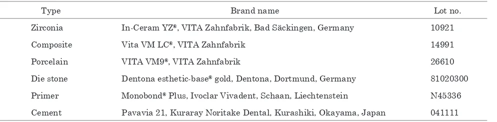
Table 1 Materials used in making crown samples for the occlusal fracture resistance test
Type Brand name Lot no.
Zirconia In-Ceram YZ¨, VITA Zahnfabrik, Bad SŠckingen, Germany 10921
Composite Vita VM LC¨, VITA Zahnfabrik 14991
Porcelain VITA VM9¨, VITA Zahnfabrik 26610
Die stone Dentona esthetic-base¨ gold, Dentona, Dortmund, Germany 81020300
Primer Monobond¨ Plus, Ivoclar Vivadent, Schaan, Liechtenstein N45336
Cement Pavavia 21, Kuraray Noritake Dental, Kurashiki, Okayama, Japan 041111
dificult to produce a test that accurately simulates the
oral environment. Although occlusal fracture resistance evaluation allows the restoration to be constructed and compared to like designs, the limitations of the data have been discussed as being not applicable to real life situations23). This is primarily due to the test
failing to reproduce crown failure as observed clinically, i.e. the mode of fracture differs and loads exceeding maximum recording bite forces are often observed24).
Such forces vary considerably depending on gender and age, but overall the molar region has a higher force25).
The test is useful in carrying out pre-clinical trials of novel materials or designs that are being investigated for future use. Using the material processed into the
deinitive crown shape and bonded to the appropriate
substructure, unlike the uniform samples in laboratory mechanical testing, is suggested as reason enough to employ such testing, rather than relying purely on standard strength tests26). The fact that the material
used to produce the crown for testing has been through a production process, is asymmetrical in shape and made out of more than one material and bonded to a tooth may have an impact on the test results of crown samples compared with standard, evenly shaped samples5).
Similarly the restoration strength may be affected by variables such as veneer thickness, substructure design, cement thickness, properties of the underlying abutment27). Mimicking the oral environment with a
comprehensive in-vitro test environment is dificult to achieve, but primary evaluations such as the occlusal fracture resistance can contribute to developing new techniques and materials28).
Finite element analysis (FEA) has been increasingly used to analyze the stress of different materials and designs saving time and resources and giving initial results for new products or explaining weak points in current ones. In dentistry, FEA has been used to investigate how different materials and restoration shapes interact with the oral cavity in a non-damaging or time consuming way and also overcomes ethical issues of in-vivo testing of new materials29-31).
This study assessed zirconia substructure crowns with both composite and ceramic veneers. The structural integrity of the crowns was assessed by subjecting them to static load and comparing their
load at fracture. FEA was also carried out to assess the stresses generated on the underlying substructure.
MATERIALS AND METHODS
Fracture resistance 1. Samples fabrication
Using the CEREC¨ CAD/CAM system (CEREC inLab,
Sirona Dental Systems, Bensheim, Germany), a zirconia substructure (0.7 mm thick) was designed using a cutback technique and from the opposing dentition. The substructure was then milled from zirconia blocks (In-Ceram YZ¨, VITA Zahnfabrik, Bad SŠckingen, Germany)
and sintered following the manufacturers instructions. Zirconia substructures were divided into two groups (n=10) based on the veneering material to: zirconia/ composite YZ/LC (VITA VM LC¨, VITA Zahnfabrik)
and zirconia/porcelain YZ/VM9 (VITA VM9¨, VITA
Zahnfabrik). A list of the materials used and their lot numbers are detailed in Table 1.
For the YZ/LC group, a light-cured composite veneer was added after the substructures had been shot-blasted with 50 μm Al2O3 and coated using
universal primer (Monobond¨ Plus, Ivoclar Vivadent,
Schaan, Liechtenstein). The YZ/VM9 veneers were produced following the manufacturerÕs guidelines. A silicone matrix (Provil Novo Putty Soft Regular Set, Heraeus Kulzer, Hanau, Germany) was used to produce the veneer overlay in order to make the crowns as consistent as possible. Crowns in both groups were
inished and polished to a clinical standard thickness of
1.2 mm. The crowns were then cemented with Pavavia 21 (Kuraray Noritake Dental, Kurashiki, Okayama, Japan) on stone (Dentona esthetic-base¨ gold, Dentona,
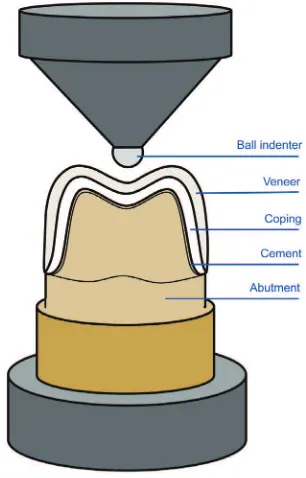
Dortmund, Germany) models before being subjected to load (Fig. 1).
2. Fracture resistance
A universal testing machine (Lloyd LRX universal testing machine, Lloyd Instruments, West Sussex, UK) was used to apply a load through a 4.2 mm diameter steel ball indenter at a crosshead speed of 1 mm/min occlusally in the middle of the crown (fossa) and the maximum load causing crown failure was recorded.
Fig. 1 Illustration of the occlusal fracture resistance test.
[image:4.595.308.543.398.556.2]Fig. 2 Occlusal fracture resistance of Zirconia/Composite and Zirconia/Porcelain crowns.
Table 2 Elastic moduli and PoissonÕs ratios for each material used for the FEA
Material Elastic modulus GPa PoissonÕs ratio
Zirconia 209.3 32) 0.32 32)
Porcelain 66.5 32) 0.21 32)
Composite 4.5 33) 0.3 34)
Die (dentine) 18.6 35) 0.31 35)
Cement 18.6 36) 0.28 36)
Superscript numbers indicate references.
1) Statistical analysis
Results were compared using LeveneÕs test for equal variance followed by WelchÕs t-test at signiicant level (p<0.05) using statistical data analyzing software IBM SPSS version 23 (IBM, Armonk, NY, USA).
Finite element analysis
The program ANSYS 11.0 (ANSYS, Canonsburg, PA, USA) was used to create a 3D crown and subjected to virtual loading to identify where the stresses were distributed in the crown. The CAD/CAM zirconia substructure designed previously was used as a guide when drawing the crown. The veneer was schematically drawn to replicate actual clinical designs.
Different layers were conigured and assigned their
characteristics according to Table 232-36).
Force was applied occlusally on a 4.2 mm diameter
radius in the middle fossa to simulate the applied force in the fracture resistance test. A load value of 500 N was distributed equally at the loading point. Maximum
irst principal stress was chosen to determine the stress
distribution in the structure after applying a virtual load to it, with the color guide showing the stress values in MPa.
RESULTS
Fracture resistance
The occlusal fracture resistance of both groups can be seen in Fig. 2. All samples were tested to failure and the composite veneered zirconia crowns showed an average load at failure of 1,465 N (±350). Although minor veneer chips were observed prior to fracture, in all sample the composite veneer remained bonded to the underlying zirconia substructure The porcelain veneered zirconia crowns showed slightly higher resistance to fracture
1,576 N (±289.2) but with no evidence of a signiicant
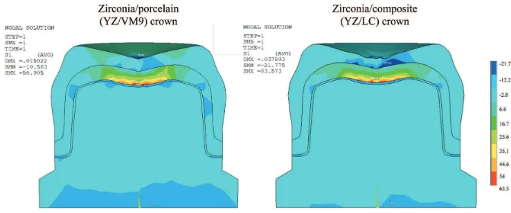
[image:4.595.54.537.635.736.2]Fig. 3 Cross sectional view of stress (MPa) distribution of the zirconia/composite and the zirconia/porcelain veneered crowns and sphere after applying a virtual load of 500 N.
Finite element analysis
Colored deformed structures representing YZ/LC and YZ/VM9 crowns are shown in Fig. 3. The color key shows the highest and lowest stress generated in the 3D crown model after being subjected to virtual load.
When a load of 500 N was applied occlusally in the middle fossa, the crown veneered with a 4.5 GPa stiff composite showed the highest tension point under the loading area in the bottom of the zirconia substructure in the range of 63.6 MPa, peaking at around −21.8 MPa in the composite veneer under the loading area as a compressive stress. These conditions were repeated for the stiffer (65 GPa) ceramic veneered crown, resulting in high tension in the bottom of zirconia at around 50.9 MPa and compressive stress peaking at about −10.6 MPa at the porcelain veneer and cement under the loading zone.
DISCUSSION
In this study, a zirconia-based crown veneered with composite was proposed to overcome some of the drawbacks associated with porcelain veneered zirconia crowns. Such crowns are made with a zirconia substructure and veneered with indirect
light cured composite. The beneits of this system
include biocompatibility and strength of the zirconia substructure and a less abrasive composite veneer that allows ease of handling and intra-oral repair. Properties of zirconia and composites have been investigated in many studies37-42), but few studies were found that
tested the performance of composite veneered zirconia crowns43-45).
One method to investigate the structural integrity of such structures is the occlusal fracture resistance or load-to-failure test, which takes into account the complexity of the crownÕs anatomy and its different
component layers. Such test conigurations and
fabrication processes differ between studies, e.g. using a ball or a bar to apply load5) or the type of the
underlying abutment46). Consequently there is a
variation in results between different investigations47).
Further variables with this testing result from the design and reproducibility of samples. It has been stated that the structure and thickness of the substructure and veneer may affect the fracture resistance of the crowns independent of the mechanical properties of the materials48). Standardizing of samples for this test was
achieved by machining the substructure using CAD/ CAM and using an index to aid the production of the hand-built outer veneer. The crowns were measured
on all sides to conirm they had been fabricated to the
expected full contour before being cemented to the die. The results obtained from an in-vitro laboratory based test cannot be directly applied to the oral environment since there are differences in magnitude and direction of load and surrounding environment. More attention is indicated to produce a test which creates conditions closer to the oral cavity, such as: mimicking the periodontal ligament49), using abutment materials with
elastic moduli close to dentine50), and using rubber sheet
under the indenter to even the stress on the crown51).
The results were measured in Newtons and all tested crowns withstood static loads exceeding 1,000 N without showing any signs of damage or chipping of the composite and porcelain veneers. The composite veneered crowns failed at 1,465.3 N compared to 1,576.4
N for the porcelain veneered crowns with no signiicant difference. This inding is in accordance with other
studies that have concluded that the fracture resistance of indirect composite zirconia restorations showed comparable results to the porcelain veneered zirconia restorations43-45). These igures signiicantly exceed the
maximum bite force recorded in the mouth.
A study by Casson et al.5) tested the fracture load of
10 human extracted teeth mounted in die stone loaded using a bar with crosshead speed of 1 mm/min and recorded an average of 754 N with a standard deviation of 150 N. Taking into account the natural teeth tested in the previous study in a manner similar to the test done
in this study, the composite veneered crowns withstood loads exceeding the natural teeth average of 754 N. A similar study by Zahran et al.52) that tested the fracture
resistance of all-ceramic crowns made out of yttrium-stabilized zirconium oxide and feldspathic ceramic gave comparable results this research. In their test, a 1.5 mm thick crowns of a 0.7 mm zirconia substructure veneered with VM9 feldspathic porcelain (n=10) gave an average fracture resistance to a ball indenter in a crosshead speed of 1 mm/min of 1,459 N (±492) and average of about 1,270 N (±109) for the other tested group of feldespathic crowns (VITA mark II, Vita Zahnfabrik) with a thickness of 1.5 mm. When comparing the Zahran et al. results it can be seen that the composite veneered zirconia crowns withstood higher loads than VITA mark II crowns. Sorrentino et al. evaluated monolithic zirconia molar crowns, and the groups with 1.5 and 1.0 mm thicknesses showed a fracture resistance of 1,554 N (±366.3) and 1,655 N (±314.6) respectively53). These
studies show that porcelain veneered zirconia and monolithic zirconia crowns exhibited fracture resistance exceeding the highest recorded bite forces and ranging between 1,400Ð1,600 N and with a common cohesive mode of fracture.
The inding of this research shows that the
light-cured composite zirconia veneered crowns showed comparable results to other zirconia based crowns but with the advantage of being repairable with the same material.
Intra-oral repair of cohesively fractured ceramic crowns with resin composite can be considered a cost and time effective method with the advantage of maintaining the restoration substructure and therefore protecting the underlying tooth54,55). The alternative is to
replace the crown, removal of which is a dificult process,
and producing an aesthetic replacement. There are some disadvantages associated with repairing ceramic restorations with resin composite, such as possible reduction in both mechanical and optical properties7).
Repairing a fractured composite veneer intraorally with the same material would be less challenging with optimal esthetics when repaired with the same material and shade.
Studies have demonstrated that the maximum bite force was 500 N56) and recommended that any
restoration in the molar area should be able to sustain an occlusal load of about 500 N57) Therefore, when
evaluating crowns in-vitro, it is thought that posterior metal-free restorations should withstand an occlusal force of at least 1,000 N, with the assumption that the mastication forces in the moist oral environment may weaken the restoration by up to half its known fracture resistance force58,59).
FEA has been used to imitate the occlusal fracture resistance test done in this study to show stress points after applying load on those structures. The
virtual schematic crowns do not necessarily relect
the actual samples due to the fabrication process involving different stages mainly by hand60). It was
assumed for this evaluation that there is a good bond
between the different layers in the virtual veneered crowns, regardless of any faults that probably exist in clinical cases. Checking the stress zones is essential in
most application ields since the stress, even if below
failure point, is considered as a major cause of crack propagation and hence of system failure36). After
applying the load to the designed structure, the result can be seen in different ways depending on the type of material and the userÕs investigation. For this study, the intention was to observe the stress generated on the crowns during testing. This virtual test can reveal compressive stress and tensile stress, which are among the causes of ceramic restoration failure61). With the
ceramic veneer, stress was distributed across different levels, the stress being highest under the point where the load was applied. When replaced by composite, higher stresses were generated at the base of zirconia based crowns under the same occlusal load. This
observation matches indings by other studies that
low stiffness veneers pass the load to the substructure material, causing a higher tensile stress in the core that eventually can initiate crack growth through the veneer layer29,60). This is also in accordance with
the results from the fracture resistance test as the composite veneered zirconia crowns fractured at lower loads than the porcelain veneered group. In this study, the composite veneered zirconia crowns showed promising results when compared to the same substructure veneered with porcelain. Further evaluation could be carried out on composite veneers when bonded to structures other than zirconia, preferably with elasticity closer to the composite and dentine to reduce the stress inducing zones between different layers of the crowns. High performance polymers such as PEEK and PEKK could be used for this purpose62).
Further testing should be carried out in conditions that simulate the oral environment, e.g. thermal cyclic loading tests and using chewing simulators, before the results can be considered for clinical application. Also, different composites with different properties could be evaluated along with the bond between the zirconia and composite veneer.
CONCLUSIONS
From this study, the following conclusions can be drawn:
1. Crowns constructed from a zirconia coping and veneered using light-cured composite gave results similar to those veneered with feldspathic
porcelain and showed no statistically signiicant
difference.
REFERENCES
1) Chevalier J, Gremillard L. Ceramics for medical applications: A picture for the next 20 years. J Eur Ceram Soc 2009; 29: 1245-1255.
2) Kosmac T, Oblak C, Jevnikar P, Funduk N, Marion L. Strength and reliability of surface treated Y-TZP dental ceramics. J Biomed Mater Res 2000; 53: 304-313.
3) Guess PC, Zavanelli RA, Silva NRFA, Bonfante EA, Coelho PG, Thompson VP. Monolithic CAD/CAM lithium disilicate versus veneered Y-TZP crowns: comparison of failure modes and reliability after fatigue. Int J Prosthodont 2010; 23: 434-442.
4) Aboushelib MN, de Jager N, Kleverlaan CJ, Feilzer AJ. Effect of loading method on the fracture mechanics of two layered all-ceramic restorative systems. Dent Mater 2007; 23: 952-959.
5) Casson AM, Glyn Jones JC, Youngson CC, Wood DJ. The
effect of luting media on the fracture resistance of a lame
sprayed all-ceramic crown. J Dent 2001; 29: 539-544. 6) Hudson JD, Goldstein GR, Georgescu M. Enamel wear
caused by three different restorative materials. J Prosthet Dent 1995; 74: 647-654.
7) Hammond BD. Critical appraisal. Intraoral repair of fractured ceramic restorations. J Esthet Restor Dent 2009; 21: 275-284.
8) Fischer J, Stawarzcyk B, Trottmann A, Hammerle CH.
Impact of thermal misit on shear strength of veneering
ceramic/zirconia composites. Dent Mater 2009; 25: 419-423.
9) Rues S, Kroger E, Muller D, Schmitter M. Effect of iring
protocols on cohesive failure of all-ceramic crowns. J Dent 2010; 38: 987-994.
10) Choi JE, Waddell JN, Torr B, Swain MV. Pressed ceramics onto zirconia. Part 1: Comparison of crystalline phases
present, adhesion to a zirconia system and lexural strength.
Dent Mater 2011; 27: 1204-1212.
11) Schmitter M, Mueller D, Rues S. Chipping behaviour of all-ceramic crowns with zirconia framework and CAD/CAM manufactured veneer. J Dent 2012; 40: 154-162.
12) Guess PC, Schultheis S, Bonfante EA, Coelho PG, Ferencz JL, Silva NRFA. All-ceramic systems: laboratory and clinical performance. Dent Clin North Am 2011; 55: 333-352. 13) Mitov G, Heintze SD, Walz S, Woll K, Muecklich F, Pospiech
P. Wear behavior of dental Y-TZP ceramic against natural
enamel after different inishing procedures. Dent Mater 2012;
28: 909-918.
14) Stober T, Bermejo JL, Rammelsberg P, Schmitter M. Enamel wear caused by monolithic zirconia crowns after 6 months of clinical use. J Oral Rehabil 2014; 41: 314-322.
15) Piconi C, Burger W, Richter HG, Cittadini A, Maccauro G, Covacci V, Bruzzese N, Ricci GA, Marmo E. Y-TZP ceramics
for artiicial joint replacements. Biomaterials 1998; 19:
1489-1494.
16) Kim HT, Han JS, Yang JH, Lee JB, Kim SH. The effect of low temperature aging on the mechanical property & phase stability of Y-TZP ceramics. J Adv Prosthodont 2009; 1: 113-117.
17) Koutayas SO, Vagkopoulou T, Pelekanos S, Koidis P, Strub JR. Zirconia in dentistry: part 2. Evidence-based clinical breakthrough. Eur J Esthet Dent 2009; 4: 348-380.
18) Sailer I, Pjetursson BE, Zwahlen M, Hammerle CHF. A systematic review of the survival and complication rates of all-ceramic and metal-ceramic reconstructions after an
observation period of at least 3 years. Part II: ixed dental
prostheses. Clin Oral Implants Res 2007; 18: 86-96. 19) Heintze SD, Rousson V. Survival of zirconia-and
metal-supported ixed dental prostheses: a systematic review. Int J
Prosthodont 2010; 23: 493-502.
20) Triwatana P, Nagaviroj N, Tulapornchai C. Clinical
performance and failures of zirconia-based ixed partial
dentures: a review literature. J Adv Prosthodont 2012; 4: 76-83.
21) Hervas-Garcia A, Martinez-Lozano MA, Cabanes-Vila J, Barjau-Escribano A, Fos-Galve P. Composite resins. A review of the materials and clinical indications. Med Oral Patol Oral Cir Bucal 2006; 11: 215-220.
22) Walton JN, Gardner FM, Agar JR. A survey of crown and
ixed partial denture failures: length of service and reasons
for replacement. J Prosthet Dent 1986; 56: 416-421.
23) Isgro G, Addison O, Fleming GJ. Transient and residual stresses induced during the sintering of two dentin ceramics. Dent Mater 2011; 27: 379-385.
24) Kelly JR. Clinically relevant approach to failure testing of all-ceramic restorations. J Prosthet Dent 1999; 81: 652-661. 25) Waltimo A, Kononen M. A novel bite force recorder and
maximal isometric bite force values for healthy young adults. Scand J Dent Res 1993; 101: 171-175.
26) Kelly JR. Perspectives on strength. Dent Mater 1995; 11: 103-110.
27) Scherrer SS, de Rijk WG. The effect of crown length on the fracture resistance of posterior porcelain and glass-ceramic crowns. Int J Prosthodont 1992; 5: 550-557.
28) Preis V, Behr M, Hahnel S, Handel G, Rosentritt M. In vitro failure and fracture resistance of veneered and full-contour zirconia restorations. J Dent 2012; 40: 921-928.
29) Ausiello P, Apicella A, Davidson CL. Effect of adhesive layer properties on stress distribution in composite restorations-a
3D inite element analysis. Dent Mater 2002; 18: 295-303. 30) Magne P. Eficient 3D inite element analysis of dental
restorative procedures using micro-CT data. Dent Mater 2007; 23: 539-548.
31) Thompson MC, Field CJ, Swain MV. The all-ceramic, inlay
supported ixed partial denture. Part 2. Fixed partial denture design: a inite element analysis. Aust Dent J 2011; 56:
302-311.
32) Borba M, de Araujo MD, de Lima E, Yoshimura HN, Cesar PF, Griggs JA, Della Bona A. Flexural strength and failure modes of layered ceramic structures. Dent Mater 2011; 27: 1259-1266.
33) VITAVM LC Working Instructions Manual. In: Co.KG VZHRG, editor. Bad SŠckingen, Germany, 2011.
34) Sakaguchi RL, Powers JM. CraigÕs restorative dental materials. 13thed. Philadelphia: Elsevier Health Sciences; 2012. p. 41.
35) Anusavice KJ, Hojjatie B, Dehoff PH. Inluence of metal
thickness on stress distribution in metal-ceramic crowns. J Dent Res 1986; 65: 1173-1178.
36) Zarone F, Sorrentino R, Apicella D, Valentino B, Ferrari M, Aversa R, Apicella A. Evaluation of the biomechanical behavior of maxillary central incisors restored by means of endocrowns compared to a natural tooth: a 3D static linear
inite elements analysis. Dent Mater 2006; 22: 1035-1044.
37) Guazzato M, Albakry M, Ringer SP, Swain MV. Strength, fracture toughness and microstructure of a selection of all-ceramic materials. Part II. Zirconia-based dental all-ceramics. Dent Mater 2004; 20: 449-456.
38) Manicone PF, Rossi Iommetti P, Raffaelli L. An overview of zirconia ceramics: basic properties and clinical applications. J Dent 2007; 35: 819-826.
39) Gargari M, Gloria F, Cappello A, Ottria L. Strength of
zirconia ixed partial dentures: review of the literature. Oral
Implantol 2010; 3: 15-24.
40) Ferracane JL. Resin composite Ñstate of the art. Dent Mater 2011; 27: 29-38.
41) Ozkurt Z, Kazazoglu E. Zirconia dental implants: A literature review. J Oral Implantol 2011; 37: 367-376.
42) Agustin-Panadero R, Roman-Rodriguez JL, Ferreiroa A,
Sola-Ruiz MF, Fons-Font A. Zirconia in ixed prosthesis. A
literature review. J Clin Exp Dent 2014; 6: 66-73.
43) Taguchi K, Komine F, Fushiki R, Blatz MB, Kamio S, Matsumura H. Fracture resistance of single-tooth implant-supported zirconia-based indirect composite-layered molar restorations. Clin Oral Implants Res 2014; 25: 983-991. 44) Komine F, Taguchi K, Fushiki R, Kamio S, Iwasaki T,
Matsumura H. In vitro comparison of fracture load of implant-supported, zirconia-based, porcelain- and
composite-layered restorations after artiicial aging. Dent Mater J 2014;
33: 607-613.
45) Kamio S, Komine F, Taguchi K, Iwasaki T, Blatz MB, Matsumura H. Effects of framework design and layering material on fracture strength of implant-supported zirconia-based molar crowns. Clin Oral Implants Res 2015; 26: 1407-1413.
46) Yucel MT, Yondem I, Aykent F, Eraslan O. Inluence of the
supporting die structures on the fracture strength of all-ceramic materials. Clin Oral Investig 2012; 16: 1105-1110. 47) Al-Makramani BM, Razak AA, Abu-Hassan MI. Comparison
of the load at fracture of Turkom-Cera to Procera AllCeram and In-Ceram all-ceramic restorations. J Prosthodont 2009; 18: 484-488.
48) Sundh A, Sjogren G. A comparison of fracture strength of yttrium-oxide- partially-stabilized zirconia ceramic crowns with varying core thickness, shapes and veneer ceramics. J Oral Rehabil 2004; 31: 682-688.
49) Soares CJ, Pizi ECG, Fonseca RB, Martins LRM. Inluence
of root embedment material and periodontal ligament simulation on fracture resistance tests. Braz Oral Res 2005; 19: 11-16.
50) Rosentritt M, Plein T, Kolbeck C, Behr M, Handel G. In vitro fracture force and marginal adaptation of ceramic crowns
ixed on natural and artiicial teeth. Int J Prosthodont 2000;
13: 387-391.
51) Tsitrou EA, Helvatjoglu-Antoniades M, van Noort R. A preliminary evaluation of the structural integrity and fracture mode of minimally prepared resin bonded CAD/CAM crowns. J Dent 2010; 38: 16-22.
52) Zahran M, El-Mowafy O, Tam L, Watson PA, Finer Y. Fracture strength and fatigue resistance of all-ceramic molar crowns
manufactured with CAD/CAM technology. J Prosthodont 2008; 17: 370-377.
53) Sorrentino R, Triulzio C, Tricarico MG, Bonadeo G, Gherlone EF, Ferrari M. In vitro analysis of the fracture resistance of CAD-CAM monolithic zirconia molar crowns with different occlusal thickness. J Mech Behav Biomed Mater 2016; 61: 328-333.
54) Han IH, Kang DW, Chung CH, Choe HC, Son MK. Effect of various intraoral repair systems on the shear bond strength of composite resin to zirconia. J Adv Prosthodont 2013; 5: 248-255.
55) Reston EG, Filho SC, Arossi G, Cogo RB, Rocha Cdos S,
Closs LQ. Repairing ceramic restorations: inal solution or
alternative procedure? Oper Dent 2008; 33: 461-466. 56) Floystrand F, Kleven E, Oilo G. A novel miniature bite force
recorder and its clinical application. Acta Odontol Scand 1982; 40: 209-214.
57) Kšrber K, Ludwig K. The maximum bite force as a critical
factor for ixed partial dentures. Dent Labor 1983; 31: 60.
58) Mehl C, Ludwig K, Steiner M, Kern M. Fracture strength of
prefabricated all-ceramic posterior inlay-retained ixed dental
prostheses. Dent Mater 2010; 26: 67-75.
59) Tinschert J, Natt G, Mautsch W, Augthun M, Spiekermann H. Fracture resistance of lithium disilicate-, alumina-, and
zirconia-based three-unit ixed partial dentures: a laboratory
study. Int J Prosthodont 2001; 14: 231-238.
60) Mollers K, Patzold W, Parkot D, Kirsten A, Guth JF, Edelhoff
D, Fischer H. Inluence of connector design and material
composition and veneering on the stress distribution of
all-ceramic ixed dental prostheses: a inite element study. Dent
Mater 2011; 27: 171-175.
61) Mollers K, Parkot D, Kirsten A, Guth JF, Edelhoff D, Fischer
H. Inluence of tooth mobility on critical stresses in all-ceramic inlay-retained ixed dental prostheses: a inite element study.
Dent Mater 2012; 28: 146-151.
62) Fuhrmann G, Steiner M, Freitag-Wolf S, Kern M. Resin bonding to three types of polyaryletherketones
(PAEKs)-durability and inluence of surface conditioning. Dent Mater