GAMETE BIOLOGY
Isolation and expression of the human gametocyte-specific factor
1 gene (
GTSF1
) in fetal ovary, oocytes, and preimplantation
embryos
John Huntriss1 &Jianping Lu1&Karen Hemmings1&Rosemary Bayne2& Richard Anderson2&Anthony Rutherford3&Adam Balen3&Kay Elder4& Helen M. Picton1
Received: 5 February 2016 / Accepted: 16 August 2016 / Published online: 19 September 2016
#The Author(s) 2016. This article is published with open access at Springerlink.com
Abstract
PurposeGametocyte-specific factor 1 has been shown in oth-er species to be required for the silencing of retrotransposons via the Piwi-interacting RNA (piRNA) pathway. In this study, we aimed to isolate and assess expression of transcripts of the gametocyte-specific factor 1 (GTSF1) gene in the human fe-male germline and in preimplantation embryos.
MethodsComplementary DNA (cDNA) libraries from hu-man fetal ovaries and testes, huhu-man oocytes and preimplanta-tion embryos and ovarian follicles isolated from an adult ovar-ian cortex biopsy were used to as templates for PCR, cloning and sequencing, and real time PCR experiments of GTSF1 expression.
Results GTSF1cDNA clones that covered the entire coding region were isolated from human oocytes and preimplantation
embryos.GTSF1mRNA expression was detected in archived cDNAs from staged human ovarian follicles, germinal vesicle (GV) stage oocytes, metaphase II oocytes, and morula and blastocyst stage preimplantation embryos. Within the adult female germline, expression was highest in GV oocytes.
GTSF1mRNA expression was also assessed in human fetal ovary and was observed to increase during gestation, from 8 to 21 weeks, during which time oogonia enter meiosis and pri-mordial follicle formation first occurs. In human fetal testis,
GTSF1expression also increased from 8 to 19 weeks.
Conclusions To our knowledge, this report is the first to de-scribe the expression of the human GTSF1gene in human gametes and preimplantation embryos.
Keywords Oocyte . Ovarian follicle .GTSF1. cue110 . FAM112B. piRNA
Introduction
The analysis of genes that are expressed in murine germ cells or whole ovaries has led to the identification of a novel gene, gametocyte-specific factor 1 (Gtsf1), also known as the com-putationally obtained undifferentiated and/or embryonic stem cell-specific 110 gene (Cue110) [1–3].Gtsf1encodes a 167-amino acid protein that is a member of the Uncharacterized Protein Family 0224 (UPF0224).
Three recent reports have revealed that in Drosophila, gametocyte-specific factor 1 (DmGTSF1), is essential for P-element-induced wimpy testis (PIWI)-interacting RNA (piRNA)-mediated transcriptional repression and histone H3K9me3-mediated repression of transposons and their neighboring genes in the ovary [4–6]. DmGTSF1 interacts with Piwi via its C-terminal tail [4] and the two CHHC zinc-CapsuleThe expression of transcripts of the human
Gametocyte-Specific Factor 1 (GTSF1) gene were assessed in fetal gonads, the female germline and in preimplantation embryos.
Electronic supplementary materialThe online version of this article (doi:10.1007/s10815-016-0795-0) contains supplementary material, which is available to authorized users.
* John Huntriss j.huntriss@leeds.ac.uk
1 Division of Reproduction and Early Development, Leeds Institute of
Cardiovascular and Metabolic Medicine, Clarendon Way, University of Leeds, Leeds LS2 9JT, UK
2 MRC Centre for Reproductive Health, University of Edinburgh,
Queen’s Medical Research Institute, Edinburgh EH16 4TJ, UK
3
Leeds Centre for Reproductive Medicine, Leeds Teaching Hospital NHS Trust, Seacroft Hospital, York Road, Leeds LS14 6UH, UK
finger motifs within the DmGTSF1 protein are required for its activity [5]. In the mouse, theGtsf1gene is essential for sper-matogenesis and has been revealed to function in transposon suppression in the mouse testes since increased expression of the long interspersed nucleotide element (line 1) and the intracisternal A-particle (IAP) retrotransposons were observed to occur inGtsf1-null male mice [7]. The same study showed thatGtsf1-null male mice are sterile due to massive apoptosis of their germ cells and the cessation of meiotic progression before the zygotene stage. In humans, these observations are reflected by the observation that the testes of high infertility risk (HIR) cryptorchidism patients have reducedGTSF1 ex-pression and corresponding over-exex-pression of LINE1 (L1) retrotransposons [8].GTSF1has also been identified as a bo-vine spermatozoal transcript [9]. Elsewhere,GTSF1transcript expression has been observed to be up-regulated in certain leukemias and in lymphomas [10–14].
The precise role of theGtsf1gene in the mammalian female germline is less clear sinceGtsf1-null female mice were ob-served to be fertile [7]. The expression ofGtsf1transcripts has been detected in adult mouse primordial and primary follicles, whilst Gtsf1 protein has been detected in germ cell clusters and primordial follicles in newborn ovaries and in the oocytes of all follicle stages [3]. Significantly, Gtsf1was identified among the transcripts that were down-regulated in ovaries from mice deficient for theNobox gene [2,3]. Whilst not essential for fertility, a role forGtsf1is implied during oogen-esis and/or early embryonic development since other genes, with diverse functions during oogenesis, are also down-regulated inNobox-deficient mice [15].
To our knowledge, this is the first report to describe cloning and expression analysis ofGTSF1transcripts in human fetal ovaries and testis, adult human ovarian follicles, GVand meta-phase II oocytes, and in preimplantation embryos.
Materials and methods
Samples
Human fetal ovaries and testes were obtained with in-formed consent, according to published methods [16]. Ethical approval was from the Lothian Research Ethics Committee (study code LREC 08/S1101/1). Briefly, human fetal gonads (gestations between 8 and 21 weeks) were obtained following medical termination of pregnancy and were subsequently snap-frozen and stored at−80 °C. RNA was extracted using the Qiagen RNeasy Mini Kit (14 weeks gestation onwards) or Qiagen RNeasy Micro Kit (8– 12 weeks gestation) (Qiagen, Crawley, UK) with 500 ng RNA used for first-strand cDNA synthesis using the Superscript Vilo Reverse Transcriptase Master Mix (Life Technologies, Paisley, UK).
Dynal lysis buffer (Life Technologies, Ltd). All samples were obtained after informed consent under ethically ap-proved protocols at the LGI, which were licensed in the UK by the Human Fertilisation and Embryology Authority.
Reverse transcription and cDNA amplification
Ovarian follicle samples, single embryos, and oocytes were collected and lysed at 80 °C in Dynal lysis buffer (Life Technologies, Ltd) and cDNA libraries were generated as pre-viously described [23]. For the ovarian follicle samples as used in Fig.1a, amplified cDNAs were generated from (i) pooled primordial follicles (n= 28), (ii) primordial/early pri-mary follicles (n= 45), (iii) primary follicles (n= 7), and (iv), secondary stage follicles (n = 7).
Messenger RNA was extracted from preimplantation embryos with oligo-dT Dynabeads, and cDNA was generated using an adaptation of existing cDNA amplification protocols, using 1 μg each of primer (primer 1: 5′
-aaacgacggccagtgaattgtaatacgactcactatagggcgct24-3′and prim-er 2: 5′-aagcagtggtatcaacgcagagtacgcggg-3′) and Superscript II RNAseH- Reverse Transcriptase (Life Technologies, Ltd) and associated reagents with incubation for 2 h at 42 °C. The cDNA was amplified by PCR using an additional 1μg of each primer, 2μl 50× Advantage 2 Polymerase (BD Clontech), in a thermal cycler for 32 cycles of 95 °C for 45 s, 65 °C for 6 min 45 s in a total volume of 50μl.
Isolation of GTSF1 transcripts and expression analysis
Expression analysis forGTSF1was performed using 0.3μl of amplified cDNA in a 12.5μl reaction volume using BIOTAQ (BIOLINE).GTSF1primers (Table1) were designed using the Primer3 programme (http://primer3.ut.ee/) and derived from human sequences found in GenBank (GTSF1
AK098819; GAPDHAF261085, NM_144594.2). PCR was performed for 32 cycles for 45 s at each step of 94, 60, and 72 °C. The housekeeping gene (glyceraldehyde-3-phosphate
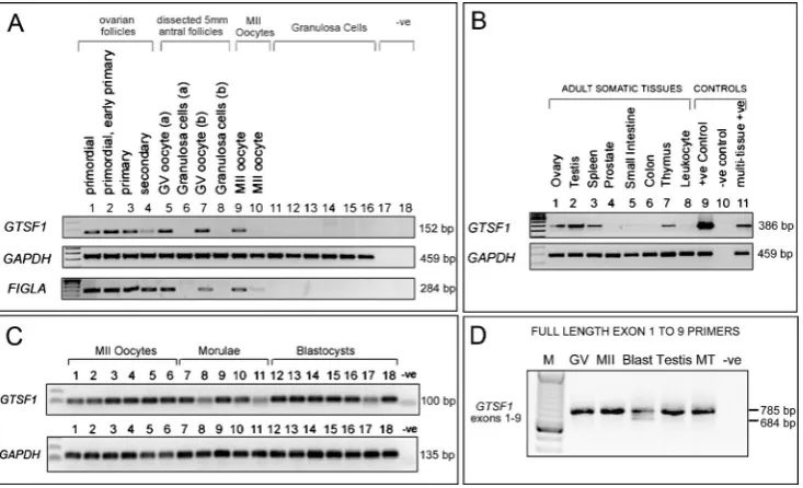
Fig. 1 Expression ofGTSF1transcripts in the human female germline, preimplantation embryos, and adult tissues.aExpression ofGTSF1 (primers 7 F and 9R2: Size = 152 base pairs),GAPDH, and FIGLA gene transcripts by reverse transcription RT-PCR in amplified cDNA samples derived from human ovarian follicles, oocytes, and granulosa cells. TheFIGLAPCR demonstrates the presence of oocyte cDNA in the ovarian follicle samples. Lane (1) Primordial follicles (n= 28 pooled follicles), (2) pooled primordial/early primary follicles (n= 45), (3) pooled primary follicles (n= 7), and (4) pooled secondary follicles (n= 7). Lanes (5) to (8) are cDNAs generated from two dissected 5 mm antral follicles (follicles a and b). Lane (5) Denuded single human GV stage oocytes isolated from the compact cumulus-enclosed oocyte complexes of a 5 mm non-luteinised antral follicle (follicle a), lane (6) granulosa cells isolated from follicle a. Lane (7) Denuded single human GV stage oocytes isolated from the compact cumulus-enclosed oocyte complexes of a 5 mm nonluteinised antral follicle (follicle b), lane 8 granulosa cells isolated from follicle b. Lanes (9) and (10) single MII oocytes, lanes 11 to 16 cDNA from cumulus granulosa cells isolated from
[image:3.595.116.483.310.533.2]dehydrogenase)GAPDH was used as a positive control to demonstrate that the cDNA libraries that were generated from each sample were successful;GAPDHprimers used in Fig.1 a, bwere taken from Weisenberger et al. 2002 [24]. The factor in the germline alpha (FIGLA) primers was from Huntriss et al. 2002 [17]. Products were run on 1.2 % agarose gels and visualised using ethidium bromide with reference to 100 base pair DNA size markers (Life Technologies, Ltd). Expression of the humanGTSF1 gene was also analysed using a range of normalized cDNA samples derived from various human tissues (Clontech MTC panels, Clontech).
Sequencing
GTSF1PCR products that were obtained using various primer combinations were cloned into TOPO TA (Life Technologies). Primer sequences are given in Table 1. The M13 primer-amplified PCR products were sequenced at the Biomolecular Analysis Facility, University of Leeds. Sequences of PCR products were obtained in both directions and were identified by the Basic Local Alignment Search Tool (BLAST) http://www.ncbi.nlm.nih.gov/BLAST.
Real-time PCR
The real-time PCR reaction data was collected using an ABI PRISM 7900HT Real-Time PCR system using the SYBR Green method (Applied Biosystems). PCR primer sequences are described in Table1. The 25μl reaction mix contained 2 ng cDNA, 10 pmol of each primer. The PCR protocol was denaturation (95 °C for 10 min), amplification (94 °C 10 s,
60 °C 30 s, and 72 °C 30 s for 45 cycles). For quantitative assessment ofGTSF1expression in the female germline, real-time PCR was performed on cDNA derived from primordial follicles, primordial/early primary follicles, primary follicles, secondary follicles, pooled cDNA from germinal vesicle (GV) stage oocytes (n= 8), pooled metaphase II oocytes (n= 8), and commercially available ovary and testis RNA (FirstChoice RNA, Ambion). Each sample was assessed in triplicate. Data are expressed as a percentage of the housekeeping gene
GAPDHusing the formula 2−ΔCT× 100, in which CT is the threshold cycle number. For real-time PCR analysis ofGTSF1
expression in human fetal ovary and testis experiments were performed as described previously [25], with the equivalent of approximately 2.5 ng of cDNA used per reaction in 10 μl reaction volumes. Significant changes in expression across gestation were determined by one-way ANOVA with Tukey’s Multiple Comparison Test (GraphPadPrism 5.0 soft-ware). Due to the limiting amounts of cDNA that were avail-able for ovarian follicle samples and human fetal gonads, we were unfortunately unable to assess further housekeeping genes across this series of samples as controls.
Results
Isolation and expression of GTSF1
A partial sequence of theGTSF1gene was originally isolated during differential display analysis from cDNAs derived from staged human ovarian follicles (J Huntriss, D Miller, HM Picton unpublished data). BLAST searches with this sequence, that were performed prior to the release of the GSTF1gene
Table 1 PCR primers for
conventional and real-time PCR Gene Primer name Sequence 5′to 3′
GTSF1 Exon1F GGAGGAAGGTGACTGTGAGG
Exon2F CACTTGGATTCAGCTTCTTC
Exon3F GACCCTGAGAAGCTATTGCA
Exon7F CCCTGCGAGCAACATAGTTA
Exon7R TAACTATGTTGCTCGCAGGG
Exon9R CTGTATCAAAGGTTTATTTGGAAGC
GTSF1real-time F ATTCAGCTTCTTCATTTCCAACA GTSF1real-time R CCTGATTTGATGGTTTTTGTCAT
FIGLA FIGLAF1* GATAAAAAATCTCAACCGTGG
FIGLAR1* CCCTCCTCTTCTTTCTTC GAPDH GAPDHF§ ACGGGAAGCTCACTGGCATGGC
GAPDHR§ TCTTACTCCTTGGAGGCCATGTAGG GAPDHreal-time F TTGTCAAGCTCATTTCCTGGTAT GAPDHreal-time R TCTCTCTTCCTCTTGTGCTCTTG
*FIGLAprimers are from Huntriss et al. [17]
§
sequence (for any species) in any gene database, initially iden-tified a 100 % match over 138 nucleotides of readable se-quence to the human sese-quences AK057504 and AK098819, both testis-derived transcripts. On translation, the partial se-quence exhibited similarity to the Xenopus D7 oocyte protein that is represented by P13007 and NP_001081517 protein se-quences. The human sequence was chosen for further study by virtue of the fact that it likely represented a novel human oo-cyte transcript with sequence similarity to the Xenopus D7 protein gene. Analysis by reverse transcription (RT)-PCR with gene-specific primers encompassing the partial sequence from AK057504 in samples derived from the adult human female germline indicated a pattern of expression consistent with be-ing derived from oocytes (Fig. 1a). Figure 1a shows that amplicons were generated in ovarian follicle cDNAs (primor-dial through to secondary stages), germinal vesicle-stage (GV) oocytes, and metaphase II (MII) oocytes, and therefore in the same samples that we detected transcripts ofFIGLA, another transcript observed in oocytes [17].
Subsequent BLAST searches identified the partial sequence as a fragment of novel gene,FAM112B(family with sequence similarity 112 member B) mapping to chromosome12q13.2. TheFAM112Bgene encodes a member of an uncharacterized protein family containing a conserved protein domain UPF0224. The gene has been more recently assigned the approved name Gametocyte-specific factor 1 and symbol (GTSF1), (GeneID: 121355, Ensembl gene ENSG00000170627). We performed further characterisation of the expression ofGTSF1 using primers designed from exons predicted in currentGTSF1
mRNA sequence NM_144594.2 and the related testis-derived mRNA sequences (AK057504 and AK098819) that pre-dated the NM_144594.2 sequence submission. Expression ofGTSF1
was examined across a range of normalized human cDNA sam-ples utilizing primers from exons 3 and 7 of NM_144594.2 (Fig.1b) and revealed expression in the ovary, testis, spleen thymus, and in the positive controls (cDNA from pooled GV oocytes (lane 9) and mRNA from multiple-tissues (lane 11). Expression ofGTSF1transcripts was also assessed in a series of individual MII oocytes and in preimplantation embryos (Fig.1c).GTSF1expression was observed in all tested samples: six additional single human MII oocytes, five morulae, and sev-en blastocysts. A PCR assay covering the sev-entire coding region was designed upon the release of the NM_144594.2 sequence, utilizing primers designed fromGTSF1exons 1 and 9. This assay amplified the appropriate-sized PCR product in samples of pooled GV oocytes, pooled MII oocytes, pooled blastocysts, testis, and mixed human adult tissues (Fig.1d). In addition, a second smaller transcript was observed in blastocysts.
GTSF1 gene and protein sequence
HumanGTSF1gene cDNA clones that were obtained from human oocytes and blastocysts were sequenced and data is
shown in Supplemental Figure 1. This experimentally-defined transcript sequence spans the entire coding region se-quence from exon 1 to 9 and the isolated sese-quence, and its translation, are in full agreement with the sequences NM_144594.2 and NP_653195 and the gene structure as rep-resented by Ensembl GTSF1-001 splice variant that contains 9 exons (Transcript ID: ENST00000305879). The two CHHC zinc-finger domains (TRM13/UPF0224_CHHC_Znf_dom) are identified in Supplemental Figure 1. In blastocysts, the smaller transcript identified in Fig.1d, that lacks exon 3, most likely represents the GTSF1-004 transcript (Transcript ID: OTTHUMT00000406189), a processed transcript that has no protein product and matches to ESTs (602638807 F1, 602552556 F1) that are derived from mucoepidermoid carcino-ma and embryonal carcinocarcino-ma, respectively. TheGTSF1cDNA sequences that were isolated from the PCR products shown in Fig. 1d, were subsequently aligned on the browser (Supplemental Figure2) using the BLAT tool on the UCSC browser (https://genome.ucsc.edu/FAQ/FAQblat.html). Shown are the blastocyst-specificGTSF1transcript variant lacking ex-on 3 (Track 1), theGTSF1transcript sequence as isolated from oocytes, blastocysts and all other stages of oogenesis and pre-implantation development (Track 2) and the UCSC gene (com-piled from RefSeq, GenBank and other sources-Track 3).
Real-time PCR analysis of
GTSF1
expression
in human fetal gonads and the adult female germline
(Fig.2b). For comparison, cDNAs derived from adult human ovary and testis were included.GTSF1expression was detect-ed in all ovarian follicles, and germinal vesicle-stage oocytes and metaphase II oocytes with the highest expression relative to GAPDH observed in germinal vesicle-stage oocytes. Finally, real-time PCR was performed using the same range of cDNA samples from adult human tissue as shown in Fig.1b to reveal that expression ofGTSF1 was highest in the testis (Fig.2c).
Conclusions
In this report, we have described the expression of the human
GTSF1gene from cDNAs derived from fetal gonads, adult human ovarian follicles, GV oocytes, metaphase II oocytes, and preimplantation embryos. The mainGTSF1transcript iso-lated from oocytes and preimplantation embryos corresponds to the GTSF1-001 transcript variant ofGTSF1that encodes a 167 amino acid protein.GTSF1is a highly conserved gene that maps to 12q13.2 and has 91 % identity between the
mouse and human proteins in agreement with earlier studies [1,3,26], with the high conservation indicative of a common function across species.
In cDNAs derived from ovarian follicles that were isolated from the adult ovary, transcripts ofGTSF1are detected from the primordial follicle stage and are expressed through to the metaphase II oocyte stage. Real-time PCR revealed that highest expression was observed in GV oocytes, with a sub-sequent decrease in MII oocytes, in agreement with data for mouse Gtsf1 gene transcripts (http://www.ncbi.nlm.nih. gov/geoprofiles/29792642). Our PCR expression data reveals that GTSF1 is also expressed during human preimplantation development, being detected in late preimplantation embryos. These observations indicate that expression is activated after zygotic gene activation, and this was supported by our observation of a blastocyst-specific tran-script variant ofGTSF1lacking exon 3. Our expression data for mature oocytes and preimplantation embryos are consis-tent with that described in the Human Embryo Resource HumER [27]. Our expression data for somatic tissues is con-sistent with microarray and RNAseq data from other sources
Fig. 2 Real-time PCR assessment ofGTSF1expression. Real-time PCR assessment ofGTSF1expression is expressed as a percentage of the housekeeping geneGAPDHand using primersGTSF1real-time F and R andGAPDHreal-time F and R.aGTSF1expression in human fetal ovaries at different stages of gestation (total tested range was between 8 and 21 weeks) that were grouped into 8–11 weeks, 14–16 weeks, and 17– 21 weeks of gestation,n= 4–5 samples per group.Barsindicate mean ± sem.bGTSF1expression in human fetal testis at different stages of gestation (total tested range was between 8 and 19 weeks) that were grouped into 8–9 weeks, 14–16 weeks, and 17–19 weeks of gestation,
[image:6.595.84.513.52.334.2]that indicate highest expression of theGTSF1transcripts with-in adult tissues occurs with-in the testis (http://biogps.org/ and http://www.ebi.ac.uk/). Our expression data for oocytes and preimplantation embryos is consistent with microarray data from http://www.ncbi.nlm.nih.gov/geoprofiles(geoprofile IDs 74253766 and 52847566). We were unable to perform analysis of the expression GTSF1 protein in our laboratory; however, GTSF1 protein expression has been described by the Human Protein Atlas ([28]: http://www.proteinatlas. org/ENSG00000170627-GTSF1/tissue). In agreement with our data for theGTSF1transcript, this resource reveals that GTSF1 protein (and RNA) expression is largely exclusive to the male and female reproductive tissues. Protein expression is described as being medium in ovarian follicles (but undetectable in ovarian stroma) enriched in the testis, but weakly expressed or undetectable in any other adult human tissues.
Here, we have reported for the first time the elevation of expression ofGTSF1gene transcripts at the time of entry into meiosis and subsequent primordial follicle formation in the human fetal ovary. This may suggest a particular requirement forGTSF1during ovarian programming and early oogenesis and also in GVoocytes in particular. In the male germ line, there is now clear evidence that Piwi-mediated retrotransposon sup-pression is essential for fertility [29–33], and this includes a requirement forGtsf1in this process [7]. Failure to suppress retrotranposons may affect genomic stability and lead to disease [34–36], and retrotransposons are abundant in germ cells and preimplantation embryos [37–42]. In contrast to the infertility observed inGtsf1-null male mice,Gtsf1-null female mice were observed to be fertile with no histological abnormalities in the ovaries [7]. Retrotransposons are known to be silenced in mu-rine primordial ovarian follicles via a piRNA-dependent mech-anism, and this is believed to occur in nuage-like structures in the oocytes of primordial follicles [43]. However, mice with nuage-mutant primordial follicles defective forMvh,Mili, or
Gaszremain fertile, despite elevated transposon expression [43]. These studies, which reflect the observations of male-specific sterility vs. female fertility inGtsf1-null mice [7], sug-gest that the PIWI-pathway in isolation may not be critical for female fertility. Retrotransposon suppression, at least that which is critical for female fertility, may be regulated in the mamma-lian female germ line by Meiosis arrest female 1 (MARF1) and accordingly,Marf1mutant females are infertile [44,45].
We acknowledge that there are a number of shortcomings in our study. Firstly, for some of the samples, particularly the hu-man ovarian follicles and fetal gonads, we were unable to obtain real-time PCR data for additional housekeeping genes across all stages because of the extremely limiting amounts of cDNA that were available to us. Accordingly, the real-time expression data is only normalised to one housekeeping gene, GAPDH. Secondly, we were only able to obtain ovarian follicles from a single patient for this study and therefore, we are not able to confirm whether the GTSF1 transcript expression patterns
observed here are representative of the patterns that would be observed in a wider range of samples from different patients.
Currently, the role ofGTSF1during mammalian oogenesis remains unclear. The function ofGTSF1/Gtsf1has also been investigated by bioinformatic analysis which has revealed that the two U11-48 K-like CHHC-type zinc finger domains (Pfam: PF05253) within the human GTSF1 protein are found in spliceosomal proteins and tRNA modifying enzymes are predicted to have an RNA binding function [26,46]. Indeed, the CHHC zinc-finger domain has been observed to specifi-cally bind the 5′ splice site of U12-type introns in spliceosomal U11-48 K proteins [46]. We hypothesize how-ever that althoughGtsf1appears dispensable for female fertil-ity in mice [7], the retention of the expression ofGTSF1/Gtsf1
in the female germline across several species including mouse [1,3], human (this report), and bovine and ovine oocytes (H.M.Picton, J.Huntriss, J.Lu, G.Liperis- unpublished data, [47,48]) is indicative of a retained function in the mammalian oocyte and/or early embryo. Of particular interest, the abun-dance of the GTSF1 protein in murine MII oocytes was ob-served to be significantly reduced upon postovulatory-ageing [49]; however, further experiments are required to understand the role of this gene in the mammalian oocyte.
Acknowledgments This work was supported by the Medical Research Council (Grant Nos. G0800250, G0801261, G1100357, and MR/ K020501/1).
Compliance with ethical standards
Conflict of interest The authors declare that they have no conflict of interest.
Open AccessThis article is distributed under the terms of the Creative C o m m o n s A t t r i b u t i on 4. 0 I n t e r n a t i o n a l L i c e n s e ( h t t p : / / creativecommons.org/licenses/by/4.0/), which permits unrestricted use, distribution, and reproduction in any medium, provided you give appropriate credit to the original author(s) and the source, provide a link to the Creative Commons license, and indicate if changes were made.
References
1. Yoshimura T, Miyazaki T, Toyoda S, Miyazaki S, Tashiro F, Yamato E, et al. Gene expression pattern of Cue110: a member of the uncharacterized UPF0224 gene family preferentially expressed in germ cells. Gene Expr Patterns. 2007;8:27–35.
2. Choi Y, Qin Y, Berger MF, Ballow DJ, Bulyk ML, Rajkovic A. Microarray analyses of newborn mouse ovaries lacking Nobox. Biol Reprod. 2007;77:312–9.
3. Krotz SP, Ballow DJ, Choi Y, Rajkovic A. Expression and lo-calization of the novel and highly conserved gametocyte-specific factor 1 during oogenesis and spermatogenesis. Fertil Steril. 2009;91:2020–4.
5. Ohtani H, Iwasaki YW, Shibuya A, Siomi H, Siomi MC, Saito K. DmGTSF1 is necessary for Piwi-piRISC-mediated transcriptional transposon silencing in the Drosophila ovary. Genes Dev. 2013;27: 1656–61.
6. Muerdter F, Guzzardo PM, Gillis J, Luo Y, Yu Y, Chen C, et al. A genome-wide RNAi screen draws a genetic framework for transpo-son control and primary piRNA biogenesis in Drosophila. Mol Cell. 2013;50:736–48.
7. Yoshimura T, Toyoda S, Kuramochi-Miyagawa S, Miyazaki T, Miyazaki S, Tashiro F, et al. Gtsf1/Cue110, a gene encoding a protein with two copies of a CHHC Zn-finger motif, is involved in spermatogenesis and retrotransposon suppression in murine tes-tes. Dev Biol. 2009;335:216–27.
8. Hadziselimovic F, Hadziselimovic NO, Demougin P, Krey G, Oakeley E. Piwi-Pathway Alteration Induces LINE-1 Transposon Derepression and Infertility Development in Cryptorchidism. Sex Dev. 2015;9:98–104.
9. Card CJ, Anderson EJ, Zamberlan S, Krieger KE, Kaproth M, Sartini BL. Cryopreserved bovine spermatozoal transcript profile as revealed by high-throughput ribonucleic acid sequencing. Biol Reprod. 2013;88:49.
10. Becker H, Marcucci G, Maharry K, Radmacher MD, Mrozek K, Margeson D, et al. Mutations of the Wilms tumor 1 gene (WT1) in older patients with primary cytogenetically normal acute myeloid leukemia: a Cancer and Leukemia Group B study. Blood. 2010;116:788–92.
11. van Kester MS, Borg MK, Zoutman WH, Out-Luiting JJ, Jansen PM, Dreef EJ, et al. A meta-analysis of gene expression data iden-tifies a molecular signature characteristic for tumor-stage mycosis fungoides. J Invest Dermatol. 2012;132:2050–9.
12. Boerkamp KM, van der Kooij M, van Steenbeek FG, van Wolferen ME, Groot Koerkamp MJ, van Leenen D, et al. Gene expression profiling of histiocytic sarcomas in a canine model: the predisposed flatcoated retriever dog. PLoS One. 2013;8:e71094.
13. Litvinov IV, Cordeiro B, Huang Y, Zargham H, Pehr K, Dore MA, et al. Ectopic expression of cancer-testis antigens in cutaneous T-cell lymphoma patients. Clin Cancer Res. 2014;20:3799–808. 14. Litvinov IV, Netchiporouk E, Cordeiro B, Dore MA, Moreau L,
Pehr K, et al. The use of transcriptional profiling to improve per-sonalized diagnosis and management of Cutaneous T-Cell Lymphoma (CTCL). Clin Cancer Res. 2015;21:2820–9.
15. Rajkovic A, Pangas SA, Ballow D, Suzumori N, Matzuk MM. NOBOX deficiency disrupts early folliculogenesis and oocyte-specific gene expression. Science. 2004;305:1157–9.
16. Bayne RAL, Kinnell HL, Coutts SM, He J, Childs AJ, Anderson RA. GDF9 is transiently expressed in oocytes before follicle for-mation in the human fetal ovary and is regulated by a novel NOBOX transcript. PLoS One. 2015;10:e0119819.
17. Huntriss J, Gosden R, Hinkins M, Oliver B, Miller D, Rutherford AJ, et al. Isolation, characterization and expression of the human Factor In the Germline alpha (FIGLA) gene in ovarian follicles and oocytes. Mol Hum Reprod. 2002;8:1087–95.
18. Huntriss J, Hinkins M, Picton HM. cDNA cloning and expression of the human NOBOX gene in oocytes and ovarian follicles. Mol Hum Reprod. 2006;12:283–9.
19. Wynn P, Picton HM, Krapez JA, Rutherford AJ, Balen AH, Gosden RG. Pretreatment with follicle stimulating hormone promotes the numbers of human oocytes reaching metaphase II by in-vitro mat-uration. Hum Reprod. 1998;13:3132–8.
20. Houghton FD, Hawkhead JA, Humpherson PG, Hogg JE, Balen AH, Rutherford AJ, et al. Non-invasive amino acid turnover pre-dicts human embryo developmental capacity. Hum Reprod. 2002;17:999–1005.
21. Ghassemifar MR, Eckert JJ, Houghton FD, Picton HM, Leese HJ, Fleming TP. Gene expression regulating epithelial intercellular
junction biogenesis during human blastocyst development in vitro. Mol Hum Reprod. 2003;9:245–52.
22. Tay JI, Rutherford AJ, Killick SR, Maguiness SD, Partridge RJ, Leese HJ. Human tubal fluid: production, nutrient composition and response to adrenergic agents. Hum Reprod. 1997;12:2451–6. 23. Huntriss JD, Hemmings KE, Hinkins M, Rutherford AJ, Sturmey RG, Elder K, et al. Variable imprinting of the MEST gene in human preimplantation embryos. Eur J Hum Genet. 2013;21:40–7. 24. Weisenberger DJ, Velicescu M, Preciado-Lopez MA, Gonzales FA,
Tsai YC, Liang G, et al. Identification and characterization of alter-natively spliced variants of DNA methyltransferase 3a in mamma-lian cells. Gene. 2002;298:91–9.
25. Bayne RA, Eddie SL, Collins CS, Childs AJ, Jabbour HN, Anderson RA. Prostaglandin E2 as a regulator of germ cells during ovarian development. J Clin Endocrinol Metab. 2009;94:4053–60. 26. Andreeva A, Tidow H. A novel CHHC Zn-finger domain found in spliceosomal proteins and tRNA modifying enzymes. Bioinformatics. 2008;24:2277–80.
27. Vassena R, Boue S, Gonzalez-Roca E, Aran B, Auer H, Veiga A, et al. Waves of early transcriptional activation and pluripotency program initiation during human preimplantation development. Development. 2011;138:3699–709.
28. Uhlén M, Fagerberg L, Hallström BM, Lindskog C, Oksvold P, Mardinoglu A, et al. Proteomics. Tissue-based map of the human proteome. Science. 2015;347(6220):1260419.
29. Ma L, Buchold GM, Greenbaum MP, Roy A, Burns KH, Zhu H, et al. GASZ is essential for male meiosis and suppression of retrotransposon expression in the male germline. PLoS Genet. 2009;5:e1000635.
30. Shoji M, Tanaka T, Hosokawa M, Reuter M, Stark A, Kato Y, et al. The TDRD9-MIWI2 complex is essential for piRNA-mediated retrotransposon silencing in the mouse male germline. Dev Cell. 2009;17:775–87.
31. Frost RJ, Hamra FK, Richardson JA, Qi X, Bassel-Duby R, Olson EN. MOV10L1 is necessary for protection of spermatocytes against retrotransposons by Piwi-interacting RNAs. Proc Natl Acad Sci U S A. 2010;107:11847–52.
32. Reuter M, Berninger P, Chuma S, Shah H, Hosokawa M, Funaya C, et al. Miwi catalysis is required for piRNA amplification-independent LINE1 transposon silencing. Nature. 2011;480:264–7. 33. Pastor WA, Stroud H, Nee K, Liu W, Pezic D, Manakov S, et al. MORC1 represses transposable elements in the mouse male germline. Nat Commun. 2014;5:5795.
34. Burns KH, Boeke JD. Human transposon tectonics. Cell. 2012;149: 740–52.
35. Shukla R, Upton KR, Munoz-Lopez M, Gerhardt DJ, Fisher ME, Nguyen T, et al. Endogenous retrotransposition activates oncogenic pathways in hepatocellular carcinoma. Cell. 2013;153:101–11. 36. Levin HL, Moran JV. Dynamic interactions between transposable
elements and their hosts. Nat Rev Genet. 2011;12:615–27. 37. Kano H, Godoy I, Courtney C, Vetter MR, Gerton GL, Ostertag
EM, et al. L1 retrotransposition occurs mainly in embryogenesis and creates somatic mosaicism. Genes Dev. 2009;23:1303–12. 38. Packer AI, Manova K, Bachvarova RF. A discrete LINE-1
tran-script in mouse blastocysts. Dev Biol. 1993;157:281–3.
39. Branciforte D, Martin SL. Developmental and cell type specificity of LINE-1 expression in mouse testis: implications for transposi-tion. Mol Cell Biol. 1994;14:2584–92.
40. Ostertag EM, DeBerardinis RJ, Goodier JL, Zhang Y, Yang N, Gerton GL, et al. A mouse model of human L1 retrotransposition. Nat Genet. 2002;32:655–60.
41. Peaston AE, Evsikov AV, Graber JH, de Vries WN, Holbrook AE, Solter D, et al. Retrotransposons regulate host genes in mouse oo-cytes and preimplantation embryos. Dev Cell. 2004;7:597–606. 42. Georgiou I, Noutsopoulos D, Dimitriadou E, Markopoulos G,
evidence for retrotransposition events in human oocytes. Hum Mol Genet. 2009;18:1221–8.
43. Lim AK, Lorthongpanich C, Chew TG, Tan CW, Shue YT, Balu S, et al. The nuage mediates retrotransposon silencing in mouse pri-mordial ovarian follicles. Development. 2013;140:3819–25. 44. Su YQ, Sugiura K, Sun F, Pendola JK, Cox GA, Handel MA, et al.
MARF1 regulates essential oogenic processes in mice. Science. 2012;335:1496–9.
45. Su YQ, Sun F, Handel MA, Schimenti JC, Eppig JJ. Meiosis arrest female 1 (MARF1) has nuage-like function in mammalian oocytes. Proc Natl Acad Sci U S A. 2012;109:18653–60.
46. Tidow H, Andreeva A, Rutherford TJ, Fersht AR. Solution structure of the U11-48K CHHC zinc-finger domain that specifically binds the 5′splice site of U12-type introns. Structure. 2009;17:294–302.
47. Baillet A, Le Bouffant R, Volff JN, Luangpraseuth A, Poumerol E, Thepot D, et al. TOPAZ1, a novel germ cell-specific expressed gene conserved during evolution across vertebrates. PLoS One. 2011;6: e26950.
48. Liperis G, Iles D, Lu J, Cotterill M, Huntriss J, Picton H. The function of Gametocyte Specific Factor 1 (GTSF1) during ovine oocyte maturation. Society for Reproduction and Fertility 2013. Abstract O027.