RESEARCH ARTICLE
Neuronal Activity Mediated Regulation
of Glutamate Transporter GLT-1 Surface
Diffusion in Rat Astrocytes in Dissociated
and Slice Cultures
Sana Al Awabdh,
1Swati Gupta-Agarwal,
1David F. Sheehan,
1James Muir,
1Rosalind Norkett,
1Alison E. Twelvetrees,
1Lewis D. Griffin,
2and Josef T. Kittler
1The astrocytic GLT-1 (or EAAT2) is the major glutamate transporter for clearing synaptic glutamate. While the diffusion dynamics of neurotransmitter receptors at the neuronal surface are well understood, far less is known regarding the surface trafficking of transporters in subcellular domains of the astrocyte membrane. Here, we have used live-cell imaging to study the mechanisms regulating GLT-1 surface diffusion in astrocytes in dissociated and brain slice cultures. Using GFP-time lapse imaging, we show that GLT-1 forms stable clusters that are dispersed rapidly and reversibly upon glutamate treatment in a transporter activity-dependent manner. Fluorescence recovery after photobleaching and single particle tracking using quan-tum dots revealed that clustered GLT-1 is more stable than diffuse GLT-1 and that glutamate increases GLT-1 surface diffu-sion in the astrocyte membrane. Interestingly, the two main GLT-1 isoforms expressed in the brain, GLT-1a and GLT-1b, are both found to be stabilized opposed to synapses under basal conditions, with GLT-1b more so. GLT-1 surface mobility is increased in proximity to activated synapses and alterations of neuronal activity can bidirectionally modulate the dynamics of both GLT-1 isoforms. Altogether, these data reveal that astrocytic GLT-1 surface mobility, via its transport activity, is modu-lated during neuronal firing, which may be a key process for shaping glutamate clearance and glutamatergic synaptic transmission.
GLIA 2016;64:1252–1264 Key words:synapse, neuron-astrocyte interaction, single particle tracking, organotypic slices
Introduction
G
lutamate is the major excitatory neurotransmitter in the mammalian central nervous system affecting neuronal and glial function by acting on glutamate receptors. Main-taining the correct level of extracellular glutamate is crucial for neuronal transmission and network activity. Glutamate clearance is achieved by diffusion away from the synaptic cleft in conjunction with glutamate uptake by excitatory amino acid transporters (EAATs). These plasma membrane transport-ers use energy from the transmembrane ionic gradients to remove glutamate from the extracellular space (Attwell et al., 1993; Danbolt, 2001; Kanner and Schuldiner, 1987; Tzingounis and Wadiche, 2007). This glutamate uptakefunction allows EAATs to terminate and shape excitatory syn-aptic transmission, and prevent neuronal excitotoxicity (Rosenberg et al., 1992; Tzingounis and Wadiche, 2007).
EAAT2/GLT-1 is highly expressed in the hippocampus and cortex and is the main astrocytic transporter involved in extracellular glutamate clearance in the adult forebrain (Chaudhry et al., 1995; Holmseth et al., 2012; Kojima et al., 1999; Ullensvang et al., 1997). Global knockout of GLT-1 results in toxic increases in extracellular glutamate concentra-tions within the CNS, inducing lethal spontaneous seizures (Rothstein et al., 1996; Tanaka et al., 1997). Regulation of GLT-1 expression, activity and trafficking modulates gluta-mate uptake and is implicated in plasticity (Omrani et al.,
View this article online at wileyonlinelibrary.com. DOI: 10.1002/glia.22997
Published online May 13, 2016 in Wiley Online Library (wileyonlinelibrary.com). Received Aug 27, 2015, Accepted for publication Apr 13, 2016.
Address correspondence to Josef T. Kittler, University College London, Gower Street, London WC1E 6BT, United Kingdom. E-mail: j.kittler@ucl.ac.uk
From the1Department of Neuroscience, Physiology and Pharmacology, University College London, United Kingdom;2Department of Computer Science, University
College London, United Kingdom
Additional Supporting Information may be found in the online version of this article.
2009) and pathology (Danbolt, 2001; Petr et al., 2015; Tzin-gounis and Wadiche, 2007) making it a key target for new therapeutics (Soni et al., 2014). Two main isoforms of GLT-1 are expressed in astrocytes in the adult brain, GLT-1a and GLT-1b (Chen et al., 2002, 2004; Holmseth et al., 2009; Sullivan et al., 2004), which differ only in their C-terminal tails. Unlike GLT-1a, the GLT-1b isoform contains a PDZ binding domain that plays a role in its trafficking via interac-tion with PDZ domain containing scaffold proteins such as DLG1, PSD95, and PICK1 (Bassan et al., 2008; Gonzalez-Gonzalez et al., 2008a; Underhill et al., 2015).
The molecular mechanisms that regulate GLT-1 expres-sion, trafficking, exocytosis and endocytosis are increasingly well understood (Gonzalez-Gonzalez et al., 2008b; Kalan-dadze et al., 2002; Peacey et al., 2009; Rothstein et al., 2005; Shih et al., 2014; Susarla and Robinson, 2008; Underhill et al., 2015). In contrast, while it is now well established that the activity-dependent regulation of neurotransmitter receptor surface diffusion dynamics in the neuronal membrane is a key mechanism for regulating synaptic signalling (Choquet and Triller, 2013; Groc et al., 2004; Levi et al., 2008; Muir et al., 2010), far less is known regarding the regulatory mech-anisms of transporter surface diffusion in subdomains of the astrocyte membrane. Recently, it was shown that GLT-1 sur-face diffusion can be increased by glutamate and that impair-ing GLT-1 surface mobility impacted the kinetics of neuronal excitatory postsynaptic currents (Murphy-Royal et al., 2015). However, whether GLT-1 isoforms exhibit differential surface diffusion on the astrocyte membrane, and the consequences of neuronal activity alterations and neuronal firing on GLT-1 surface clustering and mobility at individual activated synap-ses remain far less clear. Moreover, whether differences exist in the activity-dependent regulation of GLT-1 surface traffick-ing in astrocytes in dissociated culture compared to astrocytes in situ has also not been fully explored.
Here, using live cell imaging and fluorescence recovery after photobleaching (FRAP), we show that GLT-1 can be found as clusters on the astrocyte surface that are rapidly dis-persed upon glutamate treatment, dependent on GLT-1 trans-porter activity. Interestingly, using single particle tracking, we find that both GLT-1a and GLT-1b, exhibit a more confined surface diffusion in regions of astrocyte processes proximal to synaptic sites, with the GLT-1b isoform more stable in these regions than GLT-1a. Additionally, we demonstrate that neu-ronal activity bidirectionally regulates the surface diffusion of both GLT-1 isoforms providing a mechanism to increase GLT-1 mobility at individual activated synapses. Finally, by imaging organotypic brain slices, we also show that GLT-1 undergoes similar activity-dependent regulation in astrocyte processes in situ in intact tissue. The activity-dependent regu-lation of GLT-1 surface diffusion may play a key role in
locally regulating glutamate uptake and limiting glutamate spill over.
Materials and Methods Plasmid Constructs
Rat GLT-1a cDNA was N-terminally tagged with EGFP by PCR cloning in frame into pEGFP-C1 (Clontech) using primers (written 50–30) with the following N and C-terminal GLT-1a sequences, respectively: ATGGCATCAACCGAGGGTGC and TTATTTTT-CACGTTTCCAAGGTTCTTCCTC. GLT-1a and GLT-1b are identical over the first 551 residues, differing only in the C-termini. Thus, GLT-1b was derived from the GLT-1a cDNA using the same N-terminal primer, but replacing the C-terminal primer with one containing the GLT-1b specific sequence (lowercase) as follows: tca-tatgcaggtctcgatatccaggaatgggaaagg TACCTTGCACTCATCTATTAC GACAGAG. V5 tags were introduced by PCR into GLT-1a and 1b between two proline residues (P199 and P200) in the extracellular loop using the following primers: cctgctgggcctggacagcaccCCATCCGA GGAGGCCAATAC; gggttggggatgggcttgccAGGTGCCACCAGAACT TTCT where lowercase text corresponds to the sequence of the V5 tag. GLT-1a-V5 plasmid was a kind gift from Rattray Lab (Peacey et al., 2009). Presynaptically targeted GCaMP5 (SyGCaMP5) was cloned using SyGCaMP2 (#26124) (Dreosti et al., 2009) from Addgene as a target vector and inserting GCaMP5G from Addgene (#31788) (Akerboom et al., 2012) via the restriction sites SalI and NotI.
Preparation and Transfection of Astrocyte Cultures Primary cultures of cortical astrocytes were prepared from E18 or P0 Sprague-Dawley rats as previously described (Banker, 1998). Cells were maintained in Dulbecco’s modified Eagle’s medium DMEM GlutaMAX (Invitrogen) supplemented with 4.5 g/L glucose, 20% fetal bovine serum, 10 u/mL penicillinG, and 100mg/mL streptomy-cin at 378C with 5% CO2 in a humidified incubator. Media was exchanged the day after plating. Astrocytes were passaged when con-fluency was reached (10 days after plating). Astrocytes were trans-fected with Amaxa NucleofectorVR
technology following the manufacturer’s protocol.
Preparation and Transfection of Mixed Culture and the Neuron-Astrocyte Cocultures
neurons (Stephen et al., 2015). Transfected astrocytes were main-tained with neurons for 3 to 4 days before multi-wavelength live-imaging.
Organotypic Hippocampal Slice Preparation and Transfection
Organotypic hippocampal brain slices were prepared following the Stopini interface method (Stoppini et al., 1991). Transverse brain sli-ces (300 lm) were obtained from postnatal day 7 (P7) Sprague-Dawley rats, using a vibratome (Leica VT1200 S) and put in ice-cold dissection medium [HEPES buffered EBSS (Earle’s Balanced Salt Solution)]. Slices were cultured on sterile 0.45 lm Omnipore membrane filters (Millipore) in a humidified incubator at 378C with 5% CO2. Slices were maintained for at least 7 days in culture medium [72% MEM1glutamax (GIBCO), 25% HRS, supple-mented with 20 mM HEPES, 36 mM glucose, and 1.06% Pen-Strep (10 U mL21, 100 mg ml21) with 16% Nystatin (10,000 U ml21) prior to transfection and imaged 3–6 days later. Media was changed the day after slicing and every three days after that. Organo-typic slice cultures were biolistically transfected at 7 DIV using a Helios gene gun (BioRad; Stephen et al., 2015; Woods and Zito, 2008]. This involved coating small (0.6 mm) gold particles with up to 25 mg of GFP-GLT-1b-V5. This allowed sparse transfection of astrocytes in organotypic slices (Benediktsson et al., 2012).
FRAP Imaging and Analysis
Transfected astrocytes were perfused with imaging medium (10 mM HEPES pH57.4, 125 mM NaCl, 10 mMD-Glucose, 5 mM KCl,
2 mM CaCl2, 1 mM MgCl2, and pH57.4) at 378C and imaged 2 days later using Zeiss LSM700 confocal with a 633water objective (NA: 1.4). Movies were captured using the 488 laser at 1.5%, a 3.53optical zoom and a 2563256 pixel resolution for 250 cycles (1 second/cycle; Pathania et al., 2014). The pixel dwell time was set to 3.15 msec and pinhole size was set to 2 mm. Bleaching of the GFP-GLT-1-V5 with 100% 488-laser intensity occurred after 10 cycles. Clustered and diffuse GFP-GLT-1-V5 in astrocytes processes were selected for photobleaching. ImageJ was used for measuring the fluorescence intensity of a manually selected ROI normalized to the total fluorescence of the image to correct for photobleaching. These values were normalized to the mean of the 10 frames prior to bleaching, and the lowest value in the dataset was subtracted from all values. Finally, the recovery data points were fitted to an expo-nential recovery curve {y5a*[12exp(2b*x)]} using Mathematica (Wolfram Research) and the average time constant was calculated as
s51/b whereb is the rate constant. The mobile fraction was calcu-lated as an average of the plateaued fluorescence level, taken as the last 20 frames, and presented as a percentage of the prebleached level.
Single Particle Tracking (SPT)
Fluorescence was captured using an Olympus microscope (BX51WI) with a 603water objective (NA: 0.9) Olympus objective coupled to an EM-CCD camera (Ixon; Andor; Eckel et al., 2015; Muir et al., 2010; Muir and Kittler, 2014; Smith et al., 2012). Excitation was provided by a mercury-spiked xenon arc lamp (Cairn) or by a
metal-halide lamp (X-Cite120, EXFO). Appropriate filters were cho-sen for GFP-tagged constructs, Quantum Dots (QDs), and FM 4-64. The imaging media used for all experiments contained 125 mM NaCl, 5 mM KCl, 1 mM MgCl2, 2 mM CaCl2, 10 mMD-glucose,
and 10 mM Hepes and was adjusted to pH57.4 with NaOH before use. Cells were imaged under perfusion (1.5 mL/min) and heating (35–378C). For recovery experiments, L-Glutamate (100 mM, Sigma) alone or with TFB-TBOA (10 mM, Tocris) were applied 3 min in the perfusion during imaging. 4-AP (1 mM, Sigma) and TTX (1 mM, Tocris) were applied 20 min before imaging.
Labeling of GFP-GLT-1-V5 with QD on dissociated cultures was performed by first incubating the coverslips with the mouse anti-V5 antibody (10 mg/mL, Invitrogen) at RT for 6 min, then incubating for 2 min with an antimouse 605-nm QD (0.5 nM, Invi-trogen) in imaging media containing 10% horse serum to block nonspecific binding.
For labeling of GFP-GLT-1-V5 with QD on slice cultures, 2 mL of mouse anti-V5 antibody and 1 mL of anti-mouse 605-nm QD were mixed in a total volume of 10 mL of imaging
medium-11% BSA and vortexed for 5–10 min at RT and at the lowest speed. A 35-mm Petri dish was covered with parafilm and a drop of 150 mL of the anti-V5 QDs-labeled diluted 1 in 1000 in imag-ing medium11% BSA where the slices with the membranes have been incubated for 10 min at 37C. Slices were washed 3 times in
imaging medium and imaged thereafter. QD movies were recorded at 8.5 Hz. Labeling of active presynaptic terminals with FM 4-64 (Invitrogen) was performed by a four-step protocol in imaging media at RT: 10 mM FM 4-64 and 50 mM KCl (1 min); 2 mM FM4-64 (1 min); imaging media only (20 s); imaging media only (20 s).
Image and Statistical Analysis
All experiments were performed on astrocytes from at least three individual preparations.
For analysis of intensity measurements in clusters, analysis was performed in ImageJ by the following method: The StackReg macro was used to correct for minor coverslip drift (Thevenaz et al., 1998) then regions were drawn that captured cluster positions across the image stack. Graphs showing cluster index (V/V0) were plotted using “Mathematica” (Wolfram Research). Regions of Interest were man-ually drawn. After background subtraction, standard deviation was normalized to the first 10 frames (50 s) of the movie (Cluster Index). Images were acquired every 5 s. The standard deviation from the consecutive frames ending at t515 min was used for analysis and comparison. P values given are from two-tailed t tests. Values are given as mean SD; error bars represent SEM (standard error of the mean).
Automated QD detection and trajectory reconstruction was performed, as previously described (Muir and Kittler, 2014; Pathania et al., 2014). The mean squared displacement (MSD) versus time (t) was calculated for each QD track. Instantaneous diffusion coeffi-cients (D) were then estimated by fitting a line to the first five points of the MSD curve, using the 2D diffusion law (MSD54Dt). QD track segments were classified as synaptic if their midpoint was within 0.5mm (2 pixels) of a FM 4-64 centroid. Box plots display the interquartile range of diffusion scores, with the median score highlighted. Error bars represent the SEM. Non-Gaussian data sets were tested by nonparametric Mann-Whitney test. Indications of sig-nificance correspond to P-values P<0.05 (*), P<0.01, (**) and P<0.001 (***).
Results
GLT-1 Clustering and Surface Diffusion in Astrocytes Cultured Alone
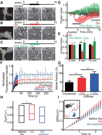
As astrocytic GLT-1 is the major astrocytic transporter involved in glutamate clearance, we first determined the effect of glutamate application on GLT-1 surface diffusion in the absence of neuronal signalling. To achieve this, we expressed GLT-1 with an N-terminal GFP tag (GFP-GLT-1) in primary astrocyte cultures and investigated GLT-1 surface clustering and diffusion dynamics on the astrocyte surface using live-cell imaging of the GFP fluorescence. We found that under basal conditions, GFP-GLT-1 formed bright fluorescent clusters in the astrocyte soma and along processes (Fig. 1A–C). Continu-ous imaging revealed that the intensities and locations of GFP-GLT-1 clusters were stable for periods of 15 min (Fig. 1A). Interestingly, GLT-1 clusters rapidly and reversibly dis-persed during acute glutamate application (100 mM, 3 min, Fig. 1B). The glutamate-induced loss of GFP-GLT-1 cluster-ing was marked by a 50% decrease in the clustercluster-ing index followed by a complete recovery after washout (Fig. 1D,E). Next, to examine the influence of transporter activity on GLT-1 clustering, we investigated the effect of a GLT-1 transporter inhibitor on glutamate induced cluster dispersal.
Intriguingly, blocking transporter activity with TFB-TBOA, a non-transportable competitive inhibitor prevented the gluta-mate induced GLT-1 cluster dispersal (Fig. 1C–E). We further explored the surface mobility of clustered GFP-GLT-1 trans-porters compared to diffuse GFP-GLT-1 using FRAP (Supp. Info. Fig. S1). Interestingly, the fluorescence recovery of dif-fuse GLT-1 was higher in comparison to clustered GLT-1 transporters, suggesting that 1 clustering increases GLT-1 stability (Fig. GLT-1F). This difference in GLT-GLT-1 mobility was confirmed by a total mobile fraction of GFP-GLT-1 of 52% in clustered GFP-GLT-1 compared with 77% for the diffuse GFP-GLT-1 (P50.002, Fig. 1G). Furthermore, glutamate treatment (100 mM, 3 min) increased GLT-1 fluorescence recovery and the mobile fraction of the diffuse GLT-1 to 100% compared to control, before treatment (P50.008, Fig. 1G). Thus, clustered GLT-1 transporters are less mobile than diffuse GLT-1 transporters, and GLT-1 cluster dispersal upon glutamate treatment correlates with increased GLT-1 mobility. Next, we investigated the lateral mobility of GLT-1 molecules on the astrocyte surface in response to glutamate treatment by using SPT with QDs. For SPT experiments we used a ver-sion of GFP-GLT-1 with a V5 extracellular tag inserted in the large GLT-1 extracellular loop (Supp. Info. Fig. S2A). By QD tracking we found that GFP-GLT-1-V5 lateral mobility was significantly increased in response to glutamate (100mM, 3 min), which was reversed upon glutamate washout (Fig. 1H). Moreover, the GLT-1 single trajectories explored a greater area upon glutamate treatment, correlating with increased GLT-1 lateral diffusion (Fig. 1H). Interestingly, despite the formation of stable GLT-1 clusters, we found a linear MSDt plot, reflecting free diffusion (Brownian motion) of GLT-1 molecules rather than a confined behavior (Fig. 1I) suggesting a large proportion of GLT-1 is found outside clusters.
Together, these data reveal that in cultured astrocytes GLT-1 can form stable clusters, while individual GLT-1 mole-cules can be highly mobile on the astrocyte surface. Gluta-mate application leads to GLT-1 cluster dispersal that is dependent on GLT-1 transporter activity, which correlates with an increase in GLT-1 surface diffusion in the astrocyte membrane.
GLT-1 Is More Stable and More Confined inside Synaptic Areas under Basal Conditions
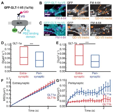
revealed by more mobile transporters on the astrocyte surface in presence of neurons (Supp. Info. Fig. S2C and S2D) as also recently reported by Murphy-Royal. GLT-1a and GLT-1b are the two main isoforms of GLT-1 expressed in forebrain and differ only in their intracellular C-terminal sequence (Fig. 2A). Unlike GLT-1a, GLT-1b has a PDZ binding domain that interacts with scaffolding proteins (Bassan et al., 2008; Gonzalez-Gonzalez et al., 2008a; Underhill et al., 2015). Hence, we next wanted to determine whether astro-cytic GLT-1a and GLT-1b transporters exhibited differential surface diffusion properties, depending on their proximity to active synapses. We expressed GFP-GLT-1a-V5 (Fig. 2B) or GFP-GLT-1b-V5 (Fig. 2C) in hippocampal astrocyte-neuron
coculture. SPT using QDs along with simultaneous labeling of the active presynaptic terminals using FM4-64 (Supp. Info. Fig. S3A), allowed us to track the GLT-1 transporters in regions of the astrocyte plasma membrane located in close proximity to synapses (hence forth termed perisynaptic loca-tions) or in astrocyte regions that were not in proximity to synapses (termed extrasynaptic locations) (Fig. 2B and Supp. Info. Fig. S2C).
[image:6.612.123.489.82.447.2]Interestingly, the median lateral diffusion of perisynaptic GLT-1a, Dperi was 0.052 mm2/s, whereas the extrasynaptic GLT-1a, Dext was 0.065 mm2/s, and for GLT-1b, Dperi was 0.047 mm2/s, whereas the extrasynaptic GLT-1b, Dext was 0.071 mm2/s (Fig. 2D,E). This reveals that astrocytic surface
FIGURE 2:GLT-1 is stable and confined inside synaptic areas under basal conditions.Astrocytes in hippocampal neuron-astrocyte mixed culture transfected with GFP-GLT-1a-V5 or GFP-GLT-1b-V5 at DIV10 and imaged at DIV13 after FM4-64 staining.A: Schematic represen-tation of GFP-GLT-1(a/b)-V5 labelled by an anti-V5 antibody/QD complex.B,C: Representative time lapse imaging illustrates GFP-GLT-1a-V5 (B) and GFP-GLT-1b-V5 (C) in a region of an astrocyte. GFP-GLT-1-V5 overlaid with FM4-64 stained synapses (left panels), or by QD-tagged 1-V5 trajectories shown in orange (middle panels), and FM4-64 stained synapses overlaid by QD-tagged GFP-GLT-1-V5 trajectories shown in orange (right panels), Scale bars 5lm.D: Instantaneous diffusion coefficients of extrasynaptic GLT-1a (red, median50.065lm2/s;n5599 trajectories) and perisynaptic GLT-1a (blue, median50.052lm2/s;n5112 trajectories), median D is
sig-nificantly decreased in perisynaptic areas (P56x1023, Mann-Whitney test).E: Instantaneous diffusion coefficients of extrasynaptic
GLT-1b (red, median50.071lm2/s;n5255 trajectories) and perisynaptic GLT-1b (blue, median50.047lm2/s;n562 trajectories). Median D
is significantly decreased in perisynaptic areas (P5831025, Mann-Whitney test). MSDt plot of extrasynaptic (F) and perisynaptic (G)
diffusion of extrasynaptic GLT-1 is more dynamic than peri-synaptic GLT-1 for both isoforms. No significant difference was observed between GLT-1a and GLT-1b median lateral diffusion. Interestingly, MSDt plot of GLT-1a and GLT-1b revealed differential properties depending on their localisation relative to the synapse (Fig. 2F,G). The MSDt plot of extrasy-naptic GLT-1a and GLT-1b exhibited linear profiles, indicat-ing Brownian diffusion, however, perisynaptic GLT-1a and GLT-1b exhibited sublinear curvature, indicative of confined motion. In addition, the differences in transporter mobility were due to a shift of the diffusive fraction without a signifi-cant change of the immobile fraction (data not shown). Fur-thermore, comparison of the extrasynaptic 1a and GLT-1b MSDt plots revealed no significant difference (Fig. 2F). However, comparison of the perisynaptic 1a and GLT-1b MSDt plots revealed that GLT-GLT-1b was significantly more confined compared to GLT-1a (Fig. 2G) and the residency time of GLT-1b within the perisynaptic area is significantly longer than GLT-1a (Supp. Info. Fig. S3B).
Together, these data showed that for both isoforms, perisynaptic GLT-1 is more stable than GLT-1 in extrasynap-tic regions under basal conditions. Moreover, perisynapextrasynap-tic GLT-1b is more stable than perisynaptic GLT-1a suggesting a role of GLT-1b PDZ interactors in its confinement.
Glutamate and Neuronal Activity Regulate the Surface Diffusion of GLT-1 in Astrocytes
Next, we were interested in further studying the effect of neuro-nal activity on 1 surface dynamics in astrocytes. As GLT-1a and GLT-1b showed similar behavior in response to the pharmacological treatments; here, we have shown the results of GLT-1a isoform only. For the following experiments, we expressed GFP-GLT-1a-V5 in hippocampal astrocyte-neuron coculture and performed single-particle tracking experiments.
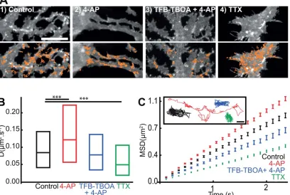
Similarly to experiments in astrocytes cultured alone (Fig. 1H,I), we noticed a rapid and reversible loss of clustered GFP-GLT-1a-V5 fluorescence (Supp. Info. Fig. S4A) and an increase in GLT-1a surface diffusion upon glutamate treatment, which are prevented when the transport activity of astrocytic glu-tamate transporters is inhibited with TFB-TBOA (Supp. Info. Fig. S4B and S4C). Since glutamate is released during neuronal firing, we studied the effect of increased neuronal activity on the GLT-1a surface dynamics via 4-AP (a nonselective voltage-dependent K1-channel blocker) treatment in astrocyte-neuron cocultures (Fig. 3). Similar to glutamate treatment, we observed a loss of clustered GFP-GLT-1a-V5 fluorescence upon 4-AP (1 mM, 20 min), which is prevented in the presence of the trans-porter inhibitor (Fig. 3A1–3). We found significantly increased GLT-1a (71%, Fig. 3B) surface diffusion following 4-AP treatment, characterized by more mobile transporters (Fig. 3C). Furthermore, inhibition of GLT-1 transporter activity with
TFB-TBOA blocked the 4-AP dependent increase in GLT-1a lat-eral diffusion (Fig. 3B), reverting it to control levels. The MSDt plot exhibited an upward linear shift, indicative of Brownian motion and increased lateral diffusion upon 4-AP treatment (Fig. 3C). These data suggest that stimulating neuronal activity triggers an increase in GLT-1 surface mobility probably due to the increased glutamate release upon 4-AP treatment (Girault et al., 1986).
In contrast, inhibiting neuronal firing with TTX (a highly selective sodium channel blocker) treatment, led to an increase in clustered GFP-GLT-1a-V5 (Fig. 3A4) and signifi-cantly decreased GLT-1a surface diffusion (35%, Fig. 3B), correlating with less mobile transporters (Fig. 3C). The MSDt plot exhibited a downward linear shift, indicative of decreased Brownian motion, but not confinement upon TTX treatment (Fig. 3C). This result confirmed that inhibition of neuronal activity decreased GLT-1 surface diffusion (Murphy-Royal et al., 2015). Glutamate and neuronal activity can also regulate the surface diffusion dynamics of GLT-1b (Supp. Info. Figs. S5 and S6). Since the majority of the GLT-1 tra-jectories in the mixed cultures were from the extrasynaptic GLT-1 population (87 and 80% of the total GLT-1a and GLT-1b trajectories, respectively), we propose that pharmaco-logical alterations in neuronal activity can influence surface diffusion of the extrasynaptic GLT-1 transporters. Together, these data showed that neuronal activity bidirectionally regu-lates GLT-1a and GLT-1b surface mobility on the surface of astrocytes in a GLT-1 transporter activity-dependent manner.
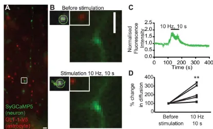
Next, to determine the effect of neuronal activity on GLT-1 surface diffusion in astrocytic processes in apposition to synap-ses undergoing activation, we performed a more physiological form of neuronal stimulation (electrical field stimulation) of hip-pocampal neurons transfected with synaptically targeted GCaMP5 (SyGCaMP5) to label stimulated synapses and co-cultured with astrocytes pre-transfected with GLT-1-V5 (Fig. 4A). By using an image splitter, we were able to simultaneously image the neuronal SyGCaMP5 signal and QD-labelled GLT-1 on the astrocyte surface (Fig. 4B). Upon field stimulation (10 Hz for 10 s, 100 APs), a 2-fold increase was observed in SyGCaMP5 signal, showing that field stimulation drives increased presynaptic calcium signal and revealing an increase in synaptic activity and position of activated synapses (Fig. 4C). Interestingly, we observed that field stimulation increased GLT-1 surface diffusion in apposition to activated synapses by 100% compared with before stimulation (Fig. 4D). The activity-dependent increase in GLT-1 surface diffusion is likely due to the glutamate released in the synaptic sites during the stimulation.
Glutamate and Neuronal Activity Regulate GLT-1 Surface Diffusion inEx-VivoBrain Slices
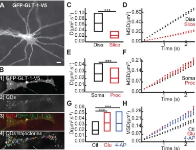
To address this, we used hippocampal organotypic brain slices transfected with GFP-GLT-1-V5 (Fig. 5A). In transfected astro-cytes, GLT-1 was expressed on the surface of the astrocytes and formed obvious clusters (Fig. 5A), corroborating previous data (Benediktsson et al., 2012). After incubating the slices with anti-V5-QD complexes, we were able to observe QD labeling through-out the slice, specific only to the transfected astrocytes (Fig. 5B1– 4). First, we found a significant decrease in GLT-1 surface diffu-sion in brain slices (slice) compared with in dissociated (diss) cul-tures (Fig. 5C, Dslicewas 0.021mm2/s and Ddisswas 0.067mm2/ s) where the transporters explored a greater area (Fig. 5D). More-over, despite a similar explored area by GLT-1 in soma compared to processes (Fig. 5E) that could be due to more compactly packed cells and extracellular matrix, we found that GLT-1 coefficient dif-fusion was significantly lower in processes (proc) compared to soma (Fig. 5E, Dprocwas at 0.021mm2/s and Dsomawas 0.027 mm2/s). This reduction could be due to the presence on astrocytic processes of more synaptic sites, where we have previously shown that GLT-1 is more confined (Fig. 2G). This data suggests that
GLT-1 is more stable in astrocytic processes in apposition to syn-apses in slice cultures under basal conditions. We then investigated the effects of glutamate (100mM, 2 min) and stimulation of neu-ronal activity with 4-AP (1 mM, 20 min) on GLT-1 surface dynamics in astrocytic processes (Fig. 5G). Interestingly, surface GLT-1 is more dynamic in brain slices when treated with gluta-mate (Dglu50.033mm2/s) or 4-AP (D4-AP50.032mm2/s), in comparison to untreated slice control (Dcontrol50.021mm2/s). Moreover, the GLT-1 single trajectories explored a greater area upon glutamate and 4-AP treatment, correlating with increased GLT-1 lateral diffusion (Fig. 5H). In addition to validating our results obtained from dissociated cultures that showed neuronal activity mediated regulation of GLT-1 surface dynamics, these data are evidence that GLT-1 modulates its surface diffusion upon stimulation of neuronal activity in brain slices.
Discussion
[image:8.612.105.513.93.367.2]GLT-1 glutamate transporters are critical modulators of extracellular glutamate in the brain (Danbolt, 2001;
FIGURE 3:Neuronal activity mediated GLT-1 surface diffusion increase is transporter activity dependent. Hippocampal neuron-astrocytes mixed culture transfected with GFP-GLT-1a-V5 at DIV10 and imaged at DIV13.A: Representative time lapse imaging illus-trates GFP-GLT-1a-V5 in a region of interest of an astrocytic process (top panel) and overlaid by QD-tagged GFP-GLT-1a-V5 trajectories shown in orange (bottom panels) in control untreated (1), treated 20 min with 1 mM 4-AP (2), 20 min with TFB-TBOA 10 lM14-AP 1mM (3) or 1mM of TTX (4), Scales bars 10lm. Instantaneous diffusion coefficients (B) and MSDt plot and single trajectories (C) of QD-tagged GFP-GLT-1-V5. Control untreated (black, median50.08 lm2/s; n51061 trajectories), after 20 min with 1 mM 4-AP (red,
median50.12lm2/s; n5779 trajectories), after 20 min of TFB-TBOA 10lM14-AP 1 mM (blue, median50.08lm2/s; n5282
trajec-tories) and after 20 min with 1mM of TTX (green, median50.05lm2/s;n51191 trajectories). Median D is significantly increased upon
4-AP (P52310214, Mann-Whitney test) and is significantly decreased upon TTX (P51.2310214, Mann-Whitney test) but is not
Tzingounis and Wadiche, 2007) and regulating their surface diffusion and clustering properties to modulate transporter number at synaptic release sites could be a rapid mechanism for locally tailoring glutamate uptake to shape glutamatergic neurotransmission. Here, by using GFP-time lapse imaging, FRAP and single-particle tracking with QD, we report the behavior of the two main GLT-1 transporter isoforms (GLT-1a and GLT-1b) in astrocytic surface domains, under both basal conditions and in response to neuronal activity. GLT-1a and GLT-1b, were both found to be highly dynamic at non-synaptic locations and to exhibit a more confined diffusion when present in astrocyte membranes adjacent to synapses, with GLT-1b more so. Moreover, exogenous glutamate appli-cation or alteration of neuronal firing led to altered GLT-1 surface diffusion in a transporter activity dependent manner and to an increase in GLT-1 mobility in proximity to acti-vated synapses. Importantly, we could also observe for the first time neuronal activity-dependent alterations in GLT1 surface mobilities in astrocyte processes in situin brain slices.
In cultured astrocytes GLT-1 transporters could form sta-ble clusters that were rapidly and reversibly dispersed upon glu-tamate treatment. Interestingly, the gluglu-tamate-dependent increase in GLT-1 de-clustering and surface diffusion was blocked by the nontransportable competitive inhibitor, TFB-TBOA, suggesting a critical role for the transport activity of
GLT-1 in regulating its surface dynamics. Indeed, application of aspartate, another glutamate transporter substrate also led to GLT-1 cluster dispersal (data not shown), reinforcing the idea that substrate binding to the GLT-1 transporter can directly affect its surface diffusion. In agreement with this, inhibition of GLT-1 transport activity with TBOA under basal conditions decreased steady state GLT-1 surface diffusion in mixed neuron-glial cultures (Murphy-Royal et al., 2015) presumably by inhibiting GLT-1 activation by endogenous glutamate in the cultures. GLT-1 is mainly found in oligomeric form on the astrocyte surface (Haugeto et al., 1996), which may facilitate the formation of the GLT-1 clusters observed in our study, although FRAP and single-particle tracking resolution do not allow us to distinguish between the different surface oligomeric GLT-1 forms. GLT-1 can also interact with scaffolds (Bassan et al., 2008; Gonzalez-Gonzalez et al., 2008a; Underhill et al., 2015), which could also contribute to cluster formation. Substrate-mediated GLT-1 cluster dispersal could be due to the change in GLT-1 conformation demonstrated in the presence of its substrates (Qu and Kanner, 2008; Yernool et al., 2004), which could facilitate oligomer disassembly or uncoupling from intracellular scaffolds.
[image:9.612.95.510.78.330.2]Although a recent study reported the surface diffusion dynamics of the GLT-1a isoform (Murphy-Royal et al, 2015), the activity-dependent behavior of GLT-1b was not known.
On studying the two isoforms, we identified two populations for both GLT-1 isoforms based on their basal surface mobili-ties in the astrocyte membrane: a perisynaptic population, characterized by a slower surface diffusion and a more mobile extrasynaptic population. This is similar to the confinement of neurotransmitter receptors observed at postsynaptic sites (Choquet and Triller, 2013), promoting the importance of specialized astrocytic subdomains proximal to synapses. The two predominant astrocytic GLT-1 isoforms, GLT-1a and GLT-1b, differ only in their extreme intracellular C-terminal sequences. Unlike GLT-1a, GLT-1b, has a PDZ binding domain that binds to PDZ containing proteins such as PICK1, PSD95, and DLG1 (Bassan et al., 2008; Gonzalez-Gonzalez et al., 2008a; Underhill et al., 2015). The confined motion seen for both perisynaptic GLT-1a and GLT-1b may be due to protein interacting motifs located in the N-terminal or C-N-terminal regions common to both isoforms. Interestingly, we observed that GLT-1b was more confined in astrocyte domains close to synapses than GLT-1a under basal
conditions, supporting the concept that the GLT-1b C-termi-nal PDZ domain interacts with scaffolding proteins, anchor-ing it to macromolecular glial complexes located in subcellular domains opposite to neuronal presynaptic sites. Furthermore, the interaction between the PICK1 scaffolding protein and GLT-1b has functional consequence on glutamate transport activity (Sogaard et al., 2013). GLT-1a is known to form heteromers with GLT-1b (Haugeto et al., 1996; Peacey et al., 2009) and since GLT-1b interacts with multiple scaf-folding proteins (Gonzalez-Gonzalez et al., 2009; Underhill et al., 2015), it could serve to stabilize perisynaptic GLT-1a, under basal conditions.
[image:10.612.117.500.86.380.2]We also explored the activity-dependence of GLT-1 syn-aptic localisation to neuronal activity. MNI-glutamate uncag-ing experiments on cultured astrocytes provided an initial suggestion that a glutamate rise could act locally to regulate GLT-1 surface mobility (Murphy-Royal et al., 2015). How-ever, in those experiments putative synapses were labeled by staining for mitochondria which also densely populate
FIGURE 5:Glutamate and neuronal activity regulate GLT-1 surface diffusion in brain slices. A: Example of astrocyte expressing GFP-GLT-1-V5 and imaged 3–5 days after transfection. Scale bar, 10lm.B: Zoomed region on an astrocytic process expressing GFP-GLT-1-V5 (B1), QDs (B2), QDs overlaid with GFP-GLT-1-GFP-GLT-1-V5 (B3) and representative QD trajectories (B4). Instantaneous diffusion coefficients (C,E,G) and MSDt plot of QD-tagged GLT-1 (D,F,H). C,D: In dissociated culture (black, medianD50.067lm2/s;n5783 trajectories) and
in slice cultures (red, median50.021lm2/s;n5325 trajectories), medianDis significantly decreased in slice cultures compared with in
dissociated cultures (P53.53 10280, Mann-Whitney test). D,E: In soma (black, median D50.027 lm2/s; n5108 trajectories) and in
processes (red, medianD50.021lm2/s;n5325 trajectories), medianDis significantly decreased in processes compared with in soma
(P50.005, Mann-Whitney test).G,H: After 2 min of glutamate 100lM (red, medianD50.033lm2/s;n5234 trajectories), and after 20
min of 4-AP 1 mM (blue, medianD50.032lm2/s;n5174 trajectories), medianDis significantly increased in both treatment conditions
dendrites and astrocyte processes themselves (Jackson and Robinson, 2015; Stephen et al., 2015) and therefore cannot equivocally label synaptic sites. To directly assess the local effect of physiologically released glutamate on GLT-1 surface diffusion, we evoked action potentials to trigger synaptic glu-tamate release while directly visualizing active synapses using SyGCaMP5 fluorescence imaging. This, for the first time, revealed that an increase in GLT-1 surface diffusion in the neighboring astrocyte processes surrounding the activated syn-apse. We speculate that during neuronal activity, synaptically released glutamate is taken up by GLT-1 transporter, resulting in a change in GLT-1 conformation, which could lead to the dissociation of GLT-1 from its scaffolding proteins to allow for increased surface mobility. It will be interesting in the future to determine the scaffold protein(s) responsible for sta-bilizing GLT-1 in astrocyte processes close to synapses but it is tempting to speculate that DLG1 or PICK1 may play a role.
To extend our findings to a more physiological setting, we used organotypic rat hippocampal brain slices, which exhibit functional local synaptic circuitry and preserved brain architecture. Interestingly, GLT-1 transporters exhibited a more confined mobility in slice cultures in comparison to dis-sociated cultures. This could be due to the more compactly packed cells and extracellular matrix, and increased number of neuron-glial contacts or due to increased expression levels of scaffold proteins within astrocytes in slices, which could restrict GLT-1 mobility. Moreover, GLT-1 transporters were more confined in astrocyte processes compared to the cell soma in organotypic slices, possibly due to a high coverage of synaptic sites on astrocytic processes in situ (Ventura and Harris, 1999). Importantly, although GLT-1 was more stable in astrocyte processes in ex vivo brain slices (compared with dissociated culture), exogenous glutamate application or phar-macological activation of neuronal activity nonetheless led to a dramatic increase in GLT-1 surface diffusion, providing compelling evidence that the activity dependent alteration of GLT-1 mobility is an important mechanism for regulating GLT-1 distribution in astrocytes in intact networks.
Although the transport cycle of GLT-1 is slow (12–70 ms/cycle) in comparison to the duration of glutamate in the synaptic cleft (Bergles and Jahr, 1998; Clements et al., 1992; Wadiche et al., 1995), efficient glutamate clearance is facili-tated by the presence of a large number of GLT-1 transporters on the astrocyte surface, which can also buffer glutamate by binding it (Wadiche et al., 1995). In rat hippocampal slices, it has been shown that perisynaptic astrocyte processes (PAPs) are present at 62% synapse, with preference towards larger synapses that contain post synaptic densities (Witcher et al., 2007). PAPs express glutamate transporters (Derouiche et al., 2012; Pannasch et al., 2014) and are highly motile processes
that are regulated by synaptically released glutamate. PAPs selectively modify their coverage of dendritic spines around potentiated synapses, which in turn is associated with an increased stability of synapses (Bernardinelli et al., 2014). While changes in surface protein levels, by processes such as endocytosis and exocytosis will occur at longer time scale of minutes to hours, rapid glutamate-dependent alterations in GLT-1 surface diffusion may act as a complementary pathway for the regulation of transporter numbers and positioning at synapses. Our data and that of others (Murphy-Royal et al., 2015), supports the hypothesis that rapid dispersal of glutamate-bound GLT-1 in regions of high extracellular gluta-mate (such as in proximity to activated synapses) could potentially leave space for positioning of unbound GLT-1 to allow for efficient and selective glutamate buffering. Rapid displacement of glutamate-bound GLT-1 molecules to allow their exchange with unbound ones, may be particularly important in PAPs where the small size of the PAPs may limit transporter availability in close proximity to synapses. Rapid increases in GLT-1 surface mobility may also contribute to limit glutamate spillover (Asztely et al., 1997) and, therefore, prevent neuronal excitotoxicity. Our data provides further support for the active role of astrocytes in regulating glutama-tergic signaling and shed light on transporter surface diffusion as an additional rapid regulatory mechanism for modulating number of transporters at synaptic release sites to regulate glutamate clearance in the CNS.
Acknowledgment
Grant sponsor: BBSRC (BB/I00274X/1 to J.T.K.); Grant sponsor: Wellcome trust grants (093239/Z/10/Z and 068817/ Z/02/Z to J.T.K.); Grant sponsor: MRC Senior Non-Clinical Fellowship (G0802377 to J.T.K.); Grant sponsor: MRC Case PhD student (to R.N.); Grant sponsor: CoMPLEX PhD pro-gram at UCL (to D.F.S. and J.M.).
The authors thank Dr Victoria Vaccaro for SyGCaMP5 cloning and Dr Souvik Modi for his precious advice for sin-gle particle tracking experiments in brain slices and Dr Ramona Eckel for her constructive comments on the manuscript.
References
Akerboom J, Chen TW, Wardill TJ, Tian L, Marvin JS, Mutlu S, Calderon NC, Esposti F, Borghuis BG, Sun XR, Gordus A, Orger MB, Portugues R, Engert F, Macklin JJ, Filosa A, Aggarwal A, Kerr RA, Takagi R, Kracun S, Shigetomi E, Khakh BS, Baier H, Lagnado L, Wang SS, Bargmann CI, Kimmel BE, Jayaraman V, Svoboda K, Kim DS, Schreiter ER, Looger LL. 2012. Optimiza-tion of a GCaMP calcium indicator for neural activity imaging. J Neurosci 32: 13819–13840.
Arancibia-Carcamo IL, Yuen EY, Muir J, Lumb MJ, Michels G, Saliba RS, Smart TG, Yan Z, Kittler JT, Moss SJ. 2009. Ubiquitin-dependent lysosomal targeting of GABA(A) receptors regulates neuronal inhibition. Proc Natl Acad Sci U S A 106:17552–17557.
Asztely F, Erdemli G, Kullmann DM. 1997. Extrasynaptic glutamate spillover in the hippocampus: Dependence on temperature and the role of active glu-tamate uptake. Neuron 18:281–293.
Attwell D, Barbour B, Szatkowski M. 1993. Nonvesicular release of neuro-transmitter. Neuron 11:401–407.
Banker G, Goslin K. 1998. Culturing nerve cells, Cambridge, MA: MIT. Bassan M, Liu H, Madsen KL, Armsen W, Zhou J, Desilva T, Chen W, Paradise A, Brasch MA, Staudinger J, Gether U, Irwin N, Rosenberg PA. 2008. Interaction between the glutamate transporter GLT1b and the synaptic PDZ domain protein PICK1. Eur J Neurosci 27:66–82.
Benediktsson AM, Marrs GS, Tu JC, Worley PF, Rothstein JD, Bergles DE, Dailey ME. 2012. Neuronal activity regulates glutamate transporter dynamics in developing astrocytes. Glia 60:175–188.
Bergles DE, Jahr CE. 1998. Glial contribution to glutamate uptake at Schaffer collateral-commissural synapses in the hippocampus. J Neurosci 18:7709–7716. Bernardinelli Y, Randall J, Janett E, Nikonenko I, Konig S, Jones EV, Flores€
CE, Murai KK, Bochet CG, Holtmaat A, Muller D. 2014. Activity-dependent structural plasticity of perisynaptic astrocytic domains promotes excitatory synapse stability. Curr Biol 24:1679–1688.
Chaudhry FA, Lehre KP, van Lookeren Campagne M, Ottersen OP, Danbolt NC, Storm-Mathisen J. 1995. Glutamate transporters in glial plasma mem-branes: Highly differentiated localizations revealed by quantitative ultrastruc-tural immunocytochemistry. Neuron 15:711–720.
Chen W, Aoki C, Mahadomrongkul V, Gruber CE, Wang GJ, Blitzblau R, Irwin N, Rosenberg PA. 2002. Expression of a variant form of the glutamate trans-porter GLT1 in neuronal cultures and in neurons and astrocytes in the rat brain. J Neurosci 22:2142–2152.
Chen W, Mahadomrongkul V, Berger UV, Bassan M, DeSilva T, Tanaka K, Irwin N, Aoki C, Rosenberg PA. 2004. The glutamate transporter GLT1a is expressed in excitatory axon terminals of mature hippocampal neurons. J Neurosci 24:1136–1148.
Choquet D, Triller A. 2013. The dynamic synapse. Neuron 80:691–703. Clements JD, Lester RA, Tong G, Jahr CE, Westbrook GL. 1992. The time course of glutamate in the synaptic cleft. Science 258:1498–1501.
Danbolt NC. 2001. Glutamate uptake. Prog Neurobiol 65:1–105.
Derouiche A, Pannicke T, Haseleu J, Blaess S, Grosche J, Reichenbach A. 2012. Beyond polarity: functional membrane domains in astrocytes and Mul-ler cells. Neurochem Res 37:2513–2523.
Dreosti E, Odermatt B, Dorostkar MM, Lagnado L. 2009. A genetically encoded reporter of synaptic activity in vivo. Nat Methods 6:883–889. Eckel R, Szulc B, Walker MC, Kittler JT. 2015. Activation of calcineurin under-lies altered trafficking of alpha2 subunit containing GABAA receptors during prolonged epileptiform activity. Neuropharmacology 88:82–90.
Edelstein A, Amodaj N, Hoover K, Vale R, Stuurman N. 2010. Computer con-trol of microscopes using microManager. Curr Protoc Mol Biol Chapt 14:20. Girault JA, Barbeito L, Spampinato U, Gozlan H, Glowinski J, Besson MJ. 1986. In vivo release of endogenous amino acids from the rat striatum: Fur-ther evidence for a role of glutamate and aspartate in corticostriatal neuro-transmission. J Neurochem 47:98–106.
Gonzalez-Gonzalez IM, Garcia-Tardon N, Cubelos B, Gimenez C, Zafra F. 2008a. The glutamate transporter GLT1b interacts with the scaffold protein PSD-95. J Neurochem 105:1834–1848.
Gonzalez-Gonzalez IM, Garcia-Tardon N, Gimenez C, Zafra F. 2008b. PKC-dependent endocytosis of the GLT1 glutamate transporter depends on ubiq-uitylation of lysines located in a C-terminal cluster. Glia 56:963–974.
Gonzalez-Gonzalez IM, Garcia-Tardon N, Gimenez C, Zafra F. 2009. Splice variants of the glutamate transporter GLT1 form hetero-oligomers that inter-act with PSD-95 and NMDA receptors. J Neurochem 110:264–274.
Groc L, Heine M, Cognet L, Brickley K, Stephenson FA, Lounis B, Choquet D. 2004. Differential activity-dependent regulation of the lateral mobilities of AMPA and NMDA receptors. Nat Neurosci 7:695–696.
Haugeto O, Ullensvang K, Levy LM, Chaudhry FA, Honore T, Nielsen M, Lehre KP, Danbolt NC. 1996. Brain glutamate transporter proteins form homomultimers. J Biol Chem 271:27715–27722.
Holmseth S, Dehnes Y, Huang YH, Follin-Arbelet VV, Grutle NJ, Mylonakou MN, Plachez C, Zhou Y, Furness DN, Bergles DE, Lehre KP, Danbolt NC. 2012. The density of EAAC1 (EAAT3) glutamate transporters expressed by neurons in the mammalian CNS. J Neurosci 32:6000–6013.
Holmseth S, Scott HA, Real K, Lehre KP, Leergaard TB, Bjaalie JG, Danbolt NC. 2009. The concentrations and distributions of three C-terminal variants of the GLT1 (EAAT2; slc1a2) glutamate transporter protein in rat brain tissue suggest differential regulation. Neuroscience 162:1055–1071.
Jackson JG, Robinson MB. 2015. Reciprocal Regulation of Mitochondrial Dynamics and Calcium Signaling in Astrocyte Processes. J Neurosci 35: 15199–15213.
Kalandadze A, Wu Y, Robinson MB. 2002. Protein kinase C activation decreases cell surface expression of the GLT-1 subtype of glutamate trans-porter. Requirement of a carboxyl-terminal domain and partial dependence on serine 486. J Biol Chem 277:45741–45750.
Kanner BI, Schuldiner S. 1987. Mechanism of transport and storage of neuro-transmitters. CRC Crit Rev Biochem 22:1–38.
Kojima S, Nakamura T, Nidaira T, Nakamura K, Ooashi N, Ito E, Watase K, Tanaka K, Wada K, Kudo Y, Miyakawa H. 1999. Optical detection of synapti-cally induced glutamate transport in hippocampal slices. J Neurosci 19:2580– 2588.
Levi S, Schweizer C, Bannai H, Pascual O, Charrier C, Triller A. 2008. Homeo-static regulation of synaptic GlyR numbers driven by lateral diffusion. Neuron 59:261–273.
Muir J, Arancibia-Carcamo IL, MacAskill AF, Smith KR, Griffin LD, Kittler JT. 2010. NMDA receptors regulate GABAA receptor lateral mobility and cluster-ing at inhibitory synapses through serine 327 on the gamma2 subunit. Proc Natl Acad Sci U S A 107:16679–16684.
Muir J, Kittler JT. 2014. Plasticity of GABAA receptor diffusion dynamics at the axon initial segment. Front Cell Neurosci
8:151-Murphy-Royal C, Dupuis JP, Varela JA, Panatier A, Pinson B, Baufreton J, Groc L, Oliet SH. 2015. Surface diffusion of astrocytic glutamate transporters shapes synaptic transmission. Nat Neurosci 18:219–226.
Omrani A, Melone M, Bellesi M, Safiulina V, Aida T, Tanaka K, Cherubini E, Conti F. 2009. Up-regulation of GLT-1 severely impairs LTD at mossy fibre– CA3 synapses. J Physiol 587:4575–4588.
Pannasch U, Freche D, Dallerac G, Ghezali G, Escartin C, Ezan P, Cohen-Salmon M, Benchenane K, Abudara V, Dufour A, L€ubke JH, Deglon N, Knott G, Holcman D, Rouach N. 2014. Connexin 30 sets synaptic strength by con-trolling astroglial synapse invasion. Nat Neurosci 17:549–558.
Pathania M, Davenport EC, Muir J, Sheehan DF, Lopez-Domenech G, Kittler JT. 2014. The autism and schizophrenia associated gene CYFIP1 is critical for the maintenance of dendritic complexity and the stabilization of mature spines. Transl Psychiatry 4:e374.
Peacey E, Miller CC, Dunlop J, Rattray M. 2009. The four major N- and C-terminal splice variants of the excitatory amino acid transporter GLT-1 form cell surface homomeric and heteromeric assemblies. Mol Pharmacol 75: 1062–1073.
Qu S, Kanner BI. 2008. Substrates and non-transportable analogues induce structural rearrangements at the extracellular entrance of the glial glutamate transporter GLT-1/EAAT2. J Biol Chem 283:26391–26400.
Rosenberg PA, Amin S, Leitner M. 1992. Glutamate uptake disguises neuro-toxic potency of glutamate agonists in cerebral cortex in dissociated cell cul-ture. J Neurosci 12:56–61.
Rothstein JD, Dykes-Hoberg M, Pardo CA, Bristol LA, Jin L, Kuncl RW, Kanai Y, Hediger MA, Wang Y, Schielke JP, Welty DF. 1996. Knockout of glutamate transporters reveals a major role for astroglial transport in excitotoxicity and clearance of glutamate. Neuron 16:675–686.
Rothstein JD, Patel S, Regan MR, Haenggeli C, Huang YH, Bergles DE, Jin L, Dykes Hoberg M, Vidensky S, Chung DS, Toan SV, Bruijn LI, Su ZZ, Gupta P, Fisher PB. 2005. Beta-lactam antibiotics offer neuroprotection by increasing glutamate transporter expression. Nature 433:73–77.
Shih J, Liu L, Mason A, Higashimori H, Donmez G. 2014. Loss of SIRT4 decreases GLT-1-dependent glutamate uptake and increases sensitivity to kainic acid. J Neurochem 131:573–581.
Smith KR, Davenport EC, Wei J, Li X, Pathania M, Vaccaro V, Yan Z, Kittler JT. 2014. GIT1 and betaPIX are essential for GABA(A) receptor synaptic sta-bility and inhibitory neurotransmission. Cell Rep 9:298–310.
Smith KR, Muir J, Rao Y, Browarski M, Gruenig MC, Sheehan DF, Haucke V, Kittler JT. 2012. Stabilization of GABA(A) receptors at endocytic zones is mediated by an AP2 binding motif within the GABA(A) receptor beta3 subu-nit. J Neurosci 32:2485–2498.
Sogaard R, Borre L, Braunstein TH, Madsen KL, MacAulay N. 2013. Func-tional modulation of the glutamate transporter variant GLT1b by the PDZ domain protein PICK1. J Biol Chem 288:20195220207.
Soni N, Reddy BV, Kumar P. 2014. GLT-1 transporter: An effective pharmaco-logical target for various neuropharmaco-logical disorders. Pharmacol Biochem Behav 127C:70–81.
Stephen TL, Higgs NF, Sheehan DF, Al Awabdh S, Lopez-Domenech G, Arancibia-Carcamo IL, Kittler JT. 2015. Miro1 Regulates Activity-Driven Posi-tioning of Mitochondria within Astrocytic Processes Apposed to Synapses to Regulate Intracellular Calcium Signaling. J Neurosci 35:15996216011.
Stoppini L, Buchs PA, Muller D. 1991. A simple method for organotypic cul-tures of nervous tissue. J Neurosci Methods 37:1732182.
Sullivan R, Rauen T, Fischer F, Wiessner M, Grewer C, Bicho A, Pow DV. 2004. Cloning, transport properties, and differential localization of two splice variants of GLT-1 in the rat CNS: Implications for CNS glutamate homeosta-sis. Glia 45:155–169.
Susarla BT, Robinson MB. 2008. Internalization and degradation of the gluta-mate transporter GLT-1 in response to phorbol ester. Neurochem Int 52:709– 722.
Tanaka K, Watase K, Manabe T, Yamada K, Watanabe M, Takahashi K, Iwama H, Nishikawa T, Ichihara N, Kikuchi T, Okuyama S, Kawashima N, Hori S, Takimoto M, Wada K. 1997. Epilepsy and exacerbation of brain injury in mice lacking the glutamate transporter GLT-1. Science 276:1699–1702. Thevenaz P, Ruttimann UE, Unser M. 1998. A pyramid approach to subpixel registration based on intensity. IEEE Trans Image Process 7:27–41.
Tzingounis AV, Wadiche JI. 2007. Glutamate transporters: confining runaway excitation by shaping synaptic transmission. Nat Rev Neurosci 8:935–947. Ullensvang K, Lehre KP, Storm-Mathisen J, Danbolt NC. 1997. Differential developmental expression of the two rat brain glutamate transporter proteins GLAST and GLT. Eur J Neurosci 9:164621655.
Underhill SM, Wheeler DS, Amara SG. 2015. Differential regulation of two isoforms of the glial glutamate transporter EAAT2 by DLG1 and CaMKII. J Neurosci 35:5260–5270.
Ventura R, Harris KM. 1999. Three-dimensional relationships between hippo-campal synapses and astrocytes. J Neurosci 19:6897–6906.
Wadiche JI, Arriza JL, Amara SG, Kavanaugh MP. 1995. Kinetics of a human glutamate transporter. Neuron 14:1019–1027.
Witcher MR, Kirov SA, Harris KM. 2007. Plasticity of perisynaptic astroglia during synaptogenesis in the mature rat hippocampus. Glia 55:13–23. Woods G, Zito K. 2008. Preparation of gene gun bullets and biolistic trans-fection of neurons in slice culture. J Vis Exp 12:675.