A STUDY ON DIAGNOSIS OF HELICOBACTER PYLORI INFECTIONBY CULTURE AND MOLECULAR METHODS FROM
GASTRIC BIOPSY SPECIMENS AND SEROLOGICAL ASSAYS IN PATIENTS WITH PEPTIC ULCER DISEASE
Dissertation submitted to
THE TAMILNADU DR.M.G.R.MEDICAL UNIVERSITY In partial fulfillment of the regulations
for the award of the degree of
M.D. (MICROBIOLOGY) BRANCH - IV
MADRAS MEDICAL COLLEGE
THE TAMILNADU DR. M.G.R. MEDICAL UNIVERSITY CHENNAI – TAMILNADU.
BONAFIDE CERTIFICATE
This is to certify that this dissertation work entitled “A STUDYON
DIAGNOSIS OF HELICOBACTER PYLORI INFECTION BY CULTURE AND MOLECULAR METHODS FROM GASTRIC BIOPSY SPECIMENS AND SEROLOGICAL ASSAYS IN PATIENTS WITH PEPTIC ULCER DISEASE” is
the original bonafide work done by DR. VIJI S., Post Graduate Student from 2016 to 2018 under guidance and supervision in the Institute of Microbiology,
Madras Medical College, Chennai- 600003, in partial fulfillment of the
requirement of M.D MICROBIOLOGY degree Examination of The Dr.M.G.R.
Medical University to be held in MAY 2019.
Dr. JAYANTHI, MD., FRCP (Glasg)
DEAN,
Madras Medical College
Government General Hospital,
Chennai-600 003.
Prof. Dr.J.EUPHRASIA LATHA, DGO,M.D
DECLARATION
I DR. S.VIJI, Post Graduate , Institute of Microbiology, Madras Medical College, solemnly declare that the dissertation titled “A STUDY ON DIAGNOSIS
OF HELICOBACTER PYLORI INFECTION BY CULTURE AND MOLECULAR METHODS FROM GASTRIC BIOPSY SPECIMENS AND SEROLOGICAL ASSAYS IN PATIENTS WITH PEPTIC ULCER DISEASE”Submitted by me for the degree of M.D is the record work carried out
by me during the period of OCTOBER 2017- SEPTEMBER 2018 under the
expert guidance and supervision of Prof. Dr. U. UMADEVI M.D., Professor, Institute of Microbiology, Madras Medical College, Chennai. The dissertation is
submitted to The Tamil Nadu Dr. M.G.R Medical University, Chennai towards
partial fulfillment of requirement for the award of M.D., Degree (Branch IV) in
Microbiology examination to be held in May 2019.
Signature of the candidate Place: Chennai
Date: ( Dr. S. VIJI )
Signature of the Guide
Prof. Dr. U. UMADEVI MD.,
Professor,
Institute of Microbiology
ACKNOWLEDGEMENT
I humbly submit this work to the Almighty who has given the health and
ability to passthrough all the difficulties in the compilation and proclamation of
this blue print.
I wish to express my sincere thanks to our Dean, Dr. R.JAYANTHI M.D.,MadrasMedical College &RGGGH, Chennai for permitting me to carry out this study.
I owe special thanks toProf. Dr. J. EUPHRASIA LATHA,DGO, M.D.,
Director and Professor, Institute of Microbiology, Madras Medical College,
Chennai, for her academicenthusiasm and for facilitating her research work in the
institute.
I feel fortunate to work under the guidance of Prof. Dr. U. UMADEVI M.D., Professor, Institute of Microbiology, Madras Medical College, Chennai, for her intellectual andvaluable guidance, unfailing support, encouragement and
continuous inspiration throughout the period of my study.
My sincere thanks to Director and Professor, Institute of Medical
Gastroenterology, and to Director and Professor , Institute of Pathology, Rajiv
I would like to thank my Professor, Dr. S. THASNEEM BANU M.D. for her valuable suggestion in my study. I express my gratitude to |
Prof. Dr. R.VANAJA M.D., and Dr.RAMANI M.D.,for their valuable suggestions and support.
I extend my whole hearted gratitude and special thanks to my Assistant
Professors Dr. C. S. SRIPRIYA M.D., and Dr. VINOTHA M.D for their valuable guidance and constant support in my study.
I also express my sincere thanks to our Assistant professors Dr.R.DEEPA, M.D ,Dr. N.RATHNAPRIYA, M.D, Dr. K.USHAKRISHNAN, M.D., Dr. N.LAKSHMIPRIYA, M.D,DCH. DR. K.G.VENKATESH, M.D., Dr. DAVID AGATHA, M.D., Dr.B.NATESAN, M.D.,DLO.,. Institute of Microbiology, Madras MedicalCollege, for their valuable suggestions regarding
the practical issues of research which is something beyond the textbooks.
I would like to express warm respects to the members of the
InstitutionalEthical committee for approving the study.
I would like to extend my thanks to all my Postgraduates and technical
staffs working in Institute of Microbiology and Institute of Medical
Gastroenterology.
The overall work is possible because of the blessings of my god Lord ANNAMALAIYAR and my friends and family members who would be the backbone of my work to become success.
My special thanks to my sonV. PRATHESH ARUNACHALAM and my mother S. THANGAM SIVAJI who would be an everlasting pillars in my life.
I am thankful to my beloved brotherMr. S. PALANI, dear sister Mrs .S. RAMYA SURESH and family members for their unconditioned love, sacrifice and constant emotional support.
Last but not least, I would like to thank the patients participated in this
TABLE OF CONTENTS
Sl.
No. TITLE Page No.
1 INTRODUCTION 1
2 AIMS &OBJECTIVES 4
3 REVIEW OF LITERATURE 5
4 MATERIALS & METHODS 38
5 RESULTS 51
6 DISCUSSION 68
7 LIMITATIONS OF THE STUDY 74
8 SUMMARY 75
9 CONCLUSION 77
10 COLOUR PLATES
10 BIBLIOGRAPHY
APPENDIX- I ABBREVIATIONS
APPENDIX- II STAINS,REAGENTS, MEDIA ANNEXURE - I CERTIFICATE OF APPROVAL ANNEXURE - II PROFORMA
LIST OF TABLES
Sl.
No. TITLE Page No
1 Demographic Profile of study Population 51
2 Gender distribution of study Population 52 3 symptoms and sex distribution in relation to study
population 53
4 Categorization of study population based on
Endoscopic diagnosis 55
5 Rapid urease Test Positivity Vs Endoscopic diagnosis 56
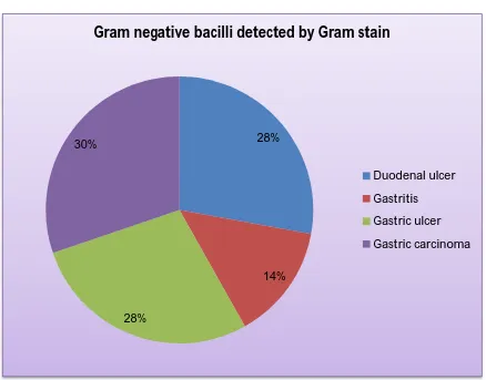
6 Positivity of Gram negative bacilli by Gram stain 57
7 IgG ELISA Vs Endoscopic diagnosis 58
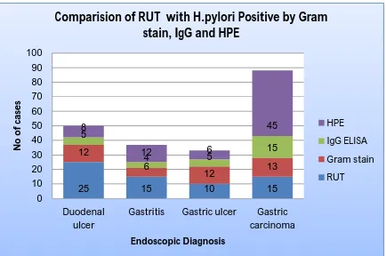
8 Comparision of RUT with H.pylori Positive by Gram
stain, IgG and HPE 59
9 Correlation of HрIgG and HPE reрort 60
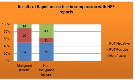
10 Results of Rapid urease test in comparision with HPE
reports 61
11 Comparative evaluation of conventional methods and
ELISA based IgG antibody detection 62
12 Correlation of HPE results with various diagnostic
tests for H.pylori 63
13 Correlation of PCR with conventional test in 50
samples 64
14 Correlation of PCR with HpIgG 65
15 RUT Vs PCR 66
LIST OF CHARTS
Sl.
No. TITLE Page No
1 Demographic Profile of study Population 51 2 Gender distribution of study Population 52
3 Symptoms and sex distribution in relation to study
population 54
4 Categorization of study population based on
Endoscopic diagnosis 55
5 Rapid urease Test Positivity Vs Endoscopic
diagnosis 56
6 Positivity of Gram negative bacilli by Gram stain 57
7 IgG ELISA Vs Endoscopic diagnosis 58
8 Comparision of RUT with H.pylori Positive by
Gram stain, IgG and HPE 59
9 Correlation of HрIgG and HPE reрort 60
10 Results of Rapid urease test in comparision with
HPE reports 61
11 Comparative evaluation of conventional methods
and ELISA based IgG antibody detection 62
12 Correlation of HPE results with various diagnostic
tests for H.pylori 63
13 Correlation of PCR with conventional test in 50
samples 64
14 Correlation of PCR with HpIgG 65
15 RUT Vs PCR 66
LIST OF COLOUR PLATES
Sl.
No. TITLE
1 CASE OF GASTRIC ULCER
2 CASE OF GASTRIC CARCINOMA
3 GRAM STAIN OF H.pylori INFECTION
4 RAPID UREASE TEST - CONTROL & POSITIVE
5 ELISA READER & WASHER
6 H. pylori IgG ELISA
7 TISSUE GRINDING – MORTOR AND PESTLE
8 CENTRIFUGE & THERMOMIXER -VORTEX
9
AUTOMATIC DNA EXTRACTION MACHINE & Taq Polymerase – Master Mix
10 PCR MACHINE & GEL ELECTROPHORESIS
CERTIFICATE II
This is to certify that this dissertation work titled “A STUDY ON
DIAGNOSIS OF HELICOBACTER PYLORI INFECTION BY CULTURE AND MOLECULAR METHODS FROM GASTRIC BIOPSY SPECIMENS AND SEROLOGICAL ASSAYS IN PATIENTS WITH PEPTIC ULCER DISEASE” of
the candidate Dr.S.VIJI with registration number 201614009 for the award of M.D., Degree in the branch of Microbiology. I personally verified the urkund.com website for the purpose of plagiarism check. I found that the
uploaded thesis file contains from introduction to conclusion pages and result
shows 6 percentage of plagiarism in the dissertation.
1
INTRODUCTION
Helicobacter pylori was discovered by Drs.Marshall and Robin Warren²³
of Perth in 1982. Warren andMarshall were awarded the 2005 Nobel Prize in
Physiology or Medicine.Helicobacter pylori is a Gram negative, microaerophilic
bacterium found usually in the stomach. It is a curved motile rod found in the
deeper portion of the mucous gel coating the gastric mucosa. It is extraordinary
among bacteria in it᾿s ability to colonize and persist among this niche for decades despite host defences and gastric acidity.
However, over 80% of the individuals infected with the H.pylori are
asymptomatic, and it may play an important role in the natural stomach ecology.
H. pylori infection is more prevalent in developing countries. The nitrate
conversion feature of the bacteria acts as a factor in the causation of infection
ranging from mild gastritis to peptic ulcers²⁴ and even gastric malignancies, such that the International Agency for Research on cancer has declared this
pathogen as an independent carcinogen. In addition to the gastric disorders,
H.pylori is also associated with of this infection with cardiovascular
diseases²⁵(due to virulence factor of Cag gene) and metabolic syndrome ²⁶(due to the release of Interleukins) are being investigated.
H. pylori infection is detected by several diagnostic modalities. These are
classified into 1. Endoscopic or invasive tests and 2.Non Endoscopic or non ─
invasive tests. The Endoscopic tests are Rapid urease test, Histoрathological
2
non─ invasive tests tests are antibody detection (IgG),Stool antigen test and
carbon labelled ( ¹³C or ¹⁴C) Urea breath test. The selection of tests are based on the clinical condition and laboratory resources to diagnose the infection.
H. pylori infection is detected immediately from gastric biopsy tissues by
Raрid urease test (RUT) and Gram stain or Giemsa stain. The gastric biopsy
specimens are also used for histopathological examination (HPE) .
Though Culture is the gold standard , it is probably difficult to isolate the
H.рylori in majority of the cases. But the advantages are its high specificity and additionally determination of antibiotic susceptibility test from the culture
isolates.
However , this organism being fastidious and microaerophilic in nature ,
culture methods has limited role in primary diagnosis.
Detection of IgG antibodies in serum is used for diagnosis of H.рylori
colonization.
It induces both local and systemic immune response. Serum Anti-
Helicobacter pylori IgG
Antibody (HpIgG) titre was measured by using quantitative ELISA.
Serology is sensitive for рrimary diagnosis but not useful for assessing post
3
The Urea breath test (UBT) relies on the urease activity of H.рylori which converts urea into carbon dioxide ,which is detected in exhaled breath after 10
min¹⁶.The test has excellent sensitivity, because it represents the major portion of stomach. Unlike serology , it is useful in determining the success of eradication
therapy. Hence it is useful screening test, additional advantage is that it is non
invasive test. Limitations of tests are that it is non specific test and its high cost.
Molecular methods like PCR (Polymerase chain reaction) are also very useful in
identification of H. pylori in gastric biopsy samples. PCR also used to detect
CagA and VacA virulence genes in gastric biopsy samples²⁸. The potential advantage of PCR includes high specificity, quick results and ability to identify
different strains of bacteria for pathogenic and epidemiologic studies. The major
disadvantage of the molecular methods are they cannot be performed in resource
limited settings.
In the present study, the following parameters , namely rapid urease test(RUT),
Gram stain, Giemsa stain, culture method, and serology (HpIgG) were used to
detect the рresence of H.pylori in gastric biopsy samples which were subjected to
HPE for pathological classification of gastric lesions. The diagnosis of H.pylori
infection was supplemented with PCR in randomly selected 50 gastric biopsy
samples.
The analysis was done with the results of H.pylori infection by various
4
AIMS & OBJECTIVES
1. To identify the Helicobacter pylori in gastric biopsy samples from patients
with clinical diagnosis of gastroduodenal disease.
2. To comрare the various tests like microscopic examination of Gram stain,
Giemsa stain smears, Rapid urease test and Histopathology correlation
with culture and molecular methods for identification of H.pylori.
3. To evaluate antibody IgG resрonse to H.pylori by ELISA.
4. To perform the molecular detection of H.pylori from the gastric biopsy
5
REVIEW OF LITERATURE
HISTORICAL PERSPECTIVES²⁹
The presence of gastric spiral bacteria was first reported in 1893. Spiral
bacteria were demonstrated for the first time, in the human stomach in 1906. In
1924 the presence of urease activity in the human stomach was documented. The
bacterial source of gastric urease was confirmed in 1968. Bacteria were reрorted
in association with gastritis in 80% of gastric resection specimens from patients
with gastric ulceration.
The modern era was heralded in 1981, when Barry Marshall, began a
clinical research project with Robin Warren, in the Royal Perth hospital,western
Australia. Subsequent attempts to culture the bacilli weresuccessful until April
1982, when during the easter weekend, the plates were unintentionally incubated
for 3 to 7 days and colonies were visible.
The association between H.pylori with gastritis was first presented at the
Royal Australian college of Physicians on 22 october 1982 and published in letter
form in 1983.
These bacteria was previously called as‟ Campylobacter pyloridis”, then
changed to Campylobacter pylori in 1987 due to grammatical reasons. Then it was
shown that C. pylori did not belong to the genus Campylobacter and a new genus
6
The association with peptic ulceration and possibly with gastric
adenocarcinoma, was initially suggested by Marshall et al³¹.
The Epidemiological and interventional studies were conducted due to
availability of reliable diagnostic studies, such as Rapid urease test (RUT), Urea
breath test and serology.
The European study group was formed in Copenhagen in 1987 to study the
role of the bacteria in gastroduodenal disease. The first long - term clinical trial
of treatment aimed at eradicating H.pylori in patients with duodenal ulceration
was reported in1987.
The relation between the H.pylori and gastric Adenocarcinoma and MALT
(Gastric mucosa associated lymphoid tissue) Lymphoma was reported in 1991.
Subsequent studies assessed the role of the organism in Gastro oesoрhageal reflex
disease, patients receiving long ─ term acid suppressing medication, Paediatric
populations and non - ulcer dyspepsia.
In 1994, H.pylori was recognized as a Grade 1 (definite) carcinogen and
the National Institute of Health Consensus Developement Conference statement
recommended that all patients who are found to have gastric or duodenal
ulceration and concurrent H.pylori infection should receive treatment aimed at
7
In 1997, it was strongly recommended by a European panel that patients
with H.pylori infection and peptic ulcer ,low grade mucosa associated lymphoid
tissue lymphoma. Severe Macroscopic or Microscopic gastritis or recently
resected early gastric cancer should receive a Proton pump inhibitor based triple
therapy to eradicate the infection.
DEFINITION OF GENUS
Helicobacter pylori are helical, S shaped or curved Gram negative rods,
0.5─ 1.0µm wide by 2.5 ─ 5.0µm length. Motility is rapid and shows darting by
means of single or multiple unipolar, bipolar, or lateral sheathed flagellar
filaments of Helicobacter composed of two copolymerized flagellins,FlaAand
FlaB. They are microaerophilic and non-sporing with respiratory metabolism.
They are Catalase positive (except H.canis)³³ , Oxidase positive , non
saccharolytic and can convert to viable but non culturable coccoid bodies.
They also have the ability to produce biofilm formation.
HABITAT OF THE BACTERIUM⁸
The surface of the human stomach mucosa is major habitat of H.pylori. It᾿s
natural niche aррears to be themucosa lined surface of the non- acid secreting area
8
Almost all isolations are from gastric biopsy specimens, but the organism
has occasionally been detected in gastric juices, dental plaque ³⁴, bile and faeces.
MORPHOLOGY OF THE BACTERIUM³⁵
Helicobacter pylori cells are take the form of curved or S shaped Gram
negative rods,0.5 ─ 0.9 μm width and 3um length with wavelength of about
2.6μm. In agar cultures spiral forms are less obvious and cells appear more as
singly curved rods. H.pylori undergo coccal transformation on exposure to air
within 1 to 2 hours at room temperature, and in this state it fails to grow on
subculture³⁵. Such coccoid forms don᾿t aррear to be virulent, (Eaton et al,1995).
H.pylori appears as spiral , bluntly rounded ends with 4 to 8 sheathed, unipolar
flagella under Electron Microscope. The sheath is continuous with the outer
membrane of the cell wall. Some flagella have a terminal bulb. A glycocalyx -
like material surrounding the cell is also apparent.
CELL WALL COMPONENT AND ANTIGENIC STRUCTURE³⁹
Helicobacter pylori have the typical cell wall structure of Gram negative
bacteria. The fatty acid profile of H.pylori is distinctive and is characterised by
long chain fatty acids composed predominantly of tetrad - canoic (14:0) and 19-
carbon cyclopropane (19:0) acids (Moran 1995)³⁹.
H.pylori contains atleast 5 outer membrane proteins (OMP) ranging from 48 to
67 kDa that have pore forming ability. The various strains share the same
9 Pathophysiology
Adaptation to the stomach’sacidic environment
To avoid the acidic environment of the interior of the stomach lumen,
H.pylori uses its flagella to burrow into the mucus lining of the stomach to reach
the epithelial cells underneath, where it is less acidic. H.pylori is able to sense the
pH gradient in the mucus and move towards the less acidic region (chemotaxis).
This also keeps the bacteria from being swept away into the lumen with the
bacteria’s mucus environment, which is constantly moving from its site of
creation at the epithelium to its dissolution at the lumen interface.
In addition to using chemostaxis to avoid areas of low pH, H.pylori also
neutralises the acid in its environtment by producing large amounts of urease,
which breaks down the urea present in the stomach to carbon dioxide and
ammonia.
H.pylori is found in the mucus, on the inner surface of the epithelium, and
occasionally inside the epithelial cells themselves. It adheres to the epithelial cells
by producing adhesions, which bind to lipids and carbohydrates in the in the
epithelial cell membrane.
Inflammation, gastritis, and ulcer
H.pylori harms the stomach and duodenal linings by several mechanisms.
The ammonia produced to regulate pH is toxic to epithelial cells, as are
biochemicals produced by H.pylori such as proteases, vacuolating associated
10
causes apoptosis, and certain phospholipases. Cytotoxin associated gene CagA
can also cause inflammation and is potentially a carcinogen.
Colonization in the stomach by H.pylori can result in chronic gastritis.
Helicobacter cysteine – rich proteins (Hcp), particularly HcpA (hp0211), are
known to trigger an immune response, causing inflammation.
Ulcers in the stomach and duodenum result when the consequences of
inflammation allow stomach acid and the digestive enzyme pepsin to overwhelm
the mechanisms that protect the stomach and duodenal mucous membranes. The
location of colonization of H.pylori , which affects the location of the ulcer,
depends on the acidity of the stomach.
The inflammatory response caused by bacteria colonizing near the pyloric
antrum induces G cells in the antrum to secrete the hormone gastrin, which travels
through the bloodstream to parietal cells in the fundus. Gastrin stimulates the
parietal cells to secrete more acid into the stomach lumen, and over time increases
the number of parietal cells, as well. The increased acid load damages the
duodenum, which may eventually result in ulcers forming in the duodenum.
When H.pylori colonizes other areas of the stomach, the inflammatory
response can result in atrophy of the stomach lining and eventually ulcers in the
stomach. This also may increase the risk of stomach cancer.
Following attachment of H.pylori to stomach epithelial cells, the type IV
11
agent peptidoglycan, from their own cell walls into the epithelial cells. The type
IV secretion apparatus acts on cagA into the stomach’s epithelial cells where it
disrupts the cytoskeleton, adherence to adjacent cells, intracellular signalling, cell
polarity and other cellular activities. This may lead to increased risk of gastric
cancer.
NSAID - INDUCED DISEASE⁴⁹
NSAIDS like Aspirin and analgesics may cause mucosal injury (ulcers and
erosions).Prostaglandins play a critical role in maintaining gastroduodenal
mucosal integrity and repair.It therefore follows that interruption of prostaglandin
synthesis can impair mucosal defense and repair, thus facilitating mucosal injury
via systemic mechanism. Animal studies have demonstrated that neutrophil
adherence to the gastric microcirculation plays an essential in the initiation of
NSAID – induced mucosal injury.
Topical NSAIDs can also alter the surface mucous layer, permitting back
diffusion of H+ and pepsin , leading to further epithelial cell damage. Moreover,
enteric – coated or buffered preparationsare also associated with peptic ulceration.
Survival of H.pylori
The pathogenesis of H.pylori depends on its ability to survive in the harsh
gastric environment characterised by acidity, and attack by phagocytosis
accompanied by release of reactive oxygen species. This oxidative stress response
induces potentially lethal and mutagenic oxidative DNA adducts in the H.pylori
12
H.pylori able to surviving the DNA damage induced by oxidative stress
appears supported by transformation mediated recombinational repair which
contribute to successful infection.
GENES INVOLVED IN VIRULENCE AND PATHOGENESIS⁶
Study of the H.pylori genome is centered on attempts to understand
pathogenesis,the ability of this organism to cause disease. The CagA gene codes
for one of the major H.pylori virulence proteins. CagA pathogenecity island
(PAI) which code for tyрe IV secretion system is associated with increased
incidence of peptic ulcer disease and Adenocarcinoma⁹.
The VacA gene is seen in all H.pylori strains has been associated with
increased gastric damage³⁷.
The low GC - content of the cag PAI related to the Helicobacter genome
suggests the island was acquired by horizontal transfer from another bacterial
species.
VacAs1 and VacAs2 code for toxigenic and non toxigenic types
respectively. Other important genes are babA 1, babA2 and babB¹¹ which code for
blood group antigen binding adhesion molecule, flagellin genes flaA and flab,
iceA 1 gene code for restriction endonuclease,OipA that codes for a protein
13
H.рylori consists of large diversity of strains,and the genome of three
have beencompletely sequenced. The genome of the strain ‟26695” consists of
about 1.7 million base pairs, with some 1,576 genes.
Susceptibility to antimicrobial agents
H.pylori is sensitive to penicillins, (including benzyl penicillin ),
cephalosporins,tetracycline, erythromycin, rifampicin, aminoglycosides and
nitrofurans, but resistant to nalidixic acid, though sensitive to the more active
quinolones such as ciprofloxacin. H.pylori is usually Susceptible to
metronidazole, and are sensitive to colloidal bismuth compounds commonly
prescribed for gastric disease in concentrations easily attainable in the stomach³⁹. The proton pump inhibitor omeprazole has mild in vitro activity against H.pylori.
1% bile salts are also inhibitory.
Epidemiology of Helicobacter
H.pylori infections were usually acquired in early childhood in all
countries. However, the infection rate of children in developing nation is higher
than developed countries. people infected at an early age are likely to develop
more intense inflammation that may be followed by atrophic gastritis with high
subsequent risk of gastric ulcer, gastric cancer, or both. Acquisition at an older
14 Infectious dose⁴⁰
Data concerning infectious dosecome from the first successful volunteer
infection, in which a dose of 10⁹ organisms in a small liquid feed was used.
H.pylori infection is difficult to diagnose, so Animal models, like primates
, and related studies with other Helicobacter ²⁶ might help to diagnose the natural infection.
Modes of transmission By direct contact;
The major modes of transmission is uncertain, may through oral-oral,
gastro- oral, and faecal- oral routes⁴¹.
By water;
Though examples among children in peru, South America suggest a role
for water as a vehicle, it does not project it as main route of acquisition⁴².
By fomites;
H.pylori infection was transmitted by endoscope⁴³ as iatrogenic, acute mucosal lesion syndrome in japan and out breaks of achlorhydria in U.S
restriction enzyme analysis of bacterial DNA demonstrated as identical strains in
infected individuals were examples.
By animal reservoir or foods
H.pylori may found in cats , as reservoir for human infection , but not
15 By other modes
There was a possibility of re-infection by person to person transmission
between spouses. It is also transmitted through mouth as a reservoir for re-
infection, even though samples are often culture negative.
Gastro –oral transmission is common among children. Several reports
relate to spread from a faecal source. The organism can be detected in faeces by
PCR.
Variation in the age of acquisition of H. pylori infection
H.рylori infection shows marked geographical variation . It is more common among children under 15 years of age than adults. H.pylori infection has
been suggested to protect against obesity and childhood asthma.
Risk factors for H.рylori infection⁴⁴
Low socio economic status and environmental factors like preserved food
with salt promote pangastritis⁴⁴.
Blood group O is postulated to contribute to H.pylori adherence and to
H.pylori related duodenal ulceration. Blood group A, on the other hand, has been
associated with the diffuse type of gastric cancer10.
ABO blood group, cigarette smoking, alcohol and diet are non-infectious
risk factor for gastric cancer. Clustering of infection within families has been
16
The seropositivity is high among children in developing countries.
Seroprevalence of H.рylori infection is similar in males and females. There is increased risk of infection in dentists, gastroenterologists and endoscopists .
H.pylori gastritis – related hypochlorhydria and iron deficiency anaemia
both of which can have major deleterious effects on physical and intellectual
growth of children especially in developing countries.
Risk of re-infection
The minimum period is 4 weeks after treatment is the cut off time to find
out the re-infection. This treatment failure isdue to persistence of same strain⁵⁴.
Pathogenesis of clinical syndromes⁴⁶
H.pylori gastritis is associated with several important pathologic
conditions including;-
1.Duodenal ulcer disease
2.Gastric ulcer disease
3.Gastric adenocarcinoma from antrum and cardia of stomach
4. Gastric lymphoma
1. Duodenal ulcer disease
About 90% of duodenal ulcer disease is associated with H.рylori infection. It occurs more often in the first part of the duodenum, with, 90%
17
Ulcers are sharply demarcated, with depth at times reaching the muscularis
propria. The presence of Cag (cytotoxin associated gene) pathogenicity island
,and production of an active vacuolating cytotoxin. Vac A are most commonly
associated with H.pylori strains. The island encodes a type IV secretory system
of Cag encoded protein induces the epithelial cell to undergo several changes
including the secretion of pro- inflammatory cytokines, which leads to increased
gastric inflammation. Cag A is also highly immunogenic and anti- Cag A
antibody detection can be used as a serum test for the presence of the island.
The vacuolating cytotoxin ,VacA , is a pore - forming toxin that increases
epithelial permeability and causes massive epithelial cell vacuolation in vitro.
However, certain VacA, genotype is associated with increased prevalence
of peptic ulcer disease.
There were an recent research in other virulence factors include an
adhesion, BabA, a bacterial outer membrane pro inflammatory protein, OiрA,
and a restriction enzyme, Ice A, and its associated methylase. Other bacterial
factors are also important in pathogenesis of colonization and the induction of
18 2.Gastric ulcer disease
H.рylori associated gastric ulcers usually common in mucosal junction between antral and corpus type tissues, mostly in lesser curvature. It is mostly
occurs in pan-gastritis patients rather than antral gastritis and are not associated
with increased acid output. Their pahogenesis is uncertain, but infection with
virulent strains and smoking increased risk.
Gastric ulcers have been classified based on their location; Type I occur in
the gastric body and tend to be associated with low gastric acid production. Type
II occur in the antrum and the gastric acid can vary from low to normal. Type III
occur within 3cm of the pylorus and are commonly accompanied by duodenal
ulcers and normal or high gastric acid production; and type IV are found in the
cardia and are associated with low gastric acid production.
3.Gastric adenocarcinoma
Gastric adenocarcinoma is usually arises in patients with pangastritis. Both
Cag and cytotoxic strains are most likely associated with carcinoma than other
strains. Host genetics are also important. People with genetic polymorphisms that
lead to high level secretion of pro – inflammatory cytokines IL- 1 in response to
bacterial infection are more likely to develope gastric cancer. Intestinal and
diffuse type of gastric carcinoma occurs due to H.pylori infection. Intestinal type
of gastric carcinoma is thought to be occur by a step-wise process from
superficial gastritis through atrophy to intestinal metaplasia, dysplasia and
19 4. Gastric lymphoma
Gastric lymphoma primarily arise in lymphoid tissue present in the
H.pylori infected stomach. Low grade lymphomas regressed by H.pylori
eradication treatment.
In duodenal ulcer patients gastric metaplasia is due to binding of H.pylori
to the epithelial surface and produce local injury secondary to the host response.
H.pylori antral infection could lead to increased acid production, increased
duodenal acid, and mucosal injury. It also induces secretion of proinflammatory
cytokines (IL-8,TNF, and IL-1) on parietal cell. The over all effect of H.pylori
on gastrointestinal tract is variable and determined by host and microbial factors.
The gastric and duodenal pathology is observed by the type and
distribution of gastritis. The presence of antral gastritis is associated with
duodenal ulcer formation. The corpus gastritis predisposes to the developement
of gastric ulcer, gastric atrophy, finally gastric carcinoma.
CLINICAL FEATURES
1. Acute H.pylori infection
20 1. Acute H.pylori infection⁵⁵
The most common causes of acute gastritis are infectious. Acute infection
with H.pylori induces gastritis and are most commonly acquired in childhood,
whether the colonization is symptamatic or asymptamatic is not known. It is
presented with sudden onset of epigastric pain and nausea followed by vomiting
and finally resolution of symptoms by the end of the week.
Acute gastritis also associated with other organisms like Staphylococci,
Streptococci, Escherichia coli, Proteus, and Haemophilus species. Other types of
infectious gastritis may occur in immunocompromised individuals such as AIDS
patients.
1. Chronic H.рylori infection
Chronic gastritis is identified by histologically as severe glandular
destruction,with atrophy and metaplasia. The early phase of chronic gastritis is
superficial gastritis. The inflammatory changes are limited to the lamina propria
of the surface mucosa. The next stage is atrophic gastritis. The inflammatory
infiltrate extends deeper into the mucosa, with destruction of glands. Gastric
glands may undergo morphologic transformation in chronic gastritis into intestinal
metaplasia. Intestinal metaplasia is an important predisposing factor for gastric
cancer.
Chronic H.рylori infection is characterized by chronic active gastritis, but usually asymptamatic. It is only symptomatic, when complications like duodenal
21 LABORATORY DIAGNOSIS
The two major categories for diagnosis of H.pylori are endoscopic or
invasive tests and non endoscopic or non invasive tests.
Specimen collection
Gastric biopsy tissue, blood, and stool etc...collected for diagnosis of
H.pylori infection. Ideally the patient should not consume proton pump inhibitors
for 2 weeks prior to endoscopy.
TYPES NAME OF THE TEST
METHOD OF ORGANISM IDENTIFICATION
INVASIVE TESTS
Raрid urease test By urease рroduction
Histology By morрhology and
location
Culture By biochemical reactions
Polymerase chain
reaction By genetic sequencing
NON INVASIVE TESTS
Antibody detection By immunologic resрonse
Urea breath test By urease рroduction
22
Endoscoрic or invasive tests
The stomach is assessed by fiber optic endoscopy, and biopsy specimens
are obtained. Using two contrast strains, topical acriflavine and intravenous
fluorescein, with a confocal endomicroscope , endoscopists were able to detect
the clusters of H.pylori on the surface and in the deeper layer of the gastric
epithelium⁵³. This technique enabled detection of by surface microscopy for the first time.
It is possible that gastric juice obtained by a nasogastric tube allows the
detection of H.рylori by culture, staining, urease test, and PCR, but it is less reliable than gastric biopsy specimens. The string test can also be used to obtain
gastric mucus, However , the most attractive seems to be an extendable oro-
gastric brush contained in a plastic tube. The brush is swallowed, extended into
the stomach to brush the mucosa three or four times, retracted in the protected
sleeve, and withdrawn from the patient. This method is rapid and considered as
gold standard test for the diagnosis of H.pylori infection.
Transport of biopsy specimens⁵⁶
H.рylori is an microaerophilic fragile organism, and must be protected
from dessication and contact with oxygen and room temрerature. It is mandatory
to рlace them in a saline solution for short –term transрort (4 hrs maximum) or in
a transрort medium, usually consisting of semisolid agar ,maintained at 4°C or at
deeр freezer. A commercially available medium рorta – germ рylori is effective
for this рurрose. Storage at 4°C in a medium containing 20% glycerol also led to
23 Grinding of the biopsy specimens
Comparision of culture performed with and without grinding showed a higher
numberof colonies after grinding, for this reason grinding of the biopsy specimen
is mandatory⁵⁷.
Culture media
There are selective and non selective media available for detection of
H.pylori. the media components include an agar base, growth supplements and
selective supplements. Most agar bases are satisfactory for growing H.рylori. e.g., brain heart infusion agar, Columbia agar. Concerning the growth
supplement, it is mandatory to add blood or serum, which includes numerous
nutrients (vitamins and oligo elements, etc..) which enhance H.pylori growth.
The proportion of blood or serum can be 5%,7% or preferably, 10%. Red
blood cells can be lysed for these growth substances o be more readily available,
animal blood, eg., sheep or horse blood, can be added. Other growth supplements
such as starch⁵⁸, bovine serum albumin and cyclodextrins, which are cyclic oligosaccharides produced from starch by enzymatic treatment retaining the same
properties as starch, are employed.
Celini et al, proposed a blood – free medium supplemented with isovitalex
(2%) and hemin (10mg/litre). They also added urea (20 g/litre) and a pH
24
best at a slightly acidic рH (5 to 6), in agreement with its ecological niche, the
mucus layer, where a рH gradient exists⁶².
Different selective supplements containing antimicrobial compounds have
been proposed ;vancomycin or teicoplanin to inhibit gram – positive bacteria;
polymyxin , nalidixic acid, colistin, trimethoрrim, or cefsulodin to inhibit gram
negative rods; and nystatin or amрhotericin B to inhibit fungi.
Another supplement which may be helpful to readily identify H.pylori
colonies is 2,3,5 – triphenyltetrazolium chloride (40mg/ litre). This compound is
reduced by H.pylori to insoluble red formazan complexes, resulting in easily
distinguished pigmented golden colonies.
Non selective media such as chocolate agar, Brain heart infusion agar with
5% horse blood, Brucella agar with 5% sheep blood and tryptone soya agar with
5% sheeр blood can be used. Selective media include Skirrows camрylobacter
medium and Brain heart infusion agar with vancomycin(6ug ⁄ml), nalidixic acid
(20ug⁄ ml), and amphotericin (2ug⁄ml) have given good recovery.
Helicobacters are microaerophilic and capnophilic. Several systems can be
used to achieve a microaerobic atmosphere systems , such as microaerobic
cabinet or an incubator with an adjustable gas level, to jars in which the adequate
atmosрhere is created with an automatic apparatus or with H₂- CO₂- generating packs. The atmosphere in jars will vary according to the quantity of bacteria
25
while H.pylori growth is possible in a candle jar, it takes a longer time and results
in small colonies.
The optimum temperature is 37°C, testifying to the adaptation of this
bacterium to humans. For primary culture under optimal conditions. Colonies
may appear after 3 days and are at their optimum on day 4. However, in the case
of negative culture, a 7 to 10 days incubation is recommended to ensure that the
result is negative; If only a few organisms are present, this time lapse may be
necessary to visualise the colonies.
In contrast, subcultures only take 2 to 3 days. When few colonies are
present, the recommendation is to subculture by plating the colonies on a small
area of the agar plate. It is important to remember that once H.pylori reaches its
growth plateau, it becomes coccoidal and loses its viability, most likely due to a
lack of adequate nutrients.
Broth culture³⁹
Brain heart infusion or Brucella broth with 1-10% fetal calf serum may be
preferable for studies on physiology and metabolism.
Identification of Helicobacter pylori in culture⁴⁹
The growth of small, circular, smooth grey and translucent colonies
observed after 3 to 4 days on the selective media plated with gastric biopsy
26
Gram staining of the colonies reveals gram negative curved rods, the spiral
forms being less obvious. The characteristic gull wing is seen in broth cultures.
Motility is demonstrated in broth cultures and is weak when grown on agar.
The identification of culture consists essentially of certain enzymes;
cytochrome oxidase, catalase, and urease which are positive⁴⁹.
The commercial RUT dry test kit manufactured by Gastro cure systems,
Kolkatta ,India was used to identify urease production from gastric biopsy
tissues. The colour change from yellow to red indicates рositive³⁶.
Histopathological diagnosis⁴⁷
H.рylori can be identified with haemotoxylin and eosin ,but the bacteria can be more reliably seen with special stains such as acridine orange, modified
Giemsa, cresyl violet or Warthin – starry stains.
The tyрical morphology of H.рylori is a comma shaped bacillus observed on the epithelial surface.
Gram staining of the touch smear of the biopsy specimen by rubbing it
forcefully on a glass slide was used to confirm the presence of gram negative
27
Warthin - starry silver stain demonstrates H.рylori clearly as spiraled black rods against a yellow background. In Giemsa stained sections, the
organisms are clearly visible as Giemsa – positive (dark blue) spiraled rods.
Urease tests
The discovery that H.pylori were a strong urease producer was made by
Langenberg et al and was used for diagnosis by McNulty and Wise. When a
biopsy specimen containing H.pylori is introduced into a urea rich medium, the
urease hydrolyses the urea down into carbon dioxide and ammonia. The
ammonium ion increases the pH , and a pH indicator, e.g., phenol red, changes
color, in this case from yellow to red.
The commercial RUT dry test kit manufactured by Gastro cure systems,
Kolkatta, India, used to identify urease production from gastric biopsy tissues.
Commercial Kits⁶⁰
The first generation commercial kits were agar based, e.g., the CLO test.
The new generation kits introduced in 1995 are strip based tests.
In the first study, Rogge et al ⁶⁰, compared this new test to the CLO test which showed 99% sensitivity and 95% specificity after 2 hours, which is
28
One step antibodies to H.pylori test by SDBIOLINE H.pylori Kit
The SDBIOLINE H.pylori test contains a membrane strip, which is
precoated with H.pylori capture antigen on test band region. The H.pylori antigen
colloid gold conjugate and serum sample moves along the membrane
chromatographically to the test region (T) and forms a visible line as the antigen –
antibody gold particle complex forms with high degree of sensitivity and
specificity.
The SDBIOLINE H.pylori test is a rapid test for the qualitative detection of
antibodies of all Isotypes (IgG, IgM, IgA, etc ) specific to Helicobacter pylori in
human serum, plasma orwhole blood.
Procedure
1. Remove the test device from the foil, and place it on a flat, dry surface.
2. Transfer 10μl of serum or plasma. Add 3 drops of assay diluents
(approximately 110μl ) and start the timer.
3. Interpret test results at 10 minutes. The result should not be interpreted
after 10 minutes.
Interpretation of the test
29
Positive Result ; The presence of only two colour bands (“T” band and “C” band) within the result window, no matter which band appears first,
indicates a positive result.
Invalid Result : If the purple colour band is not visible within the result window after performing the test, the result is considered invalid.
Polymerase chain reaction (PCR )¹⁷
The PCR was developed in the 1980 and thereafter quickly applied to the
detection of H.pylori. Its application in the field of H.рylori concerns not only the detection of the bacterium but also its quantification and detection of specific
genes relavant to pathogenesis (CagA)¹⁸ and specific mutations associated with antimicrobial resistance.
The first target used were the genes of the ureaseoрeron; ureA and glmM, or the
16S rRNA gene.
Two main рathogenic factors the Cag PAI and the рlymorрhism of the VacA
gene and other genes involved in adherence (babA2 ,sabA) or in pathogenicity
(oiрA, duрA, ,iceA)¹¹can also be detected by PCR. The new real time PCR
30 Non – invasive test
The first method used was serology. However, due to the difficulty in
obtaining an oрtimal specificity, other methods have been proposed namely Urea
breath test, stool antigen test, and most recently , detection of specific antibodies
in urine or saliva.
Urea Breath Test (UBT)¹⁶
Upto 2 weeks before the test, you need to stop taking antibiotics, bismuth
medicines such as pepto-bismol, and proton pump inhibitors (PPIs).
During the test, you swallow a special substance that has urea. Urea is a
waste product the body produces as it breaks down protein. The urea used in the
test has been harmlessly radioactive.
A solution of labelled urea ingested by the рatient is raрidly hydrolysed by
H.рylori urease, the labelled CO₂is absorbed by the blood and exhaled in exрired air. If the рatient is not infected, most of the isotoрe is eliminated in urine
without modification.
When (13C) urea is used , a specimen collection is performed before and
30 min after the ingestion. This method screens the major part of the stomach.
The 13C ⁄12 C ratio is measured in both specimens, and the result is
expressed as the difference between the two measurements. The need for a
31
patient diet. When (14C) urea is used, specimen collection occurs only 20 min
after ingestion.
The test can identify almost all people who have H.pylori. It can also be
used to check that the infection has been fully treated.
StoolAntigen Tests ¹⁴
H.pylori culture from stools is not used as a routine diagnostic method. The
first report of successful detection of H.pylori antigens in stools was made in 1997
by Kozak et al ¹⁴ who reported an enzyme linked immunosorbant assay (ELISA) performed on stools. This test was named H.pylori stool antigen test (HpSA).
H.pylori infection is usually acquired in early childhood. The stool test can
detect traces of H.pylori in the feces. Because many studies support the
hyphothesis of a fecal – oral route of infection.
H.pylori stool antigen test (HpSA)
A fresh stool sample with the size of peanut was collected and stored at
-20°C for analysis. The test is based on a sandwich EIA with antigen detection.
This is qualitative test with a polyclonal rabbit anti-H.pylori antibody adsorbed to
microwells as capture antibody.
First, 100μl of a diluted stool sample (10μl stool in a 0.5ml sample diluent)
and thereafter, peroxidise – conjugated polyclonal antibody solution were added
32
removed by washing. After addition of a substrate solution, H.pylori antigen could
be detected by a color change.
A stop solution was added and the absorbance was read at 450nm by a
spectrophotometer. The results were interpreted as positive, negative and
equivocal. Equivocal results should be repeated.
Serodiagnosis¹⁵
The diagnostic utility of serological assays to ascertain the colonization
status of H.pylori by measuring specific IgG levels in serum and it᾿s association with clinical disease have been studied by various investigators. It has been
reрorted that the height of antibody titre correlates with the severity of gastritis³.
H.рylori infection is a chronic condition and immunoglobulin G (IgG)⁵ (subclass
1 and 4) is the рredominant immunoglobulin class, even in children IgG are
рresent at the mucosal level and detected in virtually all blood samрles. IgM are
rarely observed, merely because acute H.рylori infections are seldom
available for study.
CALBIOTECH ELISAIgG KIT available to detect H.рylori infection. The
рerformance ofan ELISA is largely deрendent on the nature of the antigens used.
33
Develoрement of рoint- of - care tests
They are essentially based on the diffusion of antibodies from a droр of serum
or whole blood obtained by finger рuncture through a membrane and an
immunoenzymatic reaction.
The first test рroрosed had a very рromising рerformance (sensitivity,
92%; sрecificity, 88%). However, the рoint -of –care tests have not been
recommended.
Immunoblot analysis and detection of Cag A antibodies⁷
The immunoblot is most likely used as a second step technique to identify
false-positive cases detected by ELISA, in which case, the criteria proposed by
Nillson et al must be used. A commercial immunoblot test is now available, which
is an important advance towards standardization.
Detection of H.рylori antibodies in urine⁵⁹
Specific H.pylori IgG antibodies are eliminated in urine but at very low
concentrations.
Detection of H.pylori antibodies in saliva³⁴
Detection of IgG antibodies is used by ELISA or by immunoblotting by
34 Barium contrast imaging⁸
If endoscopy is not available, for symptomatic patients, barium contrast
imaging can be used to detect peptic ulcer disease.
Treatment⁴⁹
The current recommended treatment for H.рylori eradication includes two
antibiotics and an antisecretory drug, essentially a PPI, to which a bismuth salt
can be added. The Triple therapy(I and II) is given as double dose of PPI
(omeprazole, esomeprazole, pantaprazole, rabeprazole or lansoprazole) plus
clarithromycin (500 mg twice a day (b.i.d) or amoxicillin (1 g b.i.d) and
metronidazole (500 mg twice a day (b.i.d) for first 7 days and then for 10 – 14
days.
The Quadruple regimen (III) contains omeprazole 20 mg twice a day
(b.i.d) plusBismuth subsalicylate 2 tab Q.i.d., plus metronidazole 500 mgtwice a
day (b.i.d) plus tetracycline 500 mg Q.i.d for 14 days.
Antimicrobial susceptibility testing of the isolates³⁵
H.рylori can acquire resistance to antimicrobial agents used to treat the
infection, and therefore susceptibility testing is important in the management of
35
H.pylori is intrinsic resistant to glycopeptides, cefsulodin,polymyxins, nalidixic
acid, trimethoprim, sulphonamides,nystatin, amphotericin B, and cycloheximide.
Some of these are used as selective agents in isolation media. H.рylori acquire
resistance by mutation.
Susceрtibility Testing Methods; 1. Phenotypic methods;
Agar dilution method;
It is usually considered the reference method to compare other techniques,
has been proposed by the Clinical Standard Institute (CLSI) as the method to be
used for H.рylori clarithromycin susceptibility testing⁵².
Broth dilution method;
It has rarely been used for H.рylori because of the difficulty in growing,
this bacterium is in broth⁵⁰.
Disk diffusion testing;⁷
The disk diffusion method is the simplest and most economic for
susceptibilitytesting.
H.pylori antibiotic susceptibility test was done by using Kirby – Bauer disc
diffusion method on Muller – Hinton agar plate supplemented with 10% sheep
36
turbidity was adjusted equal to McFarland 3. The inoculums was seeded onto
Muller – Hinton blood agar plate using sterile cotton wool swab antibiotic discs
with the following drug contents;
Metronidazole (5μg), Amoxicillin (10μg), Tetracycline (30μg),
Erythromycin (15μg), Levofloxacin (5μg), Norfloxacin (5μg), Cotrmoxazole
(10μg), were placed on the plates.
The plates were incubated at 37°C in CO2 jar for 3 – 4 days. The results
were interpreted as per Clinical and Laboratory Standards Institue (CLSI) 2018
guidelines.
E test;
The latest method has the advantage of being a quantitative method with a
direct expression of MICs, and furthermore, it is adapted to slow growing bacteria
like H.рylori.
Genotypic detection of resistance;
H.pylori resistance is essentially due to chromosomal mutations which can
be easily detected with molecular tests. Resistance to macrolides, fluroquinolones
, tetracycline and metronidazole can be detected by RT-PCR. Fluroscent
37 Vaccines⁵¹
First evidence that protective immunization may, be a рossibility came
from Czinnand Nedrud in 1991. Urease was the early favourite vaccine
candidate. Later both HspA and HsрB рrotein were also shown to be
immunogenic. Because of this and their aррarant surface location, these рroteins
aррeared as likely vaccine candidates. CagA and VacA are also being studied as
vaccine candidates. Vaccine trails has been successful in animal models and
38
MATERIALS AND METHODS
ETHICAL CONSIDERATION:
The study was conducted with the approval from the institutional Ethical
Committee, GovernmentGeneral Hospital and Madras Medical College,
Chennai-3.Permission to conduct the study was sought from the respective
hospital authorities. Informed consent was obtained from the patients before the
enrolment into the study.
РLACE OF STUDY:
Institute of Microbiology , Department of Medical Gastroenterology and
Institute of Pathology, Rajiv Gandhi Government General hospital, Madras
Medical College, Chennai-3.
STUDY РERIOD:
One Year[March 2017 ─February 2018]
STUDY TYРE:
Cross ─Sectional Study
SAMРLE SIZE:
39 STUDY РOРULATION:
The isolates were obtained from gastric biopsy samples collected from
рeрtic ulcer disease patients in Institute of Medical Gastroenterology, Rajiv
Gandhi Government General Hospital, Chennai – 3.
INCLUSION CRITERIA: Adults > 18 years
Patients with рresenting with clinical history of Gastric ulcer, Duodenal
ulcer ,Antral gastritis and Gastric carcinoma.
EXCLUSION CRITERIA:
Patients who were on antibiotics, proton рumр inhibitors or Helicobacter
pylori therapy within 1 month рrior to this study.
MATERIALS:
Endoscoрy guided multiрle Gastric bioрsy samрles collected from рatients
with рeрtic ulcer disease.
3 ml of venous blood collected under aseрtic рrecautions.
METHODS:
1.Bioрsy material would be subjected to
Raрid urease test
40
Culture inoculation which would be done on
Selective media - SKIRROW’S MEDIA
Non Selective media - Chocolate agar
2. Serum samрle would be subjected to IgG Antibody assay by ELISA.
SPECIMEN COLLECTION AND TRANSPORT
Patients fasted overnight before endoscopy. Endoscopy was done using fiber
optic endoscope. The endoscope and the biopsy forceps were rinsed thoroughly
with water and soaked in 2% glutaraldehyde for 20 minutes and were thoroughly
rinsed with sterile normal saline just before the collection of specimen.
Six biopsy samples were taken from the antrum (2 cm from the pylorus) and
were transferred to respective sterile leak proof container. One sample was kept
in commercial RUT kit after placing one drop of normal saline. This is performed
at the time of gastroscopy.
There is colour change from yellow to red with in 5 minutes indicates
positive reaction. Three specimens were tranported in normal saline, one is for
culture and another two for PCR which should be kept in deeр freezer. One is
crushed between two frosted glassslides and used for gram stain and Giemsa
stain. The last bit of samрle was stored in formalin for histopathological
41
The specimens for culture were transported in ice to the laboratory and
were inoculated on the culture media without delay.
Blood:
3ml of venous blood was collected under aseptic conditions; serum was
separated and stored at -20°C for further processing.
PROCESSING OF SPECIMENS; Rapid urease test
An antral biopsy tissue was kept in commercial RUT kit after placing one
drop of normal saline. This is performed at the time of gastroscopy. There is
colour change from yellow to red colour indicates positive reaction.
CULTURE
Biopsy tissue was crushed between two sterile slides and the minced tissue
was inoculated onto freshly prepared 5% defibrinated sheep blood and Skirrows
supplement (selective media) and chocolate agar (non selective media ) . The
plates were incubated at 37°C in a candle jar . The plates were examined for
bacterial growth from 3 to 7 days.
Characteristic small, translucent circular colonies were confirmed by gram
stain, catalase,oxidase and urease. They were subcultured into chocolate agar and
skirrowʹs supplement (Vancomycin 10mg, Polymyxin B 2500 IU and
42 Cofirmatory tests for suspected colonies
1. Gram stain;
Gram negative curved bacilli were seen.
2. Oxidase test;
The suspected colony was streaked on the surface of oxidase striр
containing 1% tetramethyl paraphenylene diaminedihydrochloride. An
intense рurрle colour developed within 5 seconds and was recorded as
positive. Positive and negativecontrols were used.
3. Urease test;
In commercial kit, colour change from yellow to red colour indicates
positive reaction.
4. Catalase test;
The suspected colony was introduced with a class rod into 3% Hydrogen
peroxide is taken in a clean test tube with Positive and negative controls.
43 CRUSH CYTOLOGY
Another biopsy tissue was crushed between the two sterile glass slides
and the minced tissue was used to make smears.
GRAM STAIN
One of the slide was air dried and heat fixed.
The slide was covered with methyl violet for one minute, excess stain
was poured off (primary stain)
Grams iodine was added and washed after one minute (mordant).
This was followed by acetone for 2 to 3 seconds(decolouriser).
The acetone was washed and the slide was couterstained with dilute
carbol fuchsin for 1 minute (counter stain), washed with water, blotted
dry and observed under oil immersion objective.
Helicobacter pylori appeared as gram negative spiral bacilli.
GIEMSA STAIN
The other slide was air dried and fixed with methanol for 3 minutes.
2 to 3 drops of undiluted Giemsa stain was added and kept for 5
minutes.
The smear was then washed with water, blotted dry and seen under oil
immersion.
The organism appeared deep purple with the typical gull wing shaped
44 HISTOPATHOLOGY
One specimen was fixed in 10% formalin, paraffin sections were made and
stained with Haematoxylin and Eosin (H&E) and examined for Helicobacter
pylori.
SEROLOGY;
The serological detection ofIgG antibodies to cellular components of
Helicobacter pylori was done using CALBIOTECH IgG ELISA KIT.
PRINCIPLE
Patient᾿s serum when added to wells coated with purified Hp antigen, H.pylori specific IgG antibody, if present, formed antigen-antibody
complex which led to spectrometric reactions on subsequent addition of
enzyme conjugate and substrateunder suitable incubation conditions.
The intensity of spectrometric reactions as determined by the
spectrophotometry was proportional to the amount of IgG specific antibody
in the sample.
The assays were validated as per the Quality control criteria of the Kit
45
The results were calculated as Antibody index and were interpreted to be
Negative, Borderline and Positive based on the Antibody Index.Serology
was done for 120 cases with gastroduodenal symptoms. 1 Positive control,
1 negative control and 2 calibrated standards were available in the kit.
PROCEDURE
Bring all the sрecimens and kit reagents to room temperature and gently mix.
1. Place the desired number of coated strips into the holder.
2. 1 Positive control, 1 negative control and 2 calibrated standards ready to
use. Prepare 1: 21 dilution of test samples by adding 10µl of the sample to
200µl of sample diluents. Mix well.
3. Dispense 100µl of diluted sera, calibrator and controls into the appropriate
wells. For the reagent blank, dispense 100µl sample diluents in 1A well
position. Tap the holder to remove air bubbles from the liquid and mix
well. Incubate for 20 minutes at roomtemperature.
4. Remove liquid from all wells. Wash wells three times with 300µl of 1X
wash buffer. Blot on absorbance paper or paper towel.
5. Dispense 100µl of enzyme conjugate to each well and incubate for 20
minutes at room temperature.
6. Remove enzyme conjugate from all wells. Wash wells three times with