Secreted Oral Epithelial Cell Membrane Vesicles Induce Epstein-Barr
Virus Reactivation in Latently Infected B Cells
Zhen Lin,aKenneth Swan,bXin Zhang,cSubing Cao,aZoe Brett,dStacy Drury,d Michael J. Strong,aClaire Fewell,aAdriane Puetter,e Xia Wang,aMaryBeth Ferris,fDeborah E. Sullivan,fLi Li,cErik K. Flemingtona
Tulane University Health Sciences Center and Tulane Cancer Center, New Orleans, Louisiana, USAa
; Department of Medicine, Section of OB/GYN, Tulane University School of Medicine, New Orleans, Louisiana, USAb
; Institute of Translational Research, Ochsner Clinic Foundation, New Orleans, Louisiana, USAc
; Department of Psychiatry and Behavioral Sciences, Tulane University School of Medicine, New Orleans, Louisiana, USAd
; Department of Medicine, Section of Gastroenterology, Tulane University School of Medicine, New Orleans, Louisiana, USAe
; Department of Microbiology & Immunology, Tulane University School of Medicine, New Orleans, Louisiana, USAf
ABSTRACT
In the oral epithelium, peripheral stores of Epstein-Barr virus (EBV) are transmitted from infiltrating B cells to epithelial cells.
Once the virus is transmitted to epithelial cells, the highly permissive nature of this cell type for lytic replication allows virus
am-plification and exchange to other hosts. Since the initial transfer of EBV from B cells to epithelial cells requires transitioning of
the B-cell to a state that induces virus reactivation, we hypothesized that there might be epithelium-specific signals that allow the
infiltrating B cells to sense the appropriate environment to initiate reactivation and begin this exchange process. We previously
found that the epithelium-specific miR-200 family of microRNAs promotes EBV lytic replication. Here we show that there are
high levels of miR-200 family members in oral and tonsillar epithelia and in saliva. Analysis of cultured oral epithelial cells
(OKF6) showed that they actively secrete membrane vesicles (exosomes) that are enriched with miR-200 family members.
Cocul-turing of EBV-positive B cells with OKF6 cells induced viral reactivation. Further, treatment of EBV-positive B cells with OKF6
cell-derived membrane vesicles promoted reactivation. Using a cell system that does not naturally express miR-200 family
mem-bers, we found that enforced expression of a miR-200 family member produced membrane vesicles that were able to induce the
lytic cascade in EBV-positive B cells. We propose that membrane vesicles secreted by oral and tonsillar epithelial cells may serve
as a tissue-specific environmental cue that initiates reactivation in B cells, promoting the transfer of virus from peripheral B-cell
stores to the oral epithelium to facilitate virus amplification and exchange to other hosts.
IMPORTANCE
Epstein-Barr virus (EBV) is an important human pathogen that is causally associated with several lymphomas and carcinomas.
The switch from latency to the lytic cycle is critical for successful host infection and for EBV pathogenesis. Although the EBV
lytic cycle can be triggered by certain agents
in vitro
, the mechanisms that signal reactivation
in vivo
are poorly understood. We
previously reported that endogenously expressed miR-200 family members likely play a role in facilitating the lytic tendencies of
EBV in epithelial cells. Here we show that membrane vesicles secreted from oral epithelial cells contain miR-200 family members
and that they can be transmitted to proximal EBV-positive B cells, where they trigger reactivation. We propose that this
intercel-lular communication pathway may serve as a sensor mechanism for infiltrating B cells to recognize an appropriate environment
to initiate reactivation, thereby allowing the exchange of virus to the oral epithelium.
M
embrane vesicles (MVs), such as exosomes and
mi-crovesicles, can be actively released by cells into the
extracel-lular environment, where they can facilitate intercelextracel-lular
commu-nication. Exosomes are a class of small membrane vesicles (30 to
150 nm in diameter) of endocytic origin that are secreted from
most cell types, including epithelial cells and lymphocytes, under
both physiological and pathological conditions (
1–8
). Exosomes
are composed of proteins, lipids, and nucleic acids that are derived
from their cells of origin. Through the delivery of biologically
active components from the cells of origin to neighboring
and/or distant cells, exosomes are able to modulate many
bio-logical activities, such as tumorigenesis, immunosurveillance,
cell proliferation, and angiogenesis (
1
,
9–12
). MicroRNAs
(miRNAs) are important exosomal cargo that facilitate
signal-ing pathway alterations in recipient cells (
2
,
13–17
).
EBV-en-coded BART miRNAs are selectively enriched in EBV-positive
B-cell-derived exosomes (
13
,
14
) and have been shown to
in-hibit NLRP3 inflammasome-mediated interleukin 1

(IL-1

)
production (
14
) and to suppress the expression of the CXCL11
gene (
13
), an immunoregulatory gene involved in antiviral
ac-tivity and lymphomagenesis.
Epstein-Barr virus (EBV) causes a lifelong infection, with more
than 90% of the adult population worldwide being persistent
car-riers (
18
). EBV primarily utilizes B cells and epithelial cells in its
infection cascade (
18
,
19
). As a bona fide human tumor virus, EBV
plays an etiological role in a number of lymphoid and epithelial
malignancies, including non-Hodgkin’s and Hodgkin’s
lym-Received5 November 2015Accepted8 January 2016
Accepted manuscript posted online13 January 2016
CitationLin Z, Swan K, Zhang X, Cao S, Brett Z, Drury S, Strong MJ, Fewell C, Puetter A, Wang X, Ferris M, Sullivan DE, Li L, Flemington EK. 2016. Secreted oral epithelial cell membrane vesicles induce Epstein-Barr virus reactivation in latently infected B cells. J Virol 90:3469 –3479.doi:10.1128/JVI.02830-15.
Editor:R. M. Longnecker
Address correspondence to Erik K. Flemington, erik@tulane.edu.
Copyright © 2016, American Society for Microbiology. All Rights Reserved.
on November 7, 2019 by guest
http://jvi.asm.org/
phoma, nasopharyngeal carcinoma, and gastric carcinoma (
18
).
Similar to other herpesviruses, EBV has a biphasic infection cycle
that includes a replicative phase (lytic cycle) and a latency phase
(
19
,
20
). Following initial infection, EBV preferentially exists in
host B cells in a state of latency in which no viral production
occurs. Nevertheless, under certain circumstances, latency in B
cells can be disrupted and the virus can enter into a productive
viral replication phase (
19
).
The switch from latency to the lytic cycle in B cells is a
funda-mental component of the virus infection cycle that is critical for
virus persistence and pathogenesis. Chemicals that alter certain
intracellular regulatory pathways, such as phorbol ester, calcium
ionophores, histone deacetylase inhibitors (e.g., butyrate), and
DNA-demethylating agents (e.g., 5-aza-cytidine), can be used to
artificially induce reactivation in latently infected cell lines (
21–
24
). Possibly more physiologically relevant than these chemical
inducers, ectopic transforming growth factor

(TGF-

) or B-cell
receptor (BCR) engagement can induce reactivation in tissue
cul-ture (
25–27
). This information gives clues about some of the
regulatory pathways that facilitate reactivation, but even the
sig-nificance of TGF-

or BCR engagement in triggering virus
pro-duction
in vivo
has been a subject of debate (
19
,
28
).
Probably the most significant site of virus production is the
oral cavity, where salivary virions are produced and spread from
host to host through deep kissing or other salivary exchange
mechanisms (
29
). In contrast to the case with B cells, infection of
epithelial tissues tends to lead to productive lytic replication rather
than latency, and we (
30
) and Ellis-Connell et al. (
31
) have shown
that this may result in part through a mechanism involving the
epithelium-specific microRNA-200 family of microRNAs which
inhibit expression of a key immediate early gene repressor, ZEB1
(
32
,
33
). The propensity of epithelial cells to support lytic
replica-tion makes good sense from the standpoint of host-to-host
trans-mission because it presumably facilitates the production of high
viral titers in the oral epithelium that can be shed to the saliva.
The Hutt-Fletcher group (
34
,
35
) has shown a cross-tissue
in-fection bias for virus produced in B cells and epithelial cells, with
virus produced in B cells preferentially infecting epithelial cells
and vice versa. This speaks to the likely importance of cross-tissue
virus exchange in the EBV infection cycle. The storage site of EBV
in the persistently infected host is considered to be latently
in-fected B cells (
18
). The transfer of virus from this storage vehicle to
the oral epithelium likely occurs through reactivating B cells that
infiltrate the oral epithelium or otherwise in proximity to the oral
epithelium. The higher propensity of virus produced in B cells to
infect epithelial tissue presumably helps facilitate this process.
Nevertheless, we posited that the rarity of “random” reactivation
in B cells would present a substantial inefficiency in this
B-cell-to-epithelial-cell exchange process. We hypothesized that
epitheli-um-derived transmissible agents capable of inducing reactivation
in B cells might favor this B-cell-to-epithelial-cell exchange. Such
an agent would act to trigger B-cell reactivation when the
appro-priate epithelial milieu is detected. Since we knew that the
epithe-lium-specific miR-200 family could induce reactivation in B cells
(
30
,
31
) and since microRNAs are commonly found in shed
brane vesicles, we hypothesized that epithelium-derived
mem-brane vesicles might provide such a sensor mechanism. Here we
provide evidence that epithelium-derived membrane vesicles can
induce reactivation in latently infected B cells and that this is likely
at least partly due to the presence of miR-200 family members.
MATERIALS AND METHODS
Collection of clinical specimens.Buccal epithelium and saliva samples were collected from two healthy participants in the department of psychi-atry and behavioral sciences at the Tulane medical school in accordance with a protocol approved by the Institutional Review Board (IRB) of Tu-lane University. For the saliva samples, participants were asked to rinse their mouths well with distilled drinking water for 1 min. Five minutes after the oral rinse, participants were asked to spit⬃5 ml of saliva into a 50-ml sterile tube placed on ice. Following collection, the saliva samples were briefly vortexed and centrifuged at 2,600⫻gfor 15 min at 4°C. The cell-free supernatant was then collected and stored in a⫺80°C freezer for RNA extraction. Buccal swabs were collected by gently touching the oral mucosa with a cotton swab and were diluted in 1⫻phosphate-buffered saline (PBS). The samples were then centrifuged at 2,600⫻gfor 15 min at 4°C. Cell pellets were kept and stored in a⫺80°C freezer for RNA extrac-tion. Tonsils were obtained from two 4-year-old patients scheduled for routine adenotonsillectomy at the Ochsner Clinic Foundation in accor-dance with a protocol approved by the IRB of the Ochsner Clinic Foun-dation. The tonsil surface epithelial tissues were carefully removed and homogenized using a homogenizing device (OMNI International) ac-cording to the manufacturer’s instructions. The resulting samples were immediately subjected to total RNA isolation.
Cell culture.OKF6/TERT2 (OKF6) is a telomerase-immortalized hu-man oral epithelial cell line which retains its original oral epithelial cell morphology and was obtained from its creator, James Rheinwald (Dana-Farber Cancer Institute/Brigham and Women’s Hospital). OKF6 cells were grown in keratinocyte serum-free medium (K-SFM) supplemented with 0.2 ng/ml of human recombinant epidermal growth factor (rEGF), 25g/ml of bovine pituitary extract (BPE), and 0.5% penicillin-strepto-mycin (pen-strep; Invitrogen). Both Mutu I and Akata are EBV-positive Burkitt’s lymphoma cell lines (type I latency) and were grown in RPMI 1640 medium (ThermoFisher Scientific; catalog number SH30027) plus 10% fetal bovine serum (FBS; Invitrogen-Gibco; catalog number 16000-069) with 0.5% pen-strep (Invitrogen-Gibco; catalog number 15070-063). 293 cells are human kidney embryonic fibroblasts and were grown in a chemically defined serum-free medium (Pro293a-CDM; Lonza; catalog number 12-764Q). All cells used in this study were grown at 37°C in a humidified 5% CO2incubator.
Plasmid constructions. The pMSCV-puro-GFP-miR-429 and pMSCV-puro-GFP-CNTL retroviral expression vectors were generated as previously described (30). The pCEP4-BMRF1p-GFP vector was estab-lished by cloning a 1-kb region of the BMRF1 promoter upstream from the green fluorescent protein (GFP) reading frame in the pCEP4 vector (ThermoFisher Scientific).
Generation of stable cell lines.Retrovirus preparations were gener-ated through transient cotransfection of HEK293 cells with retroviral ex-pression vectors plus packaging vectors. Transient transfections were per-formed using a modified version of the calcium phosphate precipitation procedure (36). Forty-eight hours later, viral supernatants were col-lected and subjected to one round of centrifugation followed by filtra-tion through a 0.45-m surfactant-free cellulose acetate filter. Infec-tions were carried out in six-well plates with 1 ml of virus plus parental 293 cells. Cells were spun in six-well plates at 1,000⫻gfor 1 h at room temperature, followed by a 4-h incubation at 37°C with 5% CO2. Cells
were then cultured in fresh medium for 2 days and subsequently se-lected in medium with 1g/ml of puromycin. After selection, 293 stable cells were maintained in complete Pro293a-CDM medium plus 1g/ml of puromycin. To generate membrane vesicles and exosomes, 293 stable cells were temporarily cultured in complete Pro293a-CDM without puromycin.
To create the Mutu-BMRF1p-GFP stable cell line, the pCEP4-BMRF1p-GFP vector was transfected into Mutu I cells, using the Lonza Nucleofector kit (Lonza). Stable cell lines were then selected in medium supplemented with 50g/ml of hygromycin. After selection, the generated stable cells were maintained in RPMI 1640 medium
on November 7, 2019 by guest
http://jvi.asm.org/
supplemented with 10% FBS plus hygromycin (50g/ml) and 0.5% pen-strep.
Membrane vesicle (exosome) isolation.Membrane vesicles, includ-ing exosomes, were purified usinclud-ing the differential centrifugation method (37). Briefly, ⬃100 ml of medium was collected from cell cultures (⬃3.2⫻107
cells) and centrifuged at 3,500⫻gfor 20 min at 4°C to remove cell debris. Supernatant fractions were then collected and sub-jected to ultrafiltration using Amicon stirred cell (model number 8400; Millipore) with a molecular mass cutoff membrane of 100 kDa (Milli-pore) on ice. After ultrafiltration, the concentrates were centrifuged at 100,000⫻gfor 90 min at 4°C to generate vesicle pellets. The pellets were then washed with 12.5 ml of 1⫻PBS and centrifuged at 100,000⫻gfor 90 min at 4°C again. Finally, the pellets were resuspended in 100l of 1⫻PBS for downstream analysis.
NTA.The quantity (concentration) and size distribution of collected OKF6 cell MVs (exosomes) were analyzed by the nanoparticle tracking analysis (NTA) assay using NanoSight LM20 equipment (Malvern Instru-ments, United Kingdom). All measurements were performed at room temperature according to the manufacturer’s instructions. Samples were diluted 1:5,000 in 1⫻PBS, and triplicate samples were used to ensure measurement consistency.
RNA extraction.Total cellular or saliva RNA and MV (exosome) RNA were isolated using the miRNeasy minikit (Qiagen; catalog number 217004) or TRIzol reagent (Life Technologies; catalog number 15596-018) according to the vendors’ protocols and treated with RNase-free DNase (Life Technologies; catalog number AM1906) according to the vendor’s protocol. The quantity and quality of the isolated RNA were further analyzed using a NanoDrop 2000 spectrophotometer (Thermo Fisher) and an Agilent 2100 bioanalyzer with an RNA 6000 pico kit. Intact MV (exosome) preparations were treated with RNase before total RNA isolation to ensure that the purified MV (exosome) RNAs were from the lumens of MVs and exosomes.
Coculture analysis.One day before coculture, Mutu I cells were split to a concentration of 1⫻106cells/ml and OKF6 cells were seeded in a
6-well plate with 1⫻106cells per well. The next day, 6⫻105Mutu I cells
were collected, resuspended in 2 ml of K-SFM medium, and added to the confluent OKF6 culture. Seventy-two hours after coculture, the medium (containing Mutu I cells) was harvested and spun at 1,000⫻gand room temperature for 2 min to collect the Mutu I cells. The cells were immedi-ately lysed in 1⫻Laemmli buffer (Sigma), heated at 95°C for 15 min, and saved for Western blot analysis.
Membrane vesicle (exosome) treatment.To monitor the uptake of OKF6 MVs (exosomes) by Mutu I cells, isolated OKF6 cell-derived MVs (exosomes) were first labeled with Vybrant dye (Invitrogen) according to the vendor’s protocol. Labeled purified MVs (exosomes) were then washed in 12.5 ml of 1⫻PBS, collected by ultracentrifugation at 100,000⫻gand 4°C for 90 min, and resuspended in 100l of 1⫻PBS. Fifty microliters of labeled MV (exosome) preparation (equivalent to⬃6.5⫻1010particles) was added to a
culture of 6⫻105Mutu I cells in 0.5 ml of medium in a 24-well plate.
Twenty-four hours postincubation, cells were harvested and mounted on glass slides with Vectashield with 4=,6-diamidino-2-phenylindole (DAPI) mounting so-lution (Vector Laboratories) and visualized by fluorescence microscopy analysis.
To evaluate the functionality of purified MVs (exosomes), the isolated MVs (exosomes) from OKF6, 293-miR-429, or 293-cntl cells (⬃6.5⫻1010
particles each) were added to a culture of 6⫻105Mutu I or Akata cells in
0.5 ml of medium in a 24-well plate. Seventy-two hours postincubation, cell samples were harvested, lysed in 1⫻Laemmli buffer, and heated at 95°C for 15 min. The resulting samples were then saved for Western blot analysis.
Induction of EBV lytic cycle.Mutu-BMRF1p-GFP cells were spun down and resuspended at a concentration of 1⫻106cells/ml in fresh
RPMI1640medium(10%FBS,0.5%pen-strep).Anti-humanIgM(Sigma-Aldrich; catalog number I0759) was added to the Mutu-BMRF1p-GFP
cell suspensions to a final concentration of 100g/ml. Cells were har-vested 72 h later for fluorescence analysis.
Transmission electron microscopy and deconvolution fluorescence microscopy.For transmission electron microscopy, purified MVs (exo-somes) were washed with 1⫻PBS and collected by ultracentrifugation at 100,000⫻gand 4°C for 90 min. The vesicle pellets were then fixed in 2% paraformaldehyde in Sorensen’s buffer. Five microliters of the fixed vesi-cles was then dropped onto a Formvar-carbon-coated grid and dried at room temperature for 20 min. The grid was then carefully washed three times with Sorenson’s wash buffer (0.1 M sodium phosphate, pH 7.4). Next, the grid was fixed in 1% glutaraldehyde in Sorensen’s buffer for 5 min. The grid was then washed in water and stained with saturated aque-ous uranyl acetate for 10 min. Next, the grid was embedded in 0.4% uranyl acetate–1.8% methylcellulose on ice and dried at room temperature. The MVs (exosomes) were subsequently analyzed with a JEOL 2010 scanning transmission electron microscope. For deconvolution fluorescence mi-croscopy, slides were examined with a Leica DMRXA2 deconvolution upright microscope as described previously (38).
Quantification of miR-200 family miRNAs.The mature forms of miR-200 family miRNAs, including miR-429, miR-200b, and miR-200c, were quantified using the TaqMan microRNA assay (Applied Biosystems) as previously described (30). The expression of miR-200 family miRNAs in tonsil, buccal, and saliva samples was calculated by the comparative threshold cycle (CT) method (2⫺⌬⌬CT) versus RNU6B. The expression of
MV (exosome) miR-200 family miRNAs was calculated by directly com-paring to the averageCTvalue for the respective miR-200 family member
in cells using the following formula: 2[⫺(CTMV⫺CTcell)].
Small RNA sequencing (small RNA-seq) data sets from human serum and saliva samples were downloaded from the National Center for Biotechnology Information (NCBI) Sequence Read Archive (SRA) under accession numbers SRR2136525, SRR2136526, SRR2136528, SRR2136529, SRR2136530, and SRR2136531 for serum samples (39) and DRR006759 for a saliva sample (40). Raw sequencing reads were then analyzed with miRDeep2 (41) for the expression of mature forms of miR-200 family miRNAs, including miR-429, miR-miR-200b, and miR-miR-200c.
Real-time RT-PCR analysis.Total RNA was reverse transcribed using the SuperScript III first-strand synthesis system for reverse transcription-PCR (RT-transcription-PCR) (Invitrogen, Carlsbad, CA). Random hexamers were used along with 1g of RNA in a 20-l reaction volume according to the manufacturer’s instructions. For the incubation steps (25°C for 10 min followed by 50°C for 50 min), a Mastercycler ep (Eppendorf, Hamburg, Germany) was used. The resulting cDNA was subjected to quantitative (real-time) PCR using sequence-specific forward and reverse primers (In-tegrated DNA Technologies). For real-time PCR, 1l of the resulting cDNA was used in a 10-l reaction volume that included 5l of Sybr green and a 500 nM concentration each of forward and reverse primers. Amplification was carried out using the following conditions: 95°C for 3 min followed by 40 cycles of 95°C for 15 s and 60°C for 60 s. Melt curve analysis was performed at the end of every qRT-PCR run. Samples were tested in triplicates. No-template controls were included in each PCR run. PCRs were performed on a Bio-Rad CFX96 real-time system, and data analysis was performed using CFX Manager 3.0 software. Relative detec-tion levels were calculated by normalizing with the glyceraldehyde-3-phosphate dehydrogenase (GAPDH) gene as a reference gene. Primer sequences for Zta, Rta, and GAPDH have been previously described (38,
42).
Quantification of EBV genomes in culture medium.Seventy-two hours after incubation of Mutu I and Akata cells with OKF6 cell-derived MVs (exosomes), cells were spun down and the media were filtered through 0.45-m-pore-size surfactant-free cellulose acetate filters. Pro-teinase K was added to the media to a final concentration of 1 mg/ml and the media were incubated at 37°C for 1 h, followed by incubation at 95°C for 10 min. The media were then subjected to phenol-chloroform extrac-tion and diluted 10-fold in H2O. Two microliters of each diluted sample was used for real-time PCR analysis of viral DNA, and PCRs were carried
on November 7, 2019 by guest
http://jvi.asm.org/
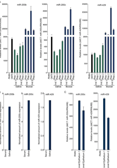
FIG 1High levels of the miR-200 family of microRNAs are present in the oral and tonsil epithelia and in saliva. (A and C) Buccal epithelium and saliva samples were collected from two healthy individuals. OKF6 is a human oral epithelial cell system. Akata is an EBV-positive Burkitt’s lymphoma cell line. Total RNA was extracted and subjected to the TaqMan microRNA assay for mature miR-200 family miRNAs. RNU6B was analyzed as a reference. The expression of miR-200 family miRNAs, including miR-200b, miR-200c, and miR-429, was determined by the comparativeCTmethod (2⫺⌬⌬
CT). (B) MiRDeep2 analysis of serum and
saliva small RNA-seq data sets for miR-200 family member expression.
on November 7, 2019 by guest
http://jvi.asm.org/
[image:4.585.96.489.65.629.2]out using two different sets of primers designed to amplify EBV genomic regions as previously described (43). Real-time PCR was conducted using an iQ5 PCR machine (Bio-Rad) with the following conditions: 95°C for 3 min followed by 40 cycles of 95°C for 30 s, 62°C for 40 s, and 72°C for 40 s using Eva green Supermix (Bio-Rad).
Western blot analysis.Cell and/or MV (exosome) lysates were sus-pended in 1⫻Laemmli buffer (Sigma) and heated at 95°C for 15 min to shear genomic DNA. The resulting samples were measured with the Bio-Rad protein assay kit (catalog number 5000002) according to the manu-facturer’s instructions. An equal weight of lysates was subjected to SDS-polyacrylamide gel electrophoresis using Bio-Rad Criterion gel system and transferred to nitrocellulose membranes (Millipore). The blots were blocked for 30 min in Tris-buffered saline containing 5% low-fat pow-dered milk and 1% FBS and then incubated with the primary antibody (in blocking buffer) overnight at 4°C. The blots were washed three times with 1⫻TBST (140 mM NaCl, 3 mM KCl, 25 mM Tris-HCl [pH 7.4], 0.1% Tween 20) (each wash was carried out for approximately 10 min). The blots were then incubated with horseradish peroxidase-conjugated sec-ondary antibody (Bio-Rad) in blocking buffer for 1 h at room tempera-ture. Blots were washed as described above and analyzed with an en-hanced chemiluminescence detection system (Perkin-Elmer) according to the manufacturer’s recommendations, and filters were exposed to Fuji Super RX films which were then processed using a Kodak X-ray processor. The following primary antibodies were used for Western blot analysis: anti-Zta (Argene; catalog number 11-007), anti-Rta (Argene; catalog number 11-008), anti-BMRF1 (Capricorn; catalog number EBV-018-48180), anti-actin (Santa Cruz; catalog number sc-1615), anti-cyto-chromec(Abcam, Epitomics), and anti-CD63 (Santa Cruz; catalog num-ber sc-15363).
RESULTS
The oral microenvironment is enriched for miR-200 family
members.
MicroRNA-200 family members are typically
ex-pressed in epithelial cells, where they help maintain the epithelial
phenotype. To assess a possible role of miR-200 family members
in signaling in the oral epithelial environment, we evaluated the
levels of the miR-200 family members in the oral cavity by
collect-ing both buccal (oral mucosal) epithelium and saliva samples
from 2 healthy individuals. Total RNAs were extracted and the
levels of representative 200 family members from two
200 gene loci, 200b plus 429 from one locus and
miR-200c from the other, were examined using a TaqMan miRNA
assay. The levels of miR-200b, miR-200c, and miR-429 in saliva
were found to be similar to the levels observed in
in vitro
-cultured
cells of OKF6, a telomerase-immortalized human oral epithelial
cell line that retains its original oral epithelial cell morphology
(
Fig. 1A
), while substantially higher miR-200b, miR-200c, and
miR-429 levels were detected in the buccal epithelial samples (
Fig.
1A
). Consistent with these findings, our analysis of small RNA-seq
data sets from normal saliva (
40
) and serum (
39
) found
substan-tially higher levels of mature miR-200b, miR-200c, and miR-429
in saliva than in serum (
Fig. 1B
). Thus, these results indicate that
mature miR-200 family members are expressed in the oral
epithe-lium and that they are present in its surrounding environment
(e.g., saliva).
We next examined the levels of miR-200 family members in
tonsillar epithelium. Fresh tonsil samples were obtained from
pa-tients undergoing routine adenotonsillectomies, and the tonsillar
epithelial tissues were excised to make total RNA. Both miR-200c
and miR-429 were found to be expressed in tonsil epithelium (
Fig.
1C
) but not in the B-cell line Akata. Together, these results
dem-onstrate the presence of miR-200 family members in the oral and
tonsillar epithelia and the surrounding environments.
Oral epithelial cells produce a microenvironment that favors
EBV lytic replication in B cells.
Since miR-200 family members
were readily detected in saliva and the oral and tonsillar epithelia
and because subepithelial and mucosal plasmacytoid B cells are
major virus producers in the oral cavity (
28
,
29
,
44–50
), we
hy-pothesized that the oral epithelial microenvironment may
pro-mote lytic replication in latently infected B cells. To examine this
possibility, cells of the telomerase-immortalized human oral
epi-thelial cell line OKF6 were seeded onto culture dishes, and 1 day
later, latently infected Mutu I cells were added to the OKF6 cell
monolayer. After 3 days of coculture, the Mutu I B cells were taken
off, leaving an intact OKF6 cell monolayer that displayed no
no-table morphological changes. The Mutu I cells were spun down
and RNA and protein were isolated and subjected to RT-PCR and
Western blot analyses, respectively. Higher expression of the
im-mediate early and early Zta, Rta, and BMRF1 genes was observed
for the biological replicates of OKF6 cocultured cells than for cells
cultured in OKF6 cell growth medium alone (
Fig. 2
). These results
suggest that latency in EBV-infected B cells can be disrupted by
this simulated oral epithelial microenvironment.
Exosome-enriched membrane vesicles (eMVs) derived from
oral OKF6 cells harbor miR-200 family members.
The induction
of reactivation in Mutu I cells cocultured with OKF6 cells
indi-cates that cell-cell contact and/or the presence of transmissible
agents can signal reactivation in these latently infected B cells.
Since membrane vesicles such as exosomes are important
extra-cellular carriers of microRNAs and since we detected miR-200
family members in saliva, we reasoned that secreted MVs carrying
miR-200 family members may function as one such signal. We
therefore assessed whether exosome-enriched MVs (eMVs)
se-creted from OKF6 cells contained miR-200 family members.
FIG 2An oral epithelial cell microenvironment favors EBV lytic replication. EBV-positive Mutu I cells were cocultured with human oral epithelial OKF6 cells for 72 h, and Mutu I cells were then harvested for qRT-PCR analysis (A) and Western blot analysis (B). For qRT-PCR, the expression of Zta and Rta was determined by the comparativeCTmethod (2⫺⌬⌬CT) using GAPDH as a
ref-erence. For Western blot analysis, the levels of the EBV lytic antigens Zta (immediate early), Rta (immediate early), and BMRF1 (early) were measured. Actin was detected as a loading control and reference. For the control group, Mutu I cells were cultured in OKF6 cell culture medium for 72 h without OKF6 cells. Two biological replicates were included in each group.
on November 7, 2019 by guest
http://jvi.asm.org/
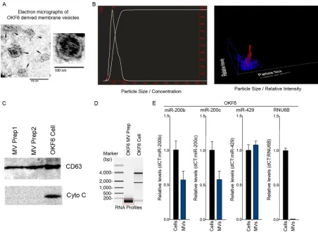
[image:5.585.40.287.64.262.2]Secreted membrane vesicles were collected and purified from
OKF6 cell culture media using the differential centrifugation
method (
37
) and were visualized with transmission electron
mi-croscopy. The isolated vesicles exhibited a typical cup-shaped
exo-somal morphology (
Fig. 3A
) with sizes ranging from
approxi-mately 30 to 100 nm, consistent with the size of exosome vesicles
(
2
). Further, subsequent nanoparticle tracking analysis (NTA)
showed a mode size distribution of
⬃
86 nm, indicating that the
MV preparations were enriched with exosome-sized vesicles (
Fig.
3B
). To further evaluate the isolated vesicles, Western blot analysis
was performed to assess the presence of exosome- and cell-specific
markers. The exosome-specific marker CD63 was readily detected
in the vesicle lysate (
Fig. 3C
). In contrast, cytochrome
c
, which is
present in intact cells, apoptotic bodies, cell debris, and other
types of vesicles, was undetectable in the vesicle lysate (
Fig. 3C
).
These results indicated that our OKF6 cell vesicle preparations
were enriched for exosomes.
As a first step to investigate the possible incorporation of
miR-200 family members in OKF6 cell-derived exosome-enriched
membrane vesicles (eMVs), total RNA was extracted from the
vesicle pellets and the RNA size distribution profile was analyzed
using an Agilent bioanalyzer. The OKF6 cell-derived eMVs were
found to be highly enriched with small RNA species (
⬍
200
nucle-otides long) (
Fig. 3D
). To specifically assess the presence of
miR-200 family members in these eMVs, equal amounts of RNAs from
the vesicle preparations and from cells were analyzed using the
TaqMan miRNA assay to quantify the levels of mature miR-200b,
miR-200c, and miR-429. All three of these family members were
readily detected in the OKF6 cell-derived eMVs, whereas another
small RNA, RNU6B, was not (
Fig. 3E
). This finding showed active
loading of these microRNAs into OKF6 cell-derived eMVs relative
to RNU6B.
OKF6 cell-derived eMVs promote EBV reactivation in B
cells.
The presence of miR-200 family members in OKF6
cell-derived eMVs raised the possibility that these MVs might facilitate
at least some of the signaling between OKF6 cells and Mutu I B
cells in our coculture experiments. To test this possibility, we first
examined whether Mutu I cells can bind and internalize OKF6
cell-derived eMVs. The green fluorescent lipid dye Vybrant was
used to label the purified OKF6 cell-derived eMVs, and the labeled
eMVs were incubated with Mutu I cells for 24 h. The Mutu I cells
were then collected and examined by fluorescence microscopy.
Visualization of exposed Mutu I cells showed binding and
inter-nalization of labeled eMVs (
Fig. 4A
).
FIG 3Characterization of membrane vesicles (exosomes) purified from OKF6 cell cultures. (A) Secretory membrane vesicles (exosomes) were isolated from the culture media of human oral epithelial OKF6 cells and analyzed by transmission electron microscopy. Representative images show that the majority of the vesicles were within the size range of exosomes (30 to 150 nm). The scale bar indicates 100 nm. (B) Nanoparticle tracking analysis (NTA) of an OKF6 cell-derived MV (exosome) preparation. (C) Equal amounts of the OKF6 whole-cell lysate and the lysates of two independent OKF6 cell-derived MV (exosome) preparations were analyzed for the indicated proteins by Western blotting. CD63 is an exosome-specific marker, and cytochromec(Cyto C) is a mitochondrial protein marker. (D) Equal amounts of OKF6 cellular RNA and RNA isolated from an OKF6 cell-derived MV (exosome) preparation were analyzed using an Agilent bioanalyzer. (E) A TaqMan microRNA assay was used to analyze mature miR-200 family miRNAs in OKF6 cells and OKF6 cell-derived MVs (exosomes).
on November 7, 2019 by guest
http://jvi.asm.org/
[image:6.585.63.515.65.395.2]To investigate whether uptake of OKF6 cell-derived eMVs can
break EBV latency in recipient B cells, we created a lytic reporter
system that expresses GFP in cells entering the lytic cascade. Initial
iterations of this reporter contained a GFP cassette driven by a
minimal promoter with multiple copies of Zta response elements.
Although these reporters were responsive to ectopic Zta, they
were poorly responsive to endogenously produced immediate
early genes (data not shown). In contrast, a reporter containing
a GFP reading frame driven by the EBV BMRF1 promoter was
found to be highly responsive to the expression of both ectopic
and endogenous EBV immediately early gene expression (data
not shown). By introducing this plasmid into Mutu I cells, we
were able to monitor initiation of the EBV lytic cycle through
the detection of increased green fluorescence signals. To assess
whether epithelium-derived eMVs can facilitate reactivation,
eMVs were isolated from OKF6 cell culture medium and
incu-bated with Mutu-BMRF1p-GFP cells. After 3 days of
incuba-tion, Mutu-BMRF1p-GFP cells were collected and analyzed by
fluorescence microscopy. As shown in
Fig. 4B
and
C
, GFP
sig-nals were detected in nearly 20% of eMV-treated cells but
rarely detected in cells treated with the control vehicle (1
⫻
PBS). Further, OKF6 cell-derived eMVs exhibited lytic
cycle-FIG 4OKF6 cell-derived eMVs break EBV latency in Mutu I cells. (A) OKF6 cell-derived eMVs were labeled with the green fluorescent dye Vybrant and incubated with Mutu I cells. Twenty-four hours postincubation, cells were examined by fluorescence microscopy. Nuclei were visualized by DAPI staining. (B and C) Mutu-BMRF1p-GFP cells were treated with OKF6 cell-derived eMVs, the control vehicle (1⫻PBS), or anti-IgM. Seventy-two hours posttreatment, cells were fixed and examined using fluorescence microscopy. Blue, Hoechst; green, GFP. Induction rates were determined by manually counting the GFP-positive cells in a total of 10 randomly selected fields from duplicate treatments for each condition. (D) Mutu I cells were incubated with purified OKF6 cell-derived eMVs or the control vehicle (1⫻PBS). Seventy-two hours postincubation, cell pellets were harvested for Western blot analysis of the EBV lytic antigens Zta (immediate early), Rta (immediate early), and BMRF1 (early). Actin was detected as a loading control and reference. Two biological replicates were included in each group. (E) The production of new virions was determined by the real-time PCR analysis of EBV genomic DNA in the cell culture media. Two biological replicates were included in each group. Two primer sets were utilized that amplify two different regions in the EBV genome (43). The amounts of EBV genomic DNA were calculated by the comparativeCTmethod (2⫺⌬⌬
CT) versus GAPDH DNA.
on November 7, 2019 by guest
http://jvi.asm.org/
[image:7.585.84.505.64.495.2]inducing potency similar to that of the anti-IgM-mediated
BCR engagement (
Fig. 4C
).
To further investigate the eMV-mediated reactivation, we
in-cubated Mutu I cells with freshly prepared OKF6 cell-derived
eMVs. Three days later, Mutu I cell pellets were collected and
examined for viral lytic gene expression. As shown in
Fig. 4D
,
expression of the viral immediate early and early Zta and Rta, and
BMRF1 genes was increased in Mutu I cells exposed to OKF6
cell-derived eMVs, indicating disruption of latency. Further,
real-time PCR analysis showed an increased level of viral genomic
DNA in the supernatant of Mutu I cells incubated with OKF6
cell-derived eMVs (
Fig. 4E
). These results further establish that
exposure of EBV-positive B cells to OKF6 cell-derived eMVs
pro-motes entry into the lytic replication cycle and the production of
secreted virions.
To ensure that the induction of reactivation by OKF6
cell-derived eMVs is not specific to a single cell line, we incubated
OKF6 cell-derived eMVs with a second EBV-positive B-cell line,
Akata. Similar to the results obtained with Mutu I cells, expression
of the immediate early and early Zta, Rta, and BMRF1 genes and
secreted virus were induced by OKF6 cell-derived eMVs (
Fig. 5
).
miR429-containing eMVs promote EBV reactivation in B
cells.
The data presented above show that OKF6 cell-derived eMV
contain miR-200 family members and that these eMVs can induce
reactivation in EBV-positive B-cell lines. To determine whether
miR-200 family members
per se
play a role in eMV-mediated
re-activation, we set out to compare the efficacies of miR-200
family-positive and miR-200 family-negative eMVs in facilitating
reacti-vation. Most nonepithelial cells, such as 293 fibroblasts, do not
express miR-200 family miRNAs (
30
). Since 293 cells have been
previously shown to actively produce and release eMVs (
51
), we
created 293 cell lines transduced in duplicate with either an empty
vector control cntl) or a miR-429 expressing retrovirus
(293-miR-429).
Mature miR-429 but not other miR-200 family members
were detected in eMVs derived from 293-miR-429 cells (
Fig.
6A
). To examine whether these miR-429-positive eMVs can
disrupt viral latency, we incubated either the 293-miR-429- or
293-cntl-derived eMVs with Mutu I cells. After 3 days of
incu-bation, cell pellets were collected and extracted proteins were
analyzed by Western blotting for viral lytic gene expression.
Exposure of Mutu I cells to miR-429-positive eMVs led to
in-creased expression of the immediate early and early Zta, Rta,
and BMRF1 genes (
Fig. 6B
). Mutu I cells incubated with eMVs
generated from empty control retrovirus-transduced 293 cells
exhibited levels of viral lytic gene expression similar to those
observed with Mutu I cells incubated with the control vehicle
(1
⫻
PBS) (
Fig. 6B
). Together, these results show that
miR-429-containing eMVs promote viral lytic reactivation in recipient B
cells.
DISCUSSION
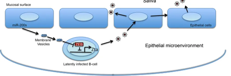
Based on the results presented here, we propose that the oral
epi-thelium-associated MV (exosome) pathway plays a role in
orches-trating EBV’s lytic replication in B cells
in vivo
. In this model, oral
epithelial cells actively load miR-200 family miRNAs into
exo-somes (and other possible membrane vesicles such as
mi-crovesicles) (
Fig. 7
) and secret these vesicles into the epithelial
microenvironment to create conditions that favor EBV lytic
rep-FIG 5OKF6 cell-derived eMVs break EBV latency in Akata cells. Akata cells were incubated with purified OKF6 cell-derived eMVs or the control vehicle (1⫻PBS). Seventy-two hours postincubation, (A) cell pellets were harvested for Western blot analysis of the EBV lytic antigens Zta (immediate early), Rta (immediate early), and BMRF1 (early). Actin was detected as a loading control and reference. Two biological replicates were included in each group. (B) The production of new virions was determined by real-time PCR analysis of EBV genomic DNA in the cell culture media. Two biological replicates were in-cluded in each group. Two primers sets were utilized that amplify two different regions in the EBV genome (43). The amounts of EBV genomic DNA were calculated by the comparativeCTmethod (2⫺⌬⌬
CT) versus GAPDH DNA.
FIG 6miR-429-containing eMVs promote lytic reactivation in Mutu I cells. (A) eMVs were purified from 293 cells stably expressing either miR-429 (293-miR429) or its control (293-cntl). RNAs were then isolated from the purified eMVs and subjected to TaqMan microRNA analysis of mature miR-200 family miRNAs. (B) Mutu I cells were incubated with purified 293-miR429 eMVs, 293-cntl eMVs, or the control vehicle (1⫻PBS). Seventy-two hours postincu-bation, cell pellets were harvested for Western blot analysis of the EBV lytic antigens Zta (immediate early), Rta (immediate early), and BMRF1 (early). Actin was detected as a loading control and reference. Two biological replicates were included in each group.
on November 7, 2019 by guest
http://jvi.asm.org/
[image:8.585.41.290.65.300.2] [image:8.585.298.546.65.256.2]lication. The transfer of eMVs containing miR-200 family
mem-bers to infiltrating and/or proximal EBV-positive B cells leads to
the disruption of latency and production of new viruses. This
facilitates the exchange of virus from the peripheral B-cell stores to
the oral epithelium, where virus is amplified and shed into the
saliva.
Emerging evidence indicates that the EBV infection cascade
relies on both the oral epithelium and B cells. One current
opinion is that the exchange of virus generated from one tissue
type to the other is an integral part of EBV’s infection cycle,
which is supported by a reciprocal infection preference for
vi-rus produced in epithelial cells versus B cells (
34
,
35
). In
agree-ment with this view, our proposed microenvironagree-ment sensing
mechanism can potentially contribute further to facilitating
the virus exchange between B cells and epithelial cells. This
sensing system would also help enable such a limited number of
virally infected B cells (approximately only one in one million
circulating B cells) to maintain constant viral replication foci at
oral epithelial sites.
Previously, our laboratory and others discovered an intimate
relationship between the miR-200 signaling pathways and the
EBV life cycle (
30
,
31
). Specifically, we found that the miR-200
family of microRNAs functions as a cellular switch that regulates
the transition from latency to the lytic cascade. The miR-200
fam-ily of microRNAs forms a double negative-feedback loop with the
ZEB1 and ZEB2 transcriptional repressors, forming a regulatory
axis that facilitates the committed transition of cells from either a
mesenchymal to epithelial phenotype or an epithelial to
mesen-chymal phenotype. Specifically, the 3
=
untranslated regions
(UTRs) of both ZEB1 and ZEB2 contain multiple seed sites for
miR-200 family members, and their expression is inhibited by
high levels of miR-200s (
52
). Conversely, both miR-200 family
member clusters are transcriptionally repressed by ZEB1 and/or
ZEB2 (
53
). This forms a self-reinforcing loop that helps establish
commitment to either high miR-200 family expression with low
ZEB1 and ZEB2 expression or high ZEB1 and ZEB2 expression
with low miR-200 family member expression. For example, once
miR-200 family member expression increases, this suppresses the
expression of their repressors, ZEB1 and ZEB2, which, in turn,
leads to a further increase in miR-200 family expression and so on.
Conversely, an increase in ZEB1 or ZEB2 is self-reinforced by
repression of miR-200 family members, causing relief from
inhi-bition by these microRNAs.
During B-cell latency, the low levels of endogenous miR-200s
allow the expression of the transcriptional suppressors ZEB1 and
ZEB2, which inhibit the viral immediate early Zta gene (
32
,
33
),
thereby promoting viral latency. In response to lytic induction
signals, an upregulation of miR-200 family miRNAs will relieve
the inhibitory effect of ZEB1 and ZEB2 on miR-200 promoters,
further enhancing the expression of miR-200 family miRNAs and
leading to further suppression of ZEB1 and ZEB2. This causes a
committed transition from high ZEB1 and ZEB2 and low
miR-200 family expression to a low ZEB1 and ZEB2 and high miR-miR-200
expression, enforcing a commitment to the loss of ZEB1 and ZEB2
suppression of the Zta promoter.
In the context of intercellular communication between
ep-ithelial cells and B cells, we propose that the internalized
miR-200s in recipient B cells will likely activate lytic replication by
relieving ZEB1- and ZEB2-mediated suppression of the viral
Zta gene. We think that miR-200s are ideal eMV signaling
mol-ecules for facilitating sustained pathway alterations. By taking
advantage of this self-reinforcing feedback mechanism, a single
dose of eMV miR-200s not only will directly inhibit ZEB1 and
ZEB2 expression but also should be able to enforce elevated
cellular expression of miR-200 family expression, thereby
en-forcing a commitment to break viral latency in recipient cells.
In our coculture system, a monolayer of OKF6 cells was
shown to disrupt viral latency in B cells. Others have argued
that both the concentration and transfer efficiency of MVs
(exosomes) in
in vivo
environments are substantially higher
than in
in vitro
simulated systems because of the high
concen-tration of cells and the low intercellular volume in the
in vivo
settings (
13
,
54
). This raises the possibility that reactivation
efficiencies
in vivo
may, in fact, be greater than those observed
in our tissue culture model.
Using a well-established differential centrifugation method,
we were able to isolate eMVs from the supernatant of OKF6 cell
culture media. The presence of exosomes in the OKF6
cell-derived vesicle preparation was confirmed by the presence of
CD63 exosomal marker and the absence of cytochrome
c
, a key
indicator for the contamination of apoptotic bodies and cell
debris. The additional morphological analysis and particle size
analysis further indicated that the majority of the isolated
ves-icles were exosomes. Nevertheless, we did detect a fraction of
microvesicle-like particles (150 to 230 nm in diameter) in our
vesicle preparations. Generally, microvesicles (or shedding
FIG 7Model of eMV miR-200-mediated communication between oral epithelium and virally infected B cells.
on November 7, 2019 by guest
http://jvi.asm.org/
[image:9.585.70.514.71.218.2]vesicles) belong to another subpopulation of membrane
vesi-cles and are more heterogeneous in size (100 to 1,000 nm in
diameter) (
2
,
55
,
56
). Microvesicles are formed by outward
budding of the plasma membrane and, like exosomes, contain
proteins, lipids, and miRNAs (
2
). Since many studies have
shown that miRNAs and other genetic materials can be carried
by both exosomes and microvesicles and functionally delivered
to recipient cells (
16
,
56
), it is possible that this fraction of
microvesicles may also contribute to the creation of a
miR-200s-rich epithelial microenvironment and cooperate with
exosomes to regulate the EBV life cycle.
Here, we have shown that eMV miR-200s can break viral
la-tency in recipient B cells, but we cannot rule out the possibility
that other epithelial signaling molecules carried by exosomes and
other membrane vesicles are also involved in this regulatory
pro-cess. Further analysis may uncover additional components within
oral epithelial secretory vesicles that also help facilitate EBV
reac-tivation.
In this study, we focused only on communication from oral
epithelial cells to virally infected B cells. It is also interesting to
consider, however, whether the virally infected B cells can
simultaneously “talk to” oral epithelial cells, particularly
through a paracrine-like manner in the microenvironment.
Previous studies have shown that EBV-infected
lymphoblas-toid B cells can actively pack both viral and cellular nucleic
acids (including miRNAs) and proteins into the exosomes,
which can influence the recipient cells (
12
,
13
,
54
). When
reaching the oral epithelial microenvironment, the virally
in-fected B cells may similarly signal the epithelial cells, thereby
modulating signaling pathways that may play a role in
facilitat-ing the needs of the virus. For instance, uptake of such vesicles
could enhance permissiveness of recipient epithelial cells to
EBV infection. This and other possible roles for B-cell-derived
exosomes in the process of transfer of virus from B cells to
epithelial cells require further investigation, however.
ACKNOWLEDGMENTS
This work was supported by a Louisiana Clinical & Translational Science Center Pilot Grant (1 U54 GM104940 from the National Institute of Gen-eral Medical Sciences of the National Institutes of Health, which funds the Louisiana Clinical and Translational Science Center) to Z.L., Ruth L. Kirschstein National Research Service Award F30CA177267 from the NCI to M.J.S., and National Institutes of Health grants R01AI101046, R01CA124311, R01CA138268, and R01AI106676 to E.K.F. (and P20GM103518 to Prescott Deininger, Tulane Cancer Center).
FUNDING INFORMATION
HHS | National Institutes of Health (NIH) provided funding to Zhen Lin under grant number 1 U54 GM104940. HHS | National Institutes of Health (NIH) provided funding to Michael J. Strong under grant number F30CA177267. HHS | National Institutes of Health (NIH) provided fund-ing to Erik K. Flemfund-ington under grant numbers R01AI101046, R01CA124311, R01CA138268, and R01AI106676.
This work was also supported through core resources from a grant awarded to a nonauthor, Prescott Deininger (Tulane Cancer Center), un-der award P20GM103518.
REFERENCES
1.Théry C, Zitvogel L, Amigorena S.2002. Exosomes: composition, bio-genesis and function. Nat Rev Immunol2:569 –579.
2.Théry C, Ostrowski M, Segura E.2009. Membrane vesicles as conveyors
of immune responses. Nat Rev Immunol9:581–593.http://dx.doi.org/10 .1038/nri2567.
3.Schageman J, Zeringer E, Li M, Barta T, Lea K, Gu J, Magdaleno S, Setterquist R, Vlassov AV.2013. The complete exosome workflow solu-tion: from isolation to characterization of RNA cargo. Biomed Res Int
2013:253957.
4.Conde-Vancells J, Rodriguez-Suarez E, Embade N, Gil D, Matthiesen R, Valle M, Elortza F, Lu SC, Mato JM, Falcon-Perez JM.2008. Charac-terization and comprehensive proteome profiling of exosomes secreted by hepatocytes. J Proteome Res 7:5157–5166. http://dx.doi.org/10.1021 /pr8004887.
5.Trajkovic K, Hsu C, Chiantia S, Rajendran L, Wenzel D, Wieland F, Schwille P, Brugger B, Simons M.2008. Ceramide triggers budding of exosome vesicles into multivesicular endosomes. Science319:1244 –1247.
http://dx.doi.org/10.1126/science.1153124.
6.Johnstone RM.2005. Revisiting the road to the discovery of exosomes. Blood Cells Mol Dis34:214 –219.http://dx.doi.org/10.1016/j.bcmd.2005 .03.002.
7.Fevrier B, Raposo G.2004. Exosomes: endosomal-derived vesicles ship-ping extracellular messages. Curr Opin Cell Biol16:415– 421.http://dx .doi.org/10.1016/j.ceb.2004.06.003.
8.Kourembanas S.2015. Exosomes: vehicles of intercellular signaling, bio-markers, and vectors of cell therapy. Annu Rev Physiol77:13–27.http://dx .doi.org/10.1146/annurev-physiol-021014-071641.
9.Meckes DG, Jr, Raab-Traub N.2011. Microvesicles and viral infection. J Virol85:12844 –12854.http://dx.doi.org/10.1128/JVI.05853-11. 10. Schorey JS, Bhatnagar S.2008. Exosome function: from tumor
immu-nology to pathogen biology. Traffic9:871– 881.http://dx.doi.org/10.1111 /j.1600-0854.2008.00734.x.
11. van Niel G, Porto-Carreiro I, Simoes S, Raposo G.2006. Exosomes: a common pathway for a specialized function. J Biochem140:13–21.http: //dx.doi.org/10.1093/jb/mvj128.
12. Meckes DG, Jr, Gunawardena HP, Dekroon RM, Heaton PR, Edwards RH, Ozgur S, Griffith JD, Damania B, Raab-Traub N.2013. Modulation of B-cell exosome proteins by gamma herpesvirus infection. Proc Natl Acad Sci U S A110:E2925–E2933.http://dx.doi.org/10.1073/pnas.1303906110. 13. Pegtel DM, Cosmopoulos K, Thorley-Lawson DA, van Eijndhoven MA,
Hopmans ES, Lindenberg JL, de Gruijl TD, Wurdinger T, Middeldorp JM.
2010. Functional delivery of viral miRNAs via exosomes. Proc Natl Acad Sci U S A 107:6328 – 6333. http://dx.doi.org/10.1073/pnas .0914843107.
14. Haneklaus M, Gerlic M, Kurowska-Stolarska M, Rainey AA, Pich D, McInnes IB, Hammerschmidt W, O’Neill LA, Masters SL.2012. Cutting edge: miR-223 and EBV miR-BART15 regulate the NLRP3 inflammasome and IL-1beta production. J Immunol189:3795–3799.http://dx.doi.org/10 .4049/jimmunol.1200312.
15. Valadi H, Ekstrom K, Bossios A, Sjostrand M, Lee JJ, Lotvall JO.2007. Exosome-mediated transfer of mRNAs and microRNAs is a novel mech-anism of genetic exchange between cells. Nat Cell Biol9:654 – 659.http: //dx.doi.org/10.1038/ncb1596.
16. Mittelbrunn M, Sanchez-Madrid F.2012. Intercellular communication: diverse structures for exchange of genetic information. Nat Rev Mol Cell Biol13:328 –335.
17. Zhang J, Li S, Li L, Li M, Guo C, Yao J, Mi S.2015. Exosome and exosomal microRNA: trafficking, sorting, and function. Genomics Pro-teomics Bioinformatics 13:17–24. http://dx.doi.org/10.1016/j.gpb.2015 .02.001.
18. Rickinson AB, Kieff E.2007. Epstein-Barr virus, p 2655–2700.InKnipe DM, Howley PM, Griffin DE, Lamb RA, Martin MA, Roizman B, Straus SE (ed), Fields virology, 5th ed. Lippincott Williams & Wilkins, Philadel-phia, PA.
19. Lin Z, Flemington EK.2010. Regulation of EBV latency by viral lytic proteins, p 167–192.InRobertson ES (ed), Epstein-Barr virus: latency and transformation. Caister Academic Press, Wymondham, Norfolk, United Kingdom.
20. Kieff E, Rickinson AB.2007. Epstein-Barr virus and its replication, p 2603–2654.InKnipe DM, Howley PM, Griffin DE, Lamb RA, Martin MA, Roizman B, Straus SE (ed), Fields virology, 5th ed. Lippincott Williams & Wilkins, Philadelphia, PA.
21. Luka J, Kallin B, Klein G.1979. Induction of the Epstein-Barr virus (EBV) cycle in latently infected cells by n-butyrate. Virology94:228 –231.
http://dx.doi.org/10.1016/0042-6822(79)90455-0.
22. zur Hausen H, Bornkamm GW, Schmidt R, Hecker E.1979. Tumor
on November 7, 2019 by guest
http://jvi.asm.org/
initiators and promoters in the induction of Epstein-Barr virus. Proc Natl Acad Sci U S A76:782–785.http://dx.doi.org/10.1073/pnas.76.2.782. 23. Ben-Sasson SA, Klein G.1981. Activation of the Epstein-Barr virus
ge-nome by 5-aza-cytidine in latently infected human lymphoid lines. Int J Cancer28:131–135.http://dx.doi.org/10.1002/ijc.2910280204.
24. Faggioni A, Zompetta C, Grimaldi S, Barile G, Frati L, Lazdins J.1986. Calcium modulation activates EpsteBarr virus genome in latently in-fected cells. Science 232:1554 –1556. http://dx.doi.org/10.1126/science .3012779.
25. Takada K.1984. Cross-linking of cell surface immunoglobulins induces Epstein-Barr virus in Burkitt lymphoma lines. Int J Cancer33:27–32.http: //dx.doi.org/10.1002/ijc.2910330106.
26. Takada K, Ono Y.1989. Synchronous and sequential activation of la-tently infected Epstein-Barr virus genomes. J Virol63:445– 449. 27. Chasserot-Golaz S, Schuster C, Dietrich JB, Beck G, Lawrence DA.
1988. Antagonistic action of RU38486 on the activity of transforming growth factor-beta in fibroblasts and lymphoma cells. J Steroid Biochem
30:381–385.http://dx.doi.org/10.1016/0022-4731(88)90127-6. 28. Laichalk LL, Thorley-Lawson DA.2005. Terminal differentiation into
plasma cells initiates the replicative cycle of Epstein-Barr virus in vivo. J Virol79:1296 –1307.http://dx.doi.org/10.1128/JVI.79.2.1296-1307.2005. 29. Hadinoto V, Shapiro M, Sun CC, Thorley-Lawson DA. 2009. The dynamics of EBV shedding implicate a central role for epithelial cells in amplifying viral output. PLoS Pathog5:e1000496.http://dx.doi.org/10 .1371/journal.ppat.1000496.
30. Lin Z, Wang X, Fewell C, Cameron J, Yin Q, Flemington EK.2010. Differential expression of the miR-200 family microRNAs in epithelial and B cells and regulation of Epstein-Barr virus reactivation by the miR-200 family member miR-429. J Virol84:7892–7897.http://dx.doi.org/10 .1128/JVI.00379-10.
31. Ellis-Connell AL, Iempridee T, Xu I, Mertz JE.2010. Cellular micro-RNAs 200b and 429 regulate the Epstein-Barr virus switch between latency and lytic replication. J Virol84:10329 –10343.http://dx.doi.org/10.1128 /JVI.00923-10.
32. Yu X, Wang Z, Mertz JE.2007. ZEB1 regulates the latent-lytic switch in infection by Epstein-Barr virus. PLoS Pathog3:e194.http://dx.doi.org/10 .1371/journal.ppat.0030194.
33. Kraus RJ, Perrigoue JG, Mertz JE.2003. ZEB negatively regulates the lytic-switch BZLF1 gene promoter of Epstein-Barr virus. J Virol77:199 – 207.http://dx.doi.org/10.1128/JVI.77.1.199-207.2003.
34. Borza CM, Hutt-Fletcher LM.2002. Alternate replication in B cells and epithelial cells switches tropism of Epstein-Barr virus. Nat Med8:594 – 599.http://dx.doi.org/10.1038/nm0602-594.
35. Jiang R, Scott RS, Hutt-Fletcher LM.2006. Epstein-Barr virus shed in saliva is high in B-cell-tropic glycoprotein gp42. J Virol80:7281–7283.
http://dx.doi.org/10.1128/JVI.00497-06.
36. Lin Z, Yin Q, Flemington E.2004. Identification of a negative regulatory element in the Epstein-Barr virus Zta transactivation domain that is reg-ulated by the cell cycle control factors c-Myc and E2F1. J Virol78:11962– 11971.http://dx.doi.org/10.1128/JVI.78.21.11962-11971.2004. 37. Zeringer E, Barta T, Li M, Vlassov AV.2015. Strategies for isolation of
exosomes. Cold Spring Harb Protoc2015:pdb.top074476.
38. Cao S, Moss W, O’Grady T, Concha M, Strong MJ, Wang X, Yu Y, Baddoo M, Zhang K, Fewell C, Lin Z, Dong Y, Flemington EK.2015. New noncoding lytic transcripts derived from the Epstein-Barr virus la-tency origin of replication, oriP, are hyperedited, bind the paraspeckle protein, NONO/p54nrb, and support viral lytic transcription. J Virol89:
7120 –7132.http://dx.doi.org/10.1128/JVI.00608-15.
39. Ignacio C, Hicks SD, Burke P, Lewis L, Szombathyne-Meszaros Z, Middleton FA.2015. Alterations in serum microRNA in humans with alcohol use disorders impact cell proliferation and cell death pathways and predict structural and functional changes in brain. BMC Neurosci16:55.
http://dx.doi.org/10.1186/s12868-015-0195-x.
40. Ogawa Y, Taketomi Y, Murakami M, Tsujimoto M, Yanoshita R.
2013. Small RNA transcriptomes of two types of exosomes in human whole saliva determined by next generation sequencing. Biol Pharm Bull36:66 –75.
41. Friedländer MR, Mackowiak SD, Li N, Chen W, Rajewsky N.2012. miRDeep2 accurately identifies known and hundreds of novel microRNA genes in seven animal clades. Nucleic Acids Res40:37–52.http://dx.doi .org/10.1093/nar/gkr688.
42. Strong MJ, Baddoo M, Nanbo A, Xu M, Puetter A, Lin Z. 2014. Comprehensive high-throughput RNA sequencing analysis reveals contamination of multiple nasopharyngeal carcinoma cell lines with HeLa cell genomes. J Virol88:10696 –10704.http://dx.doi.org/10.1128 /JVI.01457-14.
43. Yin Q, Wang X, Fewell C, Cameron J, Zhu H, Baddoo M, Lin Z, Flemington EK.2010. MicroRNA miR-155 inhibits bone morphogenetic protein (BMP) signaling and BMP-mediated Epstein-Barr virus reactiva-tion. J Virol84:6318 – 6327.http://dx.doi.org/10.1128/JVI.00635-10. 44. Tao Q, Srivastava G, Chan AC, Chung LP, Loke SL, Ho FC. 1995.
Evidence for lytic infection by Epstein-Barr virus in mucosal lymphocytes instead of nasopharyngeal epithelial cells in normal individuals. J Med Virol45:71–77.http://dx.doi.org/10.1002/jmv.1890450114.
45. Niedobitek G, Agathanggelou A, Steven N, Young LS.2000. Epstein-Barr virus (EBV) in infectious mononucleosis: detection of the virus in tonsillar B lymphocytes but not in desquamated oropharyngeal epithelial cells. Mol Pathol53:37– 42.http://dx.doi.org/10.1136/mp.53.1.37. 46. Niedobitek G, Herbst H, Young LS, Brooks L, Masucci MG, Crocker J,
Rickinson AB, Stein H.1992. Patterns of Epstein-Barr virus infection in non-neoplastic lymphoid tissue. Blood79:2520 –2526.
47. Anagnostopoulos I, Hummel M, Kreschel C, Stein H.1995. Morphol-ogy, immunophenotype, and distribution of latently and/or productively Epstein-Barr virus-infected cells in acute infectious mononucleosis: im-plications for the interindividual infection route of Epstein-Barr virus. Blood85:744 –750.
48. Niedobitek G, Agathanggelou A, Herbst H, Whitehead L, Wright DH, Young LS.1997. Epstein-Barr virus (EBV) infection in infectious mono-nucleosis: virus latency, replication and phenotype of EBV-infected cells. J Pathol182:151–159.
49. Karajannis MA, Hummel M, Anagnostopoulos I, Stein H.1997. Strict lymphotropism of Epstein-Barr virus during acute infectious mononucle-osis in nonimmunocompromised individuals. Blood89:2856 –2862. 50. Crawford DH, Ando I.1986. EB virus induction is associated with B-cell
maturation. Immunology59:405– 409.
51. Chairoungdua A, Smith DL, Pochard P, Hull M, Caplan MJ.2010. Exosome release of beta-catenin: a novel mechanism that antagonizes Wnt signaling. J Cell Biol190:1079 –1091.http://dx.doi.org/10.1083 /jcb.201002049.
52. Park SM, Gaur AB, Lengyel E, Peter ME.2008. The miR-200 family determines the epithelial phenotype of cancer cells by targeting the E-cadherin repressors ZEB1 and ZEB2. Genes Dev22:894 –907.http://dx .doi.org/10.1101/gad.1640608.
53. Burk U, Schubert J, Wellner U, Schmalhofer O, Vincan E, Spaderna S, Brabletz T.2008. A reciprocal repression between ZEB1 and members of the miR-200 family promotes EMT and invasion in cancer cells. EMBO Rep9:582–589.http://dx.doi.org/10.1038/embor.2008.74.
54. Meckes DG, Jr, Shair KH, Marquitz AR, Kung CP, Edwards RH, Raab-Traub N.2010. Human tumor virus utilizes exosomes for intercel-lular communication. Proc Natl Acad Sci U S A107:20370 –20375.http: //dx.doi.org/10.1073/pnas.1014194107.
55. Heijnen HF, Schiel AE, Fijnheer R, Geuze HJ, Sixma JJ.1999. Activated platelets release two types of membrane vesicles: microvesicles by surface shedding and exosomes derived from exocytosis of multivesicular bodies and alpha-granules. Blood94:3791–3799.
56. Cocucci E, Racchetti G, Meldolesi J.2009. Shedding microvesicles: arte-facts no more. Trends Cell Biol19:43–51.http://dx.doi.org/10.1016/j.tcb .2008.11.003.
on November 7, 2019 by guest
http://jvi.asm.org/