ResearchOnline@JCU
This file is part of the following work:
Hernández Agreda, Alejandra Isabel (2018)
Deciphering the bacterial microworld
in corals: structure, variability and persistence.
PhD Thesis, James Cook
University.
Access to this file is available from:
https://doi.org/10.25903/5c86ee53cb9a1
Copyright © 2018 Alejandra Isabel Hernández Agreda
The author has certified to JCU that they have made a reasonable effort to gain
permission and acknowledge the owners of any third party copyright material
included in this document. If you believe that this is not the case, please email
Deciphering the bacterial microworld in corals:
structure, variability and persistence
Thesis submitted by
Lic in Biology Alejandra Isabel Hernández Agreda
For the degree of Doctor in Philosophy
ARC Centre of Excellence for Coral Reef Studies
College of Public Health, Medical and Veterinary Sciences
James Cook University
i
Acknowledgements
This thesis has been possible with the collaboration and support of valuable people and
institutions that trusted me. I am profoundly thankful for my supervisors Tracy Ainsworth, Bill
Leggat, Pim Bongaerts and Andrew Hoey. I genuinely thank Tracy for hearing my ideas,
offering me multiple opportunities, and investing a lot of her time in helping me to build my
career. You encouraged and challenged me to give my best in everything I did, and in doing
so, I found out what I am capable. I am grateful to Bill for his patience in teaching laboratory
analysis and bioinformatics. It has been an enriching experience to learn from your teaching
philosophy, and I am very proud to have been one of your students. I am truly thankful to Pim
for introducing me to mesophotic reefs and population genomics, for teaching me
bioinformatics and encourage me to do open science. I am also grateful for your support, and
the opportunities you have given me. I wish to thank to Andrew for his career development
advice. More than supervisors and collaborators, you are genuinely mentors and friends. Each
of you in your way contributed to strengthening me professionally and personally. I will never
thank you enough for that.
I would like to thank the ARC Centre of Excellence for Coral Reef Studies for the opportunity
to be part of its research team. Mainly, I want to acknowledge Jennifer Lappin, Alana Grech,
and Olga Bazaka, who keep the Centre up and running; and to Janet Swanson and Vivien
Doherty, for their disposition and kindness in offering administrative support. I am grateful to
The College of Public Health, Medical and Veterinary Sciences at James Cook University,
ii
I would like to thank Australia Awards for financial support. I would also like to thank the
personnel of the International Student Support office, in particular to Alex Salvador and
Katherine Elliot, for their support and guidance during my time at JCU. I am also thankful to
Kellie Johns, Learning adviser from JCU Graduate Research School, for the effort in helping
me to strengthen my writing skills.
I also would like to thank Professor Ruth D. Gates for the support, encouragement, and career
development advice during my PhD.
I would like to extend my gratitude to University Toastmasters, for training me in public
speaking.
I also want to thank past and current members of the Symbiosis Genomics Lab for their help,
support and friendship: Sarah Geirz, Kate Quigley, Jordan M Casey, Alexander (Gus) Fordyce
and Tess Moriarty. I am especially thankful to Martina de Freitas Prazeres for generously
helping me numerous times in the field, and for her friendship and support during rough times.
I am also grateful to Marie Voisine and Chris Bligh, for helping me to organize the coral
collection and for their invaluable assistance in the lab.
Many thanks to wonderful fieldwork volunteers and even better friends: Helios Martinez,
Tiffany Nay, Connor Gervais, Floriaan Devloo-Delva, Ed Roberts, and Steph Gardner. Thanks
for the patience, the hard work and the enthusiasm. I would also thank the personnel from the
Heron Island Research Station for their assistance in the field: Elizabeth Perkins, Maureen
Roberts, Bec Tite, Ben Potts, Isaac Ashton, Abbie Taylor, Lauren Bailey.
Thanks to considerate office mates for making the office an ideal place to write: Zara Cowan,
iii
Asson-Batzel for being a friend, for encouraging me to dance as a way to preserve mental
health and for her support in editing this thesis.
I would like to thank my friends who supported, encouraged, and listened to me on a daily
basis. Thank you for your understanding and love, at JCU: Diana Pazmino, Natalia Andrade,
Diego Ortiz, Chao-Yang Kuo, Katie Sambrook, Hannah Epstein, Tess Hill, Adriana Humanes,
Natalie Wildermann and Hector Barrios; at UQ: Veronica Radice, Michelle Achlatis, Rene van
der Zande and Matheus (Matt) Mello Athayde; at HIMB: Mariana Rocha De Souza, Elizabeth
(Beth) Lenz and Tanya Brown; and in Venezuela: Maria Valentina Cedeño, Aldo Croquer, Esteban Agudo, Luis Miguel Montilla, and the Experimental Ecology Lab.
Thanks to my Australian family for giving me a home, the love, the support and patience of a
family: Vivienne Feltham, Rae and Darren Cole, and Ken and Elise Feltham.
Thanks to my family for being an example of perseverance and an endless source of love,
support and inspiration. Thanks Cesar for unconditional support and encouragement, for belief
in me and inspiring me every day.
This thesis is dedicated to my family, who taught me that only hard work and perseverance
iv
Statement of the Contribution of Others
Research funding:
• Australia Awards (AusAID)
• Higher Degree Research Enhancement Scheme 2015 and 2016 - Graduate Research
School & ARC Centre of Excellence of Coral Reef Studies, James Cook University
• Program 3 Cross-Nodal Collaborative Grant 2016 - ARC Centre of Excellence of Coral
Reef Studies, James Cook University
• Postgraduate Travel Award – ARC Centre of Excellence in Bioinformatics, The
University of Queensland
• Postgraduate Travel Award – Australian Coral Reef Society
• Postgraduate Travel Award – The Association for the Sciences of Limnology and
Oceanography
Thesis committee:
• Dr. Tracy D. Ainsworth – Australian Research Council Centre of Excellence for Coral
Reef Studies, James Cook University and School of Biological, Earth and
Environmental Science, University of New South Wales
• Associated Professor William Leggat - Australian Research Council Centre of
v
Health, Medical and Veterinary Sciences, James Cook University and School of
Environmental and Life Sciences, University of Newcastle
• Dr. Pim Bongaerts – Global Change Institute, The University of Queensland and The
California Academy of Sciences
• Dr. Andrew Hoey - Australian Research Council Centre of Excellence for Coral Reef
Studies, James Cook University
Statistical support:
Cesar Herrera Acosta
Editorial support:
Dr. Tracy D. Ainsworth
Associated Professor William Leggat
Dr. Pim Bongaerts
Cesar Herrera Acosta
vi
Abstract
Decades of research have defined the coral meta-organism as a complex microbial system due
to the diversity, abundance and variability of the associated microorganisms. Bacteria are the
most studied group of coral-associated prokaryotes, and they are located within the mucus,
skeleton, tissues and cellular spaces. Abiotic factors including light irradiance, current, pH, and
oxygen generate distinct micro-niches that differ between coral species, depths, reefs, and
bioregions. This variety in microhabitats leads to enormous configurations of hundred of
thousand bacteria, from tens of thousand phylotypes, associated with each coral species. The
variability, diversity and richness of these bacterial communities have undermined the capacity
to identify bacterial phylotypes in symbiosis with corals, to describe their functional roles and
to establish the characteristics of a healthy bacterial community in corals.
Herein, I dissect the variability, diversity and richness of bacterial communities in healthy
corals. I aim to (1) define the characteristics of a healthy coral microbiome, (2) evaluate the
presence of universal bacterial symbionts in coral-associated bacterial assemblages, and (3)
identify factors generating variability in assessments of the coral-associated bacterial
communities. In doing so, I developed a conceptual framework that extends on the core
microbiome concept, attempting to structure and understand the high diversity observed in
coral-associated microbial communities.
Initially, I compared sample preservation and preparation methodologies with samples from
the corals Goniastrea edwardsi and Isopora palifera collected from Heron Island (Southern Great Barrier Reef, Australia). I showed that preservation in DMSO and 4% paraformaldehyde
solution generate comparable composition results to traditional snap freezing in liquid nitrogen
vii
beating is the most reliable, reproducible and practical method for rapid sample preparation. I
further evaluated the bacterial communities associated with the coral polyp and coenosarcs
from the widely distributed coral Pocillopora damicornis. Although overall bacterial communities appeared similar between microhabitats, differences were evident when
comparing diversity, dispersion and core, low-abundance bacteria. These results highlight the
importance of considering rare bacteria in the coral microbiome, and the efficiency of core
microbiome concept in detecting fine-scale differences.
To address variability across broad geographic and ecological scales, I identified and quantified
the bacterial community of three depth generalist corals, Pachyseris speciosa, Mycedium elephantotus and Acropora aculeus, at distinct depth intervals (10, 20, 40, and 60-80 m), across a broad latitudinal range in two distinct bioregions (the Great Barrier Reef and the western
Coral Sea). I demonstrated that bacterial communities are comparable in richness, diversity
and taxonomic structure. In the three coral species, the response of bacterial communities
structure is reflective of differences in reef location and bioregion. I further identified
ubiquitous bacterial phylotypes (core microbiome) for each species, and determined bacteria
consistently associated with both shallow and mesophotic reefs. Coupling the core microbiome
framework with an analysis of beta-diversity, taxonomic breadth, taxonomic redundancy and
functional prediction on the databases of the three species, I further identified and quantified
the variability associated to species, bioregions, reefs and individuals. I demonstrated that
bacterial communities in corals show taxonomical, and potentially functional, redundancy in
both the resident community and the core microbiome.
Based on these results, I propose a conceptual framework defining bacterial communities in
viii
between reefs but with consistent taxonomy and function) and a core microbiome (few bacteria likely to be in symbiosis showing functional redundancy). This conceptual framework
provides structure to the observed high levels of diversity and indicates that bacterial
communities in corals are not as complex as previously considered. Given the ongoing
degradation of reef environments and the increasing frequency and severity of anthropogenic
stressors, future research should be directed towards identifying direct links of microbial
contributions to coral resistance and resilience, including an understanding of their individual
ix
Table of Contents
Acknowledgements ... i
Statement of the Contribution of Others ... iv
Abstract ... vi
Table of Contents ... ix
List of Tables ... xi
List of Figures ... xii
Publications produced during my PhD candidature ... xv
Chapter 1: General Introduction ... 18
Chapter 2: Defining the Core Microbiome in Corals’ Microbial Soup ... 28
Chapter 3: A comparative analysis of microbial DNA preparation methods for use with massive and branching coral growth forms ... 57
Chapter 4: The microbial signature provides insight into the mechanistic basis of coral success across reef habitats ... 83
Chapter 5: Rethinking the coral microbiome. Simplicity exists within a diverse microbial biosphere 103 Chapter 6: Diversity, variability and rare bacterial associations differ between the microhabitats of the coral host ... 124
Chapter 7: General Discussion ... 146
References ... 163
x
Appendix B: Chapter 2 – Supplementary table ... 188
Appendix C: Chapter 3 – Supplementary tables and figures. ... 202
Appendix D: Chapter 4 – Supplementary tables and figures ... 250
Appendix E: Chapter 5 – Supplementary tables and figures. ... 270
xi
List of Tables
Table 2-1: Comparison of the Application of the Core Microbiome Approach ... 42
xii
List of Figures
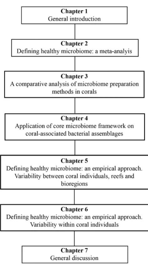
Figure 1-1: Diagram of thesis structure. ... 25
Figure 2-1: Complexity of coral host habitat and coral reef environment. ... 33
Figure 2-2: Bacterial community in corals is responsive to environmental factors and biological
events. ... 34
Figure 2-3: Microhabitat similarities between the human gut and corals. ... 39
Figure 3-1: Flow diagram of experimental design. ... 62
Figure 3-2: Number of sequences and OTUs per sample for G. edwardsi (A, C) and I. palifera
(B, D). ... 70
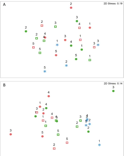
Figure 3-3: Bacterial communities are similar regardless the preservation and homogenization
method used in G. edwardsi (A), in I. palifera (B) bacterial assemblages treated with PFA-decalcified differ from the other methods. ... 72
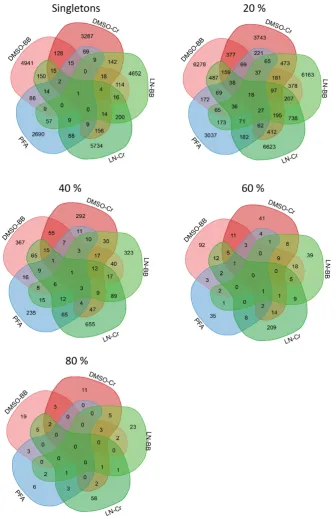
Figure 3-4: Common/shared and specific phylotypes in bacterial assemblages sampled by
different preservation and homogenization methods. ... 74
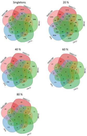
Figure 3-5: Common/shared phylotypes variate in persistence among preservation and
homogenization methods. ... 75
Figure 3-6: Variation of taxonomic composition and structure among preservation and
homogenization methods. ... 76
Figure 4-1: Host Pachyseris speciosa, a depth-generalist coral. ... 86
xiii
Figure 4-3: Comparison average of relative abundance and percentage of occurrence. ... 95
Figure 4-4: Dendrogram (Tree of life) of 97 bacteria with high percentage of occurrence (≥50%). ... 96
Figure 4-5: Presence of the eight highly persistent bacteria in coral core microbiome. ... 97
Figure 5-1: Coral microbiome conceptualised into three distinct layers. ... 105
Figure 5-2: Bacterial communities structurally differ spatially and between coral species. . 112
Figure 5-3: Coral microbiome is composed by common and species-specific phylotypes in a taxonomical stable structure across individuals. ... 114
Figure 5-4: Taxonomical structure evidenced in beta-diversity (turnover, (a)) and taxonomic breadth (b). ... 115
Figure 5-5: Different bacterial taxonomy structure on representative and highly persistent OTUs (core microbiome) encodes similar functional capabilities. ... 116
Figure 6-1: Coral microhabitats ... 126
Figure 6-2: Subsampling of polyp and coenosarc. ... 129
Figure 6-3: Diversity metrics per microhabitat. ... 135
Figure 6-4: Bacterial communities associated with polyps and coenosarcs have a similar composition. ... 136
Figure 6-5: Bacterial community structure is similar in polyps and coenosarcs. ... 137
Figure 6-6: Composition and functional prediction of core microbiome. ... 139
xv
Publications produced during my PhD candidature
Peer reviewed papers:
• Hernandez-Agreda A, Gates RD, Ainsworth TD. 2017. Defining the core microbiome in corals’ microbial soup. Trends in Microbiology, 25 (2). pp. 125-140.
• Hernandez-Agreda A, Leggat W, Bongaerts P, Ainsworth TD. 2016. The microbial signature provides insight into the mechanistic basis of coral success across reef
habitats. mBio, 7 (4). pp. 1-10.
• Hernandez-Agreda A, Leggat W, Bongaerts P, Herrera C, Ainsworth TD. Rethinking the coral microbiome. Simplicity exits within a diverse microbial biosphere. Accepted
- mBio.
• Hernandez-Agreda A, Leggat W, Ainsworth TD. A comparative analysis of microbial DNA preparation methods for use with massive and branching coral growth forms. In
review - Frontiers in Microbiology.
• Hernandez-Agreda A, Leggat W, Ainsworth TD. A place for taxonomic profiling in the study of the coral microbiome. In review - FEMS Microbiology Letters.
xvi
Book chapter:
• Miloslavich·P, Cruz-Motta JJ, Hernández A, Herrera C, [...]. 2016. Benthic Assemblages in South American intertidal rocky shores: Biodiversity services and
threats. In: Marine Benthos: Biology, Ecosystem Functions and Environmental Impact,
Chapter: 3, Publisher: Nova Science Publisher, Editor: Rafael Riosmena-Rodríguez.
Conference oral presentations:
• Australian Coral Reef Society Conference. July 2015. Daydream Island, Australia.
Hernández A, AI; T Ainsworth; P Bongaerts; B Leggat. A coral core microbiome: searching for symbiotic bacteria in corals and understanding their spatial and depth
variation.
• 13th International Coral Reef Symposium. June 2016. Honolulu, Hawaii. Hernández A, AI; P Bongaerts; W Leggat; TD Ainsworth. Exploring coralassociated bacteria over an extreme depth gradient: assessing the presence of ubiquitous symbionts.
• ASLO 2017 Aquatic Sciences Meeting. February 2017. Honolulu, Hawaii. Hernández A, AI; P Bongaerts; W Leggat; TD Ainsworth. Persistence and functional importance: identifying bacterial likely to be promotors of corals’ success.
• Coral Reef Futures Symposium 2017. June 2017. Canberra, Australia. Hernández A, AI; P Bongaerts; W Leggat; TD Ainsworth. Defining microbiome in coral’s microbial
xvii
• Origins and Function of the Animal Metaorganism. March 2018. Hernández A, AI; W Leggat; P Bongaerts; C Herrera; TD Ainsworth. Rethinking the coral microbiome:
simplicity in a diverse microbial biosphere.
Science communication pieces:
• Hernandez-Agreda A, McMahon R, Ainsworth TD, Martin J. What we have in common with corals and their unexplored microbial world, via The Conversation
18
19
The coral microbiome
Microbial communities in corals represent a complex study system due to the high diversity
and variability encountered within each coral host species and individual. Here I review the
knowledge on coral-associated microbial communities and identify factors that influence this
diversity and variability. This section provides an introduction to the topics explored in this
thesis, followed by a more in-depth review and meta-analysis in Chapter 2 (‘Defining healthy
microbiome: a meta-analysis’).
As a holobiont or meta-organism, corals are inhabited by diverse microbial communities
including microalgae, bacteria, fungi, Archaea and viruses (Blackall, Wilson et al. 2015).
Biological interactions between these microbes and the coral host are not yet fully understood,
and just recently, we have started to comprehend the dimensions in richness, diversity and
abundance of those communities. Symbiosis with the photosynthetic endosymbionts of the
genus Symbiodinium is, so far, the most studied and better understood biological interaction between coral and any member of the microbial community (Davy, Allemand et al. 2012). The
coral rely on the symbiotic relationship with these dinoflagellates for up to 95% of their nutrient
uptake (Muscatine, McCloskey et al. 1981, Muscatine, Falkowski et al. 1984), and where this
endosymbiosis is disrupted for long periods of stress, the coral can perish (Glynn 1984, Baird
and Marshall 2002, Eakin, Morgan et al. 2010). However, the remaining members of the
microbial community, the stability, and functional contribution have remained mostly
unknown despite decades of coral reef research.
In particular for bacteria, one of the factors limiting our understanding of the host-microbe
interaction is the variability that is evident in community dynamics (Rohwer, Seguritan et al.
2002, Bourne and Munn 2005). Coral-associated bacterial communities are highly variable,
20
these communities is challenging. Community variability occurs at different spatial and
temporal scales, ranging from the microbial niche within a coral colony (centimeters, (Rohwer,
Seguritan et al. 2002)) to different biogeographical regions (thousand of kilometers, i.e. genus
Porites (Wegley, Edwards et al. 2007, Li, Chen et al. 2013, Zhang, Ling et al. 2015)). Due to the broad physiological and metabolic characteristics of the prokaryotic phylum Bacteria, these
microorganisms can inhabit virtually any environment on earth (Kim and Gadd 2008). Thus,
corals represent a varied ecosystem for bacterial communities, which have been found
inhabiting the surface mucus layer (Carlos, Torres et al. 2013, Glasl, Herndl et al. 2016), coral
tissue (Ainsworth, Fine et al. 2006, van de Water, Ainsworth et al. 2015, Neave, Rachmawati
et al. 2016) and the skeleton (Yang, Lee et al. 2016). These three microhabitats represent
distinct microniches for bacteria, and consequently, the communities associated to each are
likely to differ in richness, composition and structure (Sweet, Croquer et al. 2011, Apprill,
Weber et al. 2016). While the exact ecological and biological factors influencing microbial
community structure have not yet been determined, reef environment is likely to have an
important role in generating variability on bacterial communities (Lee, Yang et al. 2012,
Morrow, Moss et al. 2012, Rodriguez-Lanetty, Granados-Cifuentes et al. 2013). Light
intensity, temperature, turbidity, pH or any environmental disturbance resulting in stress
response in the coral can potentially impact the community structure in bacteria (Thurber,
Willner-Hall et al. 2009, Littman, Willis et al. 2011, Grottoli, Dalcin Martins et al. 2018). As
evident by recent findings that distinct bacterial communities exist between organisms of the
same species located in different reefs (Morrow, Moss et al. 2012) and on the same reef but at
different depths (Glasl, Bongaerts et al. 2017). Furthermore, the vast majority of literature on
coral-associated bacteria has arisen from shallow reefs (Olson and Kellogg 2010), and such our
understanding of depth-related community variability is limited. Recent advances in diving and
21
communities of mesophotic (i.e. >30 m depth) and deep-sea corals, allowing the identification
of similarities and discrepancies with shallow coral-associated microbial communities
(Ainsworth, Krause et al. 2015, Meistertzheim, Lartaud et al. 2016, Glasl, Bongaerts et al.
2017, Gonzalez-Zapata, Bongaerts et al. 2018). Biological processes as diseases, reproduction
and competition for space are some of the factors that can also generate structural changes in
bacterial communities in corals (Barott, Rodriguez-Mueller et al. 2012, Cardenas, Rodriguez
et al. 2012, Ceh, Raina et al. 2012).
Bacterial communities on corals are defined as highly diverse (Blackall, Wilson et al. 2015,
Bourne, Morrow et al. 2016). This diversity is broadly described in literature reports of coral
species harboring more than a hundred thousand individual bacteria belonging to over 30
thousand distinct phylotypes (Ainsworth, Krause et al. 2015, Zhang, Ling et al. 2015, Meyer,
Rodgers et al. 2016), many of them not fully taxonomically identified or described. Thus, the
variability, abundance and diversity of bacterial communities in corals have impaired our
capacity of
i) identifying bacterial phylotypes potentially in stable symbiosis with corals,
ii) describing the functional roles that these symbiotic bacterial phylotypes may be
playing to support coral wellbeing, and
iii) defining the characteristics of a healthy state of a bacterial community.
Answering these questions is crucial to identify the mechanisms through which bacteria, as
physiologically and metabolically versatile organisms, may contribute to coral resilience and
recovery from disturbances. Identifying those mechanisms is crucial as disturbances to coral
reefs are increasing in frequency and magnitude (e.g. bleaching, (Hughes, Kerry et al. 2017,
22
of human populations depending on reefs. To better understand the coral bacterial community
structure, function and symbioses, we first need to identify the attributes of a normal and
healthy bacterial community. Through accurately characterizing the normal and healthy status,
we can define disturbed and dysbiotic states and identify holobiont states associated with a lack
of, or low, performance in delivering goods and services to the host. Ultimately, this knowledge
may provide avenues to enhance resilience and accelerate coral recovery (e.g. through the
active manipulation of coral microbiota).
Thesis objectives
The goal of this thesis was to advance the understanding the healthy coral microbiome by
evaluating spatial patterns in the composition and structure of bacterial communities in corals,
identifying their relevant scales of variation and proposing potential processes driving them.
To achieve this goal, I establish the following objectives:
• Objective 1: Define the characteristics of a healthy coral microbiome.
• Objective 2: Evaluate the presence of universal bacterial symbionts in coral-associated
bacterial assemblages.
• Objective 3: Identify and quantify natural and artificial factors generating variability in
coral-associated bacterial communities.
Objective 1: Define the characteristics of a healthy coral microbiome.
Due to the extreme variability, diversity and richness of microbes observed in association with
corals, the definition and characteristics of a healthy microbiome have not yet been fully
established. Firstly, I will approach this question by undertaking a meta-analysis of the
23
determine the variability of the coral microbiome to give a current status of the definition of
the healthy microbiome. Besides, this exercise exposes critical gaps in our knowledge, and
therefore an ecological framework is proposed to investigate the complex microbial systems in
corals. Following this, in Chapter 5, I will empirically determine the common attributes of a
healthy microbiome among coral species. Finally, a formal definition of the healthy
coral microbiome is proposed in Chapter 7 together with an integrative discussion of all the
chapters of this thesis.
Objective 2: Evaluate the presence of universal bacterial symbiont in coral-associated bacterial assemblages.
The core microbiome concept is one of the frameworks that has facilitated the understanding
of other complex microbial systems. In Chapter 2 this concept is extensively reviewed, and
considerations on its application in corals are proposed. Its applicability is then empirically
tested in Chapters 4, 5, 6. Discussion in regards to its applicability and utility in advance of
understanding healthy coral microbiome are discussed in Chapter 7.
Objective 3: Identify and quantify natural and artificial factors generating variability in coral-associated bacterial communities.
Factors driving the variability of coral-associated bacterial communities are reviewed as part
of the literature review (Chapter 2). However, in the meta-analysis the definition of healthy
and its dissimilarities with dysbiotic/disturbed states of the coral microbiome are resultant from
studies with different sampling and manipulation (laboratory and bioinformatics) methods.
Therefore, the natural factors generating variability in coral-associated bacteria are identified
across species, depth, reefs and bioregions in Chapter 4 and 5, and within individuals in Chapter
24
evaluated in Chapter 3, with the evaluation of preservation and homogenization methods on
the composition and structure of bacterial communities in corals.
Thesis outline
This thesis consists of seven chapters written in publication-format, intended for publication in
peer-reviewed journals (see Figure 1-1 for an outline). Chapters for publication (Chapters 2 – 7) have shared authorship with three members of my committee Tracy Ainsworth (Chapters 2
– 7), William Leggat (Chapters 3 – 6) and Pim Bongaerts (Chapters 4 and 5). Tracy Ainsworth
and William Leggat contributed to the development of the questions and design of the
samplings, funding, training on laboratory and bioinformatics tools, analyses and interpretation
of results and preparation of manuscripts for submission. Pim Bongaerts contributed to the
development of the questions and design of the samplings and preparation of manuscripts for
submission. Also, two chapters are co-authored with two collaborators; Ruth D. Gates
contributed to the conceptualization and development of Chapter 2 and Cesar Herrera Acosta
in the data analysis of Chapter 5. Data produced in this thesis have been made publically
available in the National Centre for Biotechnology Information (NCBI), with project
identification in the relevant chapters. Figures and tables illustrating the results are showed
where relevant along the thesis. Supplementary material, principally statistical analyses,
supporting results are identified and listed in the appendices. I have created all the listed figures
25
26
Chapter 1 (this chapter) represents the general thesis introduction, providing an initial introduction to microbial communities in corals and outlining thesis objectives.
Chapter 2 reviews the literature through a meta-analysis to describe the current definition of healthy state in bacterial communities and propose concepts may contribute to developing the
concept of the healthy microbiome in corals. This chapter is published in Trends in Microbiology (Hernandez-Agreda, Gates et al. 2017). I conducted the literature review and wrote the chapter, and Tracy Ainsworth and Ruth D. Gates contributed to the development of
proposals to impulse the research field and in the editing of the manuscript.
Chapter 3 evaluates the effect of preparation methods on the attributes of bacterial datasets and the perception of bacterial communities in healthy corals. This chapter is under review in
Frontiers in Microbiology. I collected the coral specimens, conducted laboratory, bioinformatics and statistical analysis and wrote the paper, Tracy Ainsworth and William
Leggat assisted in the editing of the manuscript.
Chapter 4 characterizes bacterial community on a healthy coral along of different spatial scales and in a depth gradient to identify relevant scales on the variability of bacterial communities.
This chapter also tests the concept of core microbiome developed in Chapter 2. This chapter is
published in Mbio (Hernandez-Agreda, Leggat et al. 2016). Pim Bongaerts collected coral specimens (as part of the XL Catlin Seaview Survey project) and assisted in the editing of the
manuscript. I conducted laboratory, bioinformatics and statistical analysis and wrote the paper,
Tracy Ainsworth and William Leggat assisted in the interpretation of the results and the editing
of the manuscript.
27
ecological approaches proposed in Chapter 2, to identify common characteristics among coral
individuals and establish attributes of bacterial communities in healthy corals. This chapter has
been accepted in mBio. Pim Bongaerts collected coral specimens (as part of the XL Catlin Seaview Survey project) and assisted in the editing of the manuscript. I conducted laboratory,
bioinformatics and statistical analysis and wrote the paper, Cesar Herrera contributed with the
analysis of beta-diversity, Tracy Ainsworth and William Leggat assisted in the interpretation
of the results and the editing of the manuscript.
Chapter 6 evaluates the variability of bacterial communities within microhabitats of corals. This chapter has been submitted to Applied andEnvironmental Microbiology. I collected the coral specimens, conducted laboratory and statistical analysis and wrote the paper, William
Leggat ran bioinformatics analyses and assisted in the editing of the manuscript, and Tracy
Ainsworth assisted in the editing of the manuscript.
Chapter 7 summarized the previous chapters, discusses the concept and characteristics of health on bacterial communities in corals and proposes priority research areas to progress in
the understanding of microbes’ roles in coral wellbeing. This chapter is under review in FEMS Microbiology Letters. I wrote the manuscript, Tracy Ainsworth contributed to the development of the approach for this chapter and its editing, and William Leggat and Pim Bongaerts assisted
28
Chapter 2:
Defining the Core Microbiome in Corals’
Microbial Soup
29
The coral microbiome symbioses and functional contributions of
coral-associated bacteria
Multi-organism partnerships are widespread in nature and can form the basis of organism and
ecosystem (see Glossary) success in space and time (Herre, Knowlton et al. 1999, Leigh 2010, Bordenstein and Theis 2015). Many of these symbioses have been studied in detail, and the
benefits provided by the symbiont to the host have been well documented (McFall-Ngai 2008,
Relman 2008). However, there are other systems in which symbiosis, particularly bacterial
symbiosis, are hypothesized as an underlying mechanism of the host success, but the exact
nature of the symbiosis has not, or can not yet be, determined. Corals and coral reefs are one
such ecosystem where specific bacteria, and bacterial communities, are hypothesized as
crucially important in both organism function and ecosystem dynamics (Graham, Ainsworth et
al. 2011, Blackall, Wilson et al. 2015). However, the contributions made by specific bacterial
symbionts have not yet been accurately deciphered.
The coral microbiome is one of the most complex microbial biospheres studied to date (Blackall, Wilson et al. 2015). Corals host thousands of bacterial phylotypes, in species-specific associations across broad geographical and temporal scales that have been
hypothesized as functionally significant (Ritchie and Smith 1997, Rohwer, Breitbart et al.
2001, Rohwer, Seguritan et al. 2002, Morrow, Moss et al. 2012). The coral microbiome, its
composition, spatial-temporal variability, and response to environmental change have been studied to date in over 25 coral species, from reef locations around the world, and from corals
in both healthy and diseased states (Frias-Lopez, Zerkle et al. 2002, Rohwer, Seguritan et al.
2002, Bourne and Munn 2005, Ritchie 2006, Bourne, Iida et al. 2008, Thurber, Willner-Hall et
al. 2009, Barott, Rodriguez-Brito et al. 2011, Ceh, van Keulen et al. 2011, Littman, Willis et
30
2012, Wilson, Aeby et al. 2012) (Supplementary Table B-1). As in other systems, advances in
molecular technologies have unveiled the richness and composition of bacterial communities
of the coral host, and high throughput sequencing is now universally applied to evaluate
bacterial diversity (Claesson, Wang et al. 2010, Wu, Lewis et al. 2010). This methodology
provides a representation of the bacterial community, identifies rare and less abundant species and provides insight into the conservation of bacterial phylotypes within microhabitats and between individual hosts (Pedros-Alio 2006, Sogin, Morrison et al. 2006, Sunagawa, Woodley
et al. 2010). An average of 995 distinct bacterial operational taxonomic units (OTUs) and 22,520 sequences were identified from each coral species when sequencing technologies were
first applied to the coral microbiome (Supplementary Table B-1). These estimates of
microbiome complexity have recently increased substantially as higher sequencing coverage
has been obtained. Now upwards of 100,000 bacterial OTUs and millions of reads are reported
from each coral species (Zhang, Ling et al. 2015, Hernandez-Agreda, Leggat et al. 2016,
Meyer, Rodgers et al. 2016). In general, the dominant associations that have been identified in
these studies are assumed as the healthy symbiotic state of the coral-associated bacterial
communities (Littman, Willis et al. 2011, Cardenas, Rodriguez et al. 2012, Croquer, Bastidas
et al. 2013, Lee, Davy et al. 2015).
Gamma- and Alphaproteobacteria dominate the bacterial communities of corals, other highly
abundant bacteria include members of the phyla Bacteriodetes, Firmicutes, Actinobacteria and
Cyanobacteria. Endozoicomonas are generally the highest abundance genera in the coral microbiome (Blackall, Wilson et al. 2015) (see citations in Supplementary Table B-1). The consistency of dominant associations between individuals, within and between coral species,
across spatio-temporal scales, and depth gradients, have only recently been investigated. In
fact, one of the most significant findings that have arisen from the recent deep sequencing of
31
and reef habitats. The most revealing of which is the variability that occurs between individuals
(Ainsworth, Krause et al. 2015, Hester, Barott et al. 2015, Hernandez-Agreda, Leggat et al.
2016). For example, studies investigating ubiquitous associations across individuals find that
over 60% of the identified bacterial OTUs of the coral microbiome are present in less than 10%
of individuals studied, many of which include some of the most highly abundant bacteria within
a single individual (Ainsworth, Krause et al. 2015, Hernandez-Agreda, Leggat et al. 2016).
There is also substantial variation in the occurrence (and persistence) of some of the most
abundant members of the microbiome. For example Endozoicomonas are the most abundant group in the coral microbiome but are also highly variable within and between coral species
(Bayer, Neave et al. 2013, Meyer, Paul et al. 2014, Morrow, Bourne et al. 2014) (see review (Blackall, Wilson et al. 2015)). Williams, Brown et al. (2015) have proposed that the variability
between individual seen in corals be correlated to the age of the coral colony. Thus, like other
systems studied, the coral microbiome is likely to be highly variable until the colony reaches
adulthood, at which time the microbiome stabilizes as the adult microbial signature (Williams,
Brown et al. 2015). Studies of coral reproduction have also revealed that bacterial colonization
in corals can occur principally by horizontal uptake (Apprill, Marlow et al. 2009, Littman,
Willis et al. 2009, Lema, Bourne et al. 2014) and occasionally by vertical transmission (Sharp,
Distel et al. 2012). While Neave, Rachmawati et al. (2016) suggest that substantial variation in
microbiome structure, including Endozoicomonas, could be linked to reproductive strategies of the coral host (i.e. brooding and broadcast spawning corals). The seasonal variability in the
microbiome indicates that abiotic factors strongly influenced these associations, and
correlations with carbon availability suggest a strong influence of the by-products of
32
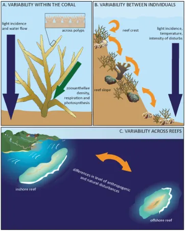
animal and the coral reef habitat that are likely to influence microbial diversity, abundance, occurrence and persistence (Figure 2-1). The bacterial community of corals is responsive to
environmental variables, biological events and also to factors that result in stress response in
the host (Figure 2-2, Supplementary Table B-1) (see review (Schwartzman and Ruby 2016)). Stress in corals manifests in response to variations in sea surface temperature, salinity,
nutrients, pH, loading of dissolved organic carbon, disease and competition for space. All of
these factors also have the potential to impact bacterial groups, OTU abundance, and may also
result in the disappearance, or appearance, of some bacteria. Biotic and abiotic factors also
influence activation and expression of genes related to virulence and secondary metabolites in
33
[image:35.595.116.481.70.522.2]34
Figure 2-2: Bacterial community in corals is responsive to environmental factors and biological events. Meta-analysis results evidence that in shallow corals bacterial community composition and abundance within different microhabitats shifts in response to changes in environmental factors and response to biological processes (see details in Supplementary Table B-1).
Bacterial communities are proposed to have important critical contributions to the health,
nutrition and nutrient cycling in the coral host (Ritchie 2006, Lesser, Falcon et al. 2007, Raina,
Tapiolas et al. 2009, Lema, Willis et al. 2012, Peixoto, Rosado et al. 2017). Bacteria in the
coral’s surface mucus layer are hypothesized as important in the provision of antibiotic activity
and likely provide population control, preventing pathogen colonization and invasion (Ritchie
2006). Diazotrophic bacteria have also been consistently found within bacteria community of the coral tissue, skeleton and mucus (within the family Cyanobacterium and order
Rhizobiales) (Shashar, Cohen et al. 1994, Rohwer, Seguritan et al. 2002, Lesser, Mazel et al.
2004, Lesser, Falcon et al. 2007, Lema, Willis et al. 2012). The ubiquitous nature of
diazotrophic bacteria in newly released larvae and juvenile corals (Lema, Bourne et al. 2014)
35
importance of nitrogen-fixing bacteria in the coral holobiont at all life history stages. The degradation of the organic compound dimethylsulfoniopropionate (DMSP) and its products
dimethylsulfide (DMS) and acrylic acid are also carried out by bacteria (Curson, Rogers et al.
2008). Genera of coral-associated bacteria Spongiobacter, Pseudomonas, Roseobacter, and
Vibrio spp. can metabolize DMSP, DMS and acrylic acid (Raina, Tapiolas et al. 2009), and provide strong evidence for the role of coral-associated bacteria in sulfur cycling. However,
differentiating the functionally important bacteria associated with corals has been limited due
to the complexity of both the coral host habitat and the coral reef environment (Figure 2-1).
The microhabitat association of only a few coral-associated bacteria has been determined.
Bacterial aggregations, dominated by members of γ-proteobacteria, have been found within
coral epidermal cell layer (Ainsworth, Fine et al. 2006, van de Water, Ainsworth et al. 2015),
and a higher abundance of aggregates has been found in corals held in captive conditions
(Ainsworth and Hoegh-Guldberg 2009). Endozoicomonas aggregations have also been localized within both of the coral cell layers (Neave, Rachmawati et al. 2016) and members of
the phylum Actinobacteria and Ralstonia sp. have also been found within the cellular space of coral that is inhabited by the endosymbiotic dinoflagellate (peri-algal space) (Ainsworth,
Krause et al. 2015). Members of the Alpha-, Gammaproteobacteria, Cyanobacteria,
Flavobacteria, and Firmicutes have been isolated from the coral gastric cavity (Agostini,
Suzuki et al. 2012). Localization of specific bacteria within cell layers and habitats may
suggest that some of these associations play an essential role in the coral nutrient uptake.
Research into the coral microbiome is however still in its infancy. Research has yet to
determine the stability of the vast majority of bacterial associations of corals and the influence
of factors including feeding strategy (autotrophy and heterotrophy), feeding time, growth stage,
immune status, and patterns of microbial succession remain unresolved. The complexity of the
36
the microbiome and need to be considered when differentiating microbial states as symbiotic,
mutualistic, commensal, parasitic, pathogenic, or dysbiotic. Overcoming the constraints that are inherent in highly complex environmental systems, such as coral reefs, and differentiating
potential symbioses will, therefore, be reliant on utilizing the theoretical and technical advances
made in more extensively studied model systems. In this review, I examine how the core
microbiome concept in conjunction with community ecology principals is a framework that
can be applied to facilitate identifying potentially important microbes in corals.
The coral microbiome: bacterial habitats
In corals, bacteria have been reported inhabiting three unique microbial habitats (Figure 2-3);
the surface mucus layer, the symbiosome space within the coral tissue, and the exposed
skeleton (Bourne and Munn 2005, Koren and Rosenberg 2006, Ritchie 2006, Sweet, Croquer
et al. 2011).
The surface mucus layer (SML, Figure 2-3B1) is an interface zone between the surrounding
seawater and the coral epithelium and plays an essential role in heterotrophic feeding, sediment
clearing, defense against pathogens, and protection during environmental stresses. The
microbial community within this zone is likely to be a highly structured biofilm, exposed to
substantial biotic and abiotic variation. Host mucus secretion and the microbial biofilm present
in this layer are released to seawater and drive microbial turnover, biofilm succession and
composition of the coral mucus-associated community (Bythell and Wild 2011). The nutrient
composition, oxygen levels, pH and rate of production of the surface mucus layer vary across
the day, between coral species, depth and as a response to environmental changes (e.g.
sedimentation, radiation). All these characteristics result in a highly variable microbial habitat
within which the bacterial community is likely also to be extremely dynamic (Sweet, Croquer
37
environmental conditions generate a physicochemical gradient of various micro-niches
available for colonization (Bolhuis, Cretoiu et al. 2014). However, it is still poorly understood
how the changes in mucus chemistry, mucus release, and colonization during regular
sedimentation on corals regulate microbial population dynamics on the surface microbial
biofilm.
The term coral tissue has been used to group the tissues layers of the coral, namely the
epithelium, mesoglea, gastroderm and calicoblastic epithelium (Figure 2-3B). The algal
endosymbiont Symbiodinium sp. resides within the gastrodermal layer, within a single gastrodermal cell where each algal cell is surrounded by the host-derived symbiosome
membrane (Yellowlees, Rees et al. 2008). The pocket created by this membrane within the
coral cell is referred to as the peri-algal space, and is an interface zone in which nutrient and
metabolite transfer between the algal symbionts and the host cell occurs (Kazandjian, Shepherd
et al. 2008). Difficulties in isolating this space have however interfered with detailed
characterization of the specific conditions that are unique to this microhabitat. Using
fluorescence in situ hybridization, bacteria from genus Propionibacterium sp. and Ralstonia sp. have beenrecently reported as potential universal coral symbionts inhabiting the cellular space of coral dinoflagellate in a high relative abundance (Ainsworth, Krause et al. 2015).
Gammaproteobacteria and specifically Endozoicomonas have also been localized to the both the epidermal (outer) and gastrodermal (inner) tissue layers (Ainsworth, Fine et al. 2006, van
de Water, Ainsworth et al. 2015, Neave, Rachmawati et al. 2016). Their localization suggests
bacteria are likely to play an essential role in corals or coral nutrient uptake, but also raises
questions such as, how bacteria can evade the coral immune systems and inhabit this space,
38
The coral skeleton (Figure 2-3B2) is a microbial habitat that is also exposed to the surrounding
seawater but unlike the SML is protected from wave and water action, sedimentation, and light
penetration (due to the overlaying algal rich coral tissues), and as a result this region is
environmentally stable (Shashar, Banaszak et al. 1997). Research in the microbial community
in this zone (the endolithic community) has focused on documenting the micro-algae and fungi presence (Le Campion-Alsumard, Golubic et al. 1995, Le Campion-Alsumard, Golubic et al.
1995, Fine and Loya 2002, Rädecker, Pogoreutz et al. 2015). The microalgae within the
endolithic layer are proposed to provide alternative energy supply to the coral host and the
community rapidly blooms following changes to the overlaying coral tissues that allow
increase light to penetrate into the skeleton (Rädecker, Pogoreutz et al. 2015). The endolithic
layer hosts an abundant bacterial community, which like the algal community blooms
following changes to the overlying coral tissues (Rosenberg, Koren et al. 2007, Rädecker,
Pogoreutz et al. 2015).
The microbial habitats of the coral tissue and skeleton structure, therefore, provide distinct and
dynamic environments within which bacterial communities can form, and in doing so
39
Figure 2-3: Microhabitat similarities between the human gut and corals. Axial view of microhabitats in A) human large intestine, and B) corals [tissue of walls (B1) and base (B2) of polyps]. The human gut and corals have structural similarities in their microhabitats distribution and characteristics. Knowledge about symbiosis and mechanisms driving microbial colonization and dynamics have significantly advanced in humans; and based on this similarity between systems, corals can follow some of those advances done in the human gut system to accelerate the understanding of coral-bacterial symbiosis.
Human gut and corals: similar systems, different drivers
The similarities between corals’ and humans’ gut microbial systems open a path to
understanding corals’ microbial symbiosis based on the progress of humans’ gut as a model
system. Composition and abundance of the bacterial community in both, human gut and corals,
vary longitudinal and axially (Ainsworth, Thurber et al. 2010, Belizario and Napolitano 2015),
although the factors promoting those variations are different. For example in the human gut,
pH changes from the esophagus (pH <4.0) through the stomach (pH 2) to the small intestine
(pH 5-7), representing a variety of habitats with different bacterial composition (Jandhyala,
Talukdar et al. 2015). Likewise, due to the structural complexity of diverse morphologies of
corals (branching, plating and laminar and massive/encrusting), factors as temperature, light,
40
microhabitats and harboring different microbial communities (Figure 2-1A,B) (Ainsworth,
Thurber et al. 2010). In an axial vision, the tissues in both human gut and corals are structurally
similar: a mucus layer as an external barrier protecting epithelial cells (Figure 2-3) (Ritchie
2006, Bythell and Wild 2011, Tuddenham and Sears 2015). However, in the human colon,
mucus has two homogeneous layers: an external and dynamic one, low adherent with bacteria
living on it; and an internal one, close to the epithelium, denser and devoid of bacteria
(Tuddenham and Sears 2015) (Figure 2-3A). Corals also possess a homogeneous mucus layer
across colony surface; nonetheless, it is inhabited by a highly abundant bacteria community
(Ritchie 2006, Bythell and Wild 2011) (Figure 2-3B1). Unlike these similarities, corals have
many differences with the human gut system; the principal is that corals are an open system,
where the host and the microbial community are vulnerable and responsive to changes in host
environment (e.g. temperature, pH, salinity, between others) and biological processes (e.g.
reproduction and competition). Whereas human gut is a closed system, where changes in
microbial community occur in response to diet or antibiotics (Backhed, Fraser et al. 2012).
The core microbiome framework
The core microbiome framework aims to identify potentially crucial microbes within microbial
communities based on the persistence of the microbe within the host, and within a niche across
spatio-temporal boundaries. Research into the identity and functional contribution of core
microbes, and a core microbiome was first applied to understanding the bacterial communities
that are associated with humans (Fierer, Hamady et al. 2008, Turnbaugh and Gordon 2009,
Turnbaugh, Hamady et al. 2009, Qin, Li et al. 2010). The Human Microbiome Project (HMP)
aimed at understanding the role of microbes in human health, and the factors influencing
41
of the human gut, unveiling both persistent bacterial functional roles and a highly variable
bacteria community (Turnbaugh, Ley et al. 2007, Turnbaugh, Hamady et al. 2009). The core
microbiome was identified as group of consistent functional microbial genes (Turnbaugh and
Gordon 2009, Turnbaugh, Hamady et al. 2009), and both the core and variable bacterial
community members were found to be influenced by the diet, genotype and developmental
stage (Turnbaugh, Hamady et al. 2009, Spor, Koren et al. 2011, Tims, Zoetendal et al. 2011).
One example of a bacterium persistently found across individuals is Faecalibacterium prausnitzii (Qin, Li et al. 2010). This bacterium contributes to human health through the production of butyrate, a short-chain fatty acid that acts in a regulatory role in colon walls, as
a defense barrier enhancer, an intestinal motility modulator and provides anti-inflammatory
action (Canani, Costanzo et al. 2011, Miquel, Martín et al. 2013, Miquel, Leclerc et al. 2015,
Quevrain, Maubert et al. 2016).
Researchers have since adopted the core microbiome concept across microbial systems (Table
2-1). Differentiating the core microbiome from the broader microbial community has been
applied to understanding the microbial role in host organisms, including plants and sponges,
and in ecosystems including soils, beaches and oceans (Table 2-1) (Turnbaugh, Hamady et al.
2009, Lundberg, Lebeis et al. 2012, Schmitt, Tsai et al. 2012, Newton, Huse et al. 2013, Vik,
Logares et al. 2013, Kembel, O’Connor et al. 2014, Shade, Jones et al. 2014, Staley, Gould et
al. 2014). As such a core microbiome is defined as the group of microbes commonly found
within a host’s microbiome, using persistence of the association as the criterion to select
42
Table 2-1: Comparison of the Application of the Core Microbiome Approach
Subject/ Species/Habitat
N. of total OTUs
N. of OTUs in core microbiome Core taxonomic level % cut-offa Justification Sequencing platform
Core phylotypes with functional role (examples)
Reference
Human
Gut - a set of microbial
functional genes
- 100 Core defined as shared microbial functional genes
454 - (Turnbaugh,
Hamady et al. 2009)
Gut 3.3 million
microbial genes, ≈ 1,000 bacteria species
75, 57 and 18 Species, strains
50, 90 and 100
Arbitrary Illumina F. prausnitzii (Canani, Costanzo et al. 2011, Miquel, Martín et al. 2013, Miquel, Leclerc et al. 2015, Quevrain, Maubert et al. 2016),
Bacteroides uniformis
(Renouf and Hendrich 2011, Li, Li et al. 2014),
Ruminococcus bromii (Ze, Duncan et al. 2012),
Bacteroides thetaiotaomicron
(Cameron, Maynard et al. 2012, Varyukhina, Freitas et al. 2012)
(Qin, Li et al. 2010)
Hands 4,742 5 Genus 100 454 (Fierer,
Hamady et al. 2008)
Plants
Arabidopsis thalianab
18,783c 97 Family - Based on OTU abundances
analysis, GLMM
454 (Lundberg,
43
Subject/ Species/Habitat
N. of total OTUs
N. of OTUs in core microbiome Core taxonomic level % cut-offa Justification Sequencing platform
Core phylotypes with functional role (examples)
Reference
A. thaliana and three relatived speciesb
88,731 9 Genus - Based on OTU abundances, as the
intersection of enriched bacteria determined by three statistical analyses
454 (Schlaeppi,
Dombrowski et al. 2014)
57 tree species, leaves
7,293 104 Family or
Genus
95 Arbitrary Illumina (Kembel,
O’Connor et al. 2014)
Sponges
32 sponges species
2,567 3 Phylum or
Class
70 Arbitrary, based on presence in the majority of sponges species
454 (Schmitt, Tsai
et al. 2012)
Corals Acropora granulosa 1,508 7 universal 149 Kingdom to Genus
30 Lowest percentage at which core OTU abundance is stable across core microbiomes
454 (Ainsworth,
Krause et al. 2015)
Leptoseris spp. 1,424 204 (Ainsworth,
Krause et al. 2015)
Montipora capitata
1,433 350 (Ainsworth,
Krause et al. 2015)
Pachyseris speciosa
173,690 8 Kingdom to
Genus
80 Arbitrary, core phylotypes were present in all spatial scales and depth considered
Illumina
44
Subject/ Species/Habitat
N. of total OTUs
N. of OTUs in core microbiome Core taxonomic level % cut-offa Justification Sequencing platform
Core phylotypes with functional role (examples)
Reference
Stylophora pistillata
560c,d,e 1 Genus
(Endozoicomo nas)
79 This study was focused in evaluating Endozoicomonas
prevalence across different geographic regions.
Illumina (Neave,
Rachmawati et al. 2016)
Pocillopora verrucosa
655 c,d,e 1 85 (Neave,
Rachmawati et al. 2016)
Antillogorgia elisabethae
502 and 281c,e,f 27 and 48f,g Kingdom to Genus
50 Arbitrary 454 (Robertson,
Haltli et al. 2016)
Anthothela grandiflora
55 c,e 1 Genus 100 454 (Lawler,
Kellogg et al. 2016)
Anthothela sp. 110 c,e 7 Phylum to
Genus
(Lawler, Kellogg et al. 2016)
Corallium rubrum 250 c,e 12 Phylum to
Family
100 Illumina (van de Water,
Melkonian et al. 2016)
Acropora cervicornis
87,668c (all coral species together) 6 universal 12 Phylum to Genus
50 Arbitrary Illumina (Chu and
Vollmer 2016)
Acropora palmata 15 (Chu and
Vollmer 2016)
Diploria labyrinthiformis
11 (Chu and
45
Subject/ Species/Habitat
N. of total OTUs
N. of OTUs in core microbiome
Core taxonomic level
% cut-offa
Justification Sequencing
platform
Core phylotypes with functional role (examples)
Reference
Diploria strigosa 11 (Chu and
Vollmer 2016)
Porites astreoides 13 (Chu and
Vollmer 2016)
Porites furcata 14 (Chu and
Vollmer 2016)
Beaches
Sand 23,670 26 Family, Genus 75 Arbitrary 454 (Newton,
Huse et al. 2013)
Water 19,411 62 (Newton,
Huse et al. 2013)
a Minimal percentage of individuals (microbiomes) where bacteria or microbial genes have to be present to be considered as part of the core microbiome. b Root system.
c Rarefied or subsampled data.
d Based on the average of the three methods applied to assign OTUs. e Average.
f Based on the amplified region (V1/V2 and V4).
46
The coral core microbiome
The core microbiome framework is still a young concept for corals. The core microbiome has
been explored in 16 coral species, across ocean depths and geographically separated locations.
Core annotations have ranged from 30% of persistence with studies using the 454-sequencing
platform, to 100% using the Illumina sequencing platform (Table 2-1). Ainsworth, Krause et
al. (2015) used a 30% core described between 149 to 350 phylotypes per coral species, and 7
shared phylotypes among coral species. Hernandez-Agreda, Leggat et al. (2016) identified 8
persistent phylotypes at different spatial scales and depth gradients using core 80% (Illumina).
van de Water, Melkonian et al. (2016) found 12 bacterial species using a 100% core in 23
individuals (Illumina) collected from five disparate reef sites over a 3-month period. The core
microbiome has also been investigated in one octocoral host (454 platform) finding 27 OTUs
at 50% persistence across 17 individuals (Robertson, Haltli et al. 2016). In the cold-water
octocoral Anthothelagrandiflora only one OTU was evident in a 100% core microbiome in 12 individuals (454 platform), further study at the genus level found between 7 OTUs in the core
microbiome (Lawler, Kellogg et al. 2016). Chu and Vollmer (2016) undertook a
spatio-temporal study of the core microbiome in 100 tagged individuals of six coral species (collected
three times during one year period from four reef habitats) identifying between 11 and 15
members of a 50% core microbiome (Illumina). These studies identify several crucial factors
that need to be considered when annotating core microbiomes, using core microbiome analysis
for identifying potential symbioses and comparing core annotations between studies.
Confounding factors include the criteria used for clustering of samples (i.e. the host taxonomic
level, reef site, reef depth, and time and season of sampling) and the bacterial taxonomic level
used for annotations (Table 2-1). Robertson, Haltli et al. (2016) also show that the targeted
47
in the number of OTUs annotated as core associations (Table 2-1). Furthermore Ainsworth,
Krause et al. (2015) show that a whole colony community approach (i.e. homogenized intact
host samples) does not reflect bacterial associations with the corals’ microhabitats (polyp
tissue, skeleton and gastrodermis). This study showed that the core microbiome within in each
microhabitat differs despite phylotypes being common across coral species.
The criteria that have been used to date to define the persistence of bacterial species within a
coral population (core microbiome) have been arbitrary. However attributing significance to
members of the microbiome solely through high or low relative abundance within a given study
while ignoring persistence, is also an arbitrary process that can be heavily impacted by
technical and biological factors (Supplementary Table B-1). Doing so overlooks key
information about the microbiome that exists within the data. Hernandez-Agreda, Leggat et al.
(2016) suggest that the coral microbiome should be conceptualized as three main components;
(i) a ubiquitous core microbiome; (ii) a dynamic site and/or species-specific community; and
(iii) a highly variable community reflective of the biotic and abiotic fluctuations. For each of
the coral systems that have been investigated to date for the presence of a core microbiome,
features such as reef location, reef habitat and patchiness of microbial habitat within the host
morphology have been overlooked. In differentiating core microbiome and potential symbioses
within highly dynamic and complex systems, such as corals, it is necessary to account for the
unique challenges of those systems. The core microbiome framework provides a means by
which to identify and analyze potentially important bacteria and bacterial groups (also defined
as Beneficial Microorganisms for corals, BMC (Peixoto, Rosado et al. 2017)) within the coral
microbial soup, based on their ubiquitous association within a coral group, reef habitat, coral
48
Technical considerations and theoretical frameworks for applying
core microbiome framework.
The criteria used to define and annotate a core microbiome across different systems have
traditionally been arbitrary, and there has not been a consensus reached in any of the organisms
in which the concept has been applied (Table 2-1). Both technical considerations and the
theoretical framework need to be considered in developing and applying the core microbiome
framework and comparing core microbiomes between studies. These factors include the study
design, target habitat and sample size, sequencing approach, replication and effort, and the
analysis tool applied. Addressing these knowledge gaps will be a crucial step in reliably
applying mechanistic models and interpreting patterns that are observed in nature.
Technical considerations
Within the application of a community framework, there are several levels of information that
are commonly reported in the study of microbiomes and should be considered in determining
criteria for the core microbiome and when comparing studies (Hamady and Knight 2009).
These factors include:
(i) Replication (number of samples),
(ii) Sampling effort (sequencing and sequences analyses),
(iii) Community membership, and
(iv) Study design.
(i) Replication:A crucial factor in defining the core microbiome is the number and quality of
49
be considered individuals and different biological units coral colonies should be similarly sized,
disparate, and randomly selected for similar reef environments. Samples from colonies should
also be collected at similar locations within the coral colony, targeting the same macro and
micro colony structures (Figure 2-1). In environmental scenarios, standardizing the number of
samples required can also be challenging due to logistical considerations such as remoteness,
field time, collection logistics and equipment needs. However, both a priori and post hoc power
analysis tests are alternative strategies for investigating adequate replication in these scenarios
(Faul, Erdfelder et al. 2007, Johnson, Barry et al. 2015). For example, post hoc power analyses
are informative about experimental design where no statistical differences have been detected,
and a small number of replicates have been used (probability of a false negative, type II error,
e.g. (Montilla, Ramos et al. 2016)); whereas a priori power analysis are useful in defining the
sample size (number of replicates, sampling effort) necessary to detect an effect or statistical
difference (e.g. (Harasti, Malcolm et al. 2015)).
(ii) Sampling effort: In microbial ecology, alpha diversity (number of taxa per samples) and
beta diversity (turnover of taxa between samples expressed as pairwise sample dissimilarity)
indexes are widely used to represent and analyze community richness (with the assumption
that OTUs are reflective of taxon (Mihaljevic 2012, Shade and Handelsman 2012)). However,
the weaknesses of alpha and beta diversity measures identified in macro-ecology are also
applicable to micro-ecology (e.g. (Tóthmérész 1995, Koleff, Gaston et al. 2003, Legendre,
Borcard et al. 2005, Bennett and Gilbert 2016)) and apply to the core microbiome framework.
The sensitivity of sample size and resolution is one of the most relevant of these. Factors such
as marker, primer and sequencing region chosen, sequencing depth selected (i.e. number of
sequences per sample relates to sampling effort), alignment and clustering algorithms used to
assign OTUs (e.g. nearest vs. furthest neighbor) (Hamady and Knight 2009, Shade and
50
and Neufeld 2015) affect the number of OTUs generated in sequencing-based estimates of
community composition. Sequencing